Embed Size (px)
Citation preview
Huperzine A Activates Wnt/b-Catenin Signaling and Enhancesthe Nonamyloidogenic Pathway in an Alzheimer TransgenicMouse Model
Chun-Yan Wang1,4, Wei Zheng1,4, Tao Wang1, Jing-Wei Xie1, Si-Ling Wang2, Bao-Lu Zhao3, Wei-Ping Teng1
and Zhan-You Wang*,1
1Key Laboratory of Medical Cell Biology of Ministry of Education, and Key Laboratory of Endocrine Diseases of Liaoning Province, China Medical
University, Shenyang, China; 2Department of Pharmaceutics, Shenyang Pharmaceutical University, Shenyang, China; 3State Key Laboratory of
Brain and Cognitive Sciences, Institute of Biophysics, Academia Sinica, Beijing, PR China
Huperzine A (HupA) is a reversible and selective inhibitor of acetylcholinesterase (AChE), and it has multiple targets when used for
Alzheimer’s disease (AD) therapy. In this study, we searched for new mechanisms by which HupA could activate Wnt signaling and
reduce amyloidosis in AD brain. A nasal gel containing HupA was prepared. No obvious toxicity of intranasal administration of HupA was
found in mice. HupA was administered intranasally to b-amyloid (Ab) precursor protein and presenilin-1 double-transgenic mice for
4 months. We observed an increase in ADAM10 and a decrease in BACE1 and APP695 protein levels and, subsequently, a reduction in
Ab levels and Ab burden were present in HupA-treated mouse brain, suggesting that HupA enhances the nonamyloidogenic APP
cleavage pathway. Importantly, our results further showed that HupA inhibited GSK3a/b activity, and enhanced the b-catenin level in the
transgenic mouse brain and in SH-SY5Y cells overexpressing Swedish mutation APP, suggesting that the neuroprotective effect of
HupA is not related simply to its AChE inhibition and antioxidation, but also involves other mechanisms, including targeting of the
Wnt/b-catenin signaling pathway in AD brain.
Neuropsychopharmacology (2011) 36, 1073–1089; doi:10.1038/npp.2010.245; published online 2 February 2011
Keywords: Alzheimer’s disease; APP/PS1 transgenic mouse; b-catenin; glycogen synthase kinase; huperzine A; Wnt signaling
��������������������������������������������
INTRODUCTION
Alzheimer’s disease (AD), which is increasing in prevalence,is a long-term condition that is expensive to treat and isnow a major public health problem for aging populationsaround the world. Pathologically, AD is characterized byextracellular b-amyloid (Ab) plaques, intracellular neurofi-brillary tangles, and selective cholinergic neuronal loss inthe brain regions involved in learning and memory.Clinically, cholinesterase inhibitors, such as donepezil,galantamine, rivastigmine, and huperzine A (HupA), whichare believed to act by blocking acetylcholine degradationand increasing the function of the surviving cholinergicneurons, have been shown to have beneficial effects by
improving general cognitive function and reducing beha-vioral disturbances (Wang et al, 2001; Ceravolo et al, 2004;Erkinjuntti et al, 2004; Sicras and Rejas-Gutierrez, 2004; vander Staay and Bouger, 2005; Alisky, 2006; Mori et al, 2006).Recently, the multiple targets of cholinesterase inhibitorsused to treat AD have received increasing attention (Lahiriet al, 2004; Zhang and Tang, 2006; Zhang et al, 2008a;Akaike et al, 2010).
The canonical Wnt signaling pathway has an importantrole in neuronal development and maintenance of thenervous system (Patapoutian and Reichardt, 2000). Recentstudies have shown that Wnt signaling is involved inneurodegenerative diseases, especially in acetylcholinester-ase (AChE) and Ab-mediated neurotoxicity (Inestrosa et al,2000; Garrido et al, 2002; De Ferrari et al, 2003). First, AChEis present in neuritic plaques in the AD brain (Geula andMesulam, 1995; Guillozet et al, 1997), and can enhance Abaggregation and plaque formation and may form AChE–Abcomplexes (Alvarez et al, 1997). In vivo Ab intrahippo-campal injection and in vitro hippocampal neuronal cultureexperiments have shown that the AChE–Ab complexesinduce neuronal death more dramatically than Ab peptide
Received 14 August 2010; revised 11 December 2010; accepted 14December 2010
*Correspondence: Professor Z-Y Wang, Key Laboratory of MedicalCell Biology of Ministry of Education, and Key Laboratory of EndocrineDiseases of Liaoning Province, China Medical University, Shenyang110001, China, Tel: + 8 61 399 889 1892, Fax: + 86 24 232566665305, E-mail: [email protected] authors contributed equally to this work.
Neuropsychopharmacology (2011) 36, 1073–1089
& 2011 American College of Neuropsychopharmacology. All rights reserved 0893-133X/11 $32.00
www.neuropsychopharmacology.org

alone (Alvarez et al, 1998; Munoz and Inestrosa, 1999; Reyeset al, 2004). Second, the levels of glycogen synthase kinase-3b (GSK3b) and b-catenin, the two key molecules in theWnt signaling pathway (Willert and Nusse, 1998; Moonet al, 2002), are altered dramatically in the brain of ADmodel mice (Zhang et al, 1998; Pei et al, 1999), andtreatment with AChE–Ab complexes reduces the level ofcytoplasmic b-catenin in cultured hippocampal neurons(Alvarez et al, 2004). Finally, treatments with a Wnt cascadeactivator (lithium) or antagonist (Frzb-1) can protectagainst Ab-induced neuronal death (Alvarez et al, 1999;Inestrosa et al, 2004). Taken together, these studiesdemonstrate that AChE–Ab-dependent neurotoxicity mayresult in a loss of neuroprotection of Wnt signalingcomponents, and that activation of Wnt signaling mayprevent cholinergic neuronal degeneration in AD. However,whether the neuroprotective effect of cholinesterase in-hibitors is related to the action of Wnt/b-catenin has notbeen fully clarified.
HupA is a novel lycopodium alkaloid extracted from theChinese folk medicine, Huperzia serrata. HupA has severalbeneficial effects for AD patients (Wang et al, 2006a) and, inChina it is one of the most commonly prescribed drugs formany forms of dementia, including AD (Zhang et al, 2008b).Apart from its well-known inhibitory effect on AChE (Zhuand Giacobini, 1995; Cheng et al, 1996; Cheng and Tang,1998), HupA is considered to have multiple neuroprotectiveeffects including anti-inflammatory and antioxidant proper-ties (Wang and Tang, 2007; Wang et al, 2008; Zhang et al,2008a), stimulation of the release of soluble a-secretase-derived fragments of APP (sAPPa) (Zhang et al, 2004; Penget al, 2006; Yan et al, 2007), protection against Ab andglutamate-induced neurotoxicity, and regulation of nervegrowth factor (Ved et al, 1997; Tang et al, 2005).Interestingly, recent studies have shown that intranasaladministration of a nasal gel containing HupA is a suitablesystem for delivery of HupA to the brain and, hence,provides a potential strategy for chronic AD therapy (Yueet al, 2007). Compared with the intravenous and oraladministration for chronic disease such as AD, intranasaldrug delivery does not require complicated medicalservices. The drug is not affected by the absorption on thebasis of digestive tract circumstances and does not undergofirst pass effect of liver (Yagi et al, 2002; Tang et al, 2008).HupA is a lipophilic weak alkaloid with a molecular weightof 242.32. It has a good permeability and can be transferredinto the central nervous system via the nasal route (Yueet al, 2007). However, the neuroprotective effects of nasalgel HupA on AD transgenic mouse brain have never beenevaluated.
In this study, human b-amyloid precursor protein andpresenilin-1 (APP/PS1) double-transgenic mice were usedto evaluate the safety of intranasal administration of HupA.Nasal gel HupA treatment did not show significant toxiceffects on mice. We further assessed the known effects ofHupA and investigated the possibility of a new mechanismof HupA on AD therapy. Our in vivo experimental resultsindicate that HupA inhibits the activity of GSK3, andincreases the level of b-catenin in APP/PS1 mouse brain.Furthermore, the effects of HupA on inhibition of GSK3 andactivation of b-catenin were confirmed using humanneuroblastoma SH-SY5Y cells stably transfected with
Swedish mutation APP (APPsw) in vitro. The present datasuggest that the neuroprotective effect of HupA is not onlyrelated to its AChE inhibition, but also involves othermechanisms, particularly targeting of the Wnt/b-cateninsignaling pathway in AD brain.
MATERIALS AND METHODS
Animals
APP/PS1 (APPswe/PSEN1dE9) double-transgenic mice andwild-type C57BL/6 mice were originally obtained from theJackson Laboratory (West Grove, PA). They were kept incages in a controlled environment (22–25 1C, 50% humidity,12-h light/dark cycle), fed a standard diet, and distilledwater was available ad libitum. All efforts were made tominimize animal suffering and the number of animals used.The experimental procedures were carried out in accor-dance with the Chinese regulations involving animalprotection and approved by the animal ethics committeeof the China Medical University.
Preparation of Nasal Gel Containing HupA andAdministration to APP/PS1 Mice
HupA was purchased from Tau Biotech (Shanghai, China).The drug was 98% pure as determined by HPLC. All otherchemicals were purchased from commercial suppliers andwere of reagent grade or better. Nasal gel containing HupAwas prepared according to a protocol described previouslywith minor modifications (Yue et al, 2007). Briefly, 100 mgHupA was dissolved in 1 ml hydrochloric acid (0.1 M). Nasalgel (8.5 ml) was made by mixing 5% (w/v) mannitol, 0.18%(w/v) methyl parahydroxybenzoate, and 0.02% (w/v) propylparahydroxy benzoate in deionized water, heating to 90 1C,and then cooling to 40 1C. Next, 0.25% (w/v) carbomer and0.1% (w/v) hydroxypropyl methylcellulose were added tothe mixture. Finally, HupA (1 ml) was mixed with nasal gel(8.5 ml) and the pH adjusted to 5.8 with trihydroxymethylaminomethane, and then deionized water was added to givea final volume of 10 ml. This resulted in nasal gel HupA witha concentration of 10 mg/ml that was ready for use.
APP/PS1 transgenic mice at 6 months of age wererandomly assigned to three groups (n¼ 7 in each group),a vehicle control group and two nasal gel HupA groups, andtreated intranasally once a day for 4 months with nasal geland nasal gel containing HupA, at doses of 167 and 500mg/kg,respectively. The doses of HupA chosen for this study werebased on a previous report showing that intranasaladministration of HupA, at 167 and 500 mg/kg, produced adose-dependent increase in the drug concentration in theCSF and blood (Yue et al, 2007). Briefly, mice were hand-restrained in a supine position. Nasal gel HupA was giveninto one nostril according to the body weight using a pipettetip attached to a 10 ml microsyringe. After treatment, micewere kept in supine position for 1 min to ensure that nasalgel was inhaled. This drug delivery process was repeateddaily through the other nostril alternately (Marks et al,2009). To evaluate the effect of nasal gel HupA on APP/PS1mouse brain, mice at the age of 10 months wereanaesthetized with sodium pentobarbital (50 mg/kg, i.p.)and killed by decapitation. The brains were removed
Huperzine A activates Wnt signalingChun-Yan Wang et al
1074
Neuropsychopharmacology
immediately and split into halves. The right hemisphere wasplaced in 4% paraformaldehyde and embedded in paraffinfor immunohistochemistry analyses, and the left hemi-sphere was kept at �80 1C for biochemical, immunoblot,RT-PCR. or ELISA analysis.
Scanning Electron Microscopy
Male C57BL/6 mice at 6 months of age were treated withnasal gel and nasal gel containing HupA at doses of 167 and500 mg/kg, respectively. A test solution was administered toone side of the nostril once a day for seven consecutivedays. At 24 h after the last administration, mice were killedand the nasal septum mucosa was removed. The sampleswere fixed with 2.5% glutaraldehyde solution followed by1% osmic acid. After being dried at the critical point ofcarbon dioxide after dehydration, and coated with goldusing an ion coater, the samples were examined and imageswere obtained using a scanning electron microscope (JSM-T300).
Nissl Staining
Nissl staining was performed to assess whether nasal gelHupA administration could affect the structure of theolfactory bulb. In brief, male APP/PS1 mice at 6 months ofage were treated with nasal gel and nasal gel containingHupA, respectively, for 1 month. Mice were anaesthetizedand killed by decapitation. The olfactory bulbs wereremoved and immersed in 4% paraformaldehyde for 24 hat 4 1C. Serial 10-mm coronal sections were prepared with afreezing microtome and stained with toluidine blueaccording to previous report (Kim et al, 2007). A total of20 sections taken at equal intervals were selected (sectionsfrom 4.0 to 4.3 mm bregma) for the measurement of the areaof olfactory granule cell layer (OGL) (Kim et al, 2007), usingImage-pro Plus 6.0 analysis software.
BrdU Staining
Male APP/PS1 mice at 6 months of age were treated withnasal gel and nasal gel containing HupA, respectively, for1 month. Afterwards, 50-bromodeoxyuridine (BrdU) label-ing was performed according to a previous report withminor modifications (Rochefort et al, 2002). Briefly, BrdU(B5002, Sigma) was administered intraperitoneally (50 mg/kg dissolved in 0.9% NaCl/0.07N NaOH) at 6 h intervalsthrice per day for 3 continuous days. Mice were transcar-dially perfused with saline 2 h after the last injection. Thebrains were removed and cryostat sections (10 mm) wereprepared. Sections were pretreated with 2 N HCl (Holicket al, 2008) for 2 h in a 37 1C water bath for DNAdenaturation. Immunofluorescence staining and assessmentof neurogenesis in the subventricular zone (SVZ) wereperformed by immunofluorescence assay with mouse anti-BrdU antibody (1 : 100, B8434, Sigma) as described pre-viously (Kuhn et al, 1996). Five sections per animal wereselected. The number and the area of BrdU-labeled cells inSVZ were measured with a confocal laser scanningmicroscope (SP2, Leica, Germany).
Evan’s Blue Leakage Assay
APP/PS1 mice were treated with nasal gel HupA asmentioned above. Quantitation of Evan’s blue (EB) leakagewas carried out according to the protocol describedpreviously (Lenzser et al, 2007; Lahoud-Rahme et al,2009). Mice were intraperitoneally injected with 2% EB(50 mg/kg) 24 h after the last intranasal treatment. After 5 h,blood samples were collected from eyes. Then, mice wereperfused with saline and the olfactory bulb, cortex, andhippocampus were separated. Samples were incubated withformamide for 72 h and centrifuged at 12 000 r.p.m. for10 min. The absorbance of the supernatants was measuredat 620 nm using a UV 1700 PharmaSpec ultraviolet spectro-photometer (Shimadzu). EB concentrations were calculatedfrom standard curves. The value was expressed as EB/specimen weight normalized to plasma EB concentration.
AChE, ChAT, GSH-PX, CAT Activity, and MDA ContentMeasurements
The cortex of the left hemisphere of APP/PS1 mice treatedwith nasal gel and nasal gel containing HupA, at doses of167 and 500 mg/kg, respectively, were weighed and aninefold volume of phosphate-buffered saline was addedand then the sample was ground gently at 4 1C. Thedetection of AChE and choline acetylase (ChAT) activity inthe cortex of APP/PS1 mice was carried out by colorimetryaccording to the instructions for the corresponding kits(Jiancheng Biology, China). The absorbance was recorded at520 and 324 nm for AChE and ChAT, respectively. Totalprotein levels were measured using a UV 1700 PharmaSpecultraviolet spectrophotometer. Glutathione peroxidase(GSH-PX), CAT, and malondialdehyde (MDA) in the tissuehomogenates were determined by colorimetry using suita-ble kits (Jiancheng Biology). The absorbance was recordedat 412, 240, and 532 nm for GSH-PX, CAT, and MDA,respectively.
Cell Culture, Drug Treatments, and MTT Assay
Human neuroblastoma SH-SY5Y cells stably transfectedwith APPsw or empty vector (neo) pCLNCXv.2 were madeusing Lipofectamine 2000 (Invitrogen) and selected by G418resistance, as reported previously (Zhang et al, 2006a, b;Zheng et al, 2009). The cells were grown in Dulbecco’sminimum essential medium (Gibco) supplemented with10% heat-inactivated fetal bovine serum (Gibco) at 37 1C ina humidified incubator containing 5% CO2, and weretransferred to serum-free medium 2 h before drug treat-ments. The cells were treated with the protease inhibitorcalphostin C or DKK-1 (R&D Systems) for 2 h and thentreated with HupA for an additional 24 h. The cells wereharvested for RT-PCR and western blot analysis.
Cell viability was measured in 96-well plates by quanti-tative colorimetric assay with MTT (Zheng et al, 2009; Yuet al, 2010). Briefly, after drug treatments, cells wereincubated with medium containing 0.5 mg/ml MTT at37 1C for 3 h and then treated with dimethylsulfoxide. Theabsorbance at 490 nm of each aliquot was determined usinga microplate reader (TECAN). Cell viability was expressed
Huperzine A activates Wnt signalingChun-Yan Wang et al
1075
Neuropsychopharmacology
as the ratio of the signal obtained from the treated andcontrol cultures.
Immunohistochemistry and Confocal Laser ScanningMicroscopy
Serial 6-mm coronal sections were prepared and the routineABC method was used to determine the distribution of Abin APP/PS1 mouse brain. Briefly, paraffin sections weredewaxed, rehydrated, and treated in 0.1 M Tris-HCl buffer(TBS, pH 7.4) containing 3% hydrogen peroxide (H2O2) for10 min. Then, sections were boiled in TEG buffer for 5 minin a microwave oven. After rinsing, the sections were treatedwith 5% bovine serum albumin for 1 h, and incubatedovernight with mouse anti-Ab (1 : 500, A5213, Sigma) at 4 1Cin a humidified chamber. Control sections were treated withidentical solutions but without primary antibody. Afterrinsing, sections were incubated with biotinylated goat anti-mouse IgG (1 : 200) for 1 h, followed by amplification withstreptavidin peroxidase for 1 h. After rinsing, the sectionswere treated with 0.025% 3,3-diaminobenzidine plus0.0033% H2O2 in TBS for 5 min. The stained sections weredehydrated, cleared, and covered with neutral balsam.Sections were examined and images were collected using alight microscope equipped with a digital camera (Olympus).Quantification was carried out by taking micrographs offive sections per brain. The number of Ab-positive plaquesin the cortex and hippocampus was calculated, and thecomparison between vehicle control and nasal gel HupAtreatment groups was made using Image-Pro Plus 6.0software. For Ab burden analysis, the percentage of the sumof Ab deposit areas compared with the total area of thecortex and hippocampus was quantified, and the data wereanalyzed with the above software.
For immunofluorescent staining, sections or culture cellswere preincubated with normal donkey serum (1 : 20,Jackson ImmunoResearch Laboratory) for 1 h and thenincubated overnight in mouse anti-OMP antibody (1 : 100,sc-67219, Santa Cruz), or a mixture of primary antibodies,mouse anti-Ab (1 : 500), and rabbit anti-b-catenin (1 : 1000,9582, Cell Signaling). Control sections were incubated withnormal serum instead of primary antibodies. After rinsing,the sections or culture cells were incubated for 2 h with amixture of secondary antibodies, Texas Red-conjugateddonkey anti-rabbit IgG (1 : 50), and FITC-conjugated donkeyanti-mouse IgG (1 : 50). After several rinses, the sections andculture cells were mounted using an anti-fading mountingmedium and examined in a confocal laser scanningmicroscope. Excitation filters for FITC (488 nm) and Texas-Red (568 nm) were selected. Images were collected andprocessed using an Adobe Photoshop program.
Western Blot
Tissue homogenates of the olfactory bulb and cortex ofAPP/PS1 mice and culture cell lysates were centrifuged at12 000 r.p.m. for 30 min at 4 1C. The supernatants werecollected and total protein levels were measured using a UV1700 PharmaSpec ultraviolet spectrophotometer. Proteins(50 mg) were separated on 10% SDS polyacrylamide gelsand transferred to PVDF membranes (Millipore). Themembranes were blocked with 5% non-fat milk in TBS
containing 0.1% Tween-20 for 1 h and then incubated with aprimary antibody for 2 h at room temperature. Antibodiesused for western blot analysis included mouse anti-OMP(1 : 500, sc-67219; Santa Cruz), rabbit anti-GAP-43 (1 : 100,sc-33705; Santa Cruz), rabbit anti-ADAM10 (1 : 1000,AB19026; Millipore), rabbit anti-APP695 (1 : 4000, AB5352;Chemicon), rabbit anti-BACE1 (1 : 1000, B0681; Sigma), ratanti-PS1 (1 : 500, MAB1563; Millipore), mouse anti-sAPPa(1 : 500, 2B3, JP11088; IBM), mouse anti-sAPPb (1 : 500, 6A1,JP10321; IBM), rabbit anti-APP-CTFs (1 : 4000, A8717;Sigma), rabbit anti-b-catenin (1 : 1000, 9582; Cell Signaling),rabbit anti-phospho-b-catenin (Ser33/37/Thr41) (1 : 1000,9561; Cell Signaling), rabbit anti-GSK3a/b (1 : 1000, 27C10,9315; Cell Signaling), rabbit anti-phospho-GSK3a/b(1 : 1000, 9327; Cell Signaling ), and mouse anti-GAPDH(1 : 10000, 0811, KC-5G5; Kang Chen). Bound secondaryantibodies were visualized by an enhanced chemilumines-cence kit (Pierce) using Chem Doc XRS with Quantity Onesoftware (Bio-Rad). Blots were repeated at least three timesfor every condition. The band intensities were quantifiedusing Image-pro Plus 6.0 analysis software.
RT-PCR
Tissue homogenates of the cortex of APP/PS1 mice andculture cell lysates were collected. Total RNA was isolatedusing Trizol reagent (Invitrogen) according to the manu-facturer’s protocol and the isolated RNA was quantied byUV spectroscopy at 260 nm. RNA purity was determinedusing the A260/A280 ratio (average 41.85). Total RNA ofeach sample was first reverse-transcribed into cDNA usingthe Reverse Transcription System (Promega). PCR ampli-fication was performed with reagents from Promega. ThecDNA solution was amplified with primers based on thehuman APP sequences. The primer sequences were: APP: 50-GACTGACCACTCGACCAGGTTCTG-30 (upstream), 50-CTTGAAGTTGGATTCTCATACCG-30 (downstream); GAPDH: 50-ACGGATTTGGTCGTATTGGG-30 (upstream), 50-CGCTCCTGGAAGATGGTGAT-30 (downstream). Amplification wasperformed as follows: APP: 35 cycles of 95 1C for 30 s,62 1C for 30 s, and 72 1C for 30 s; GAPDH: 30 cycles of 95 1Cfor 45 s, 58 1C for 45 s, and 72 1C for 60 s. The PCR productswere normalized in relation to standards of GAPDH mRNA.
Sandwich Elisa
The cortex of APP/PS1 mice treated with nasal gel and nasalgel containing HupA, at doses of 167 and 500 mg/kg,respectively, were placed in a 1 : 10 dilution of ice-cold lysisbuffer containing an inhibitor protease cocktail. Thesamples were sonicated on ice for 1 min, allowed to standovernight at 4 1C, and centrifuged at 12 000 r.p.m. for30 min. The samples were then loaded on to 96-well platesand soluble Ab was detected using soluble Ab ELISA kits(KU0821E-10, Kuregen) and Ab1-42 (KHB 3441, Invitrogen) inaccordance with the manufacturer’s instructions. The absor-bance was recorded at 450 nm using a 96-well plate reader.
Statistical Analysis
All values were expressed as mean±SEM. Statisticalsignificance between nasal gel control and nasal gel HupA
Huperzine A activates Wnt signalingChun-Yan Wang et al
1076
Neuropsychopharmacology

treatment groups was determined by one-way analysis ofvariance (ANOVA) post hoc Bonferroni or Tamhane’s T2
test when appropriate. All other comparisons were analyzedby one-way ANOVA post hoc Fisher’s PLSD. The po0.05was considered statistically significant.
RESULTS
Intranasal Administration with HupA Does Not InduceSignificant Alteration of Olfactory Bulb Structure,Neurogenesis in SVZ, and Blood–Brain Barrier Permeability
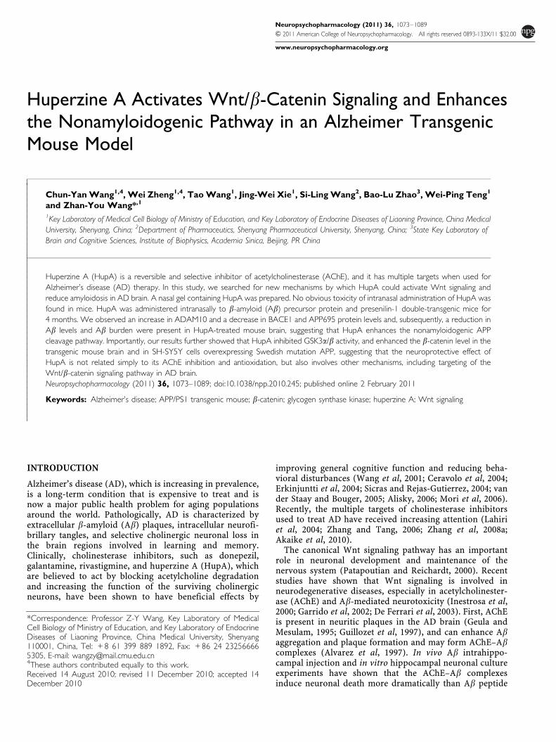
We first evaluated the potential toxicity of intranasaladministration of nasal gel HupA on mucocilia by examin-ing the morphology of mucosal cilia by scanning electronmicroscopy. No obvious changes in the structure ofmucocilia were found between the nasal gel (vehicle)- andnasal gel HupA-treated mouse nasal mucosa (Figure 1a,A1–A3), suggesting that the nasal mucocilia were notdamaged after nasal administration of HupA at doses of167 and 500 mg/kg once a day for a week.
We then assessed the olfactory bulb structure of APP/PS1mice treated with nasal gel HupA by Nissl staining andolfactory marker protein (OMP) immunofluorescence. Nisslstaining showed that the olfactory bulb exhibited similarhistology in the vehicle- and nasal gel HupA-treated (167and 500 mg/kg) mice (Figure 1a, B1–B3). Statistical analysisshowed no significant difference in the area of OGL betweenvehicle and nasal gel HupA treatment groups (Figure 1b).OMP immunostaining showed that no obvious changes inthe distribution and intensity of the immunofluorescence inthe olfactory bulb were found between the vehicle- andnasal gel HupA-treated mice (Figure 1a, C1–C3). Further-more, immunoblot showed that the expression levels ofOMP and 43 kD growth-associated protein (GAP-43) didnot exhibit marked difference between control and nasal gelHupA groups (OMP: F(2, 15) ¼ 1.082, p40.05, Figure 1cand d; GAP-43: (F(2, 15)¼ 1.128, p40.05; Figure 1c and e).
We also examined the effects of intranasal administrationof HupA or vehicle on olfactory bulb neurogenesis by BrdUstaining. We failed to find BrdU-labeled cells in theolfactory bulb of control and nasal gel HupA mice treatedwith BrdU at 6 h intervals thrice per day for 3 continuousdays (data not shown). However, BrdU-positive cells werefound in the SVZ of control and nasal gel HupA-treatedmice (Figure 1a, D1–D3). There were no significantdifferences in the number of BrdU-positive cells(F(2, 15)¼ 1.741, p40.05; Figure 1f) and the area of BrdUin the SVZ (F(2, 15)¼ 1.443, p40.05; Figure 1g).
Moreover, we evaluated whether long-term intranasaladministration might compromise the blood–brain barrier(BBB) in APP/PS1 mouse brain by EB absorbance assay. Asshown in Figure 1h, there was no significant difference inthe leakage of EB in the olfactory bulb (F(2, 15)¼ 0.171,p40.05), cortex (F(2, 15)¼ 0.107, p40.05), and hippocam-pus (F(2, 15)¼ 0.701, p40.05; Figure 1h).
Taken together, these data showed that nasal gel HupAadministration did not induce significant alterationof olfactory bulb structure, neurogenesis in the SVZ,and BBB permeability, suggesting that no obvious toxicityof intranasal administration of HupA was found in themouse brain.
Inhibition of AChE Activity and Antioxidative Effects ofHupA in APP/PS1 Mouse Brain
To assess whether intranasal administration of nasal gelHupA retains its inhibitory effects on AChE activity, braintissues of APP/PS1 mice treated with nasal gel HupA for 4months were subjected to colorimetric tests to determinethe AChE and ChAT activity in the brain. HupA treatmentdose dependently reduced AChE levels by 90.61±3.41%(po0.05) and 87.61±4.21% (po0.01) at a dose of 167 and500 mg/kg, respectively (F(2, 15)¼ 6.777, po0.05; Figure 2a),and increased ChAT levels by 111.90±12.29% (167 mg/kg;p40.05) and 123.13±11.50% (500 mg/kg; po0.05), respec-tively (F(2, 15)¼ 9.628, po0.05; Figure 2b). These datasuggest that intranasal administration of HupA dosedependently increases ChAT activity, which is a well-knowneffect of HupA using the traditional administration modes,such as intravenous and oral administration.
We then analyzed the effects of HupA on modulating theactivities of antioxidant enzymes and oxidative productionof MDA in the APP/PS1 transgenic mouse brain. Nasal gelHupA treatment increased the levels of GSH-PX to140.49±6.04% (167 mg/kg; po0.01) and 159.91±11.12%(500 mg/kg; po0.01), respectively (F(2, 15)¼ 23.391,po0.05; Figure 2c), and increased the activity of CAT to114.28±5.29% (167 mg/kg; po0.05) and 124.34±7.09%(500 mg/kg; po0.01), respectively (F(2, 15)¼ 16.971,po0.05, Figure 2d). Meanwhile, HupA reduced oxidativeproduction of MDA to 88.88±1.95% (167 mg/kg; po0.05)and 87.42±5.49% (500 mg/kg; po0.05), respectively, com-pared with control (F(2, 15)¼ 11.887, po0.05; Figure 2e).Collectively, nasal gel HupA enhanced the activities ofantioxidative enzymes and subsequently reduced theoxidative production in APP/PS1 mouse brain.
HupA Reduces Ab Burden and Inhibits Ab Generationin APP/PS1 Mouse Brain
We investigated whether nasal gel HupA could reduce Abdeposition in APP/PS1 mouse brain. Brain sections of APP/PS1 mice treated with nasal gel HupA for 4 months weresubjected to immunohistochemical analysis. Both thenumber and size of the Ab-immonoreactive neuritic plaqueswere dose dependently reduced in the cortex and hippo-campus after intranasal treatment of HupA (Figure 3a).Statistical analysis showed that HupA treatment significantlyreduced the number of Ab plaques by 77.52±12.94 and37.07±8.06% at a dose of 167 and 500mg/kg, respectively, inthe cortex (F(2, 15)¼ 70.263, po0.05), and 45.13±10.46%(167mg/kg; po0.01) and 26.73±7.15% (500mg/kg; po0.01),respectively, in the hippocampus compared with the vehiclecontrol (F(2, 15)¼ 27.656, po0.05; Figure 3b). The areas ofneuritic plaques of the HupA treatment groups were reducedby 72.07±10.56% (167mg/kg; po0.01) and 27.34±4.94%(500mg/kg; po0.01) in the cortex (F(2,15)¼ 193.938,po0.05), and 66.04±12.66% (167mg/kg; po0.01) and41.66±6.23% (500mg/kg; po0.01) in the hippocampus(F(2, 15)¼ 44.254, po0.05; Figure 3c), respectively.
Next, we measured the soluble Ab level in the cortex of thetransgenic mouse brain by sandwich ELISA. After treatmentwith nasal gel HupA for 4 months, the levels of Ab1-40 werereduced by 88.84±7.35% (167mg/kg; po0.05) and
Huperzine A activates Wnt signalingChun-Yan Wang et al
1077
Neuropsychopharmacology
80.87±8.93% (500mg/kg; po0.05), respectively (F(2, 15)¼12.329, po0.05; Figure 3d), and the levels of Ab1-42 werereduced by 76.89±10.78% (167mg/kg; po0.01) and 52.21±10.14% (500mg/kg; po0.01), respectively (F(2, 15)¼ 19.267,po0.05; Figure 3e) compared with the control group.
HupA Enhances Nonamyloidogenic Processing of APPin APP/PS1 Mouse Brain
To verify the effects of HupA on APP processing, we firstmeasured the levels of APP mRNA and APP695 protein in
APP/PS1 mouse brain following treatment with nasal gelHupA for 4 months. RT-PCR results showed that there wereno significant differences in APP mRNA levels betweenvehicle control and HupA treatment groups in APP/PS1mouse brain (F(2, 15)¼ 2.158, p40.05; Figure 4a and b).However, immunoblot analysis revealed that HupA treat-ment dose dependently reduced the levels of APP695protein by 71.22±15.16% (167 mg/kg; po0.05) and59.28±5.60% (500 mg/kg; po0.01), respectively, comparedwith the control group (F(2, 15)¼ 14.349, po0.05; Figure 4cand d).
Figure 1 Evaluation of the potential toxicity of intranasal administration of nasal gel HupA on olfactory bulb (OB) and neurogenesis in the subventricularzone (SVZ) of mice. (a; A1–A3) Scanning electron microscope (SEM) photographs showing no marked changes in the structure of nasal mucocilia betweenC57BL/6 mice given nasal gel (A1), and nasal gel containing HupA at a dose of 167 mg/kg (A2) and 500 mg/kg (A3), respectively, for 7 days. Scale bar¼ 1 mm.(B1–D3) APP/PS1 mice were treated with vehicle and nasal gel HupA, respectively, for 1 month. Nissl staining of OB exhibited similar histology betweenvehicle group (B1) and nasal gel HupA at doses of 167 mg/kg (B2) and 500 mg/kg groups (B3). Scale bar¼ 200 mm. (C1–C3) Immunostaining images showingthe distribution and expression of olfactory marker protein (OMP) in OB of APP/PS1 mice. No marked changes were found between vehicle- and HupA-treated groups. Scale bar¼ 100 mm. (D1–D3) BrdU immunofluorescent staining showed a similar neurogenesis in SVZ between vehicle and nasal gel HupAgroups. Scale bar¼ 50mm. (b) Measurement of the area of olfactory granule cell layer (OGL) in serial sections of Nissl staining. No significant differenceswere found between vehicle- and HupA-treated transgenic mice. (c) Western blot showed the protein levels of OMP and GAP-43 in the OB of vehicle- andHupA-treated APP/PS1 mice. GAPDH was used as an internal control. (d, e) Quantification of the protein levels of OMP (d) and GAP-43 (e) in the OB.Intranasal administration of HupA did not alter the expression levels of OMP and GAP-43, compared with controls. (f, g) Quantification of the number (f)and the area (g) of BrdU-labeled cells in SVZ showed no significant difference between control and HupA groups. (h) Leakage of Evan’s blue (EB) showedthat nasal gel HupA treatment did not significantly change the EB absorbance in the OB, cortex, and hippocampus in APP/PS1 mice. All values aremean±SEM (n¼ 6).
Huperzine A activates Wnt signalingChun-Yan Wang et al
1078
Neuropsychopharmacology

Figure 2 Effects of nasal gel HupA on AChE and antioxidant enzyme activity in the cortex of APP/PS1 mice. APP/PS1 transgenic mice at the age of6 months were treated with nasal gel HupA at a dose of 167 and 500 mg/kg, respectively, for 4 months. AChE and ChAT activity of brain cortex tissuehomogenate were measured by AChE and ChAT assay kits. (a) Administration of HupA significantly reduced AChE activity in the mouse brain. (b) HupAdose dependently increased ChAT activity in the mouse brain. (c) HupA significantly increased the activity of the detected antioxidant enzymes, CAT andGSH-PX (d), in APP/PS1 mouse brain. (e) Nasal gel HupA significantly reduced the production of MDA in the mouse brain. All values are mean±SEM(n¼ 6). *po0.05, **po0.01, #po0.05.
Figure 3 Nasal gel HupA treatment significantly reduces Ab plaque formation and soluble Ab production in APP/PS1 mouse brain. (a) Abimmunoreactive neuritic plaques in the cortex and hippocampus of transgenic mice treated with nasal gel and nasal gel HupA at a dose of 167 and 500 mg/kg,respectively. The number of Ab-positive plaques was significantly reduced in HupA-treated mice compared with controls. Scale bar¼ 60 mm. (b, c)Quantification of the number of Ab-positive plaques (b) and Ab burden (c) in the cortex and hippocampus of APP/PS1 mice treated with HupA. HupAsignificantly reduced the plaque number and Ab burden in the brain in a dose-dependent manner. (d, e) ELISA results showed that administration of nasal gelHupA induced a dose-dependent decrease in soluble Ab production in the cortex of APP/PS1 mice. All values are mean±SEM (n¼ 6). *po0.05,**po0.01, ##po0.01.
Huperzine A activates Wnt signalingChun-Yan Wang et al
1079
Neuropsychopharmacology

We then investigated whether nasal gel HupA wasinvolved in APP cleavage. The APP cleavage enzymes andcleavage fragments in HupA-treated transgenic mouse brainwere detected by western blot analysis (Figure 4e and i).HupA significantly increased the expression level of adisintegrin and metallopetidase 10 (ADAM10) by 210.05±57.43% (167 mg/kg; po0.01) and 245.02±11.41% (500 mg/kg; po0.01), respectively (F(2, 15)¼ 14.833, po0.05;Figure 4f). On the other hand, HupA dose dependentlyreduced the level of the b-site of APP-cleaving enzyme
(BACE1) by 76.93±4.77% (167 mg/kg; p40.05) and 56.65±9.20% (500 mg/kg; po0.01) (F(2, 15)¼ 7.406, po0.05;Figure 4g). Furthermore, HupA reduced the level of PS1by 92.81±13.46% (167 mg/kg; p40.05) and 62.09±12.00%(500 mg/kg; po0.05) compared with vehicle controls(F(2, 15)¼ 8.210, po0.05; Figure 4h).
In HupA-treated transgenic mouse brain, the proteinlevel of sAPPa was increased by 171.91±13.47% (167 mg/kg;po0.01) and 222.88±36.00% (500mg/kg; po0.01) (F(2, 15)¼26.780, po0.05; Figure 4j), in parallel with a decreased
Figure 4 Nasal gel HupA enhances the nonamyloidogenic pathway in APP/PS1 mouse brain. (a) Expression levels of APP mRNA were detected byRT-PCR in the brain of APP/PS1 transgenic mice treated with nasal gel and nasal gel HupA at a dose of 167 and 500 mg/kg, respectively. GAPDH was used asan internal reference gene. (b) HupA-treated and vehicle control APP/PS1 mice exhibited a similar APP mRNA level. (c) Western blots showed theexpression levels of APP695 protein in the vehicle- and HupA-treated transgenic mouse brain. GAPDH was used as an internal control. (d) The proteinlevels of APP695 were significantly reduced in the nasal gel HupA-treated mouse brain compared with the vehicle controls. (e) The expression levels of APPcleavage enzymes, including ADAM10, BACE1, and PS1, were examined by western blot analysis. (f–h) Analysis results showed that the protein levels ofADAM10 were significantly increased (f), whereas the levels of BACE1 were markedly reduced in the brain of nasal gel HupA-treated mice (g). HupAtreatment reduced the expression levels of PS1 protein in a dose-dependent manner, compared with vehicle controls (h). (i) Immunoblotting showed theexpression levels of APP cleavage fragments, including sAPPa, sAPPb, C83, and C99, in the vehicle- and HupA-treated transgenic mouse brain. (j–m)Quantification of sAPPa, sAPPb, C83, and C99 generation in the brain of the APP/PS1 mouse. Nasal gel HupA treatment significantly increased the levels ofsAPPa (j) and C83 (l) and reduced the levels of sAPPb (k) and C99 (m) in a dose-dependent manner. All values are mean±SEM (n¼ 6). *po0.05,**po0.01, #po0.05.
Huperzine A activates Wnt signalingChun-Yan Wang et al
1080
Neuropsychopharmacology
release of sAPPb by 79.78±6.33% (167 mg/kg; po0.05)and 62.19±8.38% (500 mg/kg; po0.01) (F(2, 15)¼ 24.423,po0.05; Figure 4k). HupA treatment led to an increasedlevel of C83 by 104.73±9.36% (167 mg/kg; p40.05)and 142.65±17.22% (500 mg/kg; po0.01) (F(2, 15)¼ 9.279,po0.05; Figure 4l), and a decreased level of C99 by 79.92±9.18% (167 mg/kg; po0.05) and 57.32±10.15% (500 mg/kg;po0.01) (F(2, 15)¼ 18.850, po0.05; Figure 4m), comparedwith vehicle controls. Taken together, these results suggestthat the a-secretase cleavage activity is markedly increasedfollowing administration of nasal gel HupA and, thus, thispromotes nonamyloidogenic processing of APP and attenu-ates cerebral amyloidosis in APP/PS1 mouse brain.
HupA Inhibits GSK3 and Stabilizes the Level ofb-Catenin in APP/PS1 Mouse Brain
As recent in vitro studies have shown that AChE–Abtoxicity may destroy the Wnt pathway and cause cholinergicneuronal loss (Inestrosa et al, 2000, 2004, 2005), weinvestigated whether HupA could target the Wnt signalingcascades by examining the expression levels of both GSK3band b-catenin, the two key components in the Wnt signalingpathway.
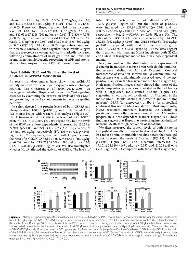
We first detected the protein levels of both GSK3b andphosphorylated GSK3b (p-GSK3b) in HupA-treated APP/PS1 mouse brains with western blot analyses (Figure 5a).HupA treatment did not affect the levels of total GSK3bprotein (F(2, 15)¼ 3.806, p40.05; Figure 5b), but the levelsof p-GSK3b were dose dependently increased by 150.56±27.94% (p40.05) and 279.93±36.42% (po0.05) at a dose of167 and 500 mg/kg, respectively (F(2, 15)¼ 44.722, po0.05;Figure 5c). Consequently, treatment with HupA increasedthe ratio of p-GSK3b/GSK3b by 156.52±66.83% (167 mg/kg;p40.05) and by 213.07±39.50% (500 mg/kg; po0.05)(F(2, 15)¼ 8.508, po0.05; Figure 5d). We also investigatedwhether HupA affected the activity of GSK3a. The levels of
total GSK3a protein were not altered (F(2, 15)¼1.938, p40.05; Figure 5e), but the levels of p-GSK3awere increased by 159.99±28.87% (po0.01) and by260.25±23.88% (po0.01) at a dose of 167 and 500 mg/kg,respectively (F(2, 15)¼ 52.075, po0.05; Figure 5f). Theratio of p-GSK3a/GSK3a was also increased by 144.29±33.22% (167mg/kg; po0.05) and 251.52±61.66% (500mg/kg;po0.01) compared with that in the control group(F(2, 15)¼ 21.678, po0.05; Figure 5g). These data suggestthat treatment with nasal gel HupA inactivates both GSK3band GSK3a in transgenic mouse brain in a dose-dependentmanner.
Next, we analyzed the distribution and expression ofb-catenin in transgenic mouse brain with double immuno-fluorescence labeling of Ab and b-catenin. Confocalmicroscopic observation showed that b-catenin immuno-fluorescence was predominantly observed around the Ab-positive plaques in the transgenic mouse brain (Figure 6a).High-magnification images clearly showed that most of theb-catenin-positive products were located in the cell bodieswith a large-sized DAPI-stained nucleus (Figure 6a),suggesting a neuronal cell localization of b-catenin in themouse brain. Double labeling of b-catenin and NeuN (forneurons), GFAP (for astrocytes), or Iba-1 (for microglias)confirmed this notion (data not shown). Most importantly,HupA treatment markedly increased the density ofb-catenin immunofluorescence around the Ab-positiveplaques in a dose-dependent manner (Figure 6a). Thesefindings suggest that HupA may protect against Ab-inducedneuronal death through activation of b-catenin.
We then measured the protein levels of both b-cateninand p-b-catenin after intranasal treatment of HupA in APP/PS1 mouse brain. Immunoblot results showed that nasal gelHupA increased the levels of b-catenin (F(2, 15)¼ 11.509,po0.05; Figure 6b), and b-catenin increased by172.01±32.14% (167 mg/kg; po0.05) and 210.21±41.04%(500 m/kg; po0.01) compared with the control (Figure 6c).
Figure 5 Nasal gel HupA upregulates the phosphorylation levels of GSK3a/b in APP/PS1 mouse brain. (a) Western blots showing the expression levels oftotal GSK3a/b and p-GSK3a/b in APP/PS1 transgenic mouse brain after HupA treatments. GAPDH was used as an internal control. (b–d) Quantification ofthe levels of GSK3b and p-GSK3b in the brain of the APP/PS1 mouse. There were no significant differences in total GSK3b levels between vehicle- andHupA-treated mouse brain (b). However, the levels of p-GSK3b were significantly increased after 500 mg HupA treatment (c). Moreover, the ratio ofp-GSK3b/GSK3b was significantly increased in 500 mg nasal gel HupA-treated mice (d). (e–g) Quantification of the levels of GSK3a and p-GSK3a in the brainof the APP/PS1 mouse. Administration of HupA did not affect the total protein levels of GSK3a (e). The levels of p-GSK3a were markedly increased afterHupA treatment (f). Nasal gel HupA induced a dose-dependent increase in the ratio of p-GSK3a/GSK3a in the transgenic mouse brain (g). All values aremean±SEM (n¼ 6). *po0.05, **po0.01, ##po0.01.
Huperzine A activates Wnt signalingChun-Yan Wang et al
1081
Neuropsychopharmacology

Furthermore, HupA treatment significantly decreased thelevels of p-b-catenin by 72.24±5.06% (167 mg/kg; po0.05)and 53.83±14.63% (500 mg/kg; po0.01), respectively(F(2, 15)¼ 19.654, po0.05; Figure 6d). Collectively, thesedata indicate that nasal gel HupA stabilizes the level ofb-catenin in APP/PS1 mouse brain.
HupA Enhances Nonamyloidogenic Processing of APPIn Vitro
To examine the effect of HupA on APP processing in vitro,APPsw-transfected SH-SY5Y cells were treated with HupA.The MTT assay showed that 0–10 mM HupA did not exhibitany significant toxic effects on the cells (Figure 7a), anddose dependently increased the levels of sAPPa aftertreatment for 18 h (F(3, 12)¼ 6.705, po0.05) and 24 h(F(3, 12)¼ 10.766, po0.05; Figure 7b). Accordingly, treat-ment with 10 mM HupA for 24 h was chosen for thesubsequent in vitro experiments.
We then examined the levels of APP cleavage enzymesand APP fragments in HupA-treated APPsw cells using
western blot analyses (Figure 7d and h). APPsw cells treatedwith 10 mM HupA showed an increased level of ADAM10, by118.39±3.64% (po0.05; Figure 7e), and a reduced level ofBACE1, by 70.13±7.84% (po0.01), in APPsw cells(Figure 7f). No statistically significant changes in theprotein levels of PS1 were detected between HupA-treatedand control cells (F(5, 24)¼ 2.017, p40.05; Figure 7g).Moreover, HupA treatment markedly increased the level ofsAPPa by 166.56±9.80% (po0.01; Figure 7i) and reducedthe level of sAPPb by 57.18±7.45% (po0.01; Figure 7j).HupA treatment significantly increased the level of C83 by110.81±7.8% (po0.05; Figure 7k), but reduced the releaseof C99 by 78.12±5.54% (po0.01; Figure 7l).
HupA Reverses PKC- and Wnt-Inhibitor-InducedInhibition of Nonamyloidogenic Processing of APPIn Vitro
Previous studies have suggested that GSK3 regulates Abformation (Pei et al, 1999; Phiel et al, 2003; Muyllaert et al,2006), and protein kinase C (PKC) is involved in the
Figure 6 Nasal gel HupA enhances the level of b-catenin in APP/PS1 mouse brain. (a) Confocal microscopic images showing the distribution of b-cateninin the HupA-treated transgenic mouse brain with double immunofluorescent staining of Ab (A1, B1, and C1) and b-catenin (A2, B2, and C2).Counterstaining of DAPI was used to show the nucleus (A3, B3, and C3). At the merged images from the three channels, it was evident that the b-cateninimmunoproducts were located around the Ab-positive neuritic plaques (A4, B4, and C4), and that the b-catenin immunofluorescence was mainly located inthe neuronal cell bodies (A5, A6, B5, B6, C5, and C6). Figure B5 shows a typical neuron with a large nucleus and containing intense b-cateninimmunofluorescence in the cell body. In the vehicle-treated control mouse brain, the density of b-catenin immunofluorescence was very weak (A2), whereasit was very intense in the HupA-treated transgenic mouse brain (B2 and C2). Scale bar¼ 50 mm (C4) and 10 mm (C6). (b) Protein levels of b-catenin andp-b-catenin (Ser33/37/Thr41) were examined by western blot in the brain of transgenic mice. GAPDH was used as an internal control. (c, d) Analysis resultsshowed that the protein levels of total b-catenin were markedly increased (c), whereas the levels of p-b-catenin were significantly decreased in HupA-treated mice compared with controls (d). All values are mean±SEM (n¼ 6). *po0.05, **po0.01.
Huperzine A activates Wnt signalingChun-Yan Wang et al
1082
Neuropsychopharmacology

inactivation of GSK-3b in the Wnt signaling pathway (Cooket al, 1996; Sheldahl et al, 1999; Chen et al, 2000). To furtherinvestigate the underlying mechanism of the effect of HupA
on the nonamyloidogenic processing of APP, we examinedthe expression levels of APP cleavage enzymes andfragments in APPsw cells preincubated with the PKC
Figure 7 HupA enhances nonamyloidogenic processing of APP in SH-SY5Y cells transfected with APPsw. (a) MTT results indicated that 0–10 mM HupAdid not have any significant toxic effects on the cells at different periods of treatment. (b) Immunoblotting detection of the protein levels of sAPPa in 0, 0.1,1.0, or 10mM HupA-treated APPsw cells. GAPDH was used as an internal control. Analysis results showed that HupA increased sAPPa release in a dose-and time-dependent manner. (c) Representative MTT results showing that there were no significant changes in cell viability in APPsw cells exposed to 10 mMHupA, 1 mM calphostin C (PKC inhibitor), and 50 ng/ml DKK-1 (Wnt signaling inhibitor). Thus, the above doses of HupA and inhibitors were used in thesubsequent experiments in culture cells. (d) Western blot analyses were carried out to examine the expression levels of APP cleavage enzymes, ADAM10,BACE1, and PS1, in APPsw cells. (e, f) HupA significantly increased the ADAM10 level and decreased the level of BACE1. There was a significant decrease inthe ADAM10 level and an increase in the BACE1 level in cultures treated with calphostin C or DKK1, whereas the levels of ADAM10 and BACE1 werereversed when cultures were preincubated with calphostin C or DKK1 followed by HupA. (g) There were no significant changes in the PS1 level in drug-treated cells compared with controls. (h) Western blots showing the expression levels of APP cleavage fragments in APPsw cells. (i–l) HupA significantlyincreased the levels of sAPPa and C83 and decreased the levels of sAPPb and C99. Calphostin C and DKK1 significantly reduced the levels of sAPPa andC83 and increased the levels of sAPPb and C99, whereas HupA reversed the inhibitor-induced reduction of sAPPa and C83 and increased that of sAPPband C99. All results are presented as the mean±SEM of at least three independent experiments. *po0.05, **po0.01 vs control group;#po0.05, ##po0.01 vs calphostin C-treated group; $po0.05, $$po0.01 vs DKK1-treated group.
Huperzine A activates Wnt signalingChun-Yan Wang et al
1083
Neuropsychopharmacology

inhibitor calphostin C (1 mM) and the Wnt signalinginhibitor DKK-1 (50 ng/ml) followed by HupA treatment.The doses of inhibitors were selected based on the MTTassay (Figure 7c).
Immunoblotting showed that calphostin C or DKK1treatment significantly reduced the levels of ADAM10 to61.73±5.56% (po0.01) and 66.04±9.23% (po0.01)(F(5, 24)¼ 20.938, po0.05) and increased the levels ofBACE1 to 122.11±3.72% (po0.05) and 119.70±8.84%(po0.05), respectively (F(5, 24)¼ 16.227, po0.05), com-pared with controls (Figure 7d–f). However, HupA treat-ment reversed the changes in ADAM10 to 148.75±7.22%(calphostin C + HupA) compared with the calphostin C-treated group (po0.01), and 132.90±8.71% (DKK1 +HupA) compared with DKK1 treatment (po0.01). HupAtreatment reversed the changes in BACE1 to 77.10±5.99%(calphostin C + HupA) compared with the calphostinC-treated group (po0.05), and 75.65±7.83% (DKK1 +HupA) compared with DKK1 treatment (po0.05), respec-tively (Figure 7d–f).
The levels of sAPPa and C83 in cultures treated withinhibitors were significantly reduced by 36.96±11.04%(calphostin C; po0.01) and 73.03±11.28% (DKK1; po0.01)(F(5, 24)¼ 34.333, po0.05; Figure 7i), and by 78.69±6.72%(calphostin C; po0.01) and 75.38±3.89% (DKK1; po0.01)(F(5, 24)¼ 15.992, po0.05; Figure 7k), respectively, com-pared with the control group. However, HupA treatmentreversed the levels of sAPPa to 342.90±42.39% (calphostinC + HupA) compared with the calphostin C-treated group(po0.01), and 182.63±22.62% (DKK1 + HupA) comparedwith DKK1 treatment (po0.01). Meanwhile, HupA treat-ment reversed the level of C83 to 115.19±4.95% (calphostinC + HupA) compared with calphostin C treatment(po0.05), and 122.43±5.08% (DKK1 + HupA) comparedwith the DKK1-treated group (po0.01; Figure 7i and k).
However, the levels of sAPPb and C99 in cultures treatedwith inhibitors significant increased by 129.03±14.96%(calphostin C; po0.01) and 125.07±11.49% (DKK1;po0.01) (F(5, 24)¼ 14.905, po0.05; Figure 7j), and by117.65±6.46% (calphostin C; po0.05) and 116.88±8.68%(DKK1; po0.05) (F(5, 24)¼ 11.765, po0.05; Figure 7l)compared with controls. In cultures preincubated withinhibitors and then treated with HupA, the levels of sAPPbwere reversed to 56.62±10.40% (calphostin C + HupA)
compared with the calphostin C-treated group (po0.01),and 57.14±13.55% (DKK1 + HupA) compared with DKK1treatment (po0.01). Meanwhile, HupA treatment reversedthe level of C99 to 84.44±4.15% (calphostin C + HupA)compared with the calphostin C-treated group (po0.05),and 84.27±5.92% (DKK1 + HupA) compared with theDKK1-treated group (po0.05). These results show thatHupA reverses PKC- and Wnt-inhibitor-induced inhibitionof nonamyloidogenic processing of APP in vitro.
HupA Inactivates GSK3 and Stabilizes the Level ofb-Catenin In Vitro
We extended our experiments to determine whether HupAcould modulate the Wnt signaling cascades in vitro. We firstexamined the expression levels of GSK3 in APPsw cellspretreated with calphostin C or DKK-1 followed bytreatment with HupA. As shown in Figure 7, the proteinlevels of total GSK3a/b were not affected by drugtreatments, but the levels of p-GSK3a/b were markedlyaltered (Figure 8a). The analysis showed that the ratio ofp-GSK3b/GSK3b in cultures treated with inhibitors sig-nificantly decreased by 64.28±7.88% (calphostin C) and62.96±3.88% (DKK1) compared with controls (po0.01;Figure 8b), whereas HupA treatment reversed the ratio ofp-GSK3b/GSK3b to 125.53±14.70% (calphostin C + HupA)compared with the calphostin C treatment group (po0.01),and 129.98±10.39% (DKK1 + HupA) compared with DKK1treatment (po0.01), respectively (F(5, 24)¼ 36.089, po0.05;Figure 8b). Furthermore, calphostin C or DKK1 treatmentsignificantly reduced the ratio of p-GSK3a/GSK3a to61.47±4.76 and 62.15±11.05%, respectively, comparedwith controls (po0.01; Figure 8c), whereas HupA treatmentincreased the levels of p-GSK3a/GSK3a to 158.10±21.46%(calphostin C + HupA) compared with calphostin C treat-ment (po0.01), and 170.09±10.29% (DKK1 + HupA)compared with the DKK1-treated group (po0.01)(F(5, 24)¼ 37.423, po0.05; Figure 8c).
We next analyzed the distribution and expression ofb-catenin in APPsw cells with double immunofluorescencelabeling of Ab and b-catenin. Consistent with our in vivodata, confocal microscopy results showed that HupAtreatment markedly increased the b-catenin immunofluor-escence in APPsw cells (Figure 9a, B2). Furthermore, both
Figure 8 HupA inhibits the GSK3a/b activity in SH-SY5Y cells transfected with APPsw. (a) Immunoblots showing the levels of GSK3a/b and p-GSK3a/b inAPPsw cells treated with HupA and inhibitors. GAPDH was used as an internal control. (b, c) HupA treatment significantly increased the ratio of p-GSK3b/GSK3b as well as p-GSK3a/GSK3a. Calphostin C or DKK1 treatment decreased the ratio of p-GSK3b/GSK3b and p-GSK3a/GSK3a, whereas HupA reversedthe ratio of p-GSK3b/GSK3b and p-GSK3a/GSK3a in cultures pretreated with Calphostin C or DKK1 followed by HupA. The data shown in (b, c) arepresented as the mean±SEM of at least three independent experiments. *po0.05, **po0.01 vs control group; #po0.05, ##po0.01 vs calphostinC-treated group; $po0.05, $$po0.01 vs DKK1-treated group.
Huperzine A activates Wnt signalingChun-Yan Wang et al
1084
Neuropsychopharmacology

calphostin C and DKK-1 treatment reduced b-cateninimmunofluorescence (Figure 9a, C2 and D2), whereasaddition of HupA reversed the reduction of b-catenin inAPPsw cells pretreated with inhibitors calphostin C andDKK-1 (Figure 9a, E2 and F2).
Immunoblotting results further confirmed the reversaleffects of HupA on the changes in b-catenin in APPsw cellspretreated with inhibitors (Figure 9b). The protein levels oftotal b-catenin were significantly reduced by 40.92±4.46%(calphostin C; po0.01) and 51.05±8.84% (DKK1; po0.01)(F(5, 24)¼ 18.382, po0.05; Figure 9c), whereas the levels ofp-b-catenin were significantly increased by 159.85±3.70%(calphostin C; po0.01) and 153.86±15.49% (DKK1;po0.01) in cultures treated with inhibitors compared withcontrols (F(5, 24)¼ 19.449, po0.05; Figure 9d). In culturespretreated with inhibitors and then treated with HupA, thelevels of b-catenin were reversed to 179.56±28.42%(calphostin C + HupA) compared with the calphostin C-treated group (po0.01), and 161.98±22.06% (DKK1 +
HupA) compared with the DKK1 treatment group (po0.01;Figure 9c), and the levels of p-b-catenin were reversed to56.02±12.41% (calphostin C + HupA) compared with thecalphostin C-treated group (po0.01), and 66.87±10.39%(DKK1 + HupA) compared with DKK1 treatment (po0.01;Figure 9d). Taken together, these data further indicate thatHupA reverses PKC- and Wnt-inhibitor-induced changes inWnt/b-catenin signaling in vitro.
DISCUSSION
It has been previously reported that the intranasal admin-istration of nasal gel containing HupA is a potentialnoninvasive strategy for the treatment of AD because itcan provide similar plasma concentrations and high CSFconcentrations of HupA compared with intravenous admin-istration (Yue et al, 2007). In this study, involving treatmentwith nasal gel HupA in APP/PS1 transgenic mice, the effects
Figure 9 HupA increases the level of b-catenin in SH-SY5Y cells transfected with APPsw. (a) Double labeling of Ab (A1–F1) and b-catenin (A2–F2) andconfocal microscopic images showing the distribution and expression of b-catenin in APPsw cells. HupA treatment significantly increased the b-cateninimmunofluorescence in APPsw cells (B2), compared with the control (A2). Calphostin C or DKK-1 treatment decreased the levels of b-catenin (C2 andD2). Additional HupA treatment reversed the reduction of b-catenin in cultures pretreated with inhibitors Calphostin C or DKK-1 (E2 and F2). Scalebar¼ 30mm. (b) Western blot analysis showing the levels of b-catenin and p-b-catenin in APPsw cells treated with HupA or inhibitors. GAPDH was used asan internal control. (c, d) The protein level of total b-catenin was significantly increased, whereas the level of p-b-catenin was significantly decreased in HupA-treated cells compared with controls. There was a significantly reduced level of total b-catenin and an increased level of p-b-catenin in cultures treated withCalphostin C or DKK1, whereas the total b-catenin level was increased and the p-b-catenin level was reduced when cultures were preincubated withinhibitors followed by HupA. Representative immunoblots from three experiments are shown. The data shown in (c, d) are presented as mean±SEM of atleast three independent experiments. **po0.05, **po0.01 vs control group; ##po0.01 vs Calphostin C-treated group; $$po0.01 vs DKK1-treated group.
Huperzine A activates Wnt signalingChun-Yan Wang et al
1085
Neuropsychopharmacology

of HupA on the specific markers were confirmed, includinginhibition of AChE, antioxidant activity, and enhancementof sAPPa release. Moreover, we showed for the first timethat HupA upregulates b-catenin expression in vivo andin vitro, suggesting that the neuroprotective effect of HupAmight be related to the regulation of the Wnt signalingpathway in the AD brain.
HupA is Involved in Regulation of the Wnt SignalingPathway
It has been generally accepted that loss of Wnt signalingfunction is involved in Ab-dependent neurodegeneration inthe AD brain (Inestrosa and Toledo, 2008b). Studies havedemonstrated that GSK3b and b-catenin, the two keycomponents of the canonical Wnt signaling pathway, arealtered dramatically in the AD model mouse brain (Zhanget al, 1998; Pei et al, 1999), and activation of Wnt signalingcan prevent neurodegeneration induced by Ab fibrils (DeFerrari et al, 2003). Interestingly, an in vivo study hasshown that AChE–Ab neurotoxicity is related to the changesin the expression levels of both GSK3b and b-catenin(Inestrosa et al, 2004). Importantly, a bifunctional (AChEinhibitor and anti-inflammatory) compound, IBU-PO,inhibits GSK3b and enhances b-catenin activity, preventingthe loss of function of the Wnt signaling pathway caused byAb toxicity (Farias et al, 2005). These findings suggest thatthe neuroprotective effect of the AChE inhibitor is relatedto modulation of the Wnt/b-catenin signaling pathway(Inestrosa et al, 2008a; Toledo et al, 2008). In this study weshowed that HupA, an AChE inhibitor, is involved in theregulation of Wnt signaling in APP/PS1 transgenic mouseand APPsw cell models. We present data showing thatHupA significantly inhibits GSK3b activity and stabilizesthe b-catenin protein level in vivo and in vitro, suggestingthat activation of the Wnt signal transduction pathway isinvolved in the neuroprotective effects of HupA against Abneurotoxicity.
On the other hand, activation of both GSK3a and GSK3bthrough their autophosphorylation has been implicated inAD pathogenesis (Caricasole et al, 2004). Although theremay be some crosslinking between GSK3a and GSK3bproteins, increased GSK3a activity is mainly involved inAPP processing and Ab generation (Phiel et al, 2003),whereas activation of GSK3b is predominantly associatedwith tau phosphorylation and neurofibrillary tangle forma-tion (Baum et al, 1996; Ma et al, 2006). Several studies haveshown that HupA has the ability to regulate APP processingin vitro (Zhang et al, 2004; Peng et al, 2007). We presentdata showing that there is a significant increase in thephosphorylation levels of both GSK3a and GSK3b proteinsin HupA-treated APP/PS1 mouse brain and APPsw-over-expressing cells, suggesting that HupA can inhibit theactivity of GSK3a/b and, hence, may inhibit Ab generationand tau phosphorylation.
HupA Regulates the Processing of APP to theNonamyloidogenic Pathway
Several lines of evidence indicate that through thea-secretase pathway, APP is cleaved within the sequenceof the Ab peptide and generates the sAPPa fragment (Esch
et al, 1990), which is beneficial for neuronal survival(Mattson et al, 1997; Wallace et al, 1997), whereas throughthe b-secretase pathway, APP is cleaved to form neurotoxicAb and is involved in the pathogenesis of AD (Haass et al,1992; Shoji et al, 1992; Bodles and Barger, 2005). Interest-ingly, the widely used AChE inhibitors, including donepezil,rivastigmine, and galantamine, have been shown to increasesAPPa release (Racchi et al, 2004). Recent studies have alsoshown that HupA can guide APP processing to thea-secretase pathway through mediating a-secretase activityand, hence, reduce Ab generation in cultured cells (Zhanget al, 2004; Peng et al, 2007). In this study, we examined theeffects of HupA on APP processing in the APP/PS1 mouse.Our data show that in the nasal gel HupA-treated transgenicmouse brain, the expression level of APP695 proteins ismarkedly reduced. HupA treatment significantly enhancesthe expression level of ADAM10, a candidate of a-secretase,and subsequently there is an increase in the levels ofa-secretase-generated sAPPa and C83 fragments in trans-genic mouse brain and in APPsw-transfected cells. Mean-while, HupA reduces the levels of BACE1 and b-secretase-generated sAPPb and C99 fragments in vivo and in vitro.Furthermore, in the HupA-treated APP/PS1 mouse brain,there is a reduction in Ab levels and Ab burden detected byELISA and immunohistochemistry, respectively. Takentogether, our results indicate that HupA modulates theprocessing of APP to the nonamyloidogenic pathway.
HupA Regulates APP Processing to theNonamyloidogenic Pathway Through Activation of PKCand Wnt/b-Catenin Signaling
Crosstalk between PKC and Wnt/b-catenin signaling isinvolved in the modulation of GSK3 activation. Severalstudies have shown that activation of PKC inhibits theactivity of GSK-3b and modulates Wnt/b-catenin signaling(Chen et al, 2000; Garrido et al, 2002). On the other hand,activation of Wnt/b-catenin signaling prevents neuronalapoptosis in a PKC-dependent manner (Chen et al, 2000; DeFerrari et al, 2003; Alvarez et al, 2004). Although themechanism of GSK3 modulation in the AD brain is far fromfully understood, the involvement of both Wnt signalingand PKC in the abnormal activation of GSK3 has causedincreasing concern. A recent study has shown that HupAmay affect the processing of APP by upregulation of PKC(Zhang et al, 2004). In our in vitro studies, we found thatHupA reverses PKC- and Wnt-inhibitor-induced inhibitionof nonamyloidogenic processing of APP, paralleled by aninactivation of GSK3 and a reversal of the level of b-catenin.As GSK3 is involved in APP processing (Phiel et al, 2003)and tau phosphorylation (Baum et al, 1996), it is reasonableto speculate that the HupA regulation of APP processing tothe nonamyloidogenic pathway is, at least partly, throughactivation of PKC and Wnt/b-catenin signaling.
Intranasal Administration of HupA May Be aReasonable Approach to AD Treatment
Drug delivery to the brain via the nasal route is a subject ofincreasing interest because the nasal mucosa offers rapidabsorption with an abundantly vascularized and relativelylarge absorptive surface area (Turker et al, 2004), does not
Huperzine A activates Wnt signalingChun-Yan Wang et al
1086
Neuropsychopharmacology
require a complicated administration method, and caneasily be carried out by medical services for chronic caresuch as in the case of AD. For the AChE inhibitors, it hasbeen reported that nasal administration of galantamine canenhance the bioavailability and reduce the emetic response(Leonard et al, 2007). Nasal gel HupA can provide similarplasma concentrations after 15 min and higher CSFconcentrations after 30 min than those following intrave-nous administration (Yue et al, 2007). In this study,intranasal administration of nasal gel HupA exhibits AChEinhibition and antioxidative effects on APP/PS1 mousebrain, which have been confirmed before by traditionaladministration methods, such as intravenous and oraladministration (Wang et al, 2000; Wang et al, 2001; Wanget al, 2006b). Our data also show that HupA regulates theprocessing of APP to the nonamyloidogenic pathway. Inaddition, we confirmed the safety of nasal gel HupAadministration by examining the structure of the mucosalcilia. We also assessed if long-term intranasal administra-tion of HupA might influence the structure of the olfactorybulb. Our data showed that intranasal administration ofHupA did not significantly alter the structure and theexpression levels of OMP and GAP-43 in the olfactory bulb.Furthermore, nasal gel HupA treatment did not alter theneurogenesis in the SVZ of APP/PS1 mouse brain. Althoughwe did not find BrdU-labeled cells in the bulb, the similardistribution of BrdU-positive cells in the SVZ in the vehicle-and nasal gel HupA-treated mouse brain indicates that thenewborn cells in the SVZ migrate to the bulb through therostral migratory stream, which might not be affected bythe administration of HupA. Moreover, the result of EBleakage assay in our study demonstrated that nasal gel HupAtreatment did not induce significant alterations of BBB in thetransgenic mouse brain. The present data, together withprevious studies (Yue et al, 2007), suggest that intranasaladministration of nasal gel HupA may represent a safe andnoninvasive therapeutic strategy for the treatment of AD.
ACKNOWLEDGEMENTS
The study was supported by the Natural Science Foundationof China (30770680), the Program for New CenturyExcellent Talents in University (NCET-04-0288), the ChinaPostdoctoral Science Foundation (2005037008), the Specia-lized Research Fund for the Doctoral Program of HigherEducation (SRFDP-20060159001), and the National BasicResearch Program of China (973 Program 2009CB930300).
DISCLOSURE
The authors declare no conflict of interest.
REFERENCES
Akaike A, Takada-Takatori Y, Kume T, Izumi Y (2010). Mechan-isms of neuroprotective effects of nicotine and acetylcholines-terase inhibitors: role of alpha4 and alpha7 receptors inneuroprotection. J Mol Neurosci 40: 211–216.
Alisky JM (2006). Cholinesterase inhibitors might alleviatemethamphetamine-induced delusions, hallucinations and cog-nitive impairment, while reducing craving and addiction. WorldJ Biol Psychiatry 7: 269.
Alvarez A, Alarcon R, Opazo C, Campos EO, Munoz FJ,Calderon FH et al (1998). Stable complexes involving acetylcho-linesterase and amyloid-beta peptide change the biochemicalproperties of the enzyme and increase the neurotoxicity ofAlzheimer’s fibrils. J Neurosci 18: 3213–3223.
Alvarez A, Opazo C, Alarcon R, Garrido J, Inestrosa NC (1997).Acetylcholinesterase promotes the aggregation of amyloid-beta-peptide fragments by forming a complex with the growingfibrils. J Mol Biol 272: 348–361.
Alvarez AR, Godoy JA, Mullendorff K, Olivares GH, Bronfman M,Inestrosa NC (2004). Wnt-3a overcomes beta-amyloid toxicity inrat hippocampal neurons. Exp Cell Res 297: 186–196.
Alvarez G, Munoz-Montano JR, Satrustegui J, Avila J, Bogonez E,Diaz-Nido J (1999). Lithium protects cultured neuronsagainst beta-amyloid-induced neurodegeneration. FEBS Lett453: 260–264.
Baum L, Hansen L, Masliah E, Saitoh T (1996). Glycogen synthasekinase 3 alteration in Alzheimer disease is related to neurofi-brillary tangle formation. Mol Chem Neuropathol 29: 253–261.
Bodles AM, Barger SW (2005). Secreted beta-amyloid precursorprotein activates microglia via JNK and p38-MAPK. NeurobiolAging 26: 9–16.
Caricasole A, Copani A, Caraci F, Aronica E, Rozemuller AJ,Caruso A et al (2004). Induction of Dickkopf-1, a negativemodulator of the Wnt pathway, is associated with neuronaldegeneration in Alzheimer’s brain. J Neurosci 24: 6021–6027.
Ceravolo R, Volterrani D, Tognoni G, Dell’Agnello G, Manca G,Kiferle L et al (2004). Cerebral perfusional effects of cholinester-ase inhibitors in Alzheimer disease. Clin Neuropharmacol 27:166–170.
Chen RH, Ding WV, McCormick F (2000). Wnt signaling to beta-catenin involves two interactive components. Glycogen synthasekinase-3beta inhibition and activation of protein kinase C. J BiolChem 275: 17894–17899.
Cheng DH, Ren H, Tang XC (1996). Huperzine A, a novelpromising acetylcholinesterase inhibitor. NeuroReport 8: 97–101.
Cheng DH, Tang XC (1998). Comparative studies of huperzine A,E2020, and tacrine on behavior and cholinesterase activities.Pharmacol Biochem Behav 60: 377–386.
Cook D, Fry MJ, Hughes K, Sumathipala R, Woodgett JR, Dale TC(1996). Wingless inactivates glycogen synthase kinase-3 via anintracellular signalling pathway which involves a protein kinaseC. EMBO J 15: 4526–4536.
De Ferrari GV, Chacon MA, Barria MI, Garrido JL, Godoy JA,Olivares G et al (2003). Activation of Wnt signaling rescuesneurodegeneration and behavioral impairments induced bybeta-amyloid fibrils. Mol Psychiatry 8: 195–208.
Erkinjuntti T, Roman G, Gauthier S (2004). Treatment of vasculardementiaFevidence from clinical trials with cholinesteraseinhibitors. J Neurol Sci 226: 63–66.
Esch FS, Keim PS, Beattie EC, Blacher RW, Culwell AR, OltersdorfT et al (1990). Cleavage of amyloid beta peptide duringconstitutive processing of its precursor. Science 248: 1122–1124.
Farias GG, Godoy JA, Vazquez MC, Adani R, Meshulam H, Avila Jet al (2005). The anti-inflammatory and cholinesterase inhibitorbifunctional compound IBU-PO protects from beta-amyloidneurotoxicity by acting on Wnt signaling components. NeurobiolDis 18: 176–183.
Garrido JL, Godoy JA, Alvarez A, Bronfman M, Inestrosa NC(2002). Protein kinase C inhibits amyloid beta peptide neuro-toxicity by acting on members of the Wnt pathway. FASEB J 16:1982–1984.
Geula C, Mesulam MM (1995). Cholinesterases and the pathologyof Alzheimer disease. Alzheimer Dis Assoc Disord 9(Suppl 2):23–28.
Guillozet AL, Smiley JF, Mash DC, Mesulam MM (1997).Butyrylcholinesterase in the life cycle of amyloid plaques. AnnNeurol 42: 909–918.
Huperzine A activates Wnt signalingChun-Yan Wang et al
1087
Neuropsychopharmacology

Haass C, Schlossmacher MG, Hung AY, Vigo-Pelfrey C, Mellon A,Ostaszewski BL et al (1992). Amyloid beta-peptide is producedby cultured cells during normal metabolism. Nature 359:322–325.
Holick KA, Lee DC, Hen R, Dulawa SC (2008). Behavioral effects ofchronic fluoxetine in BALB/cJ mice do not require adulthippocampal neurogenesis or the serotonin 1A receptor.Neuropsychopharmacology 33: 406–417.
Inestrosa NC, Alvarez A, Dinamarca MC, Perez-Acle T, ColombresM (2005). Acetylcholinesterase-amyloid-beta-peptide interac-tion: effect of Congo Red and the role of the Wnt pathway.Curr Alzheimer Res 2: 301–306.
Inestrosa NC, Alvarez A, Godoy J, Reyes A, De Ferrari GV (2000).Acetylcholinesterase-amyloid-beta-peptide interaction and Wntsignaling involvement in Abeta neurotoxicity. Acta Neurol ScandSuppl 176: 53–59.
Inestrosa NC, Dinamarca MC, Alvarez A (2008a). Amyloid-cholinesterase interactions. Implications for Alzheimer’s disease.FEBS J 275: 625–632.
Inestrosa NC, Toledo EM (2008b). The role of Wnt signaling inneuronal dysfunction in Alzheimer’s disease. Mol Neurodegener3: 9.
Inestrosa NC, Urra S, Colombres M (2004). Acetylcholinesterase(AChE)–amyloid-beta-peptide complexes in Alzheimer’s disease.The Wnt signaling pathway. Curr Alzheimer Res 1: 249–254.
Kim WR, Kim Y, Eun B, Park OH, Kim H, Kim K et al (2007).Impaired migration in the rostral migratory stream but sparedolfactory function after the elimination of programmed celldeath in Bax knock-out mice. J Neurosci 27: 14392–14403.
Kuhn HG, Dickinson-Anson H, Gage FH (1996). Neurogenesis inthe dentate gyrus of the adult rat: age-related decrease ofneuronal progenitor proliferation. J Neurosci 16: 2027–2033.
Lahiri DK, Rogers JT, Greig NH, Sambamurti K (2004). Rationalefor the development of cholinesterase inhibitors as anti-Alzheimer agents. Curr Pharm Des 10: 3111–3119.
Lahoud-Rahme MS, Stezoski J, Kochanek PM, Melick J, TishermanSA, Drabek T (2009). Blood-brain barrier integrity in a rat modelof emergency preservation and resuscitation. Resuscitation 80:484–488.
Lenzser G, Kis B, Snipes JA, Gaspar T, Sandor P, Komjati K et al(2007). Contribution of poly(ADP-ribose) polymerase to post-ischemic blood-brain barrier damage in rats. J Cereb Blood FlowMetab 27: 1318–1326.
Leonard AK, Sileno AP, Brandt GC, Foerder CA, Quay SC,Costantino HR (2007). In vitro formulation optimization ofintranasal galantamine leading to enhanced bioavailability andreduced emetic response in vivo. Int J Pharm 335: 138–146.
Ma QL, Lim GP, Harris-White ME, Yang F, Ambegaokar SS, UbedaOJ et al (2006). Antibodies against beta-amyloid reduce Abetaoligomers, glycogen synthase kinase-3beta activation and tauphosphorylation in vivo and in vitro. J Neurosci Res 83: 374–384.
Marks DR, Tucker K, Cavallin MA, Mast TG, Fadool DA (2009).Awake intranasal insulin delivery modifies protein complexesand alters memory, anxiety, and olfactory behaviors. J Neurosci29: 6734–6751.
Mattson MP, Barger SW, Furukawa K, Bruce AJ, Wyss-Coray T,Mark RJ et al (1997). Cellular signaling roles of TGF beta, TNFalpha and beta APP in brain injury responses and Alzheimer’sdisease. Brain Res Brain Res Rev 23: 47–61.
Moon RT, Bowerman B, Boutros M, Perrimon N (2002). Thepromise and perils of Wnt signaling through beta-catenin.Science 296: 1644–1646.
Mori E, Hashimoto M, Krishnan KR, Doraiswamy PM (2006).What constitutes clinical evidence for neuroprotection inAlzheimer disease: support for the cholinesterase inhibitors?Alzheimer Dis Assoc Disord 20(2 Suppl 1): S 19–S 26.
Munoz FJ, Inestrosa NC (1999). Neurotoxicity of acetylcholines-terase amyloid beta-peptide aggregates is dependent on the type
of Abeta peptide and the AChE concentration present in thecomplexes. FEBS Lett 450: 205–209.
Muyllaert D, Terwel D, Borghgraef P, Devijver H, Dewachter I, VanLeuven F (2006). Transgenic mouse models for Alzheimer’sdisease: the role of GSK-3B in combined amyloid andtau-pathology. Rev Neurol (Paris) 162: 903–907.
Patapoutian A, Reichardt LF (2000). Roles of Wnt proteins inneural development and maintenance. Curr Opin Neurobiol 10:392–399.
Pei JJ, Braak E, Braak H, Grundke-Iqbal I, Iqbal K, Winblad B et al(1999). Distribution of active glycogen synthase kinase 3beta(GSK-3beta) in brains staged for Alzheimer disease neurofibril-lary changes. J Neuropathol Exp Neurol 58: 1010–1019.
Peng Y, Jiang L, Lee DY, Schachter SC, Ma Z, Lemere CA (2006).Effects of huperzine A on amyloid precursor protein processingand beta-amyloid generation in human embryonic kidney 293APP Swedish mutant cells. J Neurosci Res 84: 903–911.
Peng Y, Lee DY, Jiang L, Ma Z, Schachter SC, Lemere CA (2007).Huperzine A regulates amyloid precursor protein processing viaprotein kinase C and mitogen-activated protein kinase pathways inneuroblastoma SK-N-SH cells over-expressing wild type humanamyloid precursor protein 695. Neuroscience 150: 386–395.
Phiel CJ, Wilson CA, Lee VM, Klein PS (2003). GSK-3alpharegulates production of Alzheimer’s disease amyloid-betapeptides. Nature 423: 435–439.
Racchi M, Mazzucchelli M, Porrello E, Lanni C, Govoni S (2004).Acetylcholinesterase inhibitors: novel activities of old molecules.Pharmacol Res 50: 441–451.
Reyes AE, Chacon MA, Dinamarca MC, Cerpa W, Morgan C,Inestrosa NC (2004). Acetylcholinesterase-Abeta complexes aremore toxic than Abeta fibrils in rat hippocampus: effect on ratbeta-amyloid aggregation, laminin expression, reactive astro-cytosis, and neuronal cell loss. Am J Pathol 164: 2163–2174.
Rochefort C, Gheusi G, Vincent JD, Lledo PM (2002). Enrichedodor exposure increases the number of newborn neurons in theadult olfactory bulb and improves odor memory. J Neurosci 22:2679–2689.
Sheldahl LC, Park M, Malbon CC, Moon RT (1999). Protein kinaseC is differentially stimulated by Wnt and Frizzled homologs in aG-protein-dependent manner. Curr Biol 9: 695–698.
Shoji M, Golde TE, Ghiso J, Cheung TT, Estus S, Shaffer LM et al(1992). Production of the Alzheimer amyloid beta protein bynormal proteolytic processing. Science 258: 126–129.
Sicras A, Rejas-Gutierrez J (2004). Drug-cholinesterase-inhibitorspersistence patterns in treated patients with dementia ofAlzheimer type: retrospective comparative analysis of donepezil,rivastigmine and galantamine. Rev Neurol 39: 312–316.
Tang LL, Wang R, Tang XC (2005). Huperzine A protects SHSY5Yneuroblastoma cells against oxidative stress damage via nervegrowth factor production. Eur J Pharmacol 519: 9–15.
Tang W, Zhang Y, Gao J, Ding X, Gao S (2008). The anti-fatigueeffect of 20(R)-ginsenoside Rg3 in mice by intranasallyadministration. Biol Pharm Bull 31: 2024–2027.
Toledo EM, Colombres M, Inestrosa NC (2008). Wnt signaling inneuroprotection and stem cell differentiation. Prog Neurobiol 86:281–296.
Turker S, Onur E, Ozer Y (2004). Nasal route and drug deliverysystems. Pharm World Sci 26: 137–142.
van der Staay FJ, Bouger PC (2005). Effects of the cholinesteraseinhibitors donepezil and metrifonate on scopolamine-inducedimpairments in the spatial cone field orientation task in rats.Behav Brain Res 156: 1–10.
Ved HS, Koenig ML, Dave JR, Doctor BP (1997). Huperzine A, apotential therapeutic agent for dementia, reduces neuronal celldeath caused by glutamate. NeuroReport 8: 963–968.
Wallace WC, Akar CA, Lyons WE (1997). Amyloid precursorprotein potentiates the neurotrophic activity of NGF. Brain ResMol Brain Res 52: 201–212.
Huperzine A activates Wnt signalingChun-Yan Wang et al
1088
Neuropsychopharmacology

Wang LM, Han YF, Tang XC (2000). Huperzine A improvescognitive deficits caused by chronic cerebral hypoperfusion inrats. Eur J Pharmacol 398: 65–72.
Wang R, Yan H, Tang XC (2006a). Progress in studies of huperzineA, a natural cholinesterase inhibitor from Chinese herbalmedicine. Acta Pharmacol Sin 27: 1–26.
Wang R, Zhang HY, Tang XC (2001). Huperzine A attenuatescognitive dysfunction and neuronal degeneration caused bybeta-amyloid protein-(1–40) in rat. Eur J Pharmacol 421: 149–156.
Wang ZF, Tang LL, Yan H, Wang YJ, Tang XC (2006b). Effects ofhuperzine A on memory deficits and neurotrophic factorsproduction after transient cerebral ischemia and reperfusion inmice. Pharmacol Biochem Behav 83: 603–611.
Wang ZF, Tang XC (2007). Huperzine A protects C6 rat gliomacells against oxygen-glucose deprivation-induced injury. FEBSLett 581: 596–602.
Wang ZF, Wang J, Zhang HY, Tang XC (2008). Huperzine A exhibitsanti-inflammatory and neuroprotective effects in a rat model oftransient focal cerebral ischemia. J Neurochem 106: 1594–1603.
Willert K, Nusse R (1998). Beta-catenin: a key mediator of Wntsignaling. Curr Opin Genet Dev 8: 95–102.
Yagi N, Taniuchi Y, Hamada K, Sudo J, Sekikawa H (2002).Pharmacokinetics of ketotifen fumarate after intravenous,intranasal, oral and rectal administration in rabbits. Biol PharmBull 25: 1614–1618.
Yan H, Zhang HY, Tang XC (2007). Involvement of M1-muscarinicacetylcholine receptors, protein kinase C and mitogen-activatedprotein kinase in the effect of huperzine A on secretory amyloidprecursor protein-alpha. NeuroReport 18: 689–692.
Yu S, Zheng W, Xin N, Chi ZH, Wang NQ, Nie YX et al (2010).Curcumin prevents dopaminergic neuronal death throughinhibition of the c-Jun N-terminal kinase pathway. RejuvenationRes 13: 55–64.
Yue P, Tao T, Zhao Y, Ren J, Chai X (2007). Huperzine A in ratplasma and CSF following intranasal administration. Int J Pharm337: 127–132.
Zhang HY, Tang XC (2006). Neuroprotective effects of huperzineA: new therapeutic targets for neurodegenerative disease. TrendsPharmacol Sci 27: 619–625.
Zhang HY, Yan H, Tang XC (2004). Huperzine A enhances the levelof secretory amyloid precursor protein and protein kinase C-alpha in intracerebroventricular beta-amyloid-(1–40) infusedrats and human embryonic kidney 293 Swedish mutant cells.Neurosci Lett 360: 21–24.
Zhang HY, Yan H, Tang XC (2008a). Non-cholinergic effects ofhuperzine A: beyond inhibition of acetylcholinesterase. Cell MolNeurobiol 28: 173–183.
Zhang HY, Zheng CY, Yan H, Wang ZF, Tang LL, Gao X et al(2008b). Potential therapeutic targets of huperzine A forAlzheimer’s disease and vascular dementia. Chem Biol Interact175: 396–402.
Zhang J, Liu Q, Chen Q, Liu NQ, Li FL, Lu ZB et al(2006a). Nicotine attenuates beta-amyloid-inducedneurotoxicity by regulating metal homeostasis. FASEB J 20:1212–1214.
Zhang J, Mori A, Chen Q, Zhao B (2006b). Fermented papayapreparation attenuates beta-amyloid precursor protein:beta-amyloid-mediated copper neurotoxicity in beta-amyloidprecursor protein and beta-amyloid precursor protein Swedishmutation overexpressing SH-SY5Y cells. Neuroscience 143:63–72.
Zhang Z, Hartmann H, Do VM, Abramowski D, Sturchler-PierratC, Staufenbiel M et al (1998). Destabilization of beta-catenin bymutations in presenilin-1 potentiates neuronal apoptosis. Nature395: 698–702.
Zheng W, Xin N, Chi ZH, Zhao BL, Zhang J, Li JY et al (2009).Divalent metal transporter 1 is involved in amyloidprecursor protein processing and Abeta generation. FASEBJ 23: 4207–4217.
Zhu XD, Giacobini E (1995). Second generation cholinesteraseinhibitors: effect of (L)-huperzine-A on cortical biogenic amines.J Neurosci Res 41: 828–835.
Huperzine A activates Wnt signalingChun-Yan Wang et al
1089
Neuropsychopharmacology