Embed Size (px)
Citation preview
Rev. sci. tech. Off. int. Epiz., 1996, 15 (1), 73-89
Husbandry practices as related to infectious and parasitic diseases of farmed ratites
T.N. TULLY & S.M. SHANE *
Summary: Over the past decade, there has been a world-wide increase in the number of farm-raised ratites. The focus of ostrich production remains in South Africa, but other countries are initiating production of this bird in addition to the emu and rhea. Ostriches, emus and rheas are being produced commercially outside their native habitat, resulting in new and unique disease presentations. The authors describe bacterial, viral and parasitic diseases which are emerging in production settings. Biosecurity, together with adequate management and nutrition, will reduce the likelihood of flock exposure and limit mortality in the event of infection.
The problem currently facing the industry is that most ratite facilities do not incorporate separate quarantine areas. Newly-introduced birds may contaminate soil and facilities with pathogens such as Mycobacterium spp. and Salmonella spp. Ratites have excellent production potential if producers can profitably multiply and rear healthy stock The authors discuss the currently-known diseases which may affect the viability of an intensive production facility.
KEYWORDS: Bacteria — Emus — Fungal diseases — Ostriches - Parasites — Ratites — Viral diseases.
I N T R O D U C T I O N
With the increase in the numbers of farm-raised ratites throughout Australia, Asia, Africa, Europe and North America, there has been a world-wide increase in the spread of parasitic and infectious diseases associated with these birds. The three most commonly raised ratite species are ostriches (Struthio camelus), emus (Dromaius novaehollandiae) and rheas (Rhea americana). The Darwin's rhea (Pterocnemia pennata) is of no significance in commercial production, as it is listed as an endangered species. The purpose of ratite production is to supply consumers with high-quality leather, low-fat red meat, feathers and other by-products (e.g. oils from rendered emu fat). As entrepreneurs raise birds to meet the potential demand, a world-wide interest in ratites has developed. The transport of birds across international borders has created the potential for the spread of infectious diseases. Regulatory authorities from many countries have recognized ratites as a threat to the health of commercial poultry. In Australia, importation restrictions permit the introduction of commercial poultry and some avian species through high-security quarantine facilities
* Louisiana State University, School of Veterinary Medicine, Veterinary Clinical Sciences, Baton Rouge, LA 70803-8414, United States of America.
74
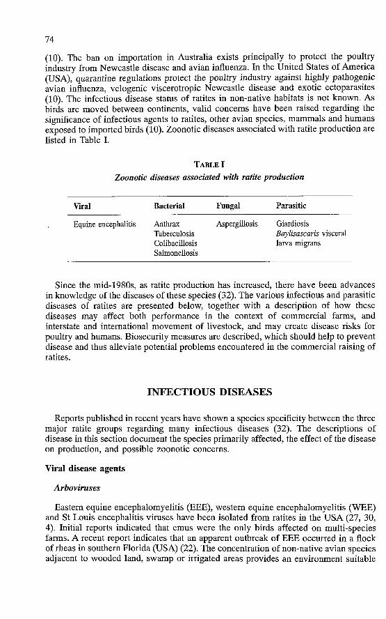
(10). The ban on importation in Australia exists principally to protect the poultry industry from Newcastle disease and avian influenza. In the United States of America (USA), quarantine regulations protect the poultry industry against highly pathogenic avian influenza, velogenic viscerotropic Newcastle disease and exotic ectoparasites (10). The infectious disease status of ratites in non-native habitats is not known. As birds are moved between continents, valid concerns have been raised regarding the significance of infectious agents to ratites, other avian species, mammals and humans exposed to imported birds (10). Zoonotic diseases associated with ratite production are fisted in Table I.
TABLE I
Zoonotic diseases associated with ratite production
Viral Bacterial Fungal Parasitic
Equine encephalitis Anthrax Aspergillosis Giardiosis Tuberculosis Baylisascaris visceral Colibacillosis larva migrans Salmonellosis
Since the mid-1980s, as ratite production has increased, there have been advances in knowledge of the diseases of these species (32). The various infectious and parasitic diseases of ratites are presented below, together with a description of how these diseases may affect both performance in the context of commercial farms, and interstate and international movement of livestock, and may create disease risks for poultry and humans. Biosecurity measures are described, which should help to prevent disease and thus alleviate potential problems encountered in the commercial raising of ratites.
INFECTIOUS DISEASES
Reports published in recent years have shown a species specificity between the three major ratite groups regarding many infectious diseases (32). The descriptions of disease in this section document the species primarily affected, the effect of the disease on production, and possible zoonotic concerns.
Viral disease agents
Arboviruses
Eastern equine encephalomyelitis (EEE), western equine encephalomyelitis (WEE) and St Louis encephalitis viruses have been isolated from ratites in the USA (27, 30, 4). Initial reports indicated that emus were the only birds affected on multi-species farms. A recent report indicates that an apparent outbreak of EEE occurred in a flock of rheas in southern Florida (USA) (22). The concentration of non-native avian species adjacent to wooded land, swamp or irrigated areas provides an environment suitable
75
for mosquitoes and other arthropod vectors. Mosquitoes transmit infection from passeriform reservoirs to emus and rheas which are accidental hosts.
The clinical manifestation of EEE in emus is a peracute haemorrhagic gastroenteritis. Cases of WEE and St Louis encephalitis develop the typical neurological signs of progressive depression and paralysis (27). The encephalitis viruses are zoonotic, and appropriate precautions should be taken when performing necropsy procedures on viraemic subjects. Ratites are not involved as amplifiers of equine encephalitis or St Louis encephalitis viruses within endemic areas. Vaccinated birds develop a protective antibody titre of unknown duration. It is currently recommended that a bivalent EEE/WEE inactivated tissue-culture origin vaccine be administered twice-yearly to emus and rheas raised in an area endemic for these arboviruses. Young birds should receive the first dose at six weeks of age, with a booster four weeks later. Unvaccinated birds should be vaccinated twice, at four-week intervals, before being subjected to the twice-yearly programme. A full dose of the equine vaccine should be administered to birds of all ages. This is not an approved vaccine for ratites in the USA, and administration is subject to the ethical and legal restraints of using the vaccine in non-approved species. Owners must receive relevant information before informal consent is granted for vaccination. Antibody response to vaccination can be monitored using the complement fixation procedure.
Avian influenza
Type A orthomyxoviruses are classified according to haemagglutinating and neuraminidase activity of glycoprotein in the viral envelope. A range of avian influenza strains have been isolated from ostriches in South Africa and rheas in the USA exhibiting various clinical syndromes ranging from high mortality to inapparent infection (2, 24).
The ability of avian influenza virus to mutate and spread among avian species makes this a potentially dangerous infection for commercial poultry. Transmission can occur from wild birds coming into contact with ratites, from newly-introduced, non-quarantined stock or indirectly via the clothing of visitors and transport trailers. The risk of exposure can be reduced by operating closed facilities with strict biosecurity measures, including control over entry of personnel (27).
Infection with influenza strains of low pathogenicity may be treated with supportive therapy. Virulent strains result in high mortality in young ostriches, which die despite administration of fluids and antibiotics (27). Prevention requires application of biosecurity measures and quarantine of newly-introduced birds, which should be monitored for the presence of antibodies using the agar gel precipitation procedure prior to purchase or delivery. It is possible that inactivated vaccines against specific strains of influenza may be permitted by the United States Department of Agriculture, Animal and Plant Health Inspection Service, which is the Federal agency responsible for control of exotic and catastrophic diseases in the USA.
Newcastle disease virus
Newcastle disease (ND) is caused by paramyxovirus type 1, which affects galliforms, passeriforms and psittacines in addition to other families, including ratites (27, 23). Velogenic viscerotropic ND (VVND) has been isolated from ostriches
76
showing high mortality in Israel and from rheas in Brazil (27). The disease manifests clinically as nervous signs, and 80% mortality occurs experimentally with infected ostrich chicks (23).
Countries which are free of VVND, such as the USA, maintain restrictions on the importation of domestic, companion and exotic bird species, together with quarantine at the point of entry. The disease is spread by direct and indirect contact with infected carriers and fomites. Air-borne dissemination of virus may occur for up to 4 km (27).
Ratites should be vaccinated only in areas where VVND is endemic. Vaccinated ratites produce antibodies which will be detected if a test and depletion control programme is implemented in a non-endemic area. This may result in compulsory slaughter or rigid and extended quarantine.
Adenovirus
Adenoviruses have been implicated as the cause of wasting disease in young ostriches in the USA (21). The clinical signs associated with these outbreaks included non-specific wasting, anorexia and depression. Specific adenoviruses are pathogenic in poultry and are responsible for haemorrhagic enteritis in turkeys, and inclusion body hepatitis/hydropericardium syndrome and egg drop syndrome in chickens (27).
Adenoviruses are transmitted vertically by the transovarian route. Adenoviral infection may remain dormant in chickens until onset of production or exposure to an environmental stress.
In the cases observed in the USA, mortality in ostriches occurred at approximately two months of age. Affected birds showed anorexia, emaciation and diarrhoea. Isolation and identification of an adenovirus from moribund or dead birds is required to confirm the diagnosis. It is noted that adenoviruses are ubiquitous, and considerable research is still required to define the role of the agent in the 'fading chick' syndrome. There is no diagnostic procedure to determine the carrier status of an asymptomatic breeder ostrich. Poultry producers in countries other than the USA administer homologous, inactivated oil-emulsion vaccine to control specific infections including egg drop syndrome, which is exogenous to the USA (27). At this time, it is considered advisable to maintain a closed breeding flock or purchase immature ostriches from a facility known to be free of the condition. Custom hatching and brooding may disseminate the infection, which can be spread by lateral exposure to 'shedders' . The total impact of adenovirus infections in the ratite industry is not known at this time, but biosecurity measures are required to prevent adenoviral infection restraining the expansion of production, especially in the large units required for commercial multiplication of slaughter stock.
Avibirnavirus
Infectious bursal disease virus (IBDV) is responsible for immunosuppression in young chickens as a result of the destruction of immature lymphocytes in the bursa of Fabricius (27, 6). Avibirnavirus identical to IBDV has been isolated from the bursa of immature ostriches in flocks in California and Florida where high flock mortality was observed (27, 6).
77
Affected birds do not show specific clinical signs, but depression and anorexia may preceed death. Isolation and identification of the virus is required for confirmation. Supportive treatment and antibiotic therapy is recommended due to immune suppression. Administration of infectious bursal disease vaccines licensed for chickens is not recommended. The response of a ratite to live attenuated chicken vaccine is unknown, and the vaccine virus could be deleterious to an unnatural recipient (27). It must be remembered that IBDV can persist for up to ninety days in biological material, necessitating thorough disinfection of brooding facilities (27). Diligent biosecurity is necessary to prevent exposure to the highly infectious agent (27, 15).
Avipoxvirus
Pox has been diagnosed in ostriches in the USA, Israel and South Africa (27, 3, 20). The virus can be transmitted by mosquitoes or by direct contact with a pox lesion. Immature ostrich chicks aged between two weeks and one year are most susceptible (27). The cutaneous form of pox is characterized by persitent proliferative lesions of 0.5-2.0 cm in size, on the eyelids, beak, wing and toes (27). The diphtheritic form produces tracheitis, stomatitis and a severe dyspnoea in affected birds.
Pox can be confirmed by histopathological examination of affected tissue, which shows the presence of intracytoplasmic Bollinger bodies. Affected birds should be separated and given supportive therapy, including systemic and topical antibiotics, to prevent secondary bacterial infections. All unaffected birds should be vaccinated with a commercial fowlpox vaccine via the intradermal route, in accordance with the recommendation of the manufacturer for reconstitution and administration (27). Control of mosquitoes may be possible but rapid diagnosis and vaccination is the most appropriate preventive measure.
Viral enteritis
A wide range of bacteria and viruses have been isolated from the gastrointestinal tract of one- to three-week-old ostriches from flocks with a high level of morbidity and mortality (27). Isolates include coronaviruses and adenoviruses which may not be the primary cause of death (27, 9). Intra-flock transmission usually occurs rapidly through faecal/oral contamination, and may be exacerbated by high stocking density or defects in biosecurity and hygiene (27). Clinical signs associated with viral enteritis are non-specific but generally include anorexia and diarrhoea. Due to the presence of opportunistic bacteria, it may be difficult to identify a viral agent during pathological examination. Virus identification by histopathology or electron microscopy and examination of faecal material may help in identifying the viral organism responsible for the clinical illness. Supportive therapy, intravenous fluid administration, tube feeding and antibiotics may help reduce flock deaths. The best way to prevent outbreaks of viral enteritis is through proper management hygiene before the introduction of new birds into a production unit, and through biosecurity in established flocks.
Borna disease virus
Borna disease virus (BDV) has been identified in Israel in ostrich chicks (two to eight weeks old) raised under intensive conditions (33). Clinical signs are initiated by paresis and general malaise, which contribute to anorexia and depression. The affected
78
birds usually die within four to eight days, due to dehydration (27, 33). Intensive farming methods seem to be a contributing factor in BDV outbreaks, with insect vectors believed to be responsible for transmission of the virus (27).
Histopathological lesions in affected birds show neuronal degeneration and lymphocytic perivascular cuffing (33). An enzyme-linked immunosorbent assay has been developed in Israel to demonstrate the presence of BDV in brain tissue of infected birds (27). Prevention of Borna disease outbreaks in intensive ostrich-raising facilities will require stress-reduction management programmes. For a Borna disease management programme, an inactivated viral vaccine must be developed, to immunize parent stock and thus provide maternal immunity to the offspring (27). At present, BDV is restricted to intensive ostrich-farming facilities in Israel, but the current popularity of ratite importation increases the likelihood of virus spread. Good husbandry, management and biosecurity will reduce the stress on birds and, consequently, reduce the risk presented by this viral organism to susceptible birds in Israel and other parts of the world which are affected.
Other viruses
For some of the viral mortality associated with disease outbreaks in ratite facilities, no aetiological agent has been identified. The capacity of ratites to harbour and grow multiple pathogens within the gastrointestinal and respiratory tracts makes the isolation of unknown viral agents difficult. Rotaviruses, reoviruses, parvoviruses and picornaviruses are all potential pathogens which may contribute to ratite morbidity and mortality, but have not yet been identified in these species (27). With increased observation and diligence by clinical veterinarians and pathologists, other ratite viral pathogens will be discovered in the future, thereby helping to ensure healthy production within the industry.
F U N G A L A N D B A C T E R I A L D I S E A S E A G E N T S
Fungi and bacteria are the primary pathogens which affect ratite species. Ratites housed in production settings are subject to underlying environmental stress, which predisposes the animals to infection with fungal and bacterial organisms. Neglecting or incorrectly applying appropriate management procedures increases the possibility of a disease outbreak. The fungal and bacterial organisms which commonly affect production ratite species are discussed below.
Fungal diseases
Aspergillosis
Aspergillus spp. has been identified as a primary cause of death in juvenile emus and ostriches (17). The most susceptible individuals are young birds in enclosed facilities with exposure to dust or to hay which is alternately wet and dry. Aspergillus spp. exposure can occur through egg contamination but, under current conditions in the industry, poorly-managed intensive chick facilities seem to be the most likely place for an outbreak to occur (27).
79
Affected chicks usually die suddenly, or exhibit severe dyspnoea prior to death. Aspergillus granulomas are detected in the lungs and/or airsacs on gross post-mortem examination. In a highly intensive farming operation, treatment may not be cost-effective, but itraconizole is the drug of choice. Although a ubiquitous organism, Aspergillus spp. can cause infection in humans, and face-masks must therefore be worn to reduce human exposure. Management procedures which reduce juvenile stress and exposure to dust and hay will alleviate much potential exposure to the fungal organism.
Zygomycosis
Basidia, Mucor and Rhizopus are genera of zygomycetes which affect the upper gastrointestinal system of ratites. Zygomycotic infections occur when the organism is ingested and is allowed to overwhelm the bird, due to suppressed immune status or concurrent bacterial, viral or parasitic infections (27).
Non-specific clinical signs accompany a zygomycotic infection, and diagnosis is usually made on post-mortem examination. Although these fungal infections have been noted in the USA and Israel, there is a real potential for exposure in other countries due to the ubiquitous nature of this organism (13). To prevent or reduce exposure to zygomycotic organisms, the producer should eliminate stress, primary viral or bacterial infections, and fungal contamination of feed and substrate (27).
Candidosis
Stomatitis characterized by white raised lesions in the oral cavity extending down the oesophagus is usually associated with a Candida spp. infection (27). The two main Candida organisms isolated from ratite species are C. albicans and C. mucor.
Candida spp. are opportunistic organisms and usually indicate a primary bacterial, viral, parasitic or management problem which is reducing the ability of the bird to fight infection. Diagnosis can be made through Gram's staining, direct scraping of the oral plaques and viewing the characteristic budding yeast forms. Treatment and prevention are achieved by using nystatin, fluconizole or ketoconizole, with adjunct supportive therapy. It is essential for the veterinarian to identify and treat the underlying primary immunosuppressive problem affecting the individual bird or flock to ensure proper treatment of the secondary fungal infection. To provide treatment in large-scale production operations, copper sulfate may be added to the drinking water at a level of 1:2,000 in a large-scale feedlot operation (27).
Bacterial diseases
Salmonellosis
Salmonella spp. are ubiquitous organisms which reflect contamination of facilities through contact with rodent or wild bird reservoirs. Various Salmonella spp. have been isolated from the gastrointestinal tracts of all ratite species, but the incidence of paratyphoid Salmonella spp. (e.g. S. Typhimurium) is very rare (27). It must be remembered that certain Salmonella organisms are zoonotic, and precautions should be taken when handling a suspect case. Emus can be infected with S. Pullorum - a pathogen which persists in game fowl - and develop antibodies against this pathogen
80
(27, 31). The importance of S. Pullorum in ratites is unknown at this time, as no documented field cases have been identified.
Salmonella organisms can be transmitted horizontally by infected birds via the faecal/oral route, or through contamination of equipment or the environment (27). Vertical transmission via faecal contamination of eggs has also been described as a method of Salmonella infection in the poultry industry (27). As Salmonella bacteria can be transmitted through eggs or contaminated equipment, care must be taken when receiving birds or eggs after interstate or international shipment. Most birds suffering from Salmonella spp. infections show no specific clinical signs, and are diagnosed through microbiological screening at necropsy.
Treatment of infected birds with an appropriate antibiotic will generally suppress the clinical infection, but may lead to an asymptomatic carrier state. To reduce the risk of subclinical carriers developing, a whole-blood S. Pullorum agglutination test should be performed prior to shipment, and routine screening of birds by cloacal swabs during quarantine is also suggested (27).
Erysipelas
Emus have been reported to be susceptible to infection by Erysipelothrix rhusiopathiae (11). Infection with this soil organism occurs via small skin lacerations, caused by trauma, or insect vectors (27). Acute deaths usually occur due to a bacterial septicaemic condition, but penicillin or quinolone antibiotics may help mildly-affected birds (27). If erysipelas is a problem within a region or at a particular production facility, ratites should be immunized using a commercial, formalin-inactivated bacterin (with aluminium hydroxide adjuvant) licensed for turkeys (27). No adverse reaction has been noted in emus vaccinated with this inactivated turkey vaccine. In areas where erysipelas is endemic, birds should receive the first vaccine subcutaneously at six weeks of age, a second dose at six months of age, and an annual booster.
Colibacillosis
Escherichia coli serotypes are widely distributed; the degree of pathogenicity depends on the condition of the bird(s) exposed (27). In general, emus seem to be more susceptible to E. coli gastroenteritis infection than other ratite species.
Transmission usually occurs via the faecal/oral route, and the agent may be identified via culturing of cloacal swabs. Appropriate antibiotics are essential in treating an E. coli infection in ratite species. Autogenous bacterins are available, which may be used when colibacillosis becomes a health problem on a farm. These bacterins use cultures isolated from affected birds to help boost immunity in the resident animals. The efficacy of these bacterins in ratite and other avian species is unknown at present. The best prevention measures for colibacillosis infections are proper management and nutrition, and stress reduction in the flock or transported birds.
Pasteurellosis
There have been rare occurrences of Pasteurella spp. isolation in ratite species (27, 1). Intensive farming may increase occurrence, and this organism should therefore be monitored closely. Environmental exposure and direct contact with recovered Pasteurella carriers provide ample opportunity for acquisition of this bacterium in an immunosuppressed ratite flock.
81
Non-specific respiratory signs may be noted in the clinically-diseased bird, while generalized vascular congestion is seen on gross necropsy specimens. At present, no vaccination measures are recommended, but high standards of biosecurity are required to prevent introduction of infections into flocks.
Mycoplasmosis
Mycoplasma cloacale has been isolated from tracheal swabs of ostriches and emus, while there has been no isolation from these species of the three pathogenic Mycoplasma spp. found in commercial poultry (27, 18). Intensification of the ratite industry, and concentration of birds in feedlots, may lead to the emergence of mycoplasmosis as a disease of economic significance due to the ease of transmission of this pathogen. Clinical signs accompanying mycoplasmal infection include nonspecific upper respiratory conditions and/or arthritis (27). Identification of Mycoplasma in other avian species is performed through isolation of the organism from joint aspirates or respiratory discharge, or through serological screening using the serum plate agglutination test. At present, a highly-sensitive polymerase chain gene probe assay can be used for identification of Mycoplasma galliseptium and M. synoviae in a few diagnostic laboratories (27). Treatment of confirmed cases is usually accomplished using tylosin or tetracycline antibiotics. Producers and transporters are encouraged to identify potential cases before the animals are placed in holding facilities, to reduce the risk of exposure of non-infected birds.
Tuberculosis
Mycobacterium avium has been diagnosed in ostriches and emus in the USA and Canada, and in emus in the USA (27, 26). M. avium persists in wild bird populations and can infect pigs, cattle and immunosuppressed humans; it is therefore classified as a zoonotic disease. Transmission of M. avium occurs through infected faeces shed by clinically-ill birds. The organism can remain viable in the soil for up to twelve months, making sanitation after a clinical case difficult for the owner. The highly-mobile ratite industry enables dissemination of the organism via transport, holding facilities, quarantine houses and auction barns.
As M. avium generally affects the avian gastrointestinal system, clinical signs are usually those of non-specific wasting. Emus may develop leukocytosis with or without cloacal prolapse. Confirmation of the infection is usually accomplished at necropsy, unless an intensive ante-mortem physical examination is performed, isolating the acid-fast organism from a bacterial granuloma or a faecal screen.
Screening tests using faecal acid-fast methods are unreliable; intradermal testing is therefore recommended, together with the maintaining of closed flocks, examination of newly-acquired breeding stock and good management. Tuberculosis is a potential zoonotic disease for which no treatment is available; a flock maintained within a production facility must therefore be free from tuberculosis.
Chlamydiosis
Chlamydia psittaci is an intracellular bacterium which has been identified as the causative agent in deaths of commercial rheas (27, 5, 12). Infected rheas usually die peracutely, while emus seem to be refractory to the organism isolated from multi-
82
species flocks. Pigeon strains of C. psittaci have been identified in outbreaks of the disease in Texas, and may have caused other rhea deaths in the southern USA (29,12) .
Diagnosis is made at necropsy through bacterial isolation. The gross pathological signs of psittacosis include splenomegaly, hepatomegaly, pericarditis and fibrinous airsacculitis. Extreme care should be taken by the veterinarian during treatment and/or necropsy of a C. psittaci case, in view of the zoonotic potential of the organism. The psittacosis bacterium is highly infectious to humans and causes severe pneumonia and death if not treated appropriately. Treatment of ratites and humans is best achieved with the tetracycline class of antibiotics. Reduction of wild bird access to feed and water areas will reduce the exposure of ratites to potential carriers of the Chlamydia organism.
Clostridial enteritis
A number of clostridial organisms have been isolated from various ratite species, including Clostridium perfringens, C. colinum, C. chauvoei and C. difficile (27, 16, 28). There is no published information on the frequency of isolation of Clostridium spp. from the intestinal tract of normal ratites, but clostridial enteritis usually occurs following proliferation of a toxin-forming Clostridium spp. in other avian species (27).
Diagnosis of clostridial enteritis is usually made at necropsy through microbiological identification. Anaerobic culture methods should be recommended by the clinical veterinarian if such procedures are not commonly performed on bacterial culture specimens by the diagnostic laboratory. Outbreaks of clostridial infections can be treated with the addition of zinc bacitracin in feed at a rate of 30 g/ton (27).
Infectious coryza
Haemophilus spp. are transmitted horizontally and have been isolated from ostriches in Israel (27, 19). Clinical signs are concentrated in the upper respiratory system and are treated with appropriate antibiotics from microbiological culture, and antibiotic sensitivity results. Proper management and quarantine of newly-acquired birds will reduce the risk of exposure and infection from this bacterial organism.
Anthrax
Although rare, Bacillus anthracis has been diagnosed in South African ostriches (29). In areas where anthrax has been identified as a cause of livestock deaths, ratite producers are advised to have any bird which dies peracutely necropsied by a veterinarian or pathologist, due to the highly zoonotic nature of this organism.
Diagnosis is usually made at necropsy. Affected birds have extreme splenomegaly, hepatomegaly and vascular congestion (27). There is no treatment for this disease, and ostriches or other ratite species should not be maintained on farms with a history of anthrax (27).
PARASITES
Parasite problems in ratite facilities can cause general ill thrift, reduction in growth, poor reproductive results and death. Effective screening prior to introduction into a production unit is essential in the prevention and control of the common ratite
83
parasites. Management procedures in reducing parasitic populations include external feather examinations, complete physical examinations, complete blood counts, direct faecal examinations and faecal floatation examinations.
Arthropods
Ticks are common parasites of the ostrich in its native African environment, and include the following genera/species: Amblyomma spp., Haemaphysalis punctata, Hyalomma spp., Rhipicephalus turanicus and Argus spp. (7). Ticks have been implicated in transmission of viral diseases, and heavy infestations cause ill thrift, slow growth and low egg production. Treatment is best achieved with 5% carbaryl dust at fourteen-day intervals (7).
Pterolichus bicaudatus (ostrich quill mite) and Struthiolipeurus struthionus (ostrich louse) may cause pruritis and/or excessive preening and feather loss. Infestation with these external parasites causes stress and predisposes birds to secondary infections and gastrointestinal disorders (e.g. impactions). Treatment for mites is accomplished with ivermectin, and lice are treated in a similar manner to tick infestations.
Miscellaneous arthropod infestations - e.g. Struthiobosca struthionis in ostriches, Struthiodiperus rheae in rheas, and Culicoides spp. and Simulium spp. in emus - cause blood loss, irritation and stress, and transmit other parasites (7). Pyrethrin sprays may help to prevent extreme exposure to many of the flying arthropod parasites.
Helminths
Helminth parasites found in ratites include the following: Baylisascaris spp., Libyostrongylus douglassi, Paraonchocerca struthionus, Struthiofilaria megalocephala, Ascaridia orthocerca, Deletrocephalus dimidiatus, Deletrocephalus casarpintoi, Dicheilonema rheae, Paradeletrocephalus minor and Chandlerella quiscali.
Baylisascaris spp. have been identified in ostriches and emus (7, 14). The definitive hosts for this parasite are skunks and raccoons, which shed the eggs in faecal material; the eggs can remain infective in the soil for several years. Birds suffering from Baylisascaris spp. infections show ataxia, muscle weakness, recumbency and death, due to visceral larval migration into the brain and spinal cord. Diagnosis is made at necropsy by observation of the parasite in brain or spinal cord tissue sections. Baylisascaris spp. cause a zoonotic parasitic disease and can provoke severe physical damage through visceral larval migration if eggs are ingested. Reduction of definitive host populations around a facility and proper feed storage will help to reduce exposure to this dangerous parasite.
A helminth parasite which affects young emus in a similar manner is Chandlerella quiscali. This parasite is transmitted by Culicoides midges, and visceral larval migration occurs before the birds reach one year of age. Generalized neurological signs accompany Chandlerella spp. infestation, and diagnosis is made at necropsy.
Libyostrongylus douglassi (wireworm) is a proventricular parasite which is extremely deadly to young ostrich chicks. Prevention of this parasite from infesting a ratite facility is very important. The life cycle is direct, and pasture rotation is recommended as a control measure. Fenbendazole, levamisole and ivermectin may be effective in the control of this deadly parasite (7, 14).

84
The other helminth parasites listed should be treated as mentioned for L. douglassi. Prevention of parasite exposure is the primary management measure recommended in all ratite operations. Once a parasite has become established on a facility, diligent testing and treatment - especially in young birds - will limit economic losses and bird mortality.
Protozoa
The blood protozoa Leucocytozoon struthionis and Plasmodium spp. have been identified in ostriches (7), and are transmitted by flying arthropod vectors. These parasites do not generally cause significant clinical illness, and treatment is usually more problematic than the infestation.
The intestinal protozoa Balantidium struthionis, Cryptosporidium spp., Histomonas meleagridis, Hexamita spp., Giardia spp. and Trichomonas spp. cause gastrointestinal problems which result in wasting, anorexia, diarrhoea and death for all ratite species, especially rheas. Direct faecal and intestinal content examinations are essential in diagnosing these parasitic diseases (7). Appropriate treatment with anti-protozoal drugs will reduce losses in an outbreak; but proper husbandry practices will limit incidence, thus reducing labour and other expenses.
Tapeworms and flukes
Houttuynia struthionis (ratite tapeworm) primarily affects younger birds. Ostriches and rheas seem to be the most common ratite species affected, and the recommended treatment is fenbendazole (8).
The adult ratite fluke (Philophthalmus gralli) is found outside the nictitating membrane in affected birds. The intermediate host of the fluke is a freshwater snail, and ratites acquire the parasite through ingestion of freshwater crustaceans or other solid objects. It is recommended that ratite exposure to standing water be restricted, thus reducing the risk of intermediate host exposure.
PREVENTION A N D REDUCTION OF RATITE DISEASES T H R O U G H APPROPRIATE M A N A G E M E N T TECHNIQUES
'All-in/all-out' management should be applied to ratite farms, to prevent transmission of infectious diseases (25). Producers can minimize the possibility of infection by maintaining separate growout breeding and trading facilities by at least 300 m (25).
Breeding stock should be maintained in a closed unit. If new birds are purchased, a thorough physical examination should be performed with appropriate diagnostic procedure before delivery. New birds should be quarantined for at least sixty days in a separate facility, and monitored for the presence of disease.
Ratite producers should limit visits to other farms. A complete body cleaning, and change of all clothing and shoes, should take place before interacting with home stock. Visitors should be provided with rubber boots and coveralls before entering pens (25). Care should be taken when decontaminating the interior and exterior of transport vehicles (25).

85
A very important policy of a ratite facility is to confine activities to one species, due to the danger of cross-transmission of infectious organisms between ratites, water fowl, exotic birds and livestock (25). All water should be free of pathogens and should be analyzed annually to confirm quality.
Bird pens should be placed in a well-drained area, and fenceposts should be on the outside of the pen. A structure placed in the middle of the pen will reduce exposure to wind and precipitation, and will protect food trays (Fig. 1). Sufficiently-high, strong wire fencing will reduce injuries to birds and prevent attack by wild animals. Fences should reach down to the ground if a perimeter fence is not in place, to reduce exposure to wild animals or stray dogs. At most ostrich facilities, producers prefer to leave a space for escape under the bottom line of the fence, and a perimeter fence should therefore be in place. Electric fences and dogs have been used with success in keeping out unwanted animals which may injure livestock.
FIG. 1
Adequate pen set-up for emus, with shelter and feeding area
In general, ratites are very healthy animals. Good management and knowledge of infectious disease is essential in maintaining a successful production unit. Preventing the spread of viral, bacterial and parasitic diseases between local facilities and internationally is imperative if the 'fledgling' ratite industry is to grow and prosper.
*
86
LES MODES D'ÉLEVAGE DES RATITES ET LEURS CONSÉQUENCES SUR LES MALADIES INFECTEEUSES ET PARASITAIRES. - T.N. Tully et S.M. Shane.
Résumé : Le nombre d'élevages de ratites dans le monde a augmenté au cours des dix dernières années. Les élevages d'autruches sont essentiellement concentrés en Afrique du Sud, mais d'autres pays envisagent également ce type de production ainsi que celle des émeus et nandous. Elevés à des fins commerciales hors de leur habitat d'origine, les autruches, émeus et nandous sont exposés à des maladies nouvelles et très particulières. Les auteurs décrivent les maladies bactériennes, virales et parasitaires qui apparaissent actuellement sur les lieux de production. Pour chacune d'entre elles, ils indiquent les mesures de prévention et de bonne conduite d'élevage à respecter pour réduire les risques d'exposition des animaux et/ou la mortalité.
Les ratites sont des animaux résistants pour peu que l'on observe des méthodes de gestion et d'alimentation appropriées. Le principal problème qui affecte actuellement cette activité est le fait que les élevages ne disposent pas de parcs de quarantaine séparés et adaptés ; cette absence est due à la réticence des propriétaires ou simplement à leur ignorance de la nécessité d'une telle mesure. Pourtant, le recours à ces parcs de quarantaine est bien plus efficace pour prévenir la contamination des sols et des locaux que n'importe quelle autre mesure de gestion. Les ratites offrent d'excellentes opportunités de production et de profit, à condition que les éleveurs prêtent attention aux problèmes sanitaires. Les auteurs présentent les maladies des ratites qui peuvent compromettre la viabilité des élevages intensifs.
MOTS-CLÉS : Autruches - Bactéries - Emeus - Maladies virales -Mycoses - Parasites - Ratites.
PRÁCTICAS DE CRÍA LIGADAS A LAS ENFERMEDADES INFECCIOSAS Y PARASITARIAS DE LAS AVES CORREDORAS DE GRANJA. - T.N. Tully y S.M. Shane.
Resumen: Durante la década pasada el número de aves corredoras criadas en granjas estabuladas ha aumentado en el mundo. El centro de la producción de avestruces sigue encontrándose en Sudáfrica, pero otros países estudian actualmente su potencial para la producción de esta ave corredora, así como de emús y de ñandús. Las avestruces, los emús y los ñandús criados con fines comerciales crecen fuera de sus hábitats nativos y son expuestos, por consiguiente, a enfermedades nuevas y exclusivas de su nuevo medio. Los autores describen las enfermedades bacterianas, virales y parasitarias que están surgiendo en los locales de producción. Para cada patología indican las prácticas de manejo y las medidas de prevención adecuadas para reducir las probabilidades de exposición del rebaño y/o los riesgos de mortalidad importante.
Las aves corredoras son animales resistentes, a condición de que sean objeto de buenas prácticas de nutrición y manejo. El problema al que actualmente se enfrenta la industria reside en la ausencia de zonas aisladas de cuarentena en las granjas, debida a la falta de voluntad de los propietarios, o a su ignorancia de la necesidad de estas zonas. El recurso a un área de
87
cuarentena hará más por prevenir la contaminación de los suelos y de las instalaciones libres de patógenos que cualquier otra medida de manejo. Las aves corredoras son excelentes animales de renta, siempre y cuando los granjeros consigan criar animales sanos. Los autores examinan las enfermedades actualmente conocidas que afectan a estas aves y que pueden incidir sobre la viabilidad de una granja dedicada a su cría intensiva.
PALABRAS CLAVE: Aves corredoras - Avestruces - Bacterias - Emús — Enfermedades fúngicas - Enfermedades virales - Parásitos.
*
REFERENCES
1. AKOHA A. ( 1 9 8 0 ) . - An outbreak of pasteurellosis in Akano Zoo. J. Wildl. Dis., 16, 3 - 5 .
2 . ALLWRIGHT D.M., BURGER W.P., GAYER A. & TERBLANCHE A.W. ( 1 9 9 3 ) . - Isolation of an influenza A virus from ostriches (Struthio camelus). Avian Pathol, 22, 5 9 - 6 5 .
3 . ALLWRIGHT D.M., BURGER W.P., GAYER A. & WESSELS J. ( 1 9 9 4 ) . - Avian pox in ostriches. Jl S. Afr. vet. Assoc., 65, 2 3 - 2 5 .
4. AYERS J.R., LESTER T.L. & ANGULO A.B. ( 1 9 9 4 ) . - An epizootic attributable to western equine encephalitis virus infection in emus in Texas. Am. vet. med. Assoc., 205, 6 0 0 - 6 0 1 .
5. CAMUS A.C., CHO D.Y., POSTON R.P., PAULSEN D.P., OLIVER J.L., LAW J.M. & TULLY T.N. ( 1 9 9 4 ) . - Chlamydiosis in commercial rheas (Rhea americana). Avian Dis., 38, 6 6 6 - 6 7 1 .
6. CHIN R.P. & WOOLCOCK P. ( 1 9 9 4 ) . - Identification of birnavirus-like particles from the intestines of 8-week-old ostriches (summary). In Proc. Western Poultry Disease Conference. Sacramento, California, 2 7 Febmary-1 March. Western Poultry Disease Conference, Davis, California, 1 1 0 .
7. CRAIG T. ( 1 9 9 3 ) . - Natural parasites of ratites. In Proc. Annual Ratite Conference. College Station, Texas, 9 - 1 0 September. College of Veterinary Medicine, Texas A&M University.
8. FECKEMA A., MALAN F., COOPER G. & VISSER E. ( 1 9 8 5 ) . - Anthelmintic efficacy of fenbendazole against Libyostrongylus douglassi and Houttuynia struthionis in ostrich. Jl S. Afr. vet. Assoc., 56, 4 7 - 4 8 .
9. FRANK R.K. & CARPENTER J.W. ( 1 9 9 2 ) . - Coronaviral enteritis in an ostrich (Struthio camelus) chick. J. Zoo Wildl. Med., 23, 1 0 3 - 1 0 7 .
10. GILCHRIST P. ( 1 9 9 3 ) . - Exotic disease-threats of importation. In Ostrich Odyssey. Proceedings of the Meeting of the Australian Ostrich Association Inc. Post-Graduate Committee in Veterinary Science, University of Sydney, Proceeding No. 2 1 7 , 8 3 - 8 8 .
11 . GRIFFITHS G. & BULLER N. ( 1 9 9 1 ) . - Erysipelothrix rhusiopathiae infection in semi-intensively farmed emu. Aust. vet. J., 68, 1 2 1 - 1 2 2 .
12. GRIMES J.E. & ARIZMENDI F. ( 1 9 9 4 ) . - Case reports of ratite chlamydiosis: an update on the chlamydias. In Proc. Association of Avian Veterinarians (AAV). Reno, Nevada, 2 8 - 3 0 September. AAV Publications, Orlando, Florida, 1 3 3 - 1 4 0 .
88
13. JEFFERY J.S., CHIN R.P., SHIVAPRASAD H.L., METEYER C.U. & DROUAL R. (1993). -Proventriculitis and ventriculitis associated with zygomycosis in ostrich chicks. Avian Dis., 38, 630-634.
14. KAZACOS K., WINTERFIELD R.W. & THACKER H.L. (1982). - Etiology and epidemiology of verminous encephalitis in an emu. Avian Dis., 26, 389-391.
15. LASHER H.N. & SHANE S.M. (1994). - Infectious bursal disease. Wld Poult. Sci. J., 50, 133-166.
16. LUBLIN A., MECHANI S., HOROWITZ H.I . & WEISMAN Y. (1993). - A paralytic-like disease of the ostrich (Struthio camelus masaicus) associated with Clostridium chauvoei infection. Vet. Rec., 132, 273-275.
17. MARKS S.L., STAUBER E.H. & ERNSTROM S.B. (1994). -Aspergillus in an ostrich. J. Am. vet. med. Assoc., 204, 784-785.
18. MOHAN R. (1993). - Mycoplasma in ratites. In Proc. Association of Avian Veterinarians (AAV). Nashville, Tennessee, 31 August-4 September. AAV Publications, Orlando, Florida, 294-296.
19. PERELMAN B. (1991). - Upper respiratory disease. In Proc. 3rd Annual Ostrich Conference. College Station, Texas, 5-6 January. College of Veterinary Medicine, Texas A&M University.
20. PERELMAN B., GUR-LAVIE A. & SAMBERG Y. (1988). - Pox in ostriches. Avian Pathol., 17, 735-739.
21. RAINES A.M. (1993). - Adenovirus infection in the ostrich (Struthio camelus). In Proc. Association of Avian Veterinarians (AAV). Nashville, Tennessee, 31 August-4 September. AAV Publications, Orlando, Florida, 304-312.
22. RANDOLPH K. (1995). - Update on equine encephalitis. In Proc. Association of Avian Veterinarians (AAV). 28 August-2 September, Philadelphia, Pennsylvania. AAV Publications, Orlando, Florida, 249-253.
23. SAMBERG Y , HADASH D., PERELMAN B. & MEROZ M. (1989). - Newcastle disease in ostrich (Struthio camelus): field case and experimental infection. Avian Pathol., 18, 221-226.
24. SELMONS R.D., FISCHBACH W.L. & GODAN S.A. (1995). - Pathogenesis of ratite-origin influenza virus infection in chickens (summary). In Annual Meeting of the American Association of Veterinary Pathologists in Proc. 132nd AVMA Conference. American Veterinary Medical Association (AVMA), Schaumberg, Illinois, 153.
25. SHANE S.M. (1994). - Disease prevention in ostrich production. Bio-Tek Industries, Inc., Atlanta, Georgia, 32 pp.
26. SHANE S.M., CAMUS A.C., STRAIN M.G., THOEN C.O. & TULLY T.N. (1993). -Tuberculosis in commercial emus (Dromaius novaehollandiae). Avian Dis., 37, 1172-1176.
27. SHANE S.M. & TULLY T.N. (1996). - Infectious diseases. In Ratite medicine, management and surgery (T.N. Tully & S.M. Shane, eds). Kreiger Publishing, Malamar, Florida (in press).
28. SHIVAPRASAD H.L. (1994). - Necrotizing hepatitis associated with Clostridium difficile in an ostrich chick. In Proc. 37th Annual Meeting of the American Association of Veterinary Laboratory Diagnosticians. Grand Rapids, Michigan, 29 October-1 November. Allen Press, Lawrence, Kansas, 67.
89
29. SNOEYENBOS G.H. (1965). - Anthrax ( H . E . Biester & L . H . Schwarte, eds). In Diseases of poultry, 5th ed. Iowa State University Press, Arnes, Iowa, 432-435.
30. TULLY T.N., SHANE S.M., POSTON R.P., ENGLAND J .J. , VICE C.A., CHO D.Y. & PANIGRAPHY B. (1992). - Viscerotropic eastern encephalitis in a flock of emus (Dromaius novaehollandiae). Avian Dis., 36, 808-819.
31. TULLY T.N. & SHANE S.M. (1993). - Salmonella Pullorum seroconversion in emus (Dromaius novaehollandiae). In Proc. Association of Avian Veterinarians (AAV). Nashville, Tennessee, 31 August-4 September. AAV Publications, Orlando, Florida, 315-317.
32. TULLY T.N. & SHANE S.M. (eds) (1996). - Ratite medicine, management and surgery. Kreiger Publishing, Malamar, Florida (in press).
33. WEISMAN Y., MALKINSON M., PERL S., ASHASH E. , MEIR R., NIR A. & LUDWIG H . (1994). - Borna disease in ostriches (abstract). In Proc. Western Poultry Disease Conference. Sacramento, California, 27 February-1 March. Western Poultry Disease Conference, Davis, California, 22.
![Basic Concepts in Public Health and Tropical Medicine [Infectious Diseases and Public Health 101] (mainly in relationship to parasitic diseases) Daniel](https://img.pdfslide.net/doc/110x75/56649d3b5503460f94a15bb0/basic-concepts-in-public-health-and-tropical-medicine-infectious-diseases.jpg)