Embed Size (px)
Citation preview
neurosurgical
focus Neurosurg Focus 41 (5):E8, 2016
The natural history of hydrocephalus after nonac-cidental head trauma (NAHT) has not been evalu-ated. This is partly due to the extremely dismal
prognosis that NAHT portends and to the fact that it often occurs within 24 hours, correlating with a significant rate of mortality. As a matter of fact, the Center for Disease Control and Prevention in a Spring 2008 report (http://www.cdc.gov/ViolencePrevention) listed NAHT as the leading cause of childhood death; the incidence reported in Pennsylvania, for example, was 14.7–26.0 cases per 100,000 person-years.11 This high incidence is not unique to the United States, as a similar rate was observed in-ternationally: 12.8–21 cases per 100,000 person-years in
England,8 for instance, and 14.7–19.6 cases per 100,000 person-years in New Zealand.10 Collectively, these sta-tistics suggest that outcomes after NAHT are universally poor, with only 50% of patients admitted for NAHT living beyond the first 24 hours after injury and with reintegra-tion into society difficult in 50% of surviving children due to severe morbidity.12 These poor outcomes may be in part due to the effects of hydrocephalus.
The incidence of ventricular dilation after traumatic head injury may be as high as 39%–44%, and on average, this pathology becomes apparent at the 4-week postinjury interval.13,15 The incidence of posttraumatic ventriculomeg-aly (PTV) is unclear, as is that of subsequent development
AbbreviAtioNs CPCT = child protection consultation team; GCS = Glasgow Coma Scale; IVH = intraventricular hemorrhage; KOSCHI = King’s Outcome Scale for Childhood Head Injury; NAHT = nonaccidental head trauma; PTV = posttraumatic ventriculomegaly; SAH = subarachnoid hemorrhage; SDH = subdural hematoma; TBI = traumatic brain injury.sUbMitteD June 28, 2016. ACCePteD August 16, 2016.iNClUDe wheN CitiNg DOI: 10.3171/2016.8.FOCUS16266.* Drs. Mittler and Schneider contributed equally to this work.
Hydrocephalus associated with childhood nonaccidental head trauma*sudhakar vadivelu, Do,1 harold l. rekate, MD,1 Debra esernio-Jenssen, MD,2 Mark A. Mittler, MD,1 and steven J. schneider, MD1
1The Cushing Neuroscience Institute and Department of Neurosurgery, Hofstra Northwell School of Medicine at Cohen Children’s Medical Center and Northwell Health System, Manhasset, New York; and 2Department of Pediatrics, University of Florida School of Medicine at Shands Children’s Hospital, Gainesville, Florida
obJeCtive The incidence of posttraumatic ventriculomegaly (PTV) and shunt-dependent hydrocephalus after nonac-cidental head trauma (NAHT) is unknown. In the present study, the authors assessed the timing of PTV development, the relationship between PTV and decompressive craniectomy (DC), and whether PTV necessitated placement of a permanent shunt. Also, NAHT/PTV cases were categorized into a temporal profile of delay in admission and evaluated for association with outcomes at discharge.MethoDs The authors retrospectively reviewed the cases of patients diagnosed with NAHT throughout a 10-year pe-riod. Cases in which sequential CT scans had been obtained (n = 28) were evaluated for Evans’ index to determine the earliest time ventricular dilation was observed. Discharge outcomes were assessed using the King’s Outcome Scale for Childhood Head Injury score.resUlts Thirty-nine percent (11 of 28) of the patients developed PTV. A low admission Glasgow Coma Scale (GCS) score predicted early PTV presentation (within < 3 days) versus a high GCS score (> 1 week). A majority of PTV/NAHT patients presented with a subdural hematoma (both convexity and interhemispheric) and ischemic stroke, but subarach-noid hemorrhage was significantly associated with PTV/NAHT (p = 0.011). Of 6 patients undergoing a DC for intractable intracranial pressure, 4 (67%) developed PTV (p = 0.0366). These patients tended to present with lower GCS scores and develop ventriculomegaly early. Only 2 patients developed hydrocephalus requiring shunt placement.CoNClUsioNs PTV presents early after NAHT, particularly after a DC has been performed. However, the authors found that only a few PTV/NAHT patients developed shunt-dependent hydrocephalus.http://thejns.org/doi/abs/10.3171/2016.8.FOCUS16266Key worDs biomarker; child abuse; excitotoxicity; hypoxia-ischemia; shaken baby syndrome; traumatic brain injury
©AANS, 2016 Neurosurg Focus Volume 41 • November 2016 1
Unauthenticated | Downloaded 01/29/22 10:10 AM UTC

s. vadivelu et al.
Neurosurg Focus Volume 41 • November 20162
to shunt-dependent hydrocephalus after NAHT. Marmarou et al.13 classically used a dye injection method to demon-strate altered CSF dynamics due to poor absorptive proper-ties associated with subarachnoid hemorrhage (SAH) or in-traventricular hemorrhage (IVH) in a majority of patients. Alternatively, operative procedures such as decompressive craniectomies have also been implicated in playing a role in promoting the development of hydrocephalus2,22 and subsequently could result in poorer outcomes.2,14,19
Here, we evaluated our cohort of patients with perpe-trator-identified NAHT to address the involvement of hy-drocephalus. We quantified the cumulative incidences of PTV and shunt-dependent hydrocephalus. We analyzed the data for epidemiological factors and radiographic signs associated with these primary findings. In addition, we questioned whether secondary factors such as a delay in seeking medical care and/or a decompressive craniec-tomy promoted hydrocephalus.
MethodsWith approval from the institutional review board, we
performed a retrospective, single-center, chart review of all patients who were evaluated by the medical child pro-tection consultation team (CPCT) and who were reported to the New York State Central Register, documented be-tween the 2001 and 2010.20 The total number of NAHT pa-tients registered was 48. Inclusion criteria included cases in which the perpetrator was specifically identified by the Child Protection Services of Nassau, Suffolk, Brooklyn, and Queens counties and in which the perpetrator was considered the primary suspect in a criminal trial or was convicted by the local/state law enforcement agencies.4,20 Fourteen cases were excluded from this review because perpetrator status could not be identified, and 6 of the 34 cases in which a perpetrator was identified were excluded because the charts could not be reviewed at the time of this study as they were under review in preparation for a crimi-nal trial or were being used during a criminal trial. The remaining 28 cases were evaluated by our hospital CPCT and pediatric neurosurgical department during the study period and included a multidisciplinary evaluation during admission.
The diagnosis of NAHT was indicated after clear evi-dence of intracranial injury was demonstrated on radio-graphs and after careful evaluation by CPCT.4,20
All admission CT scans and subsequent CT and MRI scans were collected and reviewed by both a neuroradiolo-gist and neurosurgeon. PTV was defined using the Evans’ index (≥ 0.30). Hydrocephalus requiring shunt placement was determined by the Evans’ ratio linear index for ventric-ulomegaly and persistent or worsening symptoms.15,16 We determined the temporal profile of each case by identifying the time of injury/ictus as verified by the child protection and criminal investigation teams, regardless of whether the stated mechanism was accurate. We then identified the time of admission to the pediatric emergency room. This elapsed time was then categorized into the following tem-poral classification: 1) no delay, 0–6 hours; 2) moderate de-lay, 6–12 hours; and 3) severe delay, > 12 hours.20
Outcomes assessments took into account the following
variables: serial imaging, in-hospital morbidity, and neu-rosurgical interventions. Discharge outcomes were graded using the King’s Outcome Scale for Childhood Head In-jury (KOSCHI) scores (1 = death, 2 = vegetative state, 3 = severe disability, 4 = moderate disability, and 5 = good recovery).1,20
Statistical assessment involved the Biostatistics Unit of The Feinstein Institute for Medical Research and included univariate analysis of the patient age, sex, Glasgow Coma Scale (GCS; low score of 3–8; high score of 9–15), SAH, IVH, ischemic stroke, subdural hematoma (SDH), PTV, shunt-dependent hydrocephalus, decompressive craniecto-my, admission delay, and KOSCHI score.20 For statistical purposes, scoring was represented in nonparametric ranks to preserve the order of the scoring. A Kruskal-Wallis test or Mann-Whitney U-test was performed for all univari-ate analyses, and a scatter plot was generated to represent statistical findings. Associations between categorical vari-ables (admission delay, SDH, infarct, SAH, IVH, and mid-line shift) and PTV during hospitalization were examined using Fisher’s exact test. Associations between continuous variables (age, GCS score at admission) and PTV during hospitalization were examined using exact logistic regres-sion. All data were evaluated using SAS version 9.2 statis-tical software (SAS Institute, Inc.). A p value < 0.05 was considered significant.
resultsincidence of ventriculomegaly and time of Presentation
Of the 28 children diagnosed with NAHT, we identified 11 (39%) with ventricular dilation according to the Evans’ index (Table 1). Of NAHT patients presenting with low GCS scores (n = 16), 7 developed PTV (44%), whereas of 12 NAHT patients presenting with high GCS scores, 4 developed PTV (33%) (Table 1). Despite the fact that more NAHT patients presented with low GCS scores, GCS score on arrival did not prove predictive of subsequent hy-drocephalus development. In our assessment of the earli-est time of PTV presentation, PTV occurred as early as within 1 day of trauma but more commonly within 10 days of trauma (82%; 9 of 11; Fig. 1 upper).
radiological Features Associated with PtvThe most common radiological finding observed in
our patients was SDHs; additionally, we observed a high incidence of retinal hemorrhages, a low presenting GCS score, higher male to female sex ratio, and age of less than 2 years, as previously described in our previous report.20 We investigated the association of additional radiological findings specifically to those NAHT patients who devel-oped PTV (n = 11; Table 1). Eight patients (73%) had hemi-spheric SDHs and 6 patients (55%) had interhemispheric or tentorial SDHs. Additionally, in 7 patients (64%), the CT scan displayed hypodensities suggestive of ischemic stroke, another predominant sign. Despite these trends, only SAH, seen in 3 patients (27%), proved significant (p = 0.011). Historically, SAH and IVH are thought to con-tribute to ventricular dilation, but in only 1 case of PTV (1%) was IVH observed, and this was not statistically sig-nificant (Table 1).
Unauthenticated | Downloaded 01/29/22 10:10 AM UTC

hydrocephalus in childhood nonaccidental head trauma
Neurosurg Focus Volume 41 • November 2016 3
Temporal Profile of Delay in Admission and PTVWe recently reported on the concern about delays in
obtaining acute medical care after NAHT, questioning the impact that delayed treatment may have on in-hospital discharge outcomes, including data on inpatient survival analysis. Patients who had a moderate delay in care—in other words, those who arrived to the hospital between 6 and 12 hours after onset of the first reported symptom (injury ictus)18,20,21—had demonstrated worst outcomes compared with patients who presented without delay (0–6 hours postinjury ictus) or after a significant delay (≥ 12 hours postinjury ictus).20 We questioned if time between injury and seeking medical care had any role in observa-tions of PTV. Seven (64%) of 11 patients with severe head injury (a low GCS score) had PTV, and this imaging find-ing was demonstrated within 3 days after injury. A major-ity of these patients presented without delay in arrival (0–6 hours) (Table 2). However, the other 4 patients (36%), those with high a GCS score, had PTV, but it was demonstrated at least a week after injury. When we performed a subgroup analysis based on delay in arrival, we found that cases in which patients had a no or moderate delay in seeking medi-cal care were associated with PTV development before 1 week postinjury, and in those cases in which patients ar-rived after severe delay (> 12 hours) PTV developed more than 2 weeks after injury (Fig. 1 lower). Low GCS score and no or moderate time delay in arrival for medical care were associated with earlier demonstration of PTV.
ventriculomegaly and Discharge outcomeWe considered whether development of PTV has any
impact on hospital discharge outcomes by comparing pa-
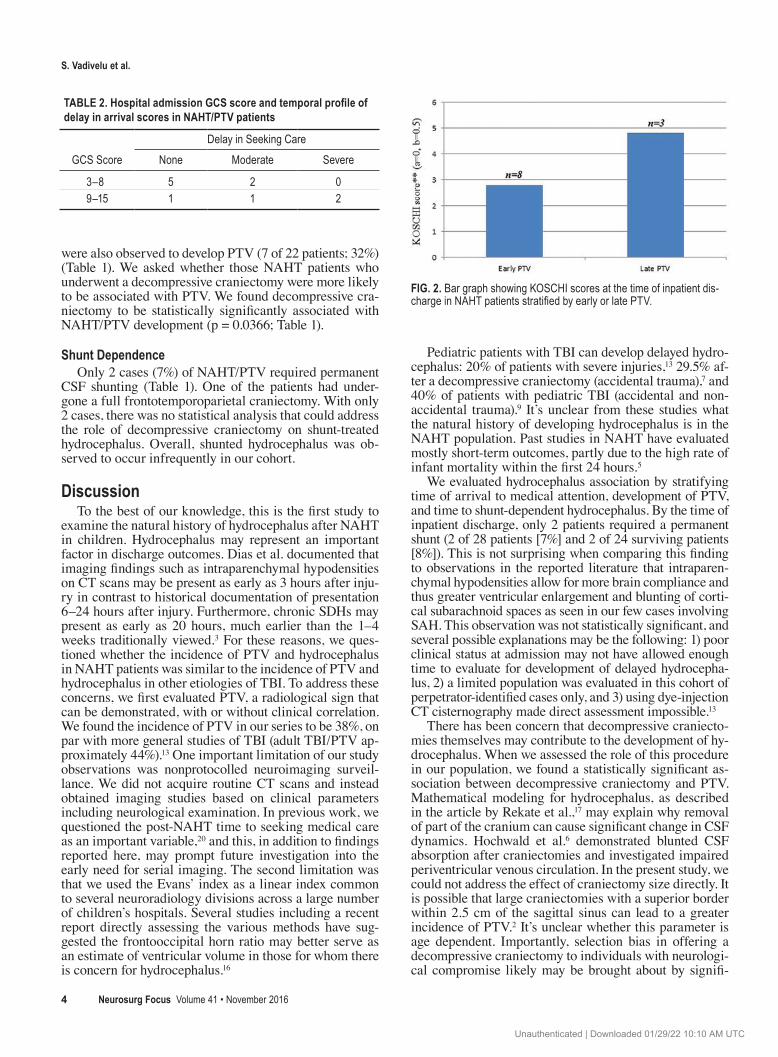
tients with and without PTV. Considering patients with early mortality who may not have undergone serial scan-ning, we were unable to perform any meaningful statistical comparison. However, when we examined those NAHT patients demonstrating PTV early (< 1 week) versus late (> 1 week), poorer outcomes at inpatient discharge were observed in patients with early PTV than in those present-ing with late PTV (KOSCHI score < 3; Fig. 2).
Ptv and Decompressive CraniectomyDecompressive craniectomies may be life-sustaining
measures in patients with traumatic brain injury (TBI). Postoperative sequelae in such patients have been a subject of recent debate, including the development hydrocepha-lus. Here, we analyzed NAHT/PTV patients who under-went decompressive craniectomy. No patient had PTV on arrival at our hospital. Large frontotemporoparietal crani-ectomies (n = 5) or a temporal craniectomy (n = 1), which included dural openings, were performed in a total 6 (21%) of 28 NAHT patients when significant cerebral edema or mass effect was present due to either intracranial findings on CT scans or intractable intracranial pressure. PTV was observed to occur in 4 of these patients (67%). NAHT pa-tients who did not undergo a decompressive craniectomy
Fig. 1. Bar graphs showing the development of PTV after NAHT pre-sentation in patients with a low or high GCS score (upper) and with or without delay in seeking acute medical care after injury (lower).
tAble 1. summary of Ptv data and their associated radiological findings
Characteristic No. of Patients p Value
PTV incidence in NAHT Overall PTV incidence 11/28 (39%) PTV incidence w/o craniectomy 7/22 (32%) PTV incidence w/ craniectomy 4/6 (67%) 0.0366PTV & GCS Mild/moderate NAHT (GCS Score 9–15) 4/12 (33%) Severe NAHT (GCS Score 3–8) 7/16 (44%)Permanent VPS placed* 2/28 (7%) VPS placed after craniectomy† 1/6 (17%)Admission CT findings‡ Convexity SDH (unilat and bilat) 8 Interhemispheric/tentorial SDH 6 SAH 3 0.011 IVH 1 Ischemic stroke 7
VPS = ventriculoperitoneal shunt.* Both cases presented with severe NAHT (GCS score of 3–8) and without delayed hospital admission.† Limited temporal craniectomy with large superior frontoparietal craniotomy.‡ Total number of PTV/NAHT cases was 11.
Unauthenticated | Downloaded 01/29/22 10:10 AM UTC
s. vadivelu et al.
Neurosurg Focus Volume 41 • November 20164
were also observed to develop PTV (7 of 22 patients; 32%) (Table 1). We asked whether those NAHT patients who underwent a decompressive craniectomy were more likely to be associated with PTV. We found decompressive cra-niectomy to be statistically significantly associated with NAHT/PTV development (p = 0.0366; Table 1).
shunt DependenceOnly 2 cases (7%) of NAHT/PTV required permanent
CSF shunting (Table 1). One of the patients had under-gone a full frontotemporoparietal craniectomy. With only 2 cases, there was no statistical analysis that could address the role of decompressive craniectomy on shunt-treated hydrocephalus. Overall, shunted hydrocephalus was ob-served to occur infrequently in our cohort.
DiscussionTo the best of our knowledge, this is the first study to
examine the natural history of hydrocephalus after NAHT in children. Hydrocephalus may represent an important factor in discharge outcomes. Dias et al. documented that imaging findings such as intraparenchymal hypodensities on CT scans may be present as early as 3 hours after inju-ry in contrast to historical documentation of presentation 6–24 hours after injury. Furthermore, chronic SDHs may present as early as 20 hours, much earlier than the 1–4 weeks traditionally viewed.3 For these reasons, we ques-tioned whether the incidence of PTV and hydrocephalus in NAHT patients was similar to the incidence of PTV and hydrocephalus in other etiologies of TBI. To address these concerns, we first evaluated PTV, a radiological sign that can be demonstrated, with or without clinical correlation. We found the incidence of PTV in our series to be 38%, on par with more general studies of TBI (adult TBI/PTV ap-proximately 44%).13 One important limitation of our study observations was nonprotocolled neuroimaging surveil-lance. We did not acquire routine CT scans and instead obtained imaging studies based on clinical parameters including neurological examination. In previous work, we questioned the post-NAHT time to seeking medical care as an important variable,20 and this, in addition to findings reported here, may prompt future investigation into the early need for serial imaging. The second limitation was that we used the Evans’ index as a linear index common to several neuroradiology divisions across a large number of children’s hospitals. Several studies including a recent report directly assessing the various methods have sug-gested the frontooccipital horn ratio may better serve as an estimate of ventricular volume in those for whom there is concern for hydrocephalus.16
Pediatric patients with TBI can develop delayed hydro-cephalus: 20% of patients with severe injuries,13 29.5% af-ter a decompressive craniectomy (accidental trauma),7 and 40% of patients with pediatric TBI (accidental and non-accidental trauma).9 It’s unclear from these studies what the natural history of developing hydrocephalus is in the NAHT population. Past studies in NAHT have evaluated mostly short-term outcomes, partly due to the high rate of infant mortality within the first 24 hours.5
We evaluated hydrocephalus association by stratifying time of arrival to medical attention, development of PTV, and time to shunt-dependent hydrocephalus. By the time of inpatient discharge, only 2 patients required a permanent shunt (2 of 28 patients [7%] and 2 of 24 surviving patients [8%]). This is not surprising when comparing this finding to observations in the reported literature that intraparen-chymal hypodensities allow for more brain compliance and thus greater ventricular enlargement and blunting of corti-cal subarachnoid spaces as seen in our few cases involving SAH. This observation was not statistically significant, and several possible explanations may be the following: 1) poor clinical status at admission may not have allowed enough time to evaluate for development of delayed hydrocepha-lus, 2) a limited population was evaluated in this cohort of perpetrator-identified cases only, and 3) using dye-injection CT cisternography made direct assessment impossible.13
There has been concern that decompressive craniecto-mies themselves may contribute to the development of hy-drocephalus. When we assessed the role of this procedure in our population, we found a statistically significant as-sociation between decompressive craniectomy and PTV. Mathematical modeling for hydrocephalus, as described in the article by Rekate et al.,17 may explain why removal of part of the cranium can cause significant change in CSF dynamics. Hochwald et al.6 demonstrated blunted CSF absorption after craniectomies and investigated impaired periventricular venous circulation. In the present study, we could not address the effect of craniectomy size directly. It is possible that large craniectomies with a superior border within 2.5 cm of the sagittal sinus can lead to a greater incidence of PTV.2 It’s unclear whether this parameter is age dependent. Importantly, selection bias in offering a decompressive craniectomy to individuals with neurologi-cal compromise likely may be brought about by signifi-
TABLE 2. Hospital admission GCS score and temporal profile of delay in arrival scores in NAht/Ptv patients
GCS ScoreDelay in Seeking Care
None Moderate Severe
3–8 5 2 09–15 1 1 2
Fig. 2. Bar graph showing KOSCHI scores at the time of inpatient dis-charge in NAHT patients stratified by early or late PTV.
Unauthenticated | Downloaded 01/29/22 10:10 AM UTC
hydrocephalus in childhood nonaccidental head trauma
Neurosurg Focus Volume 41 • November 2016 5
cant intracranial findings such as hypodensities related to stroke or intracranial hemorrhage, thus having a greater propensity toward developing PTV.
ConclusionsThis is the first report to identify the cumulative indices
of ventriculomegaly and shunt-dependent hydrocephalus after NAHT. We observed the rate of PTV in our cohort to be similar to that after accidental TBI, and this may, in part, be related to decompressive craniectomies. Furthermore, we observed a low rate of subsequent shunt-dependent hy-drocephalus. Potentially a larger collaborative prospective accrual of patients undergoing standardized hydrocepha-lus assessment and treatment with shunt placement proto-cols may confirm the observations reported here.
AcknowledgmentsWe thank Drs. Reginald Guerriero (Harvard University) and
Aditee P. Narayan (Duke University) for critical comments and Ms. Nina Kohn and the Feinstein Institute for Medical Research for statistical assistance.
references 1. Calvert S, Miller HE, Curran A, Hameed B, McCarter R,
Edwards RJ, et al: The King’s Outcome Scale for Childhood Head Injury and injury severity and outcome measures in children with traumatic brain injury. Dev Med Child Neurol 50:426–431, 2008
2. De Bonis P, Pompucci A, Mangiola A, Rigante L, Anile C: Post-traumatic hydrocephalus after decompressive craniecto-my: an underestimated risk factor. J Neurotrauma 27:1965–1970, 2010
3. Dias MS, Backstrom J, Falk M, Li V: Serial radiography in the infant shaken impact syndrome. Pediatr Neurosurg 29:77–85, 1998
4. Esernio-Jenssen D, Tai J, Kodsi S: Abusive head trauma in children: a comparison of male and female perpetrators. Pe-diatrics 127:649–657, 2011
5. Graupman P, Winston KR: Nonaccidental head trauma as a cause of childhood death. J Neurosurg 104 (4 Sup-pl):245–250, 2006
6. Hochwald GM, Epstein F, Malhan C, Ransohoff J: The rela-tionship of compensated to decompensated hydrocephalus in the cat. J Neurosurg 39:694–697, 1973
7. Jagannathan J, Okonkwo DO, Dumont AS, Ahmed H, Bahari A, Prevedello DM, et al: Outcome following decompressive craniectomy in children with severe traumatic brain injury: a 10-year single-center experience with long-term follow up. J Neurosurg 106 (4 Suppl):268–275, 2007
8. Jayawant S, Rawlinson A, Gibbon F, Price J, Schulte J, Shar-ples P, et al: Subdural haemorrhages in infants: population based study. BMJ 317:1558–1561, 1998
9. Kan P, Amini A, Hansen K, White GL Jr, Brockmeyer DL, Walker ML, et al: Outcomes after decompressive craniecto-my for severe traumatic brain injury in children. J Neuro-surg 105 (5 Suppl):337–342, 2006
10. Kelly P, Farrant B: Shaken baby syndrome in New Zealand, 2000–2002. J Paediatr Child Health 44:99–107, 2008
11. Kesler H, Dias MS, Shaffer M, Rottmund C, Cappos K, Thomas NJ: Demographics of abusive head trauma in the Commonwealth of Pennsylvania. J Neurosurg Pediatr 1:351–356, 2008
12. Ludwig S, Warman M: Shaken baby syndrome: a review of 20 cases. Ann Emerg Med 13:104–107, 1984
13. Marmarou A, Foda MA, Bandoh K, Yoshihara M, Yama-moto T, Tsuji O, et al: Posttraumatic ventriculomegaly: hy-drocephalus or atrophy? A new approach for diagnosis using CSF dynamics. J Neurosurg 85:1026–1035, 1996
14. Mazzini L, Campini R, Angelino E, Rognone F, Pastore I, Oliveri G: Posttraumatic hydrocephalus: a clinical, neuro-radiologic, and neuropsychologic assessment of long-term outcome. Arch Phys Med Rehabil 84:1637–1641, 2003
15. Poca MA, Sahuquillo J, Mataró M, Benejam B, Arikan F, Báguena M: Ventricular enlargement after moderate or se-vere head injury: a frequent and neglected problem. J Neu-rotrauma 22:1303–1310, 2005
16. Ragan DK, Cerqua J, Nash T, McKinstry RC, Shimony JS, Jones BV, et al: The accuracy of linear indices of ventricular volume in pediatric hydrocephalus: technical note. J Neuro-surg Pediatr 15:547–551, 2015
17. Rekate HL, Brodkey JA, Chizeck HJ, el Sakka W, Ko WH: Ventricular volume regulation: a mathematical model and computer simulation. Pediatr Neurosci 14:77–84, 1988
18. Starling SP, Patel S, Burke BL, Sirotnak AP, Stronks S, Ros-quist P: Analysis of perpetrator admissions to inflicted trau-matic brain injury in children. Arch Pediatr Adolesc Med 158:454–458, 2004
19. Stiver SI: Complications of decompressive craniectomy for traumatic brain injury. Neurosurg Focus 26(6):E7, 2009
20. Vadivelu S, Esernio-Jenssen D, Rekate HL, Narayan RK, Mittler MA, Schneider SJ: Delay in arrival to care in perpe-trator-identified nonaccidental head trauma: observations and outcomes. World Neurosurg 84:1340–1346, 2015
21. Vinchon M, de Foort-Dhellemmes S, Desurmont M, De-lestret I: Confessed abuse versus witnessed accidents in infants: comparison of clinical, radiological, and ophthal-mological data in corroborated cases. Childs Nerv Syst 26:637–645, 2010
22. Waziri A, Fusco D, Mayer SA, McKhann GM II, Connolly ES Jr: Postoperative hydrocephalus in patients undergoing decompressive hemicraniectomy for ischemic or hemor-rhagic stroke. Neurosurgery 61:489–494, 2007
DisclosuresThe authors report no conflict of interest concerning the materi-als or methods used in this study or the findings specified in this paper.
Author ContributionsConception and design: Vadivelu. Acquisition of data: Vadivelu, Esernio-Jenssen. Analysis and interpretation of data: Vadivelu, Rekate. Drafting the article: Vadivelu. Critically revising the article: Vadivelu, Rekate. Reviewed submitted version of manu-script: Vadivelu, Rekate, Esernio-Jenssen. Approved the final ver-sion of the manuscript on behalf of all authors: Vadivelu. Statisti-cal analysis: Vadivelu. Administrative/technical/material support: Mittler, Schneider. Study supervision: all authors.
supplemental informationPrevious PresentationsThis work was presented in part at the 2011 AANS Annual Sci-entific Meeting, Denver, Colorado, April 9–13, 2011, and the 2011 Annual Meeting of the Congress of Neurological Surgeons, Washington, DC, October 1–6, 2011.
CorrespondenceSudhakar Vadivelu, Division of Pediatric Neurosurgery, Cincin-nati Children’s Hospital Medical Center, 3333 Burnett Ave., Cin-cinnati, OH 45040. email: [email protected].
Unauthenticated | Downloaded 01/29/22 10:10 AM UTC