Embed Size (px)
Citation preview
RADIOLOGY—ORIGINAL ARTICLE
Radiographic skeletal survey for non-accidental injury:Systematic review and development of a nationalNew Zealand protocolKarin L. Phillips,1* Sonja T. Bastin,1* David Davies-Payne,1 Diana Browne,1 Helen L. Bird,1 Susan Craw,2
David Duncan,1 Philippa Depree,3 Alina Leigh,4 Andrew McLaughlin,1,5 Russell Metcalfe,1 Jean Murdoch,6
Kirsten Pearce,7 David Perry1 Iona Thomas,1 Glen D. Thomson,1 Sally Vogel,1 Francessa Wilson1 andRita L. Teele1,8
1 Department of Paediatric Radiology, Starship Children’s Hospital, Auckland, New Zealand
2 Department of Radiology, Dunedin Hospital, Dunedin, New Zealand
3 Department of Radiology, Christchurch Hospital, Christchurch, New Zealand
4 Fulford Radiology, Taranaki Base Hospital, New Plymouth, New Zealand
5 Department of Radiology, Middlemore Hospital, Auckland, New Zealand
6 Department of Radiology, Capital and Coast District Health Board, Wellington, New Zealand
7 Department of Radiology, Waitemata District Health Board, Auckland, New Zealand
8 University of Auckland School of Medicine, Auckland, New Zealand
KL Phillips MB ChB; ST Bastin MB ChB, Dip
Paeds, FRANZCR; D Davies-Payne BMedSc,
MB ChB, FRANZCR; D Browne DSR; HL BirdMB ChB, FRANZCR; S Craw MB ChB, DDR,
FRANZCR; D Duncan MB ChB, FRANZCR;
P Depree MB ChB, Dip Child Health, FRANZCR;
A Leigh MB ChB, FRANZCR; A McLaughlinBMedSc, MB ChB, FRANZCR; R Metcalfe MB
ChB, Dip Child Health, FRANZCR; J MurdochMD, FRCP(C), FRANZCR; K Pearce MB ChB,
FRANZCR; D Perry MB ChB, FRANZCR;
I Thomas MB ChB, FRANZCR; GD ThomsonMB ChB, FRANZCR; S Vogel BA, MD,
FRANZCR; F Wilson MB ChB, FRANZCR;
RL Teele MD, FRANZCR.
CorrespondenceSonja T. Bastin, Department of Paediatric
Radiology, Starship Children’s Hospital, Private
Bag 92-024, Auckland 1023, New Zealand.
Email: [email protected]
*KLP and STB contributed equally to this work
Conflict of interest: The authors have no
conflict of interest to declare.
Presented at the annual meeting of the
Australian and New Zealand Society for
Paediatric Radiology, Waiheke Island, Auckland,
New Zealand October 2013.
Submitted 31 July 2014; accepted 20
November 2014.
doi:10.1111/1754-9485.12271
Abstract
Introduction: Clinically occult fractures from non-accidental injury (NAI) arebest detected on radiographic skeletal survey. However, there are regionalvariations regarding the views included in such surveys. We undertook asystematic review of the evidence supporting skeletal survey protocols todesign a protocol that could be implemented across New Zealand.Methods: In June 2013, we searched Medline, Google Scholar, the Cochranedatabase, UpToDate and relevant reference lists for English-language publi-cations on skeletal survey in NAI from 1946. We included publications thatcontained a protocol or reported evidence supporting including, or excluding,specific views in a skeletal survey. All included publications were criticallyappraised. Based on this systematic review, a draft protocol was developedand presented to an Australian and New Zealand Society for PaediatricRadiology NAI symposium in October 2013. Feedback from the symposiumand later discussions was incorporated into the final protocol.Results: We identified 2 guidelines for skeletal survey, 13 other protocols and15 articles providing evidence for inclusion of specific images in a skeletalsurvey. The guidelines scored poorly on critical appraisal of several aspects oftheir methods. We found no studies that validate any of the protocols orcompare their performance. Evidence supporting inclusion in a skeletal surveyis limited to ribs, spine, pelvis, hands and feet, and long bone views. Our finalprotocol is a standardised, two-tiered protocol consisting of between 17 and22 views.Conclusion: A standardised protocol for radiographic skeletal survey protocolhas been developed in New Zealand. We present it here for consideration byothers.
Key words: child abuse; inflicted injury; non-accidental injury; radiography;skeletal survey; systematic review.
bs_bs_banner
Journal of Medical Imaging and Radiation Oncology 59 (2015) 54–65
© 2015 The Royal Australian and New Zealand College of Radiologists54
Introduction
In children with non-accidental injury (NAI), fracturesare the most common finding after bruising and cutane-ous injuries.1 Most fractures are clinically occult and bestdetected by a skeletal survey – a standard series ofradiographic images that visualise the entire skeleton.2,3
A skeletal survey of high technical quality with appropri-ate images allows the radiologist to accurately identifyand interpret occult skeletal injury. However, there isvariation in the views that are obtained.4–8 As identifica-tion of fractures plays a key role in the diagnosis of NAI,it is important that appropriate evidence-based protocolsfor skeletal surveys are developed and consistentlyimplemented. We undertook a systematic review of pub-lished protocols for skeletal surveys and the evidencesupporting them and, based on this review, developed aprotocol for implementation in New Zealand.
Methods
Systematic review
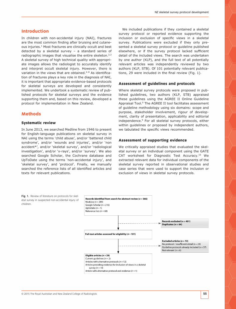
In June 2013, we searched Medline from 1946 to presentfor English-language publications on skeletal survey inNAI using the terms ‘child abuse’, and/or ‘battered childsyndrome’, and/or ‘wounds and injuries’, and/or ‘nonaccident*’, and/or ‘skeletal survey’, and/or ‘radiologicalinvestigation’, and/or ‘x-rays’, and/or ‘survey’. We alsosearched Google Scholar, the Cochrane database andUpToDate using the terms ‘non-accidental injury’, and‘skeletal survey’, and ‘protocol’. Finally, we manuallysearched the reference lists of all identified articles andtexts for relevant publications.
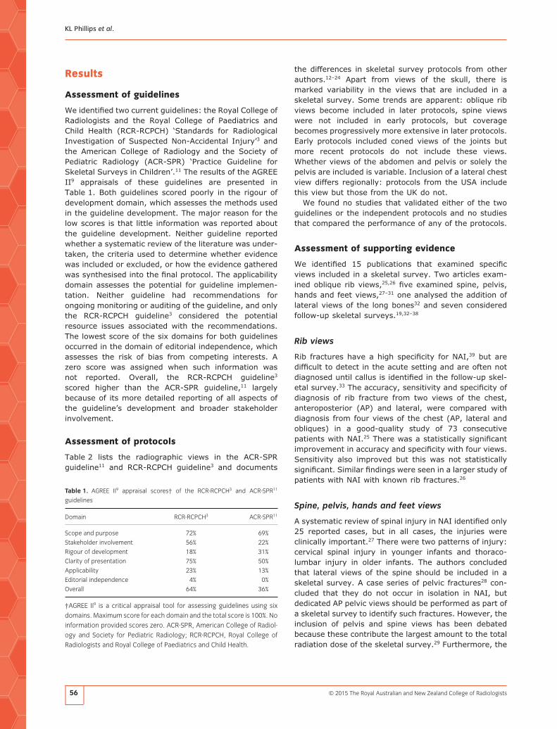
We included publications if they contained a skeletalsurvey protocol or reported evidence supporting theinclusion or exclusion of specific views in a skeletalsurvey. Publications were excluded if they only pre-sented a skeletal survey protocol or guideline publishedelsewhere, or if the survey protocol lacked sufficientdetail of the included views. The search was undertakenby one author (KLP), and the full text of all potentiallyrelevant articles was independently reviewed by twoauthors (KLP, STB). Of 101 potentially relevant publica-tions, 29 were included in the final review (Fig. 1).
Assessment of guidelines and protocols
Where skeletal survey protocols were proposed in pub-lished guidelines, two authors (KLP, STB) appraisedthese guidelines using the AGREE II Online GuidelineAppraisal Tool.9 The AGREE II tool facilitates assessmentof guideline methodology using six domains: scope andpurpose, stakeholder involvement, rigour of develop-ment, clarity of presentation, applicability and editorialindependence.9 For all skeletal survey protocols, eitherwithin guidelines or proposed by independent authors,we tabulated the specific views recommended.
Assessment of supporting evidence
We critically appraised studies that evaluated the skel-etal survey or an individual component using the GATECAT worksheet for Diagnostic Test Accuracy.10 Weextracted relevant data for individual components of theskeletal survey reported in observational studies andcase series that were used to support the inclusion orexclusion of views in skeletal survey protocols.
Fig. 1. Review of literature on protocols for skel-
etal survey in suspected non-accidental injury of
children.
NZ skeletal survey protocol development
© 2015 The Royal Australian and New Zealand College of Radiologists 55
Results
Assessment of guidelines
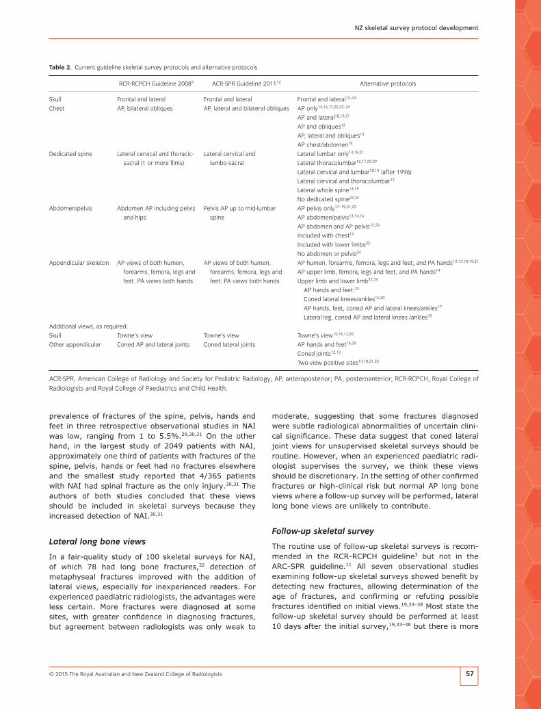
We identified two current guidelines: the Royal College ofRadiologists and the Royal College of Paediatrics andChild Health (RCR-RCPCH) ‘Standards for RadiologicalInvestigation of Suspected Non-Accidental Injury’3 andthe American College of Radiology and the Society ofPediatric Radiology (ACR-SPR) ‘Practice Guideline forSkeletal Surveys in Children’.11 The results of the AGREEII9 appraisals of these guidelines are presented inTable 1. Both guidelines scored poorly in the rigour ofdevelopment domain, which assesses the methods usedin the guideline development. The major reason for thelow scores is that little information was reported aboutthe guideline development. Neither guideline reportedwhether a systematic review of the literature was under-taken, the criteria used to determine whether evidencewas included or excluded, or how the evidence gatheredwas synthesised into the final protocol. The applicabilitydomain assesses the potential for guideline implemen-tation. Neither guideline had recommendations forongoing monitoring or auditing of the guideline, and onlythe RCR-RCPCH guideline3 considered the potentialresource issues associated with the recommendations.The lowest score of the six domains for both guidelinesoccurred in the domain of editorial independence, whichassesses the risk of bias from competing interests. Azero score was assigned when such information wasnot reported. Overall, the RCR-RCPCH guideline3
scored higher than the ACR-SPR guideline,11 largelybecause of its more detailed reporting of all aspects ofthe guideline’s development and broader stakeholderinvolvement.
Assessment of protocols
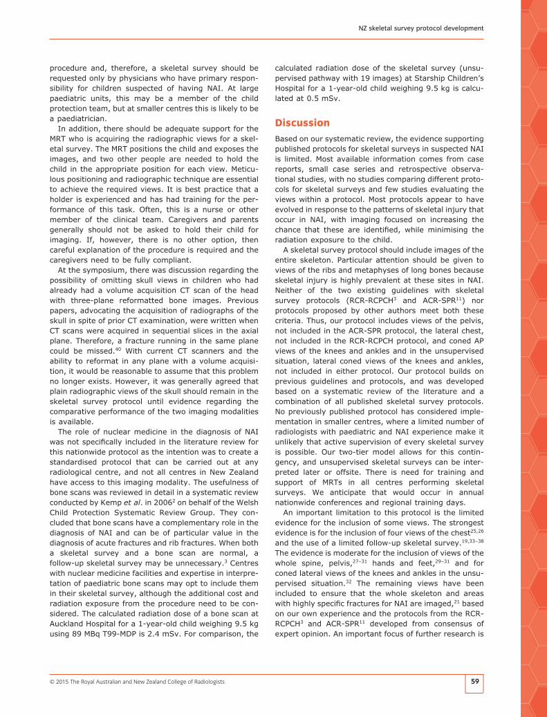
Table 2 lists the radiographic views in the ACR-SPRguideline11 and RCR-RCPCH guideline3 and documents
the differences in skeletal survey protocols from otherauthors.12–24 Apart from views of the skull, there ismarked variability in the views that are included in askeletal survey. Some trends are apparent: oblique ribviews become included in later protocols, spine viewswere not included in early protocols, but coveragebecomes progressively more extensive in later protocols.Early protocols included coned views of the joints butmore recent protocols do not include these views.Whether views of the abdomen and pelvis or solely thepelvis are included is variable. Inclusion of a lateral chestview differs regionally: protocols from the USA includethis view but those from the UK do not.
We found no studies that validated either of the twoguidelines or the independent protocols and no studiesthat compared the performance of any of the protocols.
Assessment of supporting evidence
We identified 15 publications that examined specificviews included in a skeletal survey. Two articles exam-ined oblique rib views,25,26 five examined spine, pelvis,hands and feet views,27–31 one analysed the addition oflateral views of the long bones32 and seven consideredfollow-up skeletal surveys.19,32–38
Rib views
Rib fractures have a high specificity for NAI,39 but aredifficult to detect in the acute setting and are often notdiagnosed until callus is identified in the follow-up skel-etal survey.33 The accuracy, sensitivity and specificity ofdiagnosis of rib fracture from two views of the chest,anteroposterior (AP) and lateral, were compared withdiagnosis from four views of the chest (AP, lateral andobliques) in a good-quality study of 73 consecutivepatients with NAI.25 There was a statistically significantimprovement in accuracy and specificity with four views.Sensitivity also improved but this was not statisticallysignificant. Similar findings were seen in a larger study ofpatients with NAI with known rib fractures.26
Spine, pelvis, hands and feet views
A systematic review of spinal injury in NAI identified only25 reported cases, but in all cases, the injuries wereclinically important.27 There were two patterns of injury:cervical spinal injury in younger infants and thoraco-lumbar injury in older infants. The authors concludedthat lateral views of the spine should be included in askeletal survey. A case series of pelvic fractures28 con-cluded that they do not occur in isolation in NAI, butdedicated AP pelvic views should be performed as part ofa skeletal survey to identify such fractures. However, theinclusion of pelvis and spine views has been debatedbecause these contribute the largest amount to the totalradiation dose of the skeletal survey.29 Furthermore, the
Table 1. AGREE II9 appraisal scores† of the RCR-RCPCH3 and ACR-SPR11
guidelines
Domain RCR-RCPCH3 ACR-SPR11
Scope and purpose 72% 69%
Stakeholder involvement 56% 22%
Rigour of development 18% 31%
Clarity of presentation 75% 50%
Applicability 23% 13%
Editorial independence 4% 0%
Overall 64% 36%
†AGREE II9 is a critical appraisal tool for assessing guidelines using six
domains. Maximum score for each domain and the total score is 100%. No
information provided scores zero. ACR-SPR, American College of Radiol-
ogy and Society for Pediatric Radiology; RCR-RCPCH, Royal College of
Radiologists and Royal College of Paediatrics and Child Health.
KL Phillips et al.
© 2015 The Royal Australian and New Zealand College of Radiologists56
prevalence of fractures of the spine, pelvis, hands andfeet in three retrospective observational studies in NAIwas low, ranging from 1 to 5.5%.29,30,31 On the otherhand, in the largest study of 2049 patients with NAI,approximately one third of patients with fractures of thespine, pelvis, hands or feet had no fractures elsewhereand the smallest study reported that 4/365 patientswith NAI had spinal fracture as the only injury.30,31 Theauthors of both studies concluded that these viewsshould be included in skeletal surveys because theyincreased detection of NAI.30,31
Lateral long bone views
In a fair-quality study of 100 skeletal surveys for NAI,of which 78 had long bone fractures,32 detection ofmetaphyseal fractures improved with the addition oflateral views, especially for inexperienced readers. Forexperienced paediatric radiologists, the advantages wereless certain. More fractures were diagnosed at somesites, with greater confidence in diagnosing fractures,but agreement between radiologists was only weak to
moderate, suggesting that some fractures diagnosedwere subtle radiological abnormalities of uncertain clini-cal significance. These data suggest that coned lateraljoint views for unsupervised skeletal surveys should beroutine. However, when an experienced paediatric radi-ologist supervises the survey, we think these viewsshould be discretionary. In the setting of other confirmedfractures or high-clinical risk but normal AP long boneviews where a follow-up survey will be performed, laterallong bone views are unlikely to contribute.
Follow-up skeletal survey
The routine use of follow-up skeletal surveys is recom-mended in the RCR-RCPCH guideline3 but not in theARC-SPR guideline.11 All seven observational studiesexamining follow-up skeletal surveys showed benefit bydetecting new fractures, allowing determination of theage of fractures, and confirming or refuting possiblefractures identified on initial views.19,33–38 Most state thefollow-up skeletal survey should be performed at least10 days after the initial survey,19,33–38 but there is more
Table 2. Current guideline skeletal survey protocols and alternative protocols
RCR-RCPCH Guideline 20083 ACR-SPR Guideline 201112 Alternative protocols
Skull Frontal and lateral Frontal and lateral Frontal and lateral12–24
Chest AP, bilateral obliques AP, lateral and bilateral obliques AP only14,16,17,20,22–24
AP and lateral18,19,21
AP and obliques12
AP, lateral and obliques13
AP chest/abdomen15
Dedicated spine Lateral cervical and thoracic-
sacral (1 or more films)
Lateral cervical and
lumbo-sacral
Lateral lumbar only14,19,21
Lateral thoracolumbar16,17,20,22
Lateral cervical and lumbar18,19 (after 1996)
Lateral cervical and thoracolumbar12
Lateral whole spine13,15
No dedicated spine23,24
Abdomen/pelvis Abdomen AP including pelvis
and hips
Pelvis AP up to mid-lumbar
spine
AP pelvis only17–19,21,23
AP abdomen/pelvis13,14,16
AP abdomen and AP pelvis12,20
Included with chest15
Included with lower limbs22
No abdomen or pelvis24
Appendicular skeleton AP views of both humeri,
forearms, femora, legs and
feet. PA views both hands.
AP views of both humeri,
forearms, femora, legs and
feet. PA views both hands.
AP humeri, forearms, femora, legs and feet, and PA hands12,13,18,19,21
AP upper limb, femora, legs and feet, and PA hands14
Upper limb and lower limb22,23
AP hands and feet:24
Coned lateral knees/ankles16,20
AP hands, feet, coned AP and lateral knees/ankles17
Lateral leg, coned AP and lateral knees /ankles15
Additional views, as required:
Skull Towne’s view Towne’s view Towne’s view13,16,17,20
Other appendicular Coned AP and lateral joints Coned lateral joints AP hands and feet16,20
Coned joints12,13
Two-view positive sites13,18,21,22
ACR-SPR, American College of Radiology and Society for Pediatric Radiology; AP, anteroposterior; PA, posteroanterior; RCR-RCPCH, Royal College of
Radiologists and Royal College of Paediatrics and Child Health.
NZ skeletal survey protocol development
© 2015 The Royal Australian and New Zealand College of Radiologists 57

variation in the upper time limit (21 days to 6weeks).34,36–38 Several use a more general ‘approxi-mately two weeks’19,33,35,36 and this seems reasonable forsimplification purposes. In the two studies that analysedlocation of fracture, the only new fractures identified inthe follow-up skeletal survey were in the ribs and longbones, suggesting that pelvis and spine views are unnec-essary in follow-up surveys where no initial fracture wasobserved in these sites.33,34 Not including these viewswould also reduce the radiation dose of the follow-upsurvey.
Protocol development
Based on the findings from the systematic review of theliterature, we developed a draft protocol for use in NewZealand. It needed to be sufficiently flexible to accom-modate all situations in which children with suspectedNAI would have radiographic examination. Thus, theprotocol had to be suitable for radiology departments indedicated paediatric centres and for departments ingeneral hospitals or rural centres where a supervisingradiologist, experienced in paediatric imaging, is notimmediately available. A two-tiered protocol was devel-oped, presented to participants at the NAI Symposium,Australian and New Zealand Society for Paediatric Radi-ology in October 2013 and then discussed with attend-ees. All but one of the currently practising paediatricradiologists in New Zealand were in attendance, along
with the clinical director of the only child abuse team inNew Zealand (Te Puaruruhua, Starship Children’s Hos-pital), medical radiation technologists (MRTs) with aninterest in NAI and a crown prosecuting lawyer involvedin litigation of NAI. Feedback from this meeting and fromlater discussions among all paediatric radiologists in NewZealand was incorporated into the final protocol (Fig. 2).A pictorial version of the entire radiographic skeletalsurvey is included for reference, along with a descriptionof the radiographic technique required to achieve theseviews (Appendices I and II).
The selection of one of the two pathways in the proto-col depends on the presence or absence of a supervisingradiologist at the time of the skeletal survey. If a radi-ologist supervises the survey, a Towne’s view of the skulland coned lateral views of the knees and the ankles areunnecessary unless a fracture or suspicious finding ispresent on standard views of these regions. Separateviews of the spine can be eliminated if the vertebraehave been imaged adequately (e.g. on a lateral chestview). However, in the unsupervised situation, the risk ofmissing fractures because of inadequate imaging ofthese regions outweighs the slight increase in radiationdose in our view.
There is also flexibility in the protocol for dealing withan uncooperative or large child where image quality canbe influenced by motion and divergent X-ray beam.
The strong view of symposium attendees was that askeletal survey is not a screening test but a diagnostic
Fig. 2. Consensus New Zealand protocol for
radiographic skeletal survey in suspected non-
accidental injury. AP, anteroposterior; DP,
dorsoposterior.
KL Phillips et al.
© 2015 The Royal Australian and New Zealand College of Radiologists58
procedure and, therefore, a skeletal survey should berequested only by physicians who have primary respon-sibility for children suspected of having NAI. At largepaediatric units, this may be a member of the childprotection team, but at smaller centres this is likely to bea paediatrician.
In addition, there should be adequate support for theMRT who is acquiring the radiographic views for a skel-etal survey. The MRT positions the child and exposes theimages, and two other people are needed to hold thechild in the appropriate position for each view. Meticu-lous positioning and radiographic technique are essentialto achieve the required views. It is best practice that aholder is experienced and has had training for the per-formance of this task. Often, this is a nurse or othermember of the clinical team. Caregivers and parentsgenerally should not be asked to hold their child forimaging. If, however, there is no other option, thencareful explanation of the procedure is required and thecaregivers need to be fully compliant.
At the symposium, there was discussion regarding thepossibility of omitting skull views in children who hadalready had a volume acquisition CT scan of the headwith three-plane reformatted bone images. Previouspapers, advocating the acquisition of radiographs of theskull in spite of prior CT examination, were written whenCT scans were acquired in sequential slices in the axialplane. Therefore, a fracture running in the same planecould be missed.40 With current CT scanners and theability to reformat in any plane with a volume acquisi-tion, it would be reasonable to assume that this problemno longer exists. However, it was generally agreed thatplain radiographic views of the skull should remain in theskeletal survey protocol until evidence regarding thecomparative performance of the two imaging modalitiesis available.
The role of nuclear medicine in the diagnosis of NAIwas not specifically included in the literature review forthis nationwide protocol as the intention was to create astandardised protocol that can be carried out at anyradiological centre, and not all centres in New Zealandhave access to this imaging modality. The usefulness ofbone scans was reviewed in detail in a systematic reviewconducted by Kemp et al. in 20062 on behalf of the WelshChild Protection Systematic Review Group. They con-cluded that bone scans have a complementary role in thediagnosis of NAI and can be of particular value in thediagnosis of acute fractures and rib fractures. When botha skeletal survey and a bone scan are normal, afollow-up skeletal survey may be unnecessary.3 Centreswith nuclear medicine facilities and expertise in interpre-tation of paediatric bone scans may opt to include themin their skeletal survey, although the additional cost andradiation exposure from the procedure need to be con-sidered. The calculated radiation dose of a bone scan atAuckland Hospital for a 1-year-old child weighing 9.5 kgusing 89 MBq T99-MDP is 2.4 mSv. For comparison, the
calculated radiation dose of the skeletal survey (unsu-pervised pathway with 19 images) at Starship Children’sHospital for a 1-year-old child weighing 9.5 kg is calcu-lated at 0.5 mSv.
Discussion
Based on our systematic review, the evidence supportingpublished protocols for skeletal surveys in suspected NAIis limited. Most available information comes from casereports, small case series and retrospective observa-tional studies, with no studies comparing different proto-cols for skeletal surveys and few studies evaluating theviews within a protocol. Most protocols appear to haveevolved in response to the patterns of skeletal injury thatoccur in NAI, with imaging focused on increasing thechance that these are identified, while minimising theradiation exposure to the child.
A skeletal survey protocol should include images of theentire skeleton. Particular attention should be given toviews of the ribs and metaphyses of long bones becauseskeletal injury is highly prevalent at these sites in NAI.Neither of the two existing guidelines with skeletalsurvey protocols (RCR-RCPCH3 and ACR-SPR11) norprotocols proposed by other authors meet both thesecriteria. Thus, our protocol includes views of the pelvis,not included in the ACR-SPR protocol, the lateral chest,not included in the RCR-RCPCH protocol, and coned APviews of the knees and ankles and in the unsupervisedsituation, lateral coned views of the knees and ankles,not included in either protocol. Our protocol builds onprevious guidelines and protocols, and was developedbased on a systematic review of the literature and acombination of all published skeletal survey protocols.No previously published protocol has considered imple-mentation in smaller centres, where a limited number ofradiologists with paediatric and NAI experience make itunlikely that active supervision of every skeletal surveyis possible. Our two-tier model allows for this contin-gency, and unsupervised skeletal surveys can be inter-preted later or offsite. There is need for training andsupport of MRTs in all centres performing skeletalsurveys. We anticipate that would occur in annualnationwide conferences and regional training days.
An important limitation to this protocol is the limitedevidence for the inclusion of some views. The strongestevidence is for the inclusion of four views of the chest25,26
and the use of a limited follow-up skeletal survey.19,33–38
The evidence is moderate for the inclusion of views of thewhole spine, pelvis,27–31 hands and feet,29–31 and forconed lateral views of the knees and ankles in the unsu-pervised situation.32 The remaining views have beenincluded to ensure that the whole skeleton and areaswith highly specific fractures for NAI are imaged,21 basedon our own experience and the protocols from the RCR-RCPCH3 and ACR-SPR11 developed from consensus ofexpert opinion. An important focus of further research is
NZ skeletal survey protocol development
© 2015 The Royal Australian and New Zealand College of Radiologists 59
to provide better evidence for inclusion or exclusion ofindividual views in skeletal surveys.
After the implementation of the protocol nationwide,an audit to assess uptake of the two-tiered protocol willbe conducted. Additionally, once the use of this protocolis well established, it will be important to assess theeffectiveness of the extra views in the unsupervisedsituation. This can be achieved by blinded retrospectivestudy. A study to assess identification of fractures fromradiographic views of the skull compared with imagesfrom volume acquisition cranial CT is being planned atStarship Children’s Hospital. The findings should deter-mine if there is a need for skull radiographs in thosechildren who have had cranial CT.
An important component of the medico-legal issuessurrounding NAI is the quality of radiographic imaging.Currently, there are differences in both the quality andconsistency of such surveys between different radiologydepartments both at a regional level and also betweencountries. We believe that the adoption of a standardisedprotocol in New Zealand will reduce inconsistency andimprove the quality of skeletal surveys, which in turn,will result in increased diagnostic accuracy.
In conclusion, we have developed a protocol for astandardised skeletal survey based on a systematic lit-erature review and consultation with colleagues at anational meeting. While this protocol was developed foruse in New Zealand, the protocol, or its two-tier nature,might be suitable for use in countries with a similar rangeof radiology services. We believe that standardisation ofthe radiographic skeletal survey and ongoing audit willimprove the care of children with suspected NAI.
Acknowledgement
Assistance in developing a search strategy was providedby JM Hobson, Subject Librarian at the Philson Library,University of Auckland School of Medicine.
References
1. Offiah A, van Rijn RR, Perez-Rossello JM, KleinmanPK. Skeletal imaging of child abuse (non-accidentalinjury). Pediatr Radiol 2009; 39: 461–70.
2. Kemp AM, Butler A, Morris S et al. Which radiologicalinvestigations should be performed to identifyfractures in suspected child abuse? Clin Radiol 2006;58: 702–5.
3. The Royal College of Radiologists, the Royal Collegeof Paediatrics and Child Health. Standards forradiological investigations of suspected non-accidentalinjury. London: RCR and RCPCH; 2008. [Cited 6 Jun2013.] Available from URL: https://www.rcr.ac.uk/docs/radiology/pdf/RCPCH_RCR_final.pdf.
4. Kleinman PL, Kleinman PK, Savageau JA. Suspectedinfant abuse: radiographic skeletal survey practices in
pediatric health care facilities. Radiology 2004; 233:477–85.
5. James SLJ, Halliday K, Somers J, Broderick N. Asurvey of non-accidental injury imaging in England,Scotland and Wales. Clin Radiol 2003; 58: 696–701.
6. van Rijn RR, Kieviet N, Hockstra R, Nijs HGT, BiloRAC. Radiology in suspected non-accidental injury:theory and practice in the Netherlands. Eur J Radiol2009; 71: 147–51.
7. Swinson S, Tapp M, Brindley R, Chapman S, Offiah A,Johnson K. An audit of skeletal surveys for suspectednon-accidental injury following publication of theBritish Society of Paediatric Radiology guidelines. ClinRadiol 2008; 63: 651–6.
8. Offiah AC, Hall CM. Observational study of skeletalsurveys in suspected non-accidental injury. ClinRadiol 2003; 58: 702–5.
9. Brouwers M, Kho ME, Browman GP et al. for theAGREE Next Steps Consortium. AGREE II: advancingguideline development, reporting and evaluationin healthcare. Can Med Assoc J 2010; 182:E839–42. [Cited 10 August 2013.] Available fromURL: http://www.agreetrust.org; AGREE II tool.
10. Jackson R, Ameratunga S, Broad J et al. The GATEframe: critical appraisal with pictures. ACP J Club2006; 144: A8–11. [Cited 9 July 2013.] Availablefrom URL: http://www.fmhs.auckland.ac.nz/soph/depts/epi/epiq/ebp.aspx; Gate tool.
11. American College of Radiology. ACR-SPR practiceguideline for skeletal surveys in children. 2011.[Cited 6 June 2013] Available from URL: http://www.acr.org/∼/media/ACR/Documents/PGTS/guidelines/Skeletal_Surveys.pdf.
12. Dwek JR. The radiographic approach to child abuse.Clin Orthop Relat Res 2011; 469: 776–89.
13. McPhillips M. Radiology of child abuse. In: Busuttil A,Keeling JW (eds). Paediatric Forensic Medicine andPathology. Taylor and Francis Group LLC, BocaRaton, 2008; 47–75.
14. Mandelstam SA, Cook D, Fitzgerald M, Ditchfield MR.Complementary use of radiological skeletal surveyand bone scintigraphy in detection of bony injuries insuspected child abuse. Arch Dis Child 2003; 88:387–90.
15. Carty H. Non-accidental injury: a radiologicalperspective. Hong Kong J Emer Med 2001; 8: 40–7.
16. Rao P, Carty H. Non-accidental injury: review of theradiology. Clin Radiol 1999; 54: 11–24.
17. Carty H. Non-accidental injury: a review of theradiology. Eur Radiol 1997; 7: 1365–76.
18. Nimkin K, Kleinman PK. Imaging of child abuse.Pediatr Clin North Am 1997; 44: 615–35.
19. Kleinman PK, Nimkin K, Spevak MR et al. Follow-upskeletal surveys in suspected child abuse. AJR Am JRoentgenol 1996; 167: 893–6.
20. Carty HML. The radiological features of child abuse.Current Paediatrics 1995; 5: 230–5.
21. Kleinman PK. Diagnostic imaging in infant abuse. AJRAm J Roentgenol 1990; 155: 703–12.
KL Phillips et al.
© 2015 The Royal Australian and New Zealand College of Radiologists60
22. Merten DF, Carpenter BLM. Radiologic imaging ofinflicted injury in the child abuse syndrome. PediatrClin North Am 1990; 37: 815–37.
23. Goggins M. Non-accidental injury. Radiogr Today1989; 55: 35.
24. Radkowski MA, Merten DF, Leonidas JC. The abusedchild: criteria for the radiologic diagnosis.Radiographics 1983; 3: 262–97.
25. Ingram JD, Connell J, Hay TC, Strain JD, MackenzieT. Oblique radiographs of the chest in nonaccidentaltrauma. Emerg Radiol 2000; 7: 42–6.
26. Hansen KK, Prince JS, Nixon GW. Oblique chest viewsas a routine part of skeletal surveys performed forpossible physical abuse – is this practice worthwhile?Child Abuse Negl 2008; 32: 155–9.
27. Kemp AM, Joshi AH, Mann M et al. What are theclinical and radiological characteristics of spinalinjuries from physical abuse: a systematic review.Arch Dis Child 2010; 95: 355–60.
28. Starling SP, Heller RM, Jenny C. Pelvic fractures ininfants as a sign of physical abuse. Child Abuse Negl2002; 26: 475–80.
29. Karmazyn B, Lewis ME, Jennings SG, Hibbard RA,Hicks RA. The prevalence of uncommon fractures onskeletal surveys performed to evaluate for suspectedabuse in 930 children: should practice guidelineschange? AJR Am J Roentgenol 2011; 197: W159–63.
30. Lindberg DM, Harper NS, Laskey AL, Berger RP.Prevalence of abusive fractures of the hands, feet,spine, or pelvis on skeletal survey, perhaps‘uncommon’ is more common than suggested. PediatrEmer Care 2013; 29: 26–9.
31. Kleinman PK, Morris NB, Makris J, Moles RL,Kleinman PL. Yield of radiographic skeletal surveysfor detection of hand, foot, and spine fractures insuspected child abuse. AJR Am J Roentgenol 2013;200: 641–4.
32. Karmazyn B, Duhn RD, Jennings SG et al. Long bonefracture detection in suspected child abuse:contribution of lateral views. Pediatr Radiol 2012;42: 463–9.
33. Harper NS, Eddleman S, Lindberg DM, ExSTRAInvestigators. The utility of follow-up skeletal surveysin child abuse. Pediatrics 2013; 131: e672–8.
34. Harlan SR, Nixon GW, Campbell KA, Hansen K, PrinceJS. Follow-up skeletal surveys for nonaccidentaltrauma: can a more limited survey be performed?Pediatr Radiol 2009; 39: 962–8.
35. Bennett BL, Chua MS, Care M, Kachelmeyer A,Mahabee-Gittens M. Retrospective review todetermine the utility of follow-up skeletal surveys inchild abuse evaluations when the initial skeletalsurvey is normal. BMC Res Notes 2011; 4: 354.
36. Singh R, Squires J, Fromkin JB, Berger RP. Assessingthe use of follow-up skeletal surveys in children withsuspected physical abuse. J Trauma Acute Care Surg2012; 7: 972–6.
37. Sonik A, Stein-Wexler R, Rogers KK, Coulter KP,Wootton-Gorges SL. Follow-up skeletal surveys forsuspected non-accidental trauma: can a more limitedsurvey be performed without compromisingdiagnostic information? Child Abuse Negl 2010; 34:804–6.
38. Zimmerman S, Makoroff K, Care M, Thomas A,Shapiro R. Utility of follow-up skeletal surveys insuspected child physical abuse evaluations. ChildAbuse Negl 2005; 29: 1075–83.
39. Kleinman PK. Diagnostic Imaging of Child Abuse, 2ndedn. Mosby, St Louis, MO, 1998.
40. Cohen RA, Kaufman RA, Meyers PA, Towbin RB.Cranial computed tomography in the abused childwith head injury. AJR Am J Roentgenol 1986; 146:97–102.
NZ skeletal survey protocol development
© 2015 The Royal Australian and New Zealand College of Radiologists 61
Appendices
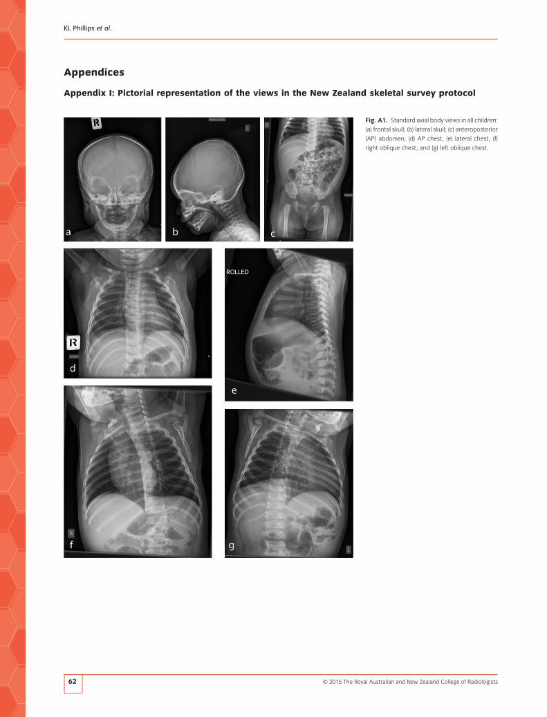
Appendix I: Pictorial representation of the views in the New Zealand skeletal survey protocol
Fig. A1. Standard axial body views in all children:
(a) frontal skull; (b) lateral skull; (c) anteroposterior
(AP) abdomen; (d) AP chest; (e) lateral chest; (f)
right oblique chest; and (g) left oblique chest.
KL Phillips et al.
© 2015 The Royal Australian and New Zealand College of Radiologists62

Fig. A2. Standard appendicular body views in all
children: (a) anteroposterior (AP) lower limbs; (b)
AP upper limb (one side only shown); (c) AP coned
ankles; (d) Dorsoposterior (DP) feet; and (e) AP
coned knees.
NZ skeletal survey protocol development
© 2015 The Royal Australian and New Zealand College of Radiologists 63

Appendix II
Table A1. Description of radiographic techniques for obtaining views in the skeletal survey protocol
Notes All pads and restraining devices should be meticulously clean.
All clothes, identification bracelets, IV cannulas or other overlying material should be removed from the region being imaged.
Modification of images will be required if a known fracture is present.
Skull: for all views Child’s body, including arms, should be wrapped in a sheet.
One holder immobilises the body; one holder positions the head.
Frontal Head holder uses 45-degree foam pads on each side of head in AP position.
Towne’s Head holder uses 45-degree foam pads on each side of the head held.
Small 15-degree pad is placed at base of skull to angle chin down
Lateral Rolled lateral or positioned supine for horizontal beam technique, view includes cervical spine
Head is elevated on flat pad; head holder uses one hand to hold foam pad on crown of head; the other hand holds
chin until immediately prior to exposure.
Chest: for all views One holder immobilises arms; one holder immobilises pelvis.
Exposure is obtained in inspiration.
AP Supine to include clavicles shoulders and entire rib cage
Upper arms held in line with shoulders with elbows flexed (arm in L-shape).
Small 15-degree pad under child’s neck; avoids superimposition of chin on clavicles
Lateral Rolled lateral, arms are held above head, thereby immobilising the head
Obliques Include clavicles, shoulders and entire rib cage
Arms are held above head, thereby immobilising the head.
Child is rolled from supine position to left/right by approximately 30 degrees.
Abdomen Supine view includes pelvis and upper femora.
One holder immobilises child’s arms above head; one holder immobilises legs.
Fig. A3. Additional views in both supervised and
unsupervised studies: (a) lateral whole spine; (b)
lateral lumbo-sacral spine; (c) Towne’s view of
skull; (d) lateral coned knee (one side only shown);
and (e) lateral coned ankle (one side only shown).
KL Phillips et al.
© 2015 The Royal Australian and New Zealand College of Radiologists64

Table A1. Continued
Lower limbs
Full-length AP legs Femurs completely imaged with a combination of this view and the supine abdominal view. Image extends to mid-feet.
If child is too large or uncooperative, image each leg separately or upper and lower legs separately.
One holder immobilised the abdomen. One holder immobilises the feet.
Knees are fully extended. Perspex/plastic ruler over the knees to immobilise
Coned AP knees One holder immobilises the pelvis. One holder immobilises the lower legs.
Coned AP ankles One holder immobilises the knees. One holder immobilises the feet.
Ankle joints are dorsiflexed with pads on soles of feet to avoid superimposition of calcaneus on distal tibiae and fibulae.
Coned lateral knees Rolled lateral
One holder immobilises the pelvis; one holder immobilises both lower legs.
Knees are positioned in 30 degrees of flexion and radiographed separately.
Coned lateral ankles Rolled lateral
One holder immobilises both lower legs. One holder immobilises the feet .
Ankles are imaged separately.
DP feet One holder immobilises lower legs. One holder immobilises toes with Perspex or plastic ruler
Upper limbs
Full-length arm Includes shoulder to hand
Arm is fully extended with elbow in AP projection and hand in supination.
If child is too large or uncooperative, upper and lower arm and hand are imaged separately.
One holder immobilises the chest. One holder keeps the child’s fingers straight with Perspex or plastic ruler.
Spine
Lateral lumbosacral spine Rolled lateral
One holder immobilises chest. One holder positions and holds hips in flexion.
Whole lateral spine Rolled lateral
Image extends from base of skull to coccyx.
One holder holds arms forward, over head but not over cervical spine.
One holder maintains hips in flexion.
AP, anteroposterior; DP, dorsoposterior; IV, intravenous.
NZ skeletal survey protocol development
© 2015 The Royal Australian and New Zealand College of Radiologists 65