Embed Size (px)
Citation preview
PICTORIAL REVIEW
Hypertrophic pyloric stenosis: tips and tricksfor ultrasound diagnosis
Sílvia Costa Dias & Sophie Swinson & Helena Torrão &
Lígia Gonçalves & Svitlana Kurochka & Carlos Pina Vaz &
Vasco Mendes
Received: 5 October 2011 /Revised: 11 February 2012 /Accepted: 19 March 2012 /Published online: 1 May 2012# European Society of Radiology 2012
Abstract We describe a systematic approach to the ultra-sound (US) examination of the antropyloric region in chil-dren. US is the modality of choice for the diagnosis ofhypertrophic pyloric stenosis (HPS). The imaging featuresof the normal pylorus and the diagnostic findings in HPS arereviewed and illustrated in this pictorial essay. Commondifficulties in performing the examination and tips to helpovercome them will also be discussed.Main Messages• Hypertrophic Pyloric Stenosis is defined by thickening of themuscular layer and failure in relaxation of the pyloric canal.
• The main diagnostic criterion is a measurement of morethan 3mm in thickness of the muscular layer.
• Abnormal elongation of the canal is characterised asgreater than 12 mm in length.
Keywords Hypertrophic pyloric stenosis . Ultrasound
Introduction
Hypertrophic pyloric stenosis (HPS) is the most frequent sur-gical condition in infants in the first few months of life [1]. Thecondition is characterised by thickening of the muscular layerand failure of the pyloric canal to relax resulting in gastric outletobstruction. Elongation of the canal and thickened mucosa arealso seen. Ultrasound (US) is the preferred diagnostic modality[2] as it is a non-invasive technique, allowing direct observationof the pyloric canal morphology and behaviour. It is importantto carry out a systematic and dynamic study and to be aware ofthe common technical difficulties and how to overcome them.
Clinical features
HPS is the most common surgical cause of vomiting ininfants. It has an incidence of 3 per 1,000 live births peryear, although wide variations have been documented withgeographic location, season and ethnic origin [3]. This dis-ease usually presents between the second and sixth weeks oflife, more commonly in the white population, in males(male:female ratio 4:1) and typically in first-born children[4]. A history of an affected first-degree relative increasesthe risk more than five-fold [5].
The US examination allows the radiologist to perform a briefclinical history, which can reveal essential clues to the diagnosis.A recent history of projectile and nonbilious vomiting, whichmay be intermittent or with every feeding is the classical com-plaint. Typically the infant has a voracious appetite.
When the vomiting persists, other clinical and biochemicalfindings may occur such as dehydration, hypochloraemic
S. Costa Dias :H. Torrão : L. Gonçalves : S. Kurochka :C. P. Vaz :V. MendesRadiology Department, Hospital de Braga,Braga, Portugal
S. Costa Dias (*)Rua Pedro Hispano, n 190, 2 Dto,4100-393 Porto, Portugale-mail: [email protected]
S. SwinsonRadiology Department, Great Ormond StreetHospital for Children NHS Trust,Great Ormond Street,London WC1N 3JH, UK
Insights Imaging (2012) 3:247–250DOI 10.1007/s13244-012-0168-x
alkalosis and unconjugated jaundice. Late clinical manifesta-tions include weight loss and visible gastric peristaltic activitywith a palpable pyloric ‘olive’. Prompt US diagnosis is im-portant as these late findings make the infants sub-optimalcandidates for surgery.
Imaging technique
US is the first modality of choice when there is clinicalsuspicion of HPS, as it is non-invasive and does not useradiation, which is a crucial advantage in children. It is alsoa commonly available with relatively low cost. US alsoallows a dynamic study with direct observation of the pylo-ric canal morphology and behaviour. The US should beperformed by an experienced radiologist. Having a system-atic approach will improve the sensitivity of the technique.
US examination of the antropyloric region
Before performing the US, some general conditions forexamining infants should be addressed, as these can affectthe quality of the examination. The key is to keep the babycomfortable, for example with US gel warmed to a suitableambient temperature. If possible the examination should beperformed after a feeding and accompanied by a parent.
A high-frequency transducer adjusted to the size of thepatient and the depth of the pylorus should be used. In themajority of the cases a 6–10 MHz linear probe will providethe depth required to visualise the pylorus [5].
Identification of the pylorus
First step: In the supine position with the transducer in atransverse position and sometimes with slight anti-clockwise
rotation, identify the gallbladder. The pylorus is usually locatedslightly medial and posterior in relation to the gallbladder(Fig. 1).
Observe the pyloric morphology
Second step: Assess the appearance and measurements ofthe pylorus (Fig. 1). The muscular layer is usually a hypo-echogenic thin layer less than 2 mm in thickness. It isimportant to be aware that tangential views and contractionscan produce pseudo-thickening.
Observe the pyloric behaviour
Third step: Visualize the passage of the gastric contentthrough the pylorus, distending the antropyloric region. Thisdynamic evaluation is vital, as a wide open pylorus withnormal passage of the gastric contents excludes HPS (Fig. 2).
Fig. 1 The pylorus (arrow) between the gastric antrum (A) and theduodenum (D) lying posterior to the gallbladder (*)
Fig. 2 Passage of the gastric content through the pylorus, distendingthe antropyloric region (arrow)
Fig. 3 The stomach distended with gas (arrow)
248 Insights Imaging (2012) 3:247–250
Tips and tricks
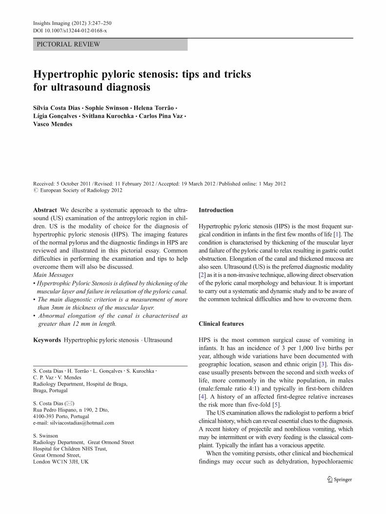
One common difficulty is a stomach filled with gas (Fig. 3).The easiest way to avoid this is by placing the infant in anoblique position with the right side down, as this will allowfluid to fill the antrum, acting as an acoustic window. Astomach completely filled with milk can also cause artefacts,other possibilities are to give the infant water or even to place anasogastric tube, empty the stomach and then fill it with water.
Another frequent problem is that a markedly distendedstomach can displace the pylorus dorsally making it verydifficult to access (Fig. 4). In this situation, moving theinfant into an oblique position with the left side down willhelp to move the pylorus to a more anterior position.
The identification of the pylorus can be difficult, but asystematic approach will improve chances of success.Remember that a normal pylorus is much harder to visualisethan a hypertrophied one.
US diagnostic criteria of HPS
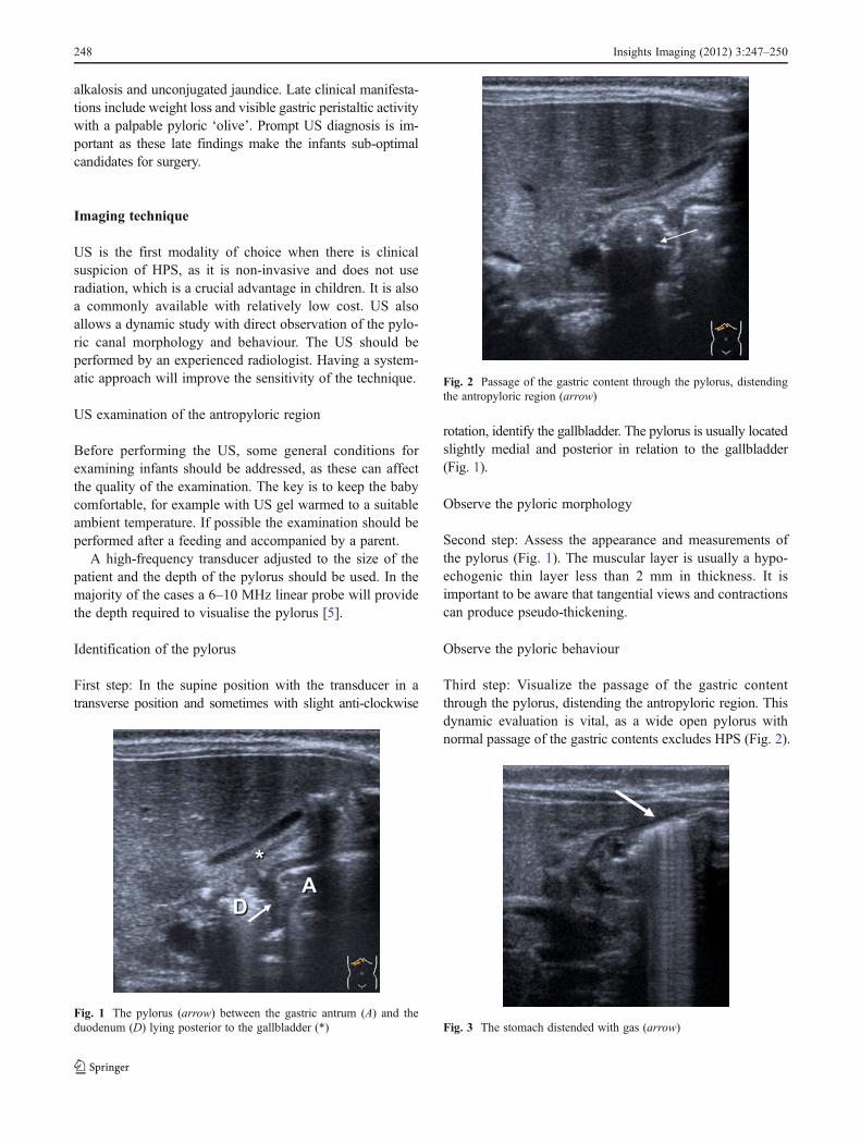
The main diagnostic criterion is measurement of the thick-ness of the muscular layer. An abnormal cut off value of
3 mm in thickness has been described in the literature(Figs. 5 and 6) [6–9].
The other principal sonographic size criterion is thelength of the pyloric canal. Abnormal elongation of thecanal is defined as greater than 12 mm in length (Fig. 6)[9], however this measure is more difficult to perform andfor this reason is considered a less reliable criterion [10].
In HPS the thickened muscle and elongated pylorus arefixed over time, which helps the operator to identify thiscondition. The appearance of the hypertrophied pylorus haspreviously been described as the cervix sign [11], as itresembles the appearance of the uterine cervix (Fig. 4).
Fig. 4 The distended stomach (*), posteriorly displacing the pylorus(arrow), which resembles the appearance of the uterine cervix
Fig. 5 The hypertrophied muscular layer
Fig. 6 Abnormal elongation of the pyloric canal (measure 1)
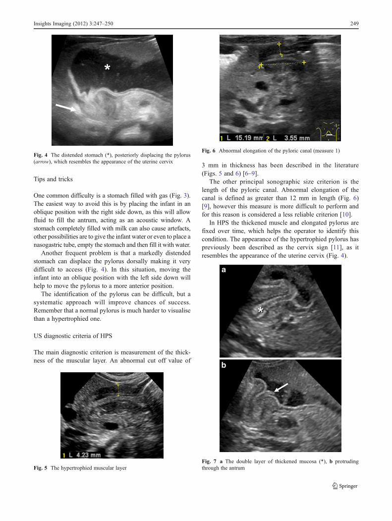
Fig. 7 a The double layer of thickened mucosa (*), b protrudingthrough the antrum
Insights Imaging (2012) 3:247–250 249

Additional US findings in HPS are hypertrophy of themucosa and a markedly distended and actively peristalsingstomach. A double internal layer of crowded and redundantmucosa may be identified (Fig. 7a), protruding through theantrum (Fig. 7b). This was classically described as the nipplesign in conventional contrast studies. The double layer ofthickened mucosa is hyperechogenic and can be confusedwith echogenic contents passing through the pylorus.
Borderline measures
Thickening of the pyloric canal may be transient due to peri-stalsis or pylorospasm. In the majority of cases of pylorospasm,the muscle is not hypertrophied. Sometimes the muscle can beslightly thick, but it usually measures less than 3 mm. Withprolonged observation, pyloric opening may be visualised.
If the muscle layer is 2–3 mm thick, and it does not relaxduring examination, clinical follow-up with repeat US isadvisable [5, 6, 12]. Particular attention should be paid topre-term infants and those in the younger age range. Inpremature infants, HPS develops at the same age as in terminfants, but their smaller size should be taken into consid-eration. Argyropoulou et al. [13] showed that normal pyloricdimensions increase with the gestational age and docu-mented an even stronger correlation with body weight,providing normal values for muscle thickness, canal lengthand canal width from prematurity to full-term infants.Haider et al. [14] performed a study with 190 infants operatedfor HPS and found a strong correlation with the ultrasoundmeasurement of the pyloric length and the weight of theinfant, which can be helpful in small and premature patients.However these authors also highlight the importance of themorphological appearance of the pylorus in premature infants.
Post-treatment imaging of HPS
The treatment of HPS is surgical pyloromyotomy. A furtherUS examination may be requested if vomiting persists fol-lowing surgery. However, the radiologist and the surgeonshould be aware that the pyloric muscle may remain thick-ened after successful surgery and can take up to 5 months toreturn to normal thickness.
In the first week after surgery, the muscle can be the samethickness or even thicker than before surgery and then thedimensions gradually return to normal. The anterior part ofthe muscle tends to normalize first, usually measuring lessthan 3 mm by 3 months. The posterior part is last tonormalise, usually after 5 months, when the pylorus regainsits appearance of an elongated ring. This order of changes isrelated to the anterior surgical approach to the muscle [15].
An upper GI examination may also be performed ifemesis continues post-operatively, in order to exclude a
duodenal leak or to assess an incomplete pyloromyotomyor gastro-oesophageal reflux [16].
Conclusion
Pyloric US examination is a dynamic investigation, which shouldbe performed in a systematic way. The radiologist should be awareof the pitfalls of the examination and how to overcome them. It isimportant to be familiar with the normal and hypertrophied pyloricappearances, as this will provide a greater diagnostic confidence,assisting in early diagnosis and improving the management ofinfants with HPS.
References
1. Ohshiro K, Puri P (1998) Pathogenesis of infantile hypertrophicpyloric stenosis: recent progress. Pediatr Surg Int 13:43–252
2. Hiorns MP (2011) Gastrointestinal tract imaging in children: currenttechniques. Pediatr Radiol 41:42–54
3. Panteli C (2009) New insights into the pathogenesis of infantilepyloric stenosis. Pediatr Surg Int 25:1043–1052
4. Chandran L, Chitkara M (2008) Vomiting in children: reassurance,red flag, or referral? Pediatr Rev 29:183–192
5. Hernanz-SchulmanM (2003) Infantile hypertrophic pyloric stenosis.Radiology 227:319–331
6. O’Keeffe FN, Stansberry SD, Swischuk LE, Hayden CK (1991)Antropiloric muscle thickness at US in infants: what is normal.Radiology 178:827–830
7. Hernanz-Schulman M, Sells LL, Ambrosino MM, Heller RM,Stein SM, Neblett WW (1994) Hypertrophic pyloric stenosis inthe infant without a palpable olive: accuracy of sonographicdiagnosis. Radiology 193:771–776
8. Rohrschneider WK, Mittnacht H, Darge K, Tröger J (1998) Pyloricmuscle in asymptomatic infants: sonographic evaluation anddiscrimination from idiopathic hypertrophic pyloric stenosis.Pediatr Radiol 28:429–434
9. Reed AA, Michael K (2010) Hypertrophic pyloric stenosis. JDiagn Med Sonography 26:157–160
10. Blumhagen JD, Maclin L, Krauter D, Rosenbaum DM, WeinbergerE (1988) Sonographic diagnosis of hypertrophic pyloric stenosis.AJR Am J Roentgenol 150:1367–1370
11. Haller JO, Cohen HL (1986)Hypertrophic pyloric stenosis: diagnosisusing US. Radiology 161:335–339
12. Hernanz-Schulman M (2009) Pyloric stenosis: role of imaging.Pediatr Radiol 39(Suppl 2):S134–S139
13. Argyropoulou MI, Hadjigeorgi CG, Kiortsis DN (1998) Antro-pyloriccanal values from early prematurity to full-term gestational age: anultrasound study. Pediatr Radiol 28:933–936
14. Haider N, Spicer R, Grie D (2002) Ultrasound diagnosis of infantilehypertrophic pyloric stenosis: determinants of pyloric length and theeffect of prematurity. Clin Radiol 57:136–139
15. Yoshizawa J, Eto T, Higashimoto Y, Saitou T, Maie M (2001)Ultrasonographic features of normalization of the pylorus afterpyloromyotomy for hypertrophic pyloric stenosis. J Pediatr Surg36:582–586
16. Nasr A, Ein SH, Connolly B (2008) Recurrent pyloric stenosis: todilate or operate? A preliminary report. J Pediatr Surg 43:E17–E20
250 Insights Imaging (2012) 3:247–250