Embed Size (px)
Citation preview
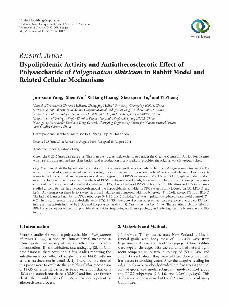
Research ArticleHypolipidemic Activity and Antiatherosclerotic Effect ofPolysaccharide of Polygonatum sibiricum in Rabbit Model andRelated Cellular Mechanisms
Jun-xuan Yang,1 Shen Wu,2 Xi-liang Huang,3 Xiao-quan Hu,4 and Yi Zhang5
1School of Traditional Chinese Medicine, Chongqing Medical University, Chongqing 400016, China2Department of Laboratory Medicine, Guiyang Medical College, Guiyang, Guizhou 550004, China3Department of Cardiology, Fuzhou City First People’s Hospital, Fuzhou, Jiangxi 344000, China4Department of Urology, Ningbo Zhenhai People’s Hospital, Ningbo, Zhejiang 315202, China5Chongqing Institute for Food and Drug Control, Chongqing Engineering Center for Pharmaceutical Processand Quality Control, China
Correspondence should be addressed to Yi Zhang; [email protected]
Received 28 June 2014; Revised 11 August 2014; Accepted 19 August 2014
Academic Editor: Qunhao Zhang
Copyright © 2015 Jun-xuan Yang et al. This is an open access article distributed under the Creative Commons Attribution License,which permits unrestricted use, distribution, and reproduction in any medium, provided the original work is properly cited.
Objective. To evaluate the hypolipidemic activity and antiatherosclerotic effect of polysaccharide of Polygonatum sibiricum (PPGS),which is a kind of Chinese herbal medicine using the rhizome part of the whole herb. Materials and Methods. Thirty rabbitswere divided into normal control group, model control group, and PPGS subgroups of 0.8, 1.6, and 3.2mL/kg/day under randomselection. In atherosclerosis model, the effects of PPGS on diverse blood lipids, foam cells number, and aortic morphology wereevaluated. In the primary culture of endothelial cells (ECs), the activities of PPGS on both ECs proliferation and ECs injury werestudied as well. Results. In atherosclerosis model, the hypolipidemic activities of PPGS were mainly focused on TC, LDL-C, andLp(a). All changes on these factors were statistically significant compared with model group (P < 0.01), except TG and HDL-C.The intimal foam cell number of PPGS subgroups (0.8, 1.6, and 3.2mL/kg/day) was significantly reduced than model control (P <0.01). In the primary culture of endothelial cells (ECs), PPGS showed no effect on cell proliferation but preferred to protect EC frominjury and apoptosis induced by H
2O2and lipopolysaccharide (LPS). Discussion and Conclusion. The antiatherosclerotic effect of
PPGS may be supported by its hypolipidemic activities, improving aortic morphology, and reducing foam cells number and ECsinjury.
1. Introduction
Plenty of studies showed that polysaccharide of Polygonatumsibiricum (PPGS), a popular Chinese herbal medicine inChina, performed variety of medical effects such as anti-inflammation [1], antioxidation, and antiaging [2]. In Chi-nese database, there were only a few studies reporting theantiatherosclerotic effect of single dose of PPGS with nocellular mechanisms in detail [3, 4]. Therefore, the aims ofthis paper were to evaluate the possible cellular mechanismof PPGS on antiatherosclerosis based on endothelial cells(ECs) and smooth muscle cells (SMCs) and finally to furtherclarify the possible role of PPGS in the development ofatherosclerosis process.
2. Materials and Methods
2.1. Animals. Thirty healthy male New Zealand rabbits ingeneral grade with body mass of 1.9–2.4 kg were fromExperimental Animal Center of Chongqing inChina. Rabbitswere kept in the cages with the condition of natural light,room temperature, relative humidity of (50 ± 3)%, andautomatic ventilation. They were fed fixed dose of food withfree access to drinking water. After the adaptive feeding for7 d, animals were randomly divided into five groups (normalcontrol group and model subgroups: model control groupand PPGS subgroups (0.8, 1.6, and 3.2mL/kg/day)). Thisstudy received the approval of Local Animal Ethics AdvisoryCommittee.
Hindawi Publishing CorporationEvidence-Based Complementary and Alternative MedicineVolume 2015, Article ID 391065, 6 pageshttp://dx.doi.org/10.1155/2015/391065
2 Evidence-Based Complementary and Alternative Medicine
2.2. Drugs, Chemicals, and Instruments. The PPGS extract(content of polysaccharide ≥90%) was purchased fromDepartment of Preparation of Chongqing Chinese MedicineHospital (Chongqing, China). The other reagents alsoincluded M199 medium and fetal bovine serum (HycloneCo., Ltd., Utah, USA), II collagenase and trypsin (InvitrogenCorporation, Grand Island, USA), CCK-8 kit (Dojindo Lab-oratories, Kyushu, Japan), crystal violet (SigmaChemical Co.,St. Louis, USA), saline (for infusion, Kelun Co., Ltd., Sichuan,China), and neonatal umbilical cord (The Affiliated Hospitalof Harbin Medical University, Harbin, China). The rabbitbasal diet and relative high cholesterol diet were preparedfrom Experimental Animal Center of Chongqing MedicalUniversity (Chongqing, China) according to the referencestudy [5].
2.3. Hypolipidemic Activity and Antiatherosclerotic Effect inHigh Fat Diet-Induced Rabbit Model. Normal control groupwas fed with basal diet, while model subgroups were fed withhigh cholesterol diet. Each rabbit was given quantitative dietof 120 g/d, in which all were basal diet for normal controlgroup and it was composed of 40 g/d high cholesterol diet and80 g/d basic diet in model subgroups. The PPGS subgroupswere also fed with different concentrations of PPGS (0.8,1.6, and 3.2mL/kg/day). The dose for animal model wasconverted from clinical dosage. During the feeding, highcholesterol diet was given firstly, and basal diet was supple-mented with free access to water for 8 weeks.
After expiration of 8-week feeding with 10 h fasting,the venous blood was obtained for lipid levels testing(total cholesterol (TC), total triglycerides (TG), high-densitylipoprotein (HDL-C), low-density lipoprotein (LDL-C), andlipoprotein (a) (Lp(a))). Then rabbits were sacrificed forstudy. In the experiments of HE staining, after conventionaldehydration, paraffin sections were prepared for HE staining.Associated pathological changes of intima and adventitiaunder optical microscopy were recorded. Under certainmag-nification (20 × 10), eyepiece micrometer and hand controlcounters were applied to count foam cells number on 5 smalllattices, taking the mean value of all slices to get cell numberon each 1mm2, which was seen as foam cell number per unitarea of intima.
2.4. The Effects of PPGS on ECs and SMCs. Human umbilicalvein endothelial cells (HUVECs) from umbilical cord wereisolated by enzymatic digestion according to the methodmentioned before [6]. Human umbilical artery smoothmuscle cells (HUASMCs) were obtained by tissue adherentmethod [7, 8]. The digestion solution of 0.25% trypsin wasprepared and added to serum medium. The supernatant wasdiscarded after centrifugation; culture fluid was added to mixthe cells and finally put them into culture flasks for cellsgrowing at 37∘C with 5% CO
2. The third generations were
used for the following experiments.200𝜇L of ECs or SMCs was seeded in 96-well plates
according to the condition of 5 × 10³ cells/hole. After 24 hincubation, adherent cells were randomly divided into fourgroups: (1) control group, the culture medium (containing20% fetal bovine serum), and (2) subgroups with different
concentrations intervention of PPGS (25, 50, and 100𝜇g/mL)in cultured cells. EC was cultured in drug-containingmedium for 12, 24, and 48 h, while HUASMC was for 12, 18,and 24 h. Each dose subgroup was given five subholes. 10𝜇LCCK-8 reagent was added into each hole for 2 h before thetermination of culture. The absorbance of cell supernatantwas detected at the wavelength of 450 nm, and cell culturemedium was used as blank control of zero absorbance. Inaddition, the migration ability of HUASMC was determinedby transwell migration chamber (8 𝜇m pore size) followingthe previous study [9]. The migration abilities of HUASMCin each group were estimated after the cells were fixed by 4%paraformaldehyde for 10min, stained by crystal violet, andrandomly selected under a fluorescence microscope with themagnification of 200x to count the number of cells migratingto the bottom of porous membrane.
2.5. The Effect on H2O2-Induced EC Injury. 100 𝜇mol/L of
H2O2was selected as testing concentration.Themain process
was introduced in another study [10]. Each treatment groupwas given drug into medium with the final concentrationof 10%. The normal control group was only administratedwith medium. The model control group contained mediumplus 100𝜇mol/L of H
2O2. The low dose treatment group was
0.3mg/kg PPGS in cell medium plus 100 𝜇mol/L H2O2. The
concentrations of PPGS in middle and high dose treatmentgroup were 0.6 and 1.2mg/kg, respectively. The cytokines(malondialdehyde (MDA) and superoxide dismutase (SOD))were tested by related testing kits.
2.6. The Effect on Lipopolysaccharide- (LPS-) Induced ECInjury. After 24 h incubation, ECs were fused in adherentmonolayer way [11]. The adherent cells were randomlydivided into five groups the same as mentioned above. Thesuspension was abandoned and renewed; related drug wasadded into each group at the same time. After cell culture for24 h at 37∘C with 5% CO
2, the old medium was abandoned.
And 3mL serum medium containing 5𝜇g/mL of LPS wasadded into each group. The cells in each group were washedwith PBS for 3 times 24 h later. Then 4% formaldehyde wasused to fix them for 30min. After washing for 3 times againwith PBS solution, Hoechst 33258 fluorescent staining withthe final concentration of 0.5𝜇g/mL was done for 10minin the dark at room temperature. The residual stainingsolution was discarded. The cellular washing was repeatedagain for 3 times. The fluorescence in the dark was observedin microscope at the wavelength of 350 nm. The cells infive different fields of picture view and cell apoptosis werecalculated for each hole.
2.7. Statistical Analysis. Results were expressed in the form ofmean ± SEM. Data were analyzed by one-way ANOVA, fol-lowed by Student’s two-tailed 𝑡-test for comparison betweentwo groups. 𝑃 < 0.05means statistically significant.
3. Result
3.1. Hypolipidemic Activity. After the treatment, the serumlevel of HDL-C and TG did not change basically. From
Evidence-Based Complementary and Alternative Medicine 3
Table 1: The hypolipidemic effects of PPGS in atherosclerosis rabbit model (𝑛 = 6).
Groups TC LDL-C Lp(a)Before After Before After Before After
Normal control 1.26 ± 0.25 1.40 ± 0.33 1.18 ± 0.26 1.26 ± 0.17 50.41 ± 10.33 67.32 ± 15.13
Model control 1.24 ± 0.31 12.18 ± 2.40 1.17 ± 0.24 10.46 ± 1.53 47.35 ± 15.16 643.72 ± 151.69
PPGS (0.8mL/kg/day) 1.31 ± 0.40 7.82 ± 4.13∗∗
1.20 ± 0.31 6.42 ± 3.48∗∗
49.52 ± 8.82 81.40 ± 26.73∗∗
PPGS (1.6mL/kg/day) 1.38 ± 0.41 5.81 ± 1.92∗∗
1.25 ± 0.27 4.61 ± 1.56∗∗
51.9 ± 14.83 47.36 ± 15.39∗∗
PPGS (3.2mL/kg/day) 1.32 ± 0.37 4.5 ± 2.11∗∗
1.22 ± 0.25 3.45 ± 0.73∗∗
46.68 ± 14.81 33.52 ± 12.68∗∗
Note: compared with model control group, ∗∗𝑃 < 0.01.
Table 2: The effects of PPGS on HUASMCs proliferation and migration (𝑛 = 6).
Groups OD values Cells migration number 24 hlater (𝑛 = 3)12 h 24 h 48 h
Normal control 0.186 ± 0.024 0.202 ± 0.022 0.215 ± 0.027 39.1 ± 4.2
PPGS (0.3mg/kg) 0.172 ± 0.020 0.171 ± 0.024∗
0.177 ± 0.019∗
35.8 ± 4.6
PPGS (0.6mg/kg) 0.159 ± 0.022∗
0.162 ± 0.017∗∗
0.169 ± 0.015∗∗
33.2 ± 4.3∗
PPGS (1.2mg/kg) 0.138 ± 0.015∗
0.141 ± 0.015∗∗
0.155 ± 0.018∗∗
30.6 ± 4.1∗∗
Note: compared with model control group, ∗𝑃 < 0.05 and ∗∗𝑃 < 0.01.
Table 1, the results of other parameters were showed. Allconcentrations of PPGS were markedly effective on bloodlipids control (𝑃 < 0.01).
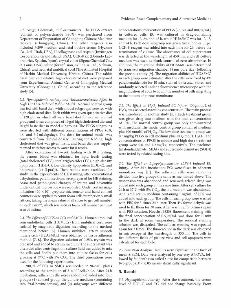
3.2. The Results of HE Staining in Atherosclerotic Model.Aortic elastic membrane in normal diet group was integral.Endothelium was close to the internal elastic membranearranged in neat rows, with smoothmuscle andmiddle elasticmembrane arranged in parallel. In model control group,aortic intima was significantly thickening with a large accu-mulation of foam cells. ECswere falling off or loosely attachedto the membrane surface. Intimal lesions had extensivelypathological changes with collagen fiber glass. Elastic fiberwas ruptured and disappeared as well. In PPGS subgroups,compared with the thickening degree of aortic intima inmodel control group, subendothelial gap was increased withvisible foam cells aggregation, but foam cells number wassignificantly less than model control group. The structure ofmedial membrane was basically integral with SMCs in thesame pole, as shown in Figure 1 (200x).
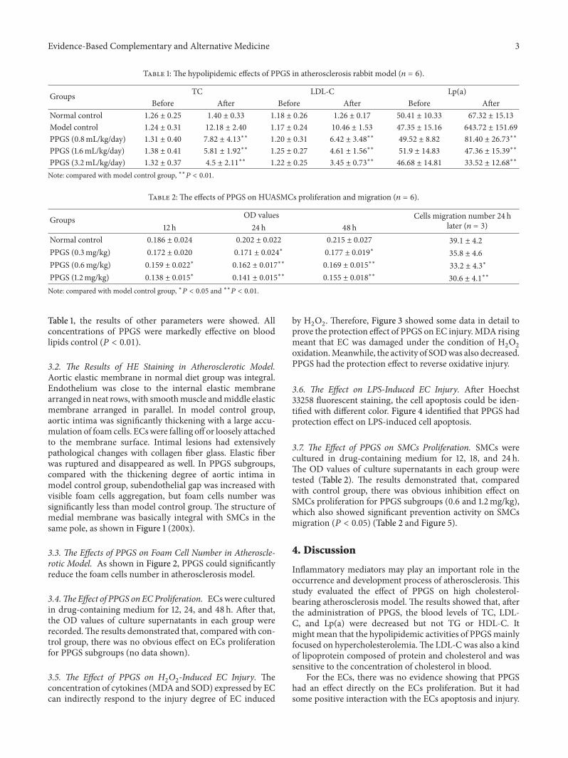
3.3. The Effects of PPGS on Foam Cell Number in Atheroscle-rotic Model. As shown in Figure 2, PPGS could significantlyreduce the foam cells number in atherosclerosis model.
3.4.The Effect of PPGS on ECProliferation. ECswere culturedin drug-containing medium for 12, 24, and 48 h. After that,the OD values of culture supernatants in each group wererecorded.The results demonstrated that, compared with con-trol group, there was no obvious effect on ECs proliferationfor PPGS subgroups (no data shown).
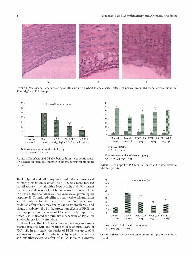
3.5. The Effect of PPGS on H2O2-Induced EC Injury. The
concentration of cytokines (MDA and SOD) expressed by ECcan indirectly respond to the injury degree of EC induced
by H2O2. Therefore, Figure 3 showed some data in detail to
prove the protection effect of PPGS on EC injury.MDA risingmeant that EC was damaged under the condition of H
2O2
oxidation.Meanwhile, the activity of SODwas also decreased.PPGS had the protection effect to reverse oxidative injury.
3.6. The Effect on LPS-Induced EC Injury. After Hoechst33258 fluorescent staining, the cell apoptosis could be iden-tified with different color. Figure 4 identified that PPGS hadprotection effect on LPS-induced cell apoptosis.
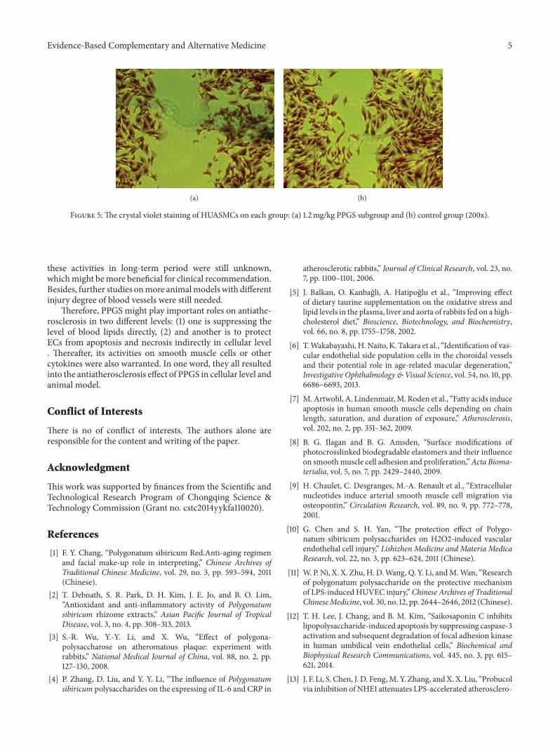
3.7. The Effect of PPGS on SMCs Proliferation. SMCs werecultured in drug-containing medium for 12, 18, and 24 h.The OD values of culture supernatants in each group weretested (Table 2). The results demonstrated that, comparedwith control group, there was obvious inhibition effect onSMCs proliferation for PPGS subgroups (0.6 and 1.2mg/kg),which also showed significant prevention activity on SMCsmigration (𝑃 < 0.05) (Table 2 and Figure 5).
4. Discussion
Inflammatory mediators may play an important role in theoccurrence and development process of atherosclerosis. Thisstudy evaluated the effect of PPGS on high cholesterol-bearing atherosclerosis model. The results showed that, afterthe administration of PPGS, the blood levels of TC, LDL-C, and Lp(a) were decreased but not TG or HDL-C. Itmight mean that the hypolipidemic activities of PPGSmainlyfocused on hypercholesterolemia.The LDL-Cwas also a kindof lipoprotein composed of protein and cholesterol and wassensitive to the concentration of cholesterol in blood.
For the ECs, there was no evidence showing that PPGShad an effect directly on the ECs proliferation. But it hadsome positive interaction with the ECs apoptosis and injury.
4 Evidence-Based Complementary and Alternative Medicine
(a) (b) (c)
Figure 1: Microscope camera drawing of HE staining on rabbit thoracic aorta (200x): (a) normal group; (b) model control group; (c)3.2mL/kg/day PPGS group.
0
5
10
15
20
25
30
35
Normal control
Model control
PPGS (0.8mL/kg/day)
Foam cells number/mm2
∗∗
∗∗
∗∗
PPGS (1.6mL/kg/day)
PPGS (3.2mL/kg/day)
Note: compared with model control group,∗P < 0.05 and ∗∗
P < 0.01
Figure 2:The effects of PPGS after being administered continuouslyfor 8 weeks on foam cells number in atherosclerosis rabbit model(𝑛 = 6).
The H2O2-induced cell injury may result into necrosis based
on strong oxidation reaction. And LPS was more focusedon cell apoptosis by inhibiting NOS activity and NO contentboth inside and outside of cell, but increasing the intracellularROS level [12]. For another distinction based on physiologicalresponse,H
2O2-induced cell injurymay lead to inflammation
and thrombosis for its acute oxidation. But the chronicoxidation effect of LPSmay finally lead to atherosclerosis andplaque instability [13]. So the protection effects of PPGS onboth apoptosis and necrosis of ECs were really impressive,which also indicated the primary mechanism of PPGS onatherosclerosis for the first time.
It was learnt that PPGSwas composed of single monosac-charide fructose with the relative molecular mass (Mr) of7247 [14]. In this study, the purity of PPGS was up to 90%and was good enough to evaluate the hypolipidemic activityand antiatherosclerotic effect of PPGS initially. However,
Normal control
Model control
PPGS (0.3mg/kg)
PPGS (0.6mg/kg)
PPGS (1.2mg/kg)
∗∗
∗∗
∗∗∗∗
∗
∗
MDA (nmol/L)SOD (U/mL)
05
10152025303540
Note: compared with model control group,∗P < 0.05 and ∗∗
P < 0.01
Figure 3: The impact of PPGS on EC injury and related cytokinesreleasing (𝑛 = 6).
05
1015202530354045
Normal control
Model control
PPGS (0.3mg/kg)
PPGS (0.6mg/kg)
PPGS (1.2mg/kg)
∗∗∗∗
∗∗
Apoptosis rate (%)
Note: compared with model control group,∗P < 0.05 and ∗∗
P < 0.01
Figure 4:The impact of PPGS on EC injury and apoptotic condition(𝑛 = 6).
Evidence-Based Complementary and Alternative Medicine 5
(a) (b)
Figure 5: The crystal violet staining of HUASMCs on each group: (a) 1.2mg/kg PPGS subgroup and (b) control group (200x).
these activities in long-term period were still unknown,whichmight bemore beneficial for clinical recommendation.Besides, further studies onmore animalmodels with differentinjury degree of blood vessels were still needed.
Therefore, PPGS might play important roles on antiathe-rosclerosis in two different levels: (1) one is suppressing thelevel of blood lipids directly, (2) and another is to protectECs from apoptosis and necrosis indirectly in cellular level. Thereafter, its activities on smooth muscle cells or othercytokines were also warranted. In one word, they all resultedinto the antiatherosclerosis effect of PPGS in cellular level andanimal model.
Conflict of Interests
There is no of conflict of interests. The authors alone areresponsible for the content and writing of the paper.
Acknowledgment
This work was supported by finances from the Scientific andTechnological Research Program of Chongqing Science &Technology Commission (Grant no. cstc2014yykfa110020).
References
[1] F. Y. Chang, “Polygonatum sibiricum Red.Anti-aging regimenand facial make-up role in interpreting,” Chinese Archives ofTraditional Chinese Medicine, vol. 29, no. 3, pp. 593–594, 2011(Chinese).
[2] T. Debnath, S. R. Park, D. H. Kim, J. E. Jo, and B. O. Lim,“Antioxidant and anti-inflammatory activity of Polygonatumsibiricum rhizome extracts,” Asian Pacific Journal of TropicalDisease, vol. 3, no. 4, pp. 308–313, 2013.
[3] S.-R. Wu, Y.-Y. Li, and X. Wu, “Effect of polygona-polysaccharose on atheromatous plaque: experiment withrabbits,” National Medical Journal of China, vol. 88, no. 2, pp.127–130, 2008.
[4] P. Zhang, D. Liu, and Y. Y. Li, “The influence of Polygonatumsibiricum polysaccharides on the expressing of IL-6 and CRP in
atherosclerotic rabbits,” Journal of Clinical Research, vol. 23, no.7, pp. 1100–1101, 2006.
[5] J. Balkan, O. Kanbagli, A. Hatipoglu et al., “Improving effectof dietary taurine supplementation on the oxidative stress andlipid levels in the plasma, liver and aorta of rabbits fed on a high-cholesterol diet,” Bioscience, Biotechnology, and Biochemistry,vol. 66, no. 8, pp. 1755–1758, 2002.
[6] T.Wakabayashi, H. Naito, K. Takara et al., “Identification of vas-cular endothelial side population cells in the choroidal vesselsand their potential role in age-related macular degeneration,”Investigative Ophthalmology &Visual Science, vol. 54, no. 10, pp.6686–6693, 2013.
[7] M. Artwohl, A. Lindenmair,M. Roden et al., “Fatty acids induceapoptosis in human smooth muscle cells depending on chainlength, saturation, and duration of exposure,” Atherosclerosis,vol. 202, no. 2, pp. 351–362, 2009.
[8] B. G. Ilagan and B. G. Amsden, “Surface modifications ofphotocrosslinked biodegradable elastomers and their influenceon smoothmuscle cell adhesion and proliferation,”Acta Bioma-terialia, vol. 5, no. 7, pp. 2429–2440, 2009.
[9] H. Chaulet, C. Desgranges, M.-A. Renault et al., “Extracellularnucleotides induce arterial smooth muscle cell migration viaosteopontin,” Circulation Research, vol. 89, no. 9, pp. 772–778,2001.
[10] G. Chen and S. H. Yan, “The protection effect of Polygo-natum sibiricum polysaccharides on H2O2-induced vascularendothelial cell injury,” Lishizhen Medicine and Materia MedicaResearch, vol. 22, no. 3, pp. 623–624, 2011 (Chinese).
[11] W. P. Ni, X. X. Zhu, H. D.Wang, Q. Y. Li, andM.Wan, “Researchof polygonatum polysaccharide on the protective mechanismof LPS-inducedHUVEC injury,”Chinese Archives of TraditionalChineseMedicine, vol. 30, no. 12, pp. 2644–2646, 2012 (Chinese).
[12] T. H. Lee, J. Chang, and B. M. Kim, “Saikosaponin C inhibitslipopolysaccharide-induced apoptosis by suppressing caspase-3activation and subsequent degradation of focal adhesion kinasein human umbilical vein endothelial cells,” Biochemical andBiophysical Research Communications, vol. 445, no. 3, pp. 615–621, 2014.
[13] J. F. Li, S. Chen, J. D. Feng,M. Y. Zhang, andX. X. Liu, “Probucolvia inhibition ofNHE1 attenuates LPS-accelerated atherosclero-
6 Evidence-Based Complementary and Alternative Medicine
sis and promotes plaque instability in vivo,” Experimental andMolecular Pathology, 2014.
[14] X. Zhang, G. Borjihan, and N. Zhaorigetu, “Determinationof relative molecular mass and composition for Polygonatumsibiricum polysaccharide by high performance liquid chro-matography,” Chinese Journal of Chromatography, vol. 23, no. 4,pp. 394–396, 2005 (Chinese).

Submit your manuscripts athttp://www.hindawi.com
Stem CellsInternational
Hindawi Publishing Corporationhttp://www.hindawi.com Volume 2014
Hindawi Publishing Corporationhttp://www.hindawi.com Volume 2014
MEDIATORSINFLAMMATION
of
Hindawi Publishing Corporationhttp://www.hindawi.com Volume 2014
Behavioural Neurology
EndocrinologyInternational Journal of
Hindawi Publishing Corporationhttp://www.hindawi.com Volume 2014
Hindawi Publishing Corporationhttp://www.hindawi.com Volume 2014
Disease Markers
Hindawi Publishing Corporationhttp://www.hindawi.com Volume 2014
BioMed Research International
OncologyJournal of
Hindawi Publishing Corporationhttp://www.hindawi.com Volume 2014
Hindawi Publishing Corporationhttp://www.hindawi.com Volume 2014
Oxidative Medicine and Cellular Longevity
Hindawi Publishing Corporationhttp://www.hindawi.com Volume 2014
PPAR Research
The Scientific World JournalHindawi Publishing Corporation http://www.hindawi.com Volume 2014
Immunology ResearchHindawi Publishing Corporationhttp://www.hindawi.com Volume 2014
Journal of
ObesityJournal of
Hindawi Publishing Corporationhttp://www.hindawi.com Volume 2014
Hindawi Publishing Corporationhttp://www.hindawi.com Volume 2014
Computational and Mathematical Methods in Medicine
OphthalmologyJournal of
Hindawi Publishing Corporationhttp://www.hindawi.com Volume 2014
Diabetes ResearchJournal of
Hindawi Publishing Corporationhttp://www.hindawi.com Volume 2014
Hindawi Publishing Corporationhttp://www.hindawi.com Volume 2014
Research and TreatmentAIDS
Hindawi Publishing Corporationhttp://www.hindawi.com Volume 2014
Gastroenterology Research and Practice
Hindawi Publishing Corporationhttp://www.hindawi.com Volume 2014
Parkinson’s Disease
Evidence-Based Complementary and Alternative Medicine
Volume 2014Hindawi Publishing Corporationhttp://www.hindawi.com