Embed Size (px)
Citation preview
HYPOXAEMIA AMONG CHILDREN WITH SEVERE OR VERY SEVERE PNEUMONIA AT KENYATTA NATIONAL HOSPITAL.
BY:
DR. SAMSON K. MUGANE
SUPERVISORS:
PROF. RACHEL MUSOKE
PROF. E. MALECHE-OBIMBO
DR. GRACE IRIMU
This research dissertation is submitted in partial fulfillment for award of Master of
Medicine in Paediatrics and Child Health of the University of Nairobi.
University of NAIROBI Library
0407218 7
U N IV ER S ITY O F NAIROBI MEDICAL library
1
DECLARATION
I declare that this dissertation is my own original work and has not been presented for a
degree in any other university.
Date......
Dr. Samson K. Mugane
This dissertation is submitted for examination with our approval as university
supervisors:
Sign....... ....................................... D a t e . ^ L ^ ^ . .® r^ .(Q .
Prof. Rachel Musoke
M.B.Ch.B. MMed (Paediatrics), FABM.
Associate Professor, Department of Paediatrics and Child Health. University of Nairobi.
Sign... ... Date . .f? ? .......®
Prof. E. Maleche-Obimbo
M.B.Ch.B. MMed (Paediatrics). M.P.H (Epidemiology).
Associate Professor, Department of Paediatrics and Child Health. University of Nairobi.
I Sign.......( S k — * .......
Dr. Grace Irimu
M.B.Ch.B, MMed (Paediatrics),
Senior Lecturer, Department of Paediatrics and Child Health, University of Nairobi.

d e d ic a t io n
To my beloved wife Dorothy and daughter Joy who have been a great source of
inspiration.

ACKNOWLEDGEMENTS
The success of this work is a result o f inputs and help from various persons to whom I am
very grateful.
To the Almighty God. the source and sustainer of life.
To my wife Dorothy for her unwavering support.
To my supervisors for their guidance and patience during the entire period from the
inception of the proposal to the writing of the book.
To Dr. Mike English of KEMRI/Wellcome Trust for his support and encouragement,
fo members of the ’Childhood Pneumonia Group' for the team spirit during the entire
period of data collection.
To Dr. Ambrose Agweyu and Mr. Moses Mwangi of KEMRI who helped with data
analysis.
4
TABLE QF CONTENTS
DECLARATION.......................................................................................................................... 2
DEDICATION............................................................................................................................... 3
ACKNOWLEDGEMENTS.........................................................................................................4
TABLE OF CONTENTS............................................................................................................. 5
LIST OF ABBREVIATIONS AND SYMBOLS..................................................................... 7
LIST OF TABLES......................................................................................................................... 8
LIST OF FIGURES...................................................................................................................... 9
LIST OF FIGURES...................................................................................................................... 9
ABSTRACT..................................................................................................................................10
1.0 BACKGROUND AND LITERATURE REVIEW...................................................... 12
2.0 STUDY JUSTIFICATION AND OBJECTIVES...................................................... 25
2.1 Problem Statement.....................................................................................................25
2.2 Justification................................................................................................................. 26
2.3 U tility........................................................................................................................... 27
2.4 Study Objectives........................................................................................................ 27
3.0 M ETHODOLOGY........................................................................................................ 29
3.1 Study A rea .................................................................................................................. 29
3.2 Study Population........................................................................................................ 29
3.3 Study Design............................................................................................................... 29
3.4 Sample Size Estim ation............................................................................................ 29
3.5 Sampling M ethod....................................................................................................... 30
5
3.6 Inclusion Criteria:..................................................................................................... 30
3.7 Exclusion C riteria..................................................................................................... 30
3.8 Case Definitions.........................................................................................................31
3.9 Equipment...................................................................................................................32
3.10 Study Procedures.......................................................................................................32
3.11 Data Analysis............................................................................................................. 33
3.12 Ethical Considerations.............................................................................................. 34
4.0 RESULTS....................................................................................................................... 37
5.0 DISCUSSION.................................................................................................................53
6.0 STUDY LIMITATIONS.............................................................................................. 58
7.0 CONCLUSIONS............................................................................................................ 58
8.0 RECOMMENDATIONS.............................................................................................. 59
REFERENCES............................................................................................................................60
APPENDICES.............................................................................................................................66
APPENDIX 1: QUESTIONNAIRE..................................................................................66
.APPENDIX 2: PNEUMONIA MANAGEMENT PROTOCOL...................................68
APPENDIX 3: INFORMATION AND CONSENT FO RM ............................................69
.APPENDIX 4: BUDGET................................................................................................... 75
MST OF ABBREVIATIONS AND SYMBOLS
ALRI Acute lower respiratory infections
SaO: .Arterial haemoglobin oxygen saturation
SpO: Arterial haemoglobin oxygen saturation by pulse oximetry
P a 0 2 Arterial blood oxygen tension
WHO World Health Organization
SD Standard deviation
GoK Government of Kenya
AVPU Alert, Voice, Pain. Unconscious
HIV Human Immunodeficiency virus
AIDS Acquired immune deficiency syndrome
PCP Pneumocystis jirovecii pneumonia
LIP Lymphocytic interstitial pneumonia
KNH Kenvatta National Hospital
PITC Provider Initiated Testing and Counseling
PEU Peadiatric Emergency Unit
DNA PCR Deoxyribonucleic Acid Polymerase Chain Reaction
KEMRI Kenya Medical Research Institute
SOPS Standard operating procedures
7
1 1ST OF TABLES.
Table 1: Prevalence of Hypoxaemia Among Children With Severe or Very
Severe Pneumonia.......................................................................................... 21
Table 2: Age and Gender Distribution of Children Enrolled...................................... 39
Table 3: Distribution of Clinical Features at Admission............................................. 40
Table 4: Distribution of Pneumonia Severity in Study Population............................ 41
Table 5: Sensitivity, Specificity and Predictive Values of GoK Criteria for
Oxygen Therapy.............................. 45
Table 6a: GoK Clinical Signs and their Ability to Predict Hypoxaemia.................... 46
Table 6b Other Clinical Signs and their Ability to Predict Hypoxaemia................... 48
Table 7: Association Between HIV infection and Hypoxaemia................................ 48
Table 8: Multivariate Logistic Regression Showing Ability’ of Clinical Signs to
Predict Hypoxaemia......................................................................................... 49
Table 9: Mortality Among Hvpoxaemic and Non hypoxaemic Children................. 50
Table 10: Association Between HIV Infection and Mortality...................................... 52
8
11ST OF FIGURES
Figure 1: Flow Chart Showing Patient Screening and Enrolment....................... 38
Figure 2: Prevalence o f Hypoxemia in Study Population...................................... 41
Figure 3: Prevalence of Hvpoxaemia Stratified by Severity of Pneumonia........ 42
Figure 4: Distribution of Oxygen Saturation Levels in Study
Population................................................................................................... 43
Figure 5: Distribution of Oxygen Saturation Levels Stratified by Severity of
Pneumonia................................................................................................... 44
Figure 6: Relationship between Hypoxaemia and Mortality (Oxygen saturation
<90%)......................................................................................... 51
Figure 7: Relationship between Hypoxaemia and Mortality (Oxygen saturation
<85%)...................................................................................... 51
9
a r e t r a c t
Background: Pneumonia is the leading cause of childhood morbidity and mortality' in
developing countries with hvpoxaemia as the most common and fatal complication.
Oxygen therapy is an important intervention for children with hypoxaemia. In many
settings in Kenya, clinical signs are used to identify children who require oxygen. The
Government of Kenya (GoK) has provided criteria for oxygen therapy. It states that
oxygen should be administered to a child with any of these signs: cyanosis, inability to
drink/breastfeed, impaired consciousness, grunting or head nodding. While there is data
exploring the utility of clinical signs to identify hypoxaemic children, this GoK ‘decision
rule' has never been evaluated. There is paucity of information on some of the signs
included in the GoK criteria and little local information on the prevalence of hypoxaemia
among children with severe forms of pneumonia.
Objectives: To determine the prevalence of hypoxaemia and evaluate the sensitivity and
specificity of the GoK criteria for oxygen therapy for children with severe or very severe
pneumonia admitted at Kenyatta National Hospital, Nairobi, Kenya, to determine
whether Human Immunodeficiency Virus (HIV) infection was a risk factor for
hypoxaemia and to evaluate the association between hypoxaemia and short term in
patient mortality.
Methodology: This was a hospital based short longitudinal survey. We enrolled 343
children aged two to 59 months, assessed them for presence of clinical signs associated
with hypoxaemia, measured their arterial oxygen saturation using a portable hand held
10
pulse oximeter and had them tested for HIV infection. We followed up the children for
five days to determine mortality outcome.
Results: Prevalence of hypoxaemia was 50.7% in the study population. Stratified by severity,
39.7% and 59.4% of children with severe and very severe pneumonia respectively were
hypoxaemic. Cyanosis and grunting were found to be independent predictors of hypoxaemia. The
GoK criteria had a sensitivity of 65.5% and a specificity of 53.8% for detecting children who
required oxygen therapy. Thirty one children (9.0%) were HIV infected. Oxygen saturation of
<85% was associated with increased mortality (OR 3.3. 95% CI= 1.5 to 7.1, P=0.005).
Conclusions: Hypoxaemia is frequent, occurring in 50.7% of children hospitalized with
severe or very severe pneumonia at Kenyatta National Hospital. The GoK criteria for
oxygen therapy have a low sensitivity (65.5%) and specificity (53.8%) for predicting
hypoxaemia. Severe hypoxaemia (Sp02 <85%) is associated with a 3.3 fold increased
mortality.
Recommendations: The Government of Kenya should consider promoting the use of
pulse oximetry in all public hospitals to detect hypoxaemia. A cost-benefit study on the
use of pulse oximeters vis-a-vis continued use of clinical signs to determine which
children require oxygen therapy should be carried out.
11
1 0 BACKGROUND AND LITERATURE REVIEW
Acute lower respiratory tract infections (ALRI), particularly pneumonia, are the leading
causes of childhood morbidity and mortality in developing countries.' ALRI cause more
than 2 million child deaths worldwide each year, mostly from pneumonia. Ninety percent
of these deaths occur in less-developed countries. 2'3 It is estimated that 1.9 million
children died from ALRI throughout the world in the year 2000 of which 70% occurred
in Africa and Southeast A sia4 Hypoxaemia is the most common and fatal complication
of ALRI. Onyango et al in a study to determine short term mortality (death within 5 days
of admission) among children with ALRI in Nairobi, Kenya found that hypoxaemic
5children were 4.3 times more likely to die than those without.’ Weber et al in a similar
study in the Gambia found that the relative risk for death among hypoxaemic children
with ALRI was 4.6 [95% Confidence Interval, 2.2 to 9.6]. (p=0.0007)]. The case fatality
rate was inversely related to the arterial haemoglobin oxygen saturation (SaCb)/" Early
detection of hypoxaemia and appropriate initiation of oxygen therapy is therefore an
important intervention to improve outcome.
Childhood pneumonia in developing countries, unlike in developed countries is caused
more commonly by bacteria than viruses, the commonest aetiological agents being
Streptococcus pneumoniae and Haemophilus influenzae''. Bacterial pneumonia is
associated with higher mortality rates than viral pneumonia.2'1 However, due to
difficulties associated with diagnosing bacterial pneumonia in developing countries,7 the
vH orld Health Organization (WHO) has promoted the use of a clinical case-definition to
12
guide initiation of empiric antibiotic therapy. For children who have cough or difficult
breathing, the WHO acute respiratory infection case management guidelines require only
an assessment of the respiratory rate and the presence of visible and audible signs of
respiratory distress. Very severe pneumonia is present when there is cough or difficult
breathing plus any of the following danger signs: cyanosis, inability to drink/breastfeed,
impaired level of consciousness, grunting or head nodding. Cough or difficult breathing
with lower chest wall in-drawing and none of the above danger signs is categorized as
severe pneumonia. Pneumonia is defined as the presence of cough and tachypnea (>60
breaths per minute for infants up to 2 months of age. ^50 breaths per minute for children
2 to 11 months and >40 breaths per minute for children 12 to 59 months) without any of
the signs of severe pneumonia syndromes8. The WHO acute respiratory infection
algorithm has been shown to have a sensitivity and specificity of 80% for the diagnosis of
• 4,9pneumonia.
Defining Hypoxemia
Hypoxaemia refers to low oxygen level in blood. The best definition would be the level
of blood oxygen associated with increased morbidity, risk of death or delayed recovery
rather than a certain level o f arterial haemoglobin oxygen saturation (SaCV) below normal
for the population. This is because SaCb varies with altitude. The arterial haemoglobin
oxygen saturation by pulse oximetry (SpCb) bears a relationship to arterial blood oxygen
tension (Pa02). At higher altitudes, the partial pressure of oxygen reduces and the normal
range of SpC>2 progressively reduces. The mean SpC>2 at sea level is 97-99% with the
13
lower limit (two standard deviations below the mean) of 94%. Thus the normal range of
SpO: is 94-100%. 10
The WHO defines hypoxaemia as any oxygen saturation <90% and does not take into
account the variation in normal oxygen saturation with altitude11. An Sp02 of 90%
corresponds to an arterial oxygen tension (Pa02) of 60-70 mmHg. Below this, the
haemoglobin oxygen dissociation curve falls steeply such that further decrease in arterial
oxygen tension is associated with greater decrease in arterial oxygen saturation. An Sp02
of <90% is therefore considered by most clinicians as an appropriate indication for
administering oxygen. It has however been suggested that at higher altitudes, the
threshold for initiating oxygen therapy may be lower .1(l
A number of studies have been done with the aim of defining hypoxaemia based on
altitude specific normal values. Haemoglobin oxygen saturation values 2-3 standard
deviations (SD) below the population mean has been used by a number of investigators to
define hypoxaemia. Duke et al measured Sp02 of 151 well children aged between one to
60 months in Goroka Hospital in the highlands of Papua New Guinea at an altitude of
1600 metres. The mean S p02 wras 95.7% (SD 2.7%).1_ Nairobi lies at an altitude of 1670
meters above sea level. Although no evaluation has been done among healthy children in
Nairobi to establish normal values, it is likely to be comparable to that of children in
Goroka as the two regions lie at almost the same altitude.
14
Subhi et al performed a systematic review of literature addressing normal values of
oxygen saturation in children aged one week to 12 years. An SpC>2 of 90% corresponded
to the 2.5th centile for a population of healthy children living at an altitude of
approximately 2500 metres above sea level. This decreased to 85% at an altitude of about
3°00 metres. He concluded that at altitudes greater than 2500 metres above sea level an
S p O o f <85% can be used to indicate the need for oxygen.1''
Mechanism of Hypoxaemia
The principle mechanism of the hypoxia of acute respiratory infection is a mismatch
between ventilation and perfusion. The infectious organism, whether viral or bacterial,
causes areas of pneumonic consolidation, which become inappropriately under
oxygenated relative to their hvper-perfusion. The mismatch is not redressed by vascular
redistribution to the unaffected parts of the lung as most pneumonia in children is of a
bronchopneumonic distribution rather than showing the lobar pattern seen in adults.
Moreover, lung compliance decreases as consolidation develops, leading to increased
work of breathing. Dehydration from fever, panting, and inability to drink leads to
haemoconcentration. peripheral underperfusion and metabolic acidosis which worsen
tissue hypoxia. 14
Hypoxaemia also occurs in non-ALRI conditions. There are several reasons why children
with non-ALRI illnesses have hypoxaemia. In meningitis, for example, upper airway
obstruction may occur from retained secretions, increased or decreased upper airway tone
and bradypnoea or apnoea which may occur because of the brain injury or from chest
wall rigidity during convulsions. In septicaemia, hypoxaemia may occur from
intrapulmonary shunting of blood, pulmonary hypertension or pulmonary congestion.
Systemic oxygen transport is the product of cardiac output and systemic oxygen content.
Cardiac failure of any aetiology resulting in reduced cardiac output leads to decreased
oxvsen transport and tissue hypoxia. Congenital heart defects in which there is mixing of
oxygenated and deoxygenated blood through right to left shunts also cause hypoxaemia.
Detecting Hypoxaemia.
The most reliable method of detecting hypoxaemia is by arterial blood gas analysis or
determination of the arterial haemoglobin oxygen saturation by a pulse oximeter. The
principle of pulse oximetry is based on the red and infrared light absorption
characteristics of oxygenated and deoxygenated haemoglobin. A transcutaneous sensor is
used to measure the percentage of haemoglobin that is fully saturated with oxygen (SpCb).
Pulse oximetry uses spectrophotometry and plethysmography. The pulse oximeter
consists of a computerized unit and a sensor probe, which is attached to the patient's
finger, toe or earlobe. The sensor emits two different wavelengths of light, red (600-750
nanometer wavelength light band) and infrared (850-1000 nanometer wavelength light
band). These lights are absorbed by hemoglobin and transmitted through tissues to a
photo detector. Oxygenated haemoglobin absorbs more infrared light and allows more
red light to pass through to the photo detector. Deoxygenated haemoglobin absorbs more
red light and allows more infrared light to pass through to the photo detector. The amount
of light transmitted is convened to a digital value. The ratio of absorbed red to infrared
light indicates the degree of oxygenation.
16
The height of the plethysmographic (pulse) wave signifies the arterial pulsation. The
signal between the pulse waves (baseline) is subtracted from the signal at the peak of the
plethysmographic wave, the difference being due to inflowing arterial blood, so reflecting
the saturation of arterial blood. A microprocessor compares the absorption of light at the
peak (arterial pulse) and trough (baseline) at both red and infrared waveforms of light. 1(
The accuracy of SpC>2 measurements requires consideration of a number of factors that
include hemoglobin level, arterial blood flow to the vascular bed. temperature of the digit
or the area where the oximetry sensor is located and the amount of ambient light detected
by the sensor1".
Predicting Hypoxaemia
Despite a strong case for arterial blood gas analysis and use of pulse oximeters to reliably
detect hypoxaemia, equipment to make these measurements are expensive, need constant
maintenance and are not widely available in developing countries. As a result clinical
signs continue to be used to identify severely ill children who require oxygen therapy.
Many studies have been carried out to determine clinical signs that best predict
hypoxaemia in children with ALRI. Some of these studies have been done in developing
countries.
Usen et al studied possible clinical predictors of hypoxaemia among sixty7 nine children
aged between two months and 5 years admitted to hospital in Gambia with ALRI and an
Sp02 < 90%. These children were compared with 67 children matched for age and
17
diagnosis from the same referral hospital with an Sp02 of 90% or above (control group
1) and 44 children admitted to a secondary care hospital with ALRI (control group 2).
Using multiple logistic regression analysis, drowsiness, cyanosis, head nodding,
decreased air entry and nasal flaring were found to be independent predictors of
hvpoxaemia. Using a simple model of cyanosis or head nodding or not crying, the
sensitivity to predict hvpoxaemia was 59% and specificity 94%. Over half of the children
with hvpoxaemia could be identified with a combination of these three signs: extreme
respiratorv distress, cyanosis and severely compromised general status.10
Onvango et al followed 256 children aged seven days to 36 months with acute respiratory
infection at Kenyatta National Hospital in Nairobi, Kenya to determine w'hich clinical
signs best predicted hvpoxaemia. The most common diagnosis in the study population
was pneumonia (53%) and bronchiolitis (33%). Fifty nine percent of the children
admitted were hypoxaemic (SpCU <90%). For children aged 3-11 months, the best
predictor of hvpoxaemia with a sensitivity of 70% was a respiratory rate of >70 breaths
per minute [Odds Ratio (OR ) 2.6; p=0.001]. For children aged 12 months and older, the
sole best predictor was a respiratory rate of at least 60 breaths per minute with a
sensitivity of 70% [OR 5.1; p=0.001)]"
Lozano et al studied children in Bogota (2640 m above sea level), Colombia to assess the
usefulness of clinical signs in the diagnosis of hvpoxaemia. Two hundred children aged 7
days to 36 months presenting to an urban emergency room with cough lasting less than
seven days were studied. .An Sp02 <88% w?as used to define hypoxaemia. One hundred
18
and twenty five (63%) children had hvpoxaemia. Rapid breathing as perceived by the
child's mother, chest retractions, nasal flaring, and crepitations were associated with
• 17hypoxaemia.
Smyth et al conducted a prospective study in children with ALRI to determine which
clinical signs identified children with hypoxaemia and at risk of death. Of 158 children
studied. 55 were found to be hypoxaemic and 23 died. For children under 1 yr of age. a
respiratory rate of >70 breaths per minute was the only significant predictor of
hypoxaemia (p<0.01, sensitivity 63%, specificity 89%). In older children only the
presence of crepitations/bronchial breathing was predictive of hypoxaemia (p=0.018,
sensitivity 75%, specificity 57%).18
Where pulse oximeters are not available, the WHO recommends the following clinical
signs as indicators for oxygen therapy for children 2 months or older: presence of
cyanosis, inability to drink, severe chest-wall in-drawing and a respiratory rate of over 70
breaths per minute.8
The Government of Kenya (GoK) adapted its criteria for oxygen therapy from the WHO.8
This is outlined in the Ministry of Health Basic Paediatric Protocol10 and provides advice
on which children should receive oxygen therapy. It states that oxygen should be
administered to all children with any one or more of these danger signs that define very
severe pneumonia: cyanosis, inability to drink/breastfeed, impaired consciousness,
grunting or head nodding. For assessment of level of consciousness, the GoK has adapted
19
a simplified approach for determining the level of consciousness in children. It is a scale
with four categories of levels of consciousness. A fully conscious child is classified as
alert (A). Any child who is not alert is classified as able to respond to voice (V), to pain
(P) or as unconscious (U). This categorization is commonly referred to as the AVPU
coma scale. Any child who is categorized as not alert (AVPU<A) should receive oxygen.
Prevalence of Hypoxemia in Childhood Pneumonia
In the developing world each year there are an estimated 150 million episodes of
pneumonia. 11 to 20 million of which require hospitalization. It is estimated that between
1.5 and 2.7 million children get hypoxaemia annually.
Most studied done to determine the prevalence of hypoxaemia have adopted the WHO
recommended threshold of an Sp02 of <90% to define hypoxaemia. Subhi R. et al
performed a systematic review in the year 2008 of both published and unpublished
studies that reported the prevalence of hypoxaemia in ALRI. Median prevalence of
hypoxaemia among studies reviewed was 13% but prevalence in various studies varied
widely ranging from 6.9% to 100% 20. Selected studies reporting the prevalence of
hypoxaemia in children with severe or very severe pneumonia are displayed in Table 1
below.
20
Table 1: Prevalence of Hypoxaemia Among Children With Severe or Very
Severe Pneumonia.
A u th o r /
Y e a r
Setting Altitude (metres above sea level)
Definitionofhypoxemia SpO: (%)
WHO category7 of pneumonia severin’
Proportion of children withhypoxemia
Percentagewithhypoxemia
Fu et al]l (2006) Tertiary 7 out of 9 <90 low alt SevereMexico. Bogota hospitals sites at sea <88 high alt pneumonia 80/843 9.5%(Multicentre) level
—---- -— ■ r>r"Wandi et ar Tertiary(2006) hospitals 1600 <90 Severe and very 315/578 54.2%Papua New severe pneumoniaGuineaBasnet et aT Tertiary 1300 <90 Severe 20/25 80%(2006) hospitalNepal Very severe 20/20 100%Ashraf et al~4 Primary Severe and very(2008) care 0 <90 severe pneumonia 143/251 57%BangladeshBose et al~ Tertiary Severe and very(2006) hospital 0 <90 severe pneumonia 56/300 18.7%IndiaBrooks et Tertiary Severe and veryal’6 (2004) hospital 0 <90 severe pneumonia 37/270 13.7%Bangladesh
Gessner et al" Secondary Severe and very(2003) hospital 0 <90 severe pneumonia 1616/4306 37.5%IndonesiaSinghi et a P T ertiary Severe 86/331 26%(2003) hospital 0 <90India V ery severe 93/126 73.8%
Mwaniki et al Secondary Severe 156/2267 6.9%(Unpublished) hospital 0 <90Kenya Very severe 291/2525 11.5%
Nadjm et al Secondary Severe 21/259 8.1%(Unpublished) hospital 0 <90Tanzania Very severe 38/445 8.5%
21
Studies reviewed showed wide variations in prevalence of hypoxaemia between
oeocraphical regions and at different altitudes. Most studies were done in Asia and only
few studies reporting the prevalence of hypoxaemia in WHO-defined pneumonia from
Africa were included in the review. Studies from Africa reported lower prevalence of
hypoxaemia than similar studies from Asia even at similar altitudes and within
comparable classifications of pneumonia severity. Majority of studies from Africa were
conducted in secondary level hospitals as compared to the predominantly tertiary hospital
settim: of Asian studies. Tertiary hospitals serve as referral centres and may represent
populations with more severe disease. High altitude has been found to be associated with
a higher prevalence and severity of hypoxaemia than at sea level despite comparable
clinical diagnostic criteria and an adjustment of the definition of hypoxaemia for a lower
normal SpCh at altitude. ' *" . There is however no evidence that geographical location
is an independent determinant of hypoxaemia. Regional variations in pathogen aetiology,
host epidemiology, prevalence and severity of co-morbidities and environmental factors
have been identified as possible contributing factors to the regional differences in
prevalence observed.2(1
Pneumonia and Human Immunodeficiency Virus (HIV)/ Acquired Immune
Deficiency Syndrome (AIDS) co-morbidity
Pneumonia is the leading cause of hospital admissions and the commonest cause of death
in children infected with HIV. ' 1 Sub-Saharan Africa is the most heavily affected region
by HIV and AIDS in the world. An estimated 22 million people were living with HIV at
the end of 2007 and approximately 1.9 million additional people were infected with HIV
22
during that same year.'1 Estimates for 2006 showed there were 2.5 million children
infected globally with 2.25 million of these in Sub Saharan Africa. In Kenya, the
prevalence of HIV and AIDS in 2007 was 7.4%, a rise from 5.9% in 2006 according to
the report of Kenya Aids Indicator Survey of 2007. It is estimated that between 100,000
to 150.000 children are HIV infected in Kenya."
Most children infected with HIV present with recurrent bacterial pneumonia caused by
Streptococcus pneumoniae and Haemophilus influenzae. Pneumocystis jirovecii
pneumonia, previously known as Pneumocystis carinii pneumonia (PCP) has been
identified as the most common opportunistic form of pneumonia among these children in
Africa. The prevalence of PCP among HIV infected children hospitalized with
pneumonia in Africa has varied from 10-49%.14 °
Lymphocytic interstitial pneumonitis (LIP) is the most common chronic lower respiratory
abnormality in HIV infected children occurring in approximately 25% of these children.
Other causes of pneumonia in HIV infected children include cytomegalovirus,
Mycobacterium tuberculosis and fungal infections/1'
Although the common presentation of the HIV infected child with pneumonia may be
similar to that of the uninfected, the high prevalence of opportunistic infections
contributes to increased morbidity and mortality. In addition to a strong association with
PCP. HIV infection has been shown to be significantly and independently associated with
fatal outcome. 4 Data from a number of studies that investigated pneumonia mortality in
32
23
Zimbabwe showed that the risk of dying was three times higher in HIV infected children
treated for pneumonia compared to HIV uninfected children/'0
A. study by Bii et al shoved HIV seropositivity of 60% among children with severe
pneumonia at Kenvatta National Hospital (K N H / Confirmatory Deoxyribonucleic Acid
Polvmerase Chain Reaction (DNA PCR) was not done for children 18 months and below.
Maina et al found a prevalence of HIV infection of 18.9% among children with WHO
classified severe or very severe pneumonia admitted at KNH j8 Confirmatory DNA PCR
was done for children aged 18 months and below in this study.
24
-> n STUDY JUSTIFICATION AND OBJECTIVES
2 i Problem Statement
\LRI remain a major killer of children in developing countries with hypoxaemia as the
most common and fatal complication. The Government of Kenya criteria as outlined in
the Basic Paediatric Protocol provides guidance on which children should receive oxygen
if only clinical evaluation is available. Signs indicating the need for oxygen therapy
include any of the following signs: cyanosis, inability to drink/breastfeed. altered level
of consciousness (AVPU<A), grunting or head nodding. All these signs indicate a
classification of very severe pneumonia. The following observations have been made:
a: The sensitivity and specificity of this ‘decision rule" has never been
prospectively evaluated
b. Head nodding has only previously been evaluated as an indicator of
hypoxaemia in two studies in The Gambia (Weber et al and Usen et al)6 16
c. AVPU coma scale findings have never been evaluated as indicators of
hypoxaemia
d. No previous studies of oxygen saturation have included all of these
indicators
e. No previous studies have been of sufficient size to examine more extreme
definitions of hypoxaemia such as saturations < 80%.
In a recent systematic review of studies on prevalence of hypoxemia it was stated that
the prevalence in Africa was lower than in Asia. In that review no studies from high
25
altitude in Africa were included either because they were of insufficient quality with
potential for serious bias or they included children with upper respiratory tract infections
making the reported prevalence hard to interpret. In the work of Onyango et al in 1993.
WHO criteria was not used to stratify the patients and thus no estimates of prevalence in
severe or very severe pneumonia was possible.
Children with severe forms of pneumonia with HIV co-morbidity are three times more
likely to die than HIV uninfected. Whether HIV infection is a risk factor for hvpoxaemia
in this group of children has never been evaluated.
2.2 Justification
There is need for increased awareness of the burden of hvpoxaemia and the need for
oxygen therapy as an important intervention for reducing child mortality. Since pulse
oximeters are not readily available and the decision to provide oxygen therapy in most
health facilities in Kenya is based on the GoK clinical criteria, there is need to
prospectively evaluate this 'decision rule’.
At present, the evidence most likely to be cited internationally is that only 13% of
African children with severe or very severe pneumonia require oxygen therapy based on
the findings of the recent systematic review which stated that the prevalence in Africa
was lower than in Asia.211 Such data may seriously underestimate the need for oxygen in
KNH and in other high altitude areas of Africa if earlier work is indicative of true
prevalence. A large, comprehensive study is the best way to estimate true prevalence.
26
It is useful to know whether HIV infection is a risk factor for hypoxaemia. Through the
plTC strategy, all sick children admitted at KNH are tested for HIV infection. Screening
for HIV seropositivity is done using rapid antibody tests at the Paediatric Emergency
Unit (PEU) which is the point of admission. Where a confirmatory test is required, this is
done in the admission ward.
2.3 UtilityEvaluation of the GoK criteria for oxygen therapy will provide useful information that
will enable judicious use of oxygen in our health facilities.
Data on the burden of hypoxaemia will help KNH to plan for adequate oxygen supply
and develop efficient delivery systems.
If HIV infection is found to be a risk factor for hypoxaemia then it might be an additional
criterion for provision of oxygen where only clinical evaluation is available. This is
especially if it is found to be independently associated with hypoxaemia.
2.4 Study Objectives
Overall Objective
To determine the prevalence of hypoxaemia and evaluate the Government of Kenya
criteria for oxygen therapy for children with WHO classified severe or very severe
pneumonia.
27
Primary Objectives:
1. To determine the prevalence of hypoxaemia among children with WHO
classified severe or very severe pneumonia.
2. To determine the sensitivity and specificity of the Government of Kenya
criteria for oxygen therapy for children with severe or very severe pneumonia.
Specific criteria of interest include cyanosis, inability to drink/breastfeed,
altered level of consciousness (AVPU<A), grunting and head nodding.
Secondary Objectives
1. To determine whether HIV infection is a risk factor for hypoxaemia among
children with severe or very severe pneumonia
2. To evaluate the association between hypoxaemia and short term in-patient
mortality among children with severe or very severe pneumonia.
28
3.0 M E T H O D O L O G Y
3 i Study Area
The study was carried out at the PEU and the general paediatric wards in KNH which is
the largest tertian' hospital in Kenya. It is located in Nairobi and receives patients from
all parts of the country and also serves as a primary and secondary care hospital for
acutely ill children from the city and its environs. All children admitted are tested for
HIV according to the PITC policy unless a guardian opts not to have his/her child tested.
There are trained staff who counsel and test patients either at the PEU or in the ward.
3.2 Study Population
All children aged between two to 59 months with cough/ difficult breathing for a duration
not exceeding 14 days were screened and recruited into the study.
3.3 Study Design
This was a short longitudinal survey.
3.4 Sample Size Estimation
The sample size was determined using the Fisher's formula for prevalence studies.
Prevalence study calculation: N = Z 2 p (1 - p )
D2
Where:
N - minimum sample size
2 = standard normal deviate for 95% confidence interval (= 1.96)
29
p = estimated prevalence of hypoxaemia among children with severe or very severe
pneumonia.
D = degree of precision (5%)
Estimated prevalence of 54.5%, based on a study by Wandi et al22 in Papua New Guinea
(altitude 1600 metres above sea level) was used to calculate the required sample size
civing a minimum of 381 children.
3.5 Sampling Method
Comprehensive sampling was used. The study was part of a larger study on pneumonia at
KNH (Childhood Pneumonia Study), conducted by a group of eight Master of Medicine
in Paediatrics students of the University of Nairobi. The principal investigator together
with the other co-investigators of the ‘Childhood Pneumonia Study' provided 24 hour
coverage of the PEU for a period of four months between June and September 2009 and
recruited all patients who met the inclusion criteria.
3.6 Inclusion Criteria:
All children aged two to 59 months with a diagnosis of severe or very severe pneumonia
according to WHO classification were screened.
tenal or cardiac disease or primary neurological abnormality such as cerebral palsy,
children with wheeze who after bronchodilator therapy at the PEU were
30
classified as having severe pneumonia, children with upper airway obstruction producing
stridor, children in shock due to severe dehydration and those arriving at the PEU already
on continuous oxygen therapy.
3 8 Case Definitions
Ven' severe pneumonia: Cough or difficult breathing plus one or more of the following
danaer signs: cyanosis, inability to drink/breastfeed. altered level of consciousness
(AVPU<A), head nodding or grunting.
Severe pneumonia: cough or difficult breathing with chest-wall indrawing with or
without tachypnea (respiratory rate of >50 per minute for infants 2-11 months and >40
per minute for children 12- 59 months).
Hypoxaemia: Oxygen saturation <90% by pulse oximetry.
HIV in fection:
Children >18 months:
Two rapid HIV test kits (Determine and Bioline) were used to detect antibodies to HIV.
A child was considered infected if both tests were positive and negative if both were
negative. If one test was positive and one negative, a confirmatory HIV test using
Microparticle Enzyme Immunoassay (MEIA) technique was done at the University of
Nairobi laboratory at the Department of Paediatrics.
Children <18 months:
Two rapid HIV tests using Determine and Bioline test kits were carried out. Where any of
the tests turned positive, Deoxyribonucleic Acid Polymerase Chain Reaction (DNA PCR)
test was performed at the University of Nairobi laboratory to confirm HIV infection.
31
Short term mortality: Death within five days of admission.
39 Equipment
A portable hand held battery powered pulse oximeter (Nellcor NPB-40 manufactured by
Mallinckrodt Inc USA) with Nellcor sensors (Oxiband®. Model OXI-P/I) and sensor
cables were used to measure arterial oxygen saturation. The sensors are reusable oxygen
transducers with disposable non-sterile adhesive.
3.10 Study Procedures
Patient Enrolment
Patient recruitment was carried out at the PEU. The principal investigator together with a
team of seven co-investigators of the 'Childhood Pneumonia Study’ provided 24 hour
coverage of the PEU each day for a period of four months and screened all children
presenting with cough or difficult breathing. All children who met the inclusion criteria
were enrolled into the study. All investigators had undertaken the five day Emergency
Triage and Treatment plus Admission Care (ETAT+)"9 course, a programme for
dissemination of GoK guidelines. They had also undergone training on the use of the
pulse oximeter as well as on all standard case definitions and procedures required for this
study. The training was facilitated by trainers from the Kenya Medical Research Institute
(KEMRiyWellcome Trust Research Programme.
Clinical Assessment
All children with history of cough and difficult breathing were assessed for presence of
pneumonia and severity classification per the WHO management guidelines. For any
32
child who met the inclusion criteria, the investigator explained the nature of the study to
the parent or guardian and sought written consent for their child to participate.
Demographic data and clinical history was obtained and recorded in a pre-coded
questionnaire. The investigator performed a thorough, focused physical examination and
recorded the findings in the questionnaire (appendix 1).
Determination o f Oxygen Saturation
A portable battery powered pulse oximeter (Nellcor NPB-40) with sensor probes of
various sizes was used. The examiner explained to the parent or guardian briefly on pulse
oximetry and its value. Ensuring the child was comfortably positioned and calm, the
examiner selected an appropriately sized sensor probe for patient age and weight and
attached the probe on the selected site ( toe or finger) ensuring a good capillary refill at a
point closest to the selected site. The probe was held in position until a steady reading
was obtained with a good pulse wave and heart rate demonstrated. The value was
recorded in the questionnaire.
Patient Follow-up
Patients were followed up in the general admission wards for 5 days and outcome
(survival or death) during this period recorded.
•11 Data Analysis
Data from the pre-coded questionnaire was entered into a computer database (Epidata
Version 3.1) and verified. Categorical data was tabulated. Proportions were calculated
J J
within 95% confidence interval and means, with standard deviations and standard errors,
or medians, with inter-quartile ranges, derived as appropriate to provide descriptive
summaries of the data.
Results were presented in descriptive form using frequency tables, pie charts, graphs
and cross tabulation. Association between clinical features being evaluated and the
presence of hypoxemia were independently tested using Chi-square statistics or Fisher’s
Exact testing where numbers were small. The same applied in determining the
relationship between hvpoxaemia and mortality. Multivariate analysis was performed
using binary logistic regression. Occurrence of hvpoxaemia was modeled using clinical
features and mortality outcome. Determination of whether HIV infection was an
independent risk factor for hypoxaemia was also explored in the multivariate analysis.
Analysis was conducted using Statistical Package for Social Sciences (SPSS) version
11.5.
3.12 Ethical Considerations
The study was designed to comply with international ethical guidelines and those of
KNH and was carried out after approval by the Department o f Peadiatrics and Child
Health. University of Nairobi and KNH Scientific and Ethics Committee.
34
Risks and Benefits to Subjects
Risks
]s!o experimental investigations or products were employed in this study. jdeasunng
blood oxygen saturation using a pulse oximeter is entirely non-invasive and carries no
risk. The risks of serious adverse consequences of blood taking in this study 'ere veiT
low. Blood taking for the purpose of the study was co-ordinated with that req^reci f°r
routine hospital care to avoid additional discomfoii attributable to the study.
Benefits
Patients were carefully evaluated in a standardized way which enabled standardized
therapeutic decisions. Patients also benefited from early identification of hvpox&em a by
pulse oximetry which is not routine at KNH PEU enabling early initiation of oxy&en
therapy. Children found to be HIV exposed or infected were referred to the KNH
Comprehensive Care Clinic (CCC) or linked to any other CCC for further management
and follow7 up.
Confidentiality
Confidentiality of patient information and HIV results was maintained. On admission
every child was allocated a unique identifying number which was used as the linking
identifier for clinical and laboratory databases. This database was only accessible to
investigators. Access to study data after the completion of the study for reaS°ns not
specified in this application was not permitted without a further application to the fO\TH
Scientific and Ethics Committee.
mS?: o fm e d i c a l l i b r a r y
35
Information Sharing
Clinically important findings and laboratory results were made readily available to the
medical team managing the children. The purpose and nature of this study was explained
to KNH staff at ward-based meetings and within the Department of Paediatrics,
University of Nairobi by the investigators prior to the study's start. The study findings
were presented to both the University and KNH staff and will be shared more widely
with the Ministry of Health and other parties.
Informed Consent
Consent was obtained in writing and after adequate explanation (see appended consent
form) for enrolment in this study. A participant w'as free to withdraw from the study at
any stage without penalty.
36
4.0 RESULTS
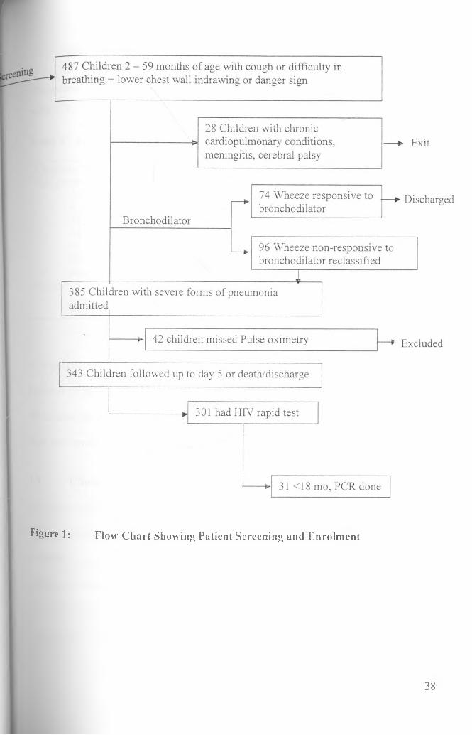
4.1 Patient Enrolment
We screened a total of 487 children presenting at the PEU with cough or difficulty
breathing between June and September 2009. Twenty eight children did not meet the
inclusion criteria. Seventy four children with wheeze had good response to
bronchodilator therapy and were therefore excluded.. The rest of the children (385) were
assessed and admitted to the general paediatric wards. Fourty two children however
failed to have oxygen saturation determined by pulse oximetry and were therefore
excluded. We followed up 343 children in the wards for five days to determine mortality
outcome.
Testing for HIV infection was done for 301 children either at the PEU or in the admission
ward. Thirty one children aged 18 months and below with a positive HIV antibody test
had DNA PCR test.
Figure 1 is a flow chart showing patient screening and enrolment.
37
r
Figure 1: Flow C hart Showing Patient Screening and Enrolm ent
38
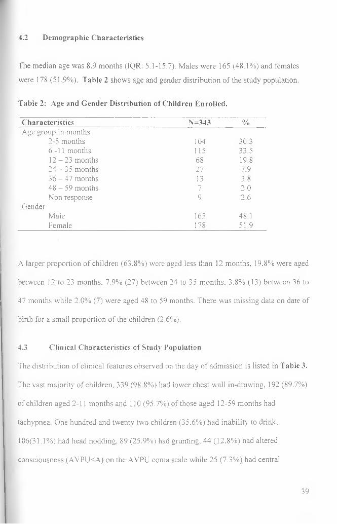
4.2 Demographic Characteristics
The median age was 8.9 months (IQR: 5.1-15.7). Males were 165 (48.1%) and females
were 178 (51.9%). Table 2 shows age and gender distribution of the study population.
Table 2: Age and Gender Distribution of Children Enrolled.
Characteristics N=343 %Age group in months
2-5 months 104 30.36-11 months 115 33.51 2 -2 3 months 68 19.82 4 -3 5 months 27 7.936 - 4 7 months 13 3.848 - 59 months 7 2.0Non response 9 2.6
GenderMale 165 48.1Female 178 51.9
A larger proportion of children (63.8%) were aged less than 12 months, 19.8% were aged
between 12 to 23 months, 7.9% (27) between 24 to 35 months. 3.8% (13) between 36 to
47 months while 2.0% (7) were aged 48 to 59 months. There was missing data on date of
birth for a small proportion of the children (2.6%).
4.3 Clinical Characteristics of Study Population
The distribution of clinical features observed on the day of admission is listed in Table 3.
The vast majority of children. 339 (98.8%) had lower chest wall in-drawing, 192 (89.7%)
of children aged 2-11 months and 110 (95.7%) of those aged 12-59 months had
tachypnea. One hundred and twenty two children (35.6%) had inability to drink,
106(31.1%) had head nodding, 89 (25.9%) had grunting. 44 (12.8%) had altered
consciousness (AVPU<A) on the AVPU coma scale while 25 (7.3%) had central
39
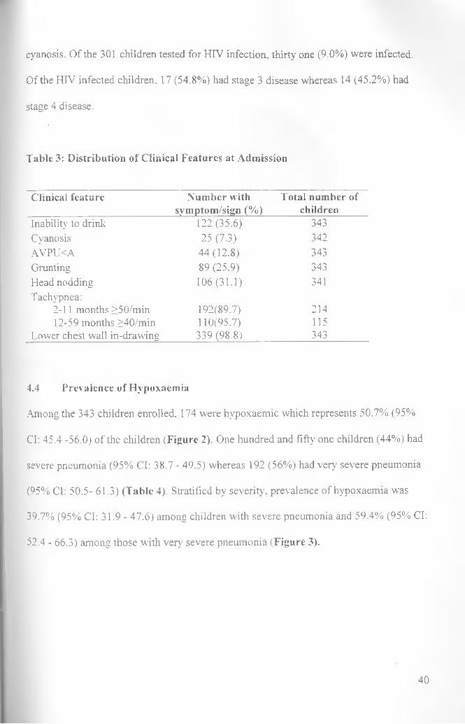
cyanosis. Of the 301 children tested for HIV infection, thirty one (9.0%) were infected.
Of the HIV infected children, 17 (54.8%) had stage 3 disease whereas 14 (45.2%) had
stage 4 disease.
Table 3: Distribution of Clinical Features at Admission
Clinical feature Number with symptom/sign (%)
Total number of children
Inability' to drink 122 (35.6) 343Cyanosis 25 (7.3) 342AVPU<A 44(12.8) 343Grunting 89 (25.9) 343Head nodding 106 (31.1) 341Tachypnea:
2-11 months >50/min 192(89.7) 21412-59 months >40/min 110(95.7) 115
Lower chest wall in-drawing 339 (98.8) 343
4.4 Prevalence of Hypoxaemia
Among the 343 children enrolled, 174 were hvpoxaemic which represents 50.7% (95%
Cl: 45.4 -56.0) of the children (Figure 2). One hundred and fifty one children (44%) had
severe pneumonia (95% Cl: 38.7 - 49.5) whereas 192 (56%) had very severe pneumonia
(95% Cl: 50.5- 61.3) (Table 4). Stratified by severity, prevalence of hypoxaemia was
39.7% (95% Cl: 31.9- 47.6) among children with severe pneumonia and 59.4% (95% Cl:
52.4 - 66.3) among those with very severe pneumonia (Figure 3).
40
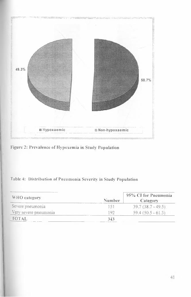
Figure 2: Prevalence of Hypoxemia in Study Population
Table 4: Distribution of Pneumonia Severity in Study Population
WHO category
Severe pneumonia Very severe pneumoniat o t a l _____________
Number151192
95% Cl for Pneumonia ______ Category______
39.7 (38.7 -49. 59.4 (50.5 - 61.
343
in co
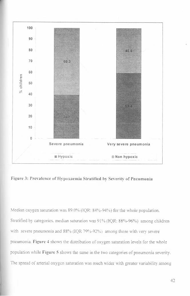
Figure 3: Prevalence of Hypoxaemia Stratified by Severity of Pneumonia
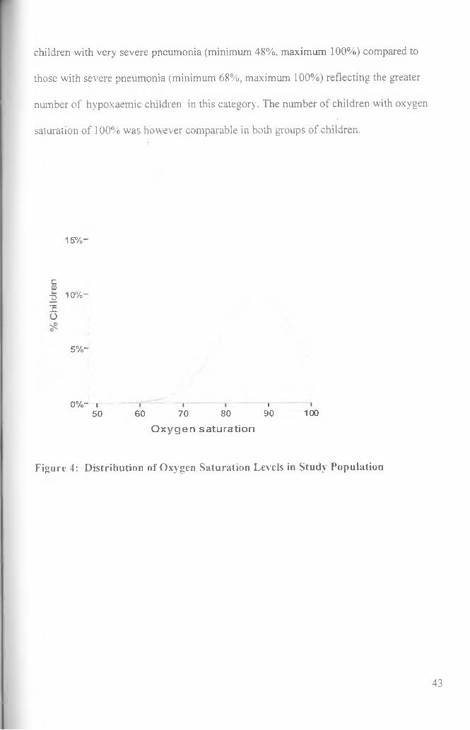
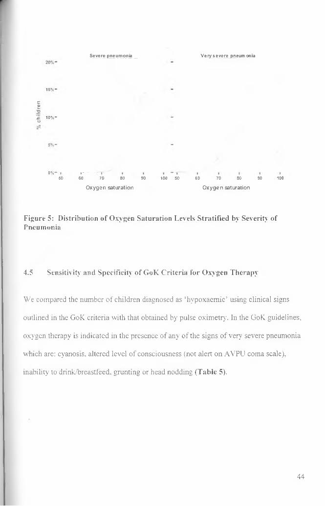
Median oxygen saturation was 89.0% (IQR: 84%-94%) for the whole population.
Stratified by categories, median saturation was 91% (IQR: 88%-96%) among children
with severe pneumonia and 88% (IQR 79%-92%) among those with very severe
pneumonia. Figure 4 shows the distribution of oxygen saturation levels for the whole
population while Figure 5 shows the same in the two categories of pneumonia severity.
The spread of arterial oxygen saturation was much wider with greater variability among
42
children with very severe pneumonia (minimum 48%, maximum 100%) compared to
those with severe pneumonia (minimum 68%, maximum 100%) reflecting the greater
number of hvpoxaemic children in this category. The number of children with oxygen
saturation of 100% was however comparable in both groups of children.
15% -
E0-q 10%-
Zo
5% -
0 % “ i -
50 60 70 80
O x y g e n s a tu ra tio n
i90 100
Figure 4: Distribution of Oxygen Saturation Levels in Study Population
43
Very severe pneumonia
15%
ca
'£ 10%-u
0% I*6050 70 80 90 100 50 60 70 80 90 100
Oxygen saturation Oxygen saturation
Figure 5: Distribution of Oxygen Saturation Levels Stratified by Severity of Pneumonia
4.5 Sensitivity and Specificity of GoK Criteria for Oxygen Therapy
We compared the number of children diagnosed as ‘hypoxaemic’ using clinical signs
outlined in the GoK criteria with that obtained by pulse oximetry. In the GoK guidelines,
oxygen therapy is indicated in the presence of any of the signs of very severe pneumonia
which are: cyanosis, altered level of consciousness (not alert on AVPU coma scale),
inability to drink/breastfeed, grunting or head nodding (Table 5).
44
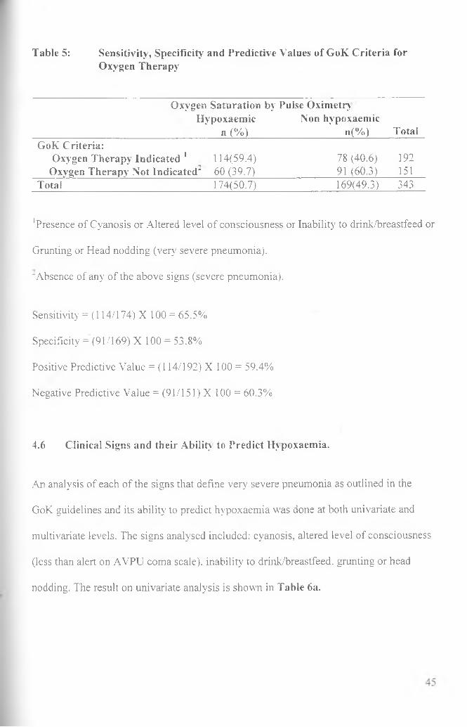
Table 5: Sensitivity, Specificity and Predictive Values of GoK Criteria for Oxygen Therapy
Oxygen Saturation by Pulse OximetryHypoxaemic Non hypoxaemic
n (%) n ( % ) TotalGoK Criteria:
Oxygen Therapy Indicated 1 114(59.4) 78 (40.6) 192Oxygen Therapy Not Indicated' 60 (39.7) 91 (60.3) 151
Total 174(50.7) 169(49.3) 343
'Presence of Cyanosis or Altered level of consciousness or Inability to drink/breastfeed or
Grunting or Head nodding (very severe pneumonia).
"Absence of any of the above signs (severe pneumonia).
Sensitivity = (114/174) X 100 = 65.5%
Specificity = (91/169) X 100 = 53.8%
Positive Predictive Value = (114/192) X 100 = 59.4%
Negative Predictive Value = (91/151) X 100 = 60.3%
4.6 Clinical Signs and their Ability to Predict Hypoxaemia.
An analysis of each of the signs that define very severe pneumonia as outlined in the
GoK guidelines and its ability to predict hypoxaemia was done at both univariate and
multivariate levels. The signs analysed included: cyanosis, altered level of consciousness
(less than alert on AVPU coma scale), inability to drink/breastfeed. grunting or head
nodding. The result on univariate analysis is shown in Table 6a.
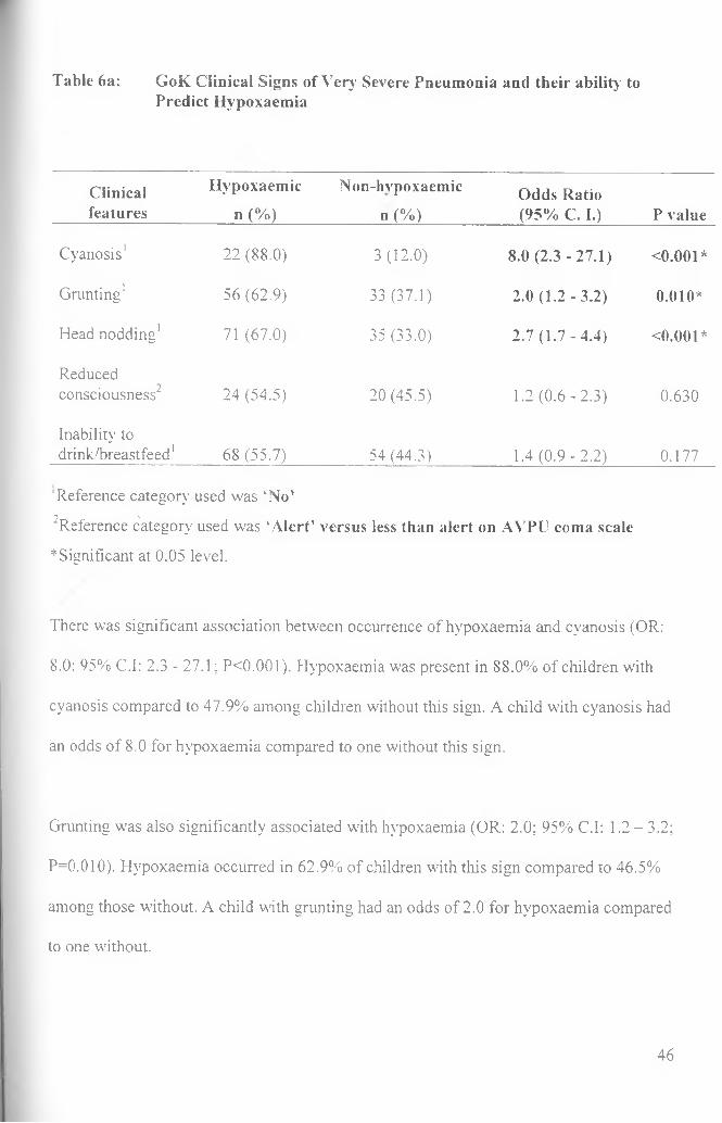
Table 6a: GoK Clinical Signs of Very Severe Pneumonia and their ability toPredict Hypoxaemia
Clinicalfeatures
Hypoxaemic
n (%)
Non-hvpoxaemic
n (%)Odds Ratio(95% C. I.) P value
Cyanosis1 22 (88.0) 3 (12.0) 8.0 (2.3 - 27.1) <0.001*
Grunting1 56 (62.9) 33 (37.1) 2.0 (1 .2-3 .2) 0.010*
Head nodding1 71 (67.0) 35 (33.0) 2.7 (1 .7-4 .4) <0.001*
Reducedconsciousness2 24 (54.5) 20 (45.5) 1.2 (0 .6-2 .3) 0.630
Inability to drink/breastfeed1 68 (55.7) 54 (44.3) 1.4 (0 .9-2 .2) 0.177
‘Reference category used was ‘No’9 N■Reference category used was ‘Alert’ versus less than alert on AVPU coma scale
*Significant at 0.05 level.
There was significant association between occurrence of hypoxaemia and cyanosis (OR:
8.0; 95% C.I: 2.3 - 27.1; PO.OOl). Hypoxaemia was present in 88.0% of children with
cyanosis compared to 47.9% among children without this sign. A child with cyanosis had
an odds of 8.0 for hypoxaemia compared to one without this sign.
Grunting was also significantly associated with hypoxaemia (OR: 2.0; 95% C.I: 1.2 - 3.2;
P=0.010). Hypoxaemia occurred in 62.9% of children with this sign compared to 46.5%
among those without. A child with grunting had an odds of 2.0 for hypoxaemia compared
to one without.
46
Similarly, there was a significant association between occurrence of hypoxaemia and
head nodding (OR: 2.7; 95% C.1:1.7 — 4.4; PO.OOl). A child with this sign had an odds
of 2.7 for hypoxaemia than one without.
Altered level of consciousness and inability to drink/breastfeed were not significantly
associated with hypoxaemia. Hypoxaemia was present in 54.5% of children with altered
level of consciousness and in 50.2% of children who were alert (OR: 1.2; 95% C.I: 0.6 -
2.3; P=0.630). Hypoxaemia occurred in 55.7% of children who were unable to
drink/breastfeed and in 48.0% of those who were able (OR: 1.4; 95% C.I: 0.9 - 2.2;
P=0.177). A child who was unable to drink/breastfeed had an odds of 1.4 for hypoxaemia
although this did not achieve statistical significance.
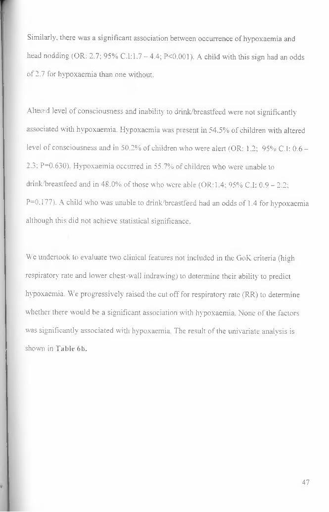
We undertook to evaluate two clinical features not included in the GoK criteria (high
respiratory rate and lower chest-wall indrawing) to determine their ability to predict
hypoxaemia. We progressively raised the cut off for respiratory rate (RR) to determine
whether there would be a significant association with hypoxaemia. None of the factors
was significantly associated with hypoxaemia. The result of the univariate analysis is
shown in Table 6b.
47
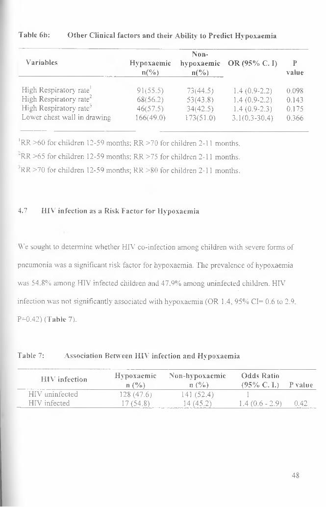
Table 6b: Other Clinical factors and their Ability to Predict Hypoxaemia
Non-Variables Hypoxaemic
’ n(%)hvpoxaemic
■»(%)OR (95% C. I) P
value
High Respiratory rate1 91(55.5) 73(44.5) 1.4 (0.9-2.2) 0.098High Respiratory rate2 68(56.2) 53(43.8) 1.4 (0.9-2.2) 0.143High Respiratory rate-5 46(57.5) 34(42.5) 1.4 (0.9-2.3) 0.175Lower chest wall in drawing 166(49.0) 173(51.0) 3.1(0.3-30.4) 0.366
*RR >60 for children 12-59 months; RR >70 for children 2-11 months.
'RR >65 for children 12-59 months; RR >75 for children 2-11 months.
'RR >70 for children 12-59 months; RR >80 for children 2-11 months.
4.7 HIV infection as a Risk Factor for Hypoxaemia
We sought to determine whether HIV co-infection among children with severe forms of
pneumonia was a significant risk factor for hypoxaemia. The prevalence of hypoxaemia
was 54.8% among HIV infected children and 47.9% among uninfected children. HIV
infection was not significantly associated with hypoxaemia (OR 1.4, 95% CI= 0.6 to 2.9,
P=0.42) (Table 7).
Table 7: Association Between HIV' infection and Hypoxaemia
HIV7 infection Hvpoxaemic Non-hvpoxaemic Odds Ration (%) n (%) (95% C. I.) P value
HIV7 uninfected HIV7 infected
128 (47.6) 17(54.8)
141 (52.4) 14(45.2)
11.4 (0 .6-2 .9) 0.42
48
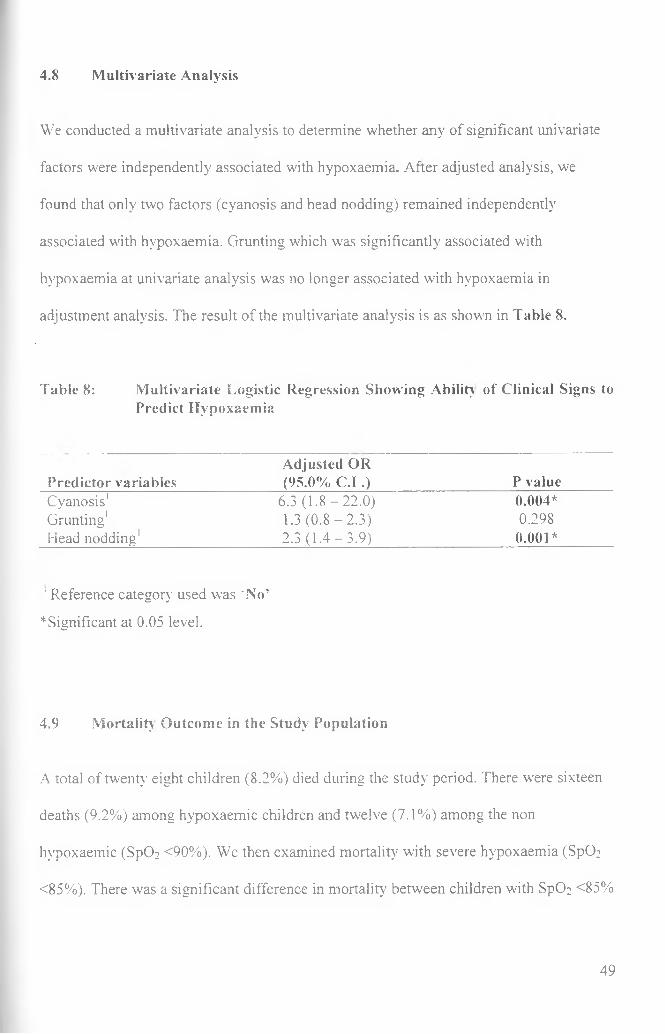
4.8 Multivariate Analysis
We conducted a multivariate analysis to determine whether any of significant univariate
factors were independently associated with hypoxaemia. After adjusted analysis, we
found that only two factors (cyanosis and head nodding) remained independently
associated with hypoxaemia. Grunting which was significantly associated with
hypoxaemia at univariate analysis was no longer associated with hypoxaemia in
adjustment analysis. The result of the multivariate analysis is as shown in Table 8.
Table 8: Multivariate Logistic Regression Showing Ability of Clinical Signs toPredict Hypoxaemia
Predictor variablesAdjusted OR (95.0% C.I .) P value
Cyanosis1 6.3 (1 .8 -22 .0 ) 0.004*Grunting1 1.3 (0 .8 -2 .3 ) 0.298Head nodding1 2.3 (1.4 - 3.9) 0.001*
1 Reference category used was ‘No’
* Significant at 0.05 level.
4.9 Mortality Outcome in the Study Population
A total of twenty eight children (8.2%) died during the study period. There were sixteen
deaths (9.2%) among hypoxaemic children and twelve (7.1%) among the non
hypoxaemic (SpO: <90%). We then examined mortality with severe hypoxaemia (Sp02
<85%). There was a significant difference in mortality between children with Sp02 <85%
49
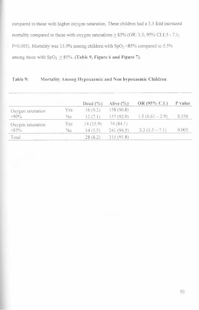
compared to those with higher oxygen saturation. These children had a 3.3 fold increased
mortality compared to those with oxygen saturations > 85% (OR: 3.3; 95% 0 :1 .5 -7.1;
P=0.005). Mortality was 15.9% among children with Sp02 <85% compared to 5.5%
among those with Sp02 > 85%. (Table 9, Figure 6 and Figure 7).
Table 9: Mortality Among Hypoxaemic and Non hypoxaemic Children
Dead (%) Alive (%) OR (95% C.I.) P value
Oxygen saturation Yes 16(9.2) 158 (90.8)<90% No 12(7.1) 157 (92.9) 1.3 (0.61 -2 .9 ) 0.556
Oxygen saturation Yes 14(15.9) 74 (84.1)<85% No 14(5.5) 241 (94.5) 3.3 (1.5 - 7.1) 0.005
Total 28 (8.2) 315(91.8)
50
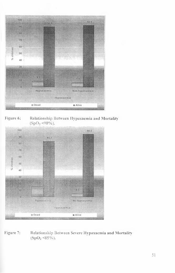
Figure 6: Relationship Between Hypoxaemia and Mortality(S p 0 2 <90%).
Figure 7: Relationship Between Severe Hypoxaemia and Mortality(S p 02 <85%).
51
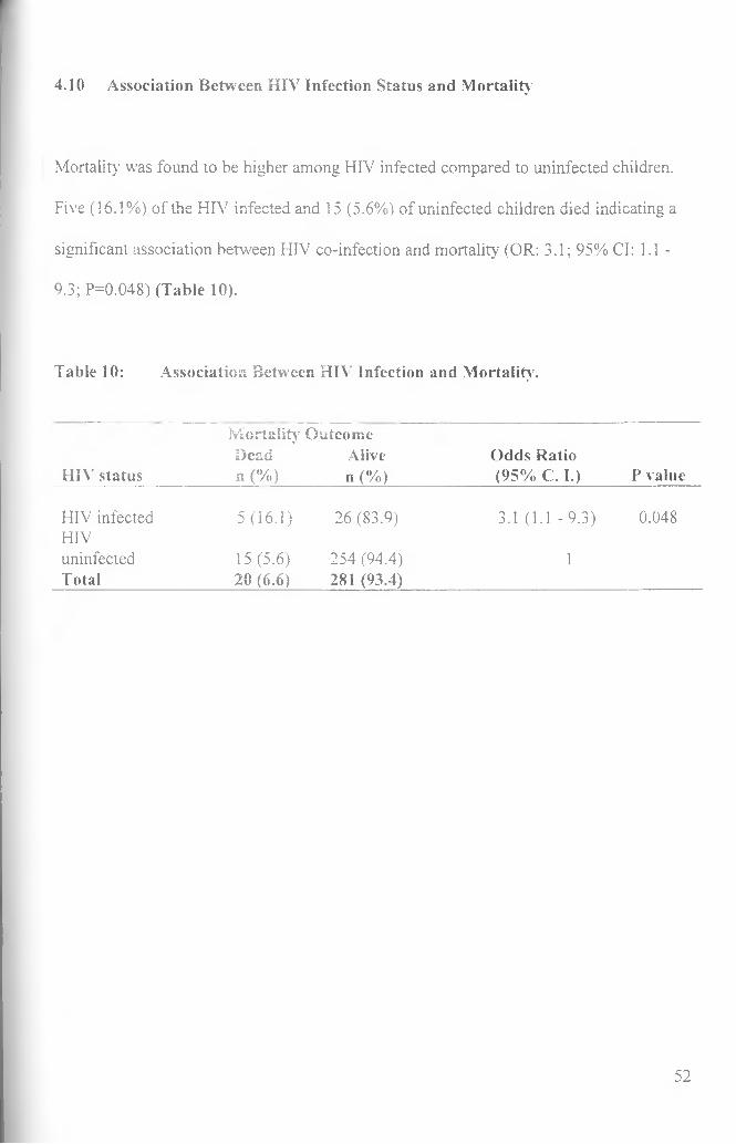
4.10 Association Between HIV Infection Status and Mortality
Mortality was found to be higher among HIV infected compared to uninfected children.
Five (16.1%) of the HIV infected and 15 (5.6%) of uninfected children died indicating a
significant association between HIV co-infection and mortality (OR: 3.1; 95% Cl: 1.1 -
9.3; P=0.048) (Table 10).
Table 10: Association Between HIV Infection and Mortality.
M ortality Outcome
HIV statusDead n (%)
Aliven (%)
Odds Ratio (95% C. I.) P value
HIV infected HIV
5 (16.1) 26 (83.9) 3.1 (1.1 -9 .3) 0.048
uninfectedTotal
15 (5.6) 20 (6.6)
254 (94.4) 281 (93.4)
1
52
5.0 DISCUSSION
In our study, there was an equal distribution between males (48.1%) and females
(51.9%). %). Majority of the children (63.8%) were less than one year of age indicating
that infants are the most vulnerable to severe forms of pneumonia.
The study indicates that hypoxaemia is a frequent occurrence among children with severe
or very severe pneumonia. Prevalence of 50.7% in KNH, Nairobi (altitude 1630m) is
comparable to that of a study by Wandi et al in Papua New Guinea (altitude 1600m) of
54.2% in this group of patients.
Prevalence in our study was however higher than reported by many other studies. Fu et
ah reported a prevalence of 9.5% in a multicentred study which included Mexico and
Bogota among other sites. Seven of these sites were at sea level and a threshold of <90%
was used to determine hypoxaemia. Two sites were at high altitude and threshold for
hypoxaemia was <88%. Brooks et aP(' reported a prevalence of 13.7% in Bangladesh
(altitude 0 m) and Bose et al0 a prevalence of 18.7% in India (altitude 0 m).
Stratified by severity, prevalence of hypoxaemia was 39.7% and 59.4% among children
with severe and very severe pneumonia respectively. This is much lower than that
reported by Basnet et aP in Nepal (1300m altitude) in which prevalence was 80% and
100% among children with severe and very severe pneumonia respectively. In that study,
a total of 250 children categorized as having cough/cold, pneumonia, severe pneumonia
or very severe pneumonia were recruited and assessed. Only 45 children had severe or
very severe pneumonia. Twenty five children had severe pneumonia and twenty had very
severe pneumonia. With such a small number, it is likely that there was selection for very
sick children. Compared to a study by Singhi et al“8 in India (altitude 0 m), prevalence of
hvpoxaemia in our study was higher among children with severe pneumonia (39.7%
compared to 26%) but lower (59.4% compared to 73.8%) among children with very
severe pneumonia.
A large number of the above studies were conducted at low altitude (seven out of nine
sites in Fu et al21, Brooks et al~6, Bose et al“‘\ Singhi et aF8). Low altitude has been
associated with lower prevalence of hypoxaemia. In a systematic review by Subhi et al2"
of both published and unpublished studies reporting the prevalence of hypoxaemia in
ALRJ, median prevalence of hypoxaemia among studies reviewed was 13% but
prevalence in various studies varied widely ranging from 6.9% to 100%. There were
however, wide variations between different regions even at similar altitudes and cut-off
values for hypoxaemia which means there could be other factors in our set up
contributing to the high prevalence of hypoxaemia. Factors that have been thought to
contribute to the wide variations in prevalence include: regional variations in pathogen
aetiology, host epidemiology, prevalence and severity of co-morbidities and
environmental factors.~ We did not explore the effect o f co-morbidities in our study.
54
Hvpoxaemia as a complication of pneumonia has been widely studied. Most studies on
hvpoxaemia in developing countries have mainly been on the ability of clinical signs to
predict hvpoxaemia x 16, ,7' 18
Most of the signs seen in pneumonia have been found to be associated with hvpoxaemia
by different studies. Drowsiness, cyanosis, decreased air entry,16 head nodding.0 16 nasal
flaring,16 1 grunting/ 18 chest retractions,''1 crepitations.1 ' 18 and a respiratory rate of
>70 breaths per minute for children three to 11 months or >60 breaths per minute for
those 12 months or older0 have all been reported to be predictors of hvpoxaemia. No
single sign has however been found to predict hvpoxaemia with both high sensitivity and
specificity. Despite this, clinical signs including some on which there is limited data
(head nodding and AVPU coma scale) have been included in the GoK guidelines as
indicators for oxygen therapy.
Our evaluation of the GoK criteria for oxygen therapy (presence of any of these signs:
cyanosis, inability to drink/breastfeed altered level of consciousness (AVPU<A),
grunting or head nodding) found this 'decision rule' to have both low sensitivity (65.5%)
and specificity (53.8%). This means using this criteria about one third (34.5%) of
children with hvpoxaemia would miss oxygen therapy with increased risk of mortality7
and almost half of the children (46.7%) who do not require oxygen would be given
oxygen leading to wastage. Administering oxygen to children who do not require it may
not be a problem in centres such as KNH where oxygen is harvested from the atmosphere
by use of oxygen concentrators and so may be relatively cheaper but is a challenge for
55
small hospitals which depend on oxygen supplied in cylinders. This is expensive and
sometimes hospitals run out of oxygen posing increased risk of mortality for those who
require it. The government funds public health facilities in Kenya. It is regrettable that a
lot of money is spent on purchase of oxygen much of which ends up being used by
children who do not require it. Even at KNH where concentrators are used and therefore
relatively cheaper compared to use of oxygen cylinders, the increased demand for oxygen
is an additional cost to the hospital. Oxygen is an expensive and precious resource that
should be used judiciously.
We evaluated the five clinical signs recommended by the GoK as indicators of oxygen
therapy. We found cyanosis, grunting and head nodding to be associated with
hypoxaemia at univariate level but at multivariate analysis only cyanosis (OR: 9.1; 95%
CI:1.99 - 44.0, P=0.06) and grunting (OR: 2.1; 95% Cl: 1.2 - 3.6. P=0.013) were
independently associated with hypoxaemia.
Altered level of consciousness (AVPU<A) may be associated with other complications
such as hypoglycemia which may occur in severely ill children. It is possible that some of
the children had altered consciousness due to hypoglycemia. We did not collect data on
hypoglycaemia and therefore cannot verify its effect on our findings. Similarly, other
complications of severe illness other than hypoxaemia may result in inability to
drink/breastfeed.
56
Our study, unlike other previous studies did not show a significant association between
hypoxaemia (arterial oxygen saturation <90%) and mortality but did show a significant
association wdien the cut off was reduced to <85% (OR: 3.3; 95% Cl: 1.5 - 7.1, P=0.005).
In the study by Onyango et a f hypoxaemic children were 4.3 times more likely to die
than non hypoxaemic children. Weber et a f in a similar study in the Gambia found that
the relative risk for death among hypoxaemic children with ALRI w'as 4.6 and the case
fatality rate w’as inversely related to the arterial haemoglobin oxygen saturation. All
children with hypoxaemia received oxygen and management for pneumonia according to
the standard protocols. We believe other factors such as treatment failure and co
morbidities may have contributed significantly to mortality in our cohort of patients.
Thirty one children (9.0%) were HIV infected. Although our results suggested a possible
association between HIV infection and hypoxaemia. the number of HIV infected children
in this study was small so we were not powered to make this conclusion. HIV co-infected
children had an odds of 3.1 for mortality than uninfected children. This compares with
Zimbabwean studies which showed the risk of mortality among children treated for
pneumonia was three times higher in those with HIV co-infection.30 Although not the
main focus o f this study, our finding validates the recommendation by the GoK to treat
HIV co-infected children more aggressively. HIV infected children w7ith either severe or
very severe pneumonia are treated with both crystalline penicillin and gentamycin as first
line therapy whereas for the uninfected, gentamycin is given to only those with a
classification of very severe pneumonia.
57
6.0 STUDY LIMITATIONS
1. We had limited time over which we had to complete the study as dictated by the
Postgraduate academic calendar. A longer duration of data collection would have
yielded a larger sample size that would allow greater precision in reporting of
estimates for proportions or odds ratios. In addition, a longer study period would
reduce the possible influence of seasonal variation on reported estimates and
hence improve generalisability.
2. Kenyatta National Hospital, being a tertiary facility receives patients referred
from lower-level hospitals. Our study population may have comprised of “sicker"
patients than those attending other hospitals in Kenya thus limiting the
applicability of our results on prevalence of hypoxaemia to these other health
facilities.
7.0 CONCLUSIONS
1. Prevalence of hypoxaemia was high, occurring in 50.7% of children
hospitalized with severe forms of pneumonia. Prevalence was highest (59.4%)
among children hospitalized with very severe pneumonia.
2. The Government of Kenya criteria for oxygen administration (presence of any
of these signs: cyanosis, inability to drink/breastfeed, altered level of
consciousness (AVPU<A), grunting or head nodding) have a low sensitivity'
(65.5%) and specificity (53.8%) for predicting hypoxaemia among children
with severe forms of pneumonia.
58
J>. Severe hvpoxaemia (SpC>2 <85%) is associated with a 3.3 fold increased
mortality among children hospitalised with severe forms of pneumonia.
8.0 RECOMMENDATIONS
Based on findings in this study, we recommend the following:
1. That the government of Kenya promotes the use of pulse oximeters for
detection of hvpoxaemia in all public hospitals.
2. That the Government consider carrying out a cost-benefit study on the use of
pulse oximeters vis-a-vis continued use of clinical signs to determine w'hich
children require oxygen therapy.
59
REFERENCES
E W orld Health Organization. Life in the 21st century. A vision for all. Geneva:
W'HO, 1998:66.
2. Murray CJ and Lopez AD. Mortality> by cause for eight regions o f the world:
Global burden o f disease study. The Lancet. 1997; 349:1269-1276.
3. Mulholland K. Magnitude o f the problem o f childhood pneumonia. The Lancet.
1999:354:590 - 592.
4. WTlliams BG, Gouws E, Boschi-Pinto C, Bryce J and Dye C. Estimates o f
world-wide distribution o f child deaths from acute respiratory infections. The
Lancet, 2002;2:25-32.
5. Onyango FE, Steinhoff MC. W7afula EM, W7ariua S, Musia J and Kitonyi J.
Hypoxaemia in young Kenyan children M’ith acute lower respiratory infection.
BMJ, 1993;306:612-615.
6. W'eber MW7. Usen S, Palmer A. Jaffar S and Mulholland EK. Predictors o f
hypoxaemia in hospital admissions with acute lower respiratory tract infection in
a developing country. Arch Dis Child. 1997;76 310-314.
7. Korppi M, Koskela M, Makela P and Launiala K. Bacterial coinfection in
children hospitalized with respiratory syncytial virus infections. Pediatr Infect Dis
1989; 8:687-92.
8. W7HO programme for the control of acute respiratory infections in children.
Acute respiratory infections in children: case management in small hospitals in
developing countries. Geneva: W7HO,1990, W7HO/ARI/90.5
60
9. Bryce J, Boschi-Pinto C, Shibuya K and Black RE. WHO, WHO estimates o f the
causes o f death in children. The Lancet 2005;365:1147-1152.
10. World Health Organization. The clinical use o f oxygen: Guidelines for
appropriate oxygen technology in hospitals with limited resources. WHO draft,
2008.
11. W'orld Health Organization. Pocket hook o f hospital care for children: Guidelines
for the management o f common illnesses with limited resources. Geneva: WHO,
2005.
12. Duke T, Blacken AJ, Sails A.T and Bonkowsky J. Hypoxaemia in acute
respiratory and non-respiratory illnesses in neonates and children in a
developing country. Arch. Dis. Child, 2002; 86:108-112.
13. Subhf R., Smith K and Duke T. When should oxygen be given to children at high
altitude? A systematic review to define altitude-specific hypoxaemia. Arch Dis
Child, 2009;94:6-10.
14. C-hantarojanasiri ND and Rogers M. Lower Airways Disease. In: Textbook o f
Pediatrics and Intensive Care. Ed. Rogers MC. Wulliam and Wilkins,
Baltimore. 1987. First edition pp 199-235.
15. Sandra LS. Oxygen Saturation Monitoring by Pulse Oximetry. In: AACN
Procedure Manual for Critical Care. Ed. Debra SL and Karen KC. Saunders WrB.
2001. Fourth edition p77.
61
16. Usen S, Weber M, Mulholland K, Jaffar S, Oparaugo A, Omosigho C, Adegbola
R and Greenwood B. Clinical predictors o f hypoxaemia in Gambian children with
acute lower respiratory infection: prospective cohort study. BMJ. 1999:318:86-
91.
17. Lozano JM, Steinhoff M. Ruiz JG.. Mesa ML. Martinez N and Dussan B. Clinical
predictors o f acute radiological pneumonia and hypoxaemia at high altitude.
Arch Dis Child, 1994;71:323-7.
18. Smyth A. Carty H and Hart CA. Clinical predictors o f hypoxaemia in children
with pneumonia. Ann Trop Paediatr, 1998. 18:31-40.
19. Republic o f Kenya. Basic paediatric protocols. Ed. Ministry of Health, 2004.
20. Subhi R. Adamson M. W eber M. Smyth K and Duke T. The prevalence o f
hypoxaemia among ill children in developing countries: A systematic review.
Lancet Infect Dis, 2009;9:219-27.
21.. Fu LY, Ruthazer R. Wilson I, Patel A , Fox LM. Tuan TA, Jeena P. Chisaka N,
Hassan M,. Lozano J, Maulen-Radovan I, Thea DM. Qazi S and Hibberd P. Brief
hospitalization and pulse oximetry for predicting amoxicillin treatment failure in
children M’ith severe pneumonia. Pediatrics, 2006;! 18 :e 1822-30.
22. Wandi F, Peel D and Duke T. Hypoxaemia among children in rural hospitals in
Papua New Guinea: epidemiology: and resource availability:—a study to support a
national oxygen programme. Ann Trop Paediatr, 2006;26:277-84.
23. Basnet S, Adhikari RK and Gurung CK. Hypoxaemia in children with pneumonia
and its clinical predictors. Indian .1 Pediatr, 2006;73:777-79.
62

24. Ashraf H, Jahan SA, Alam NH, . Mahmud R, Kamal SM, Salam MA and Gyr
N. Day-care management o f severe and very severe pneumonia, without
associated co-morbidities such as severe malnutrition, in an urban health clinic in
Dhaka. Bangladesh. Arch Dis Child. 2008. 93:490-4.
25. Bose A. Coles CL, Gunavathi J, Moses, P. Raghupathy P, Kirubakaran C. Black
RE. Brooks W A and Santosham M. Efficacy o f zinc in the treatment o f severe
pneumonia in hospitalized children <2 y old. Am J Clin Nutr, 2006:83:1089-96;
quiz 1207.
26. Brooks WA, Yunus M, Santosham M. Wahed MA, Nahar K. Yeasmin S and
Black RE. Zinc for severe pneumonia in very young children: Double-blind
Placebo-Controlled trial. The Lancet. 2004:363:1683-1688.
27. Djelantik, 1G, Gessner BD, Sutanto A, Steinhoff M, . Linehan M Moulton LH
and Arjoso S. Case fatality proportions and predictive factors for mortality
among children hospitalized with severe pneumonia in a rural developing country
setting. J Trop Pediatr, 2003;49:327-32.
28. Singhi S and Kaur H. Prevalence and predictors o f hypoxaemia in acute
infections presenting to pediatric emergency department. Indian J Crit Care Med,
2003;7:118-123.
29. Reuland DS, Steinhoff MC, Gilman RH. Bara M., Olivares EG. Jabra, A. and
Finkelstein D. Prevalence and prediction o f hypoxemia in children with
respiratory infections in the Peruvian Andes. J Pediatr, 1991:119:900-6.
63
30. World Health Organization. Management o f children with pneumonia and HIV in
low-resource settings. Report of a consultative meeting in Harare, Zimbabwe, 30-
31 January, 2003.
31. UN AIDS. http://data.unaids.org/pub/GlobalReport/2008/JCl 511 GR08
Executive summary en.pclf (Accessed 23 rd January 2009.
32. National Aids and Sexually Transmitted Infections Control Programme. Kenya
AIDS indicator survey. Ministry of Health . 2008.
33. Central Bureau of Statistics. Ministry of Health. Kenya Medical Research
Institute. National Council for Population and Development, Centers for Disease
Control Prevention. ORC Macro. Calverton, Maryland. USA. Kenya
Demographic Health Survey. 2008.
34. Graham SM. Kamanga PIS, Walsh AL, Hart CA and Molyneux ME. Clinical
presentation and outcome o f Pneumocystis carinii pneumonia in Malawian
children. The Lancet, 2000:355:369-73.
35. Ruffini D and Mahdi A. The high burden o f Pneumocystis carinii pneumonia in
African HIV-1-infected children hospitalized for severe pneumonia. AIDS. 2002;
16:105-12.
36. \o g e r R and Chadwick EG. Acquired Immunodeficiency Syndrome. In: Nelson
Textbook o f Pediatrics. Eds. Kliegman RM., Behrman RE, Jenson HB and
Stanton BF. Saunders Elsevier, Pennsylvania. 2007. 18th edition p p l431-1434.
37. Bii C, Kose J and Taguchi H. Pneumocystis jirovechii and microbiological
findings in children with severe pneumonia in Nairobi, Kenya. Int J Tuberc Lung
Dis 2006;10:1286-91.
64
38. Maina B. Short term mortality Outcome o f pneumonia in children admitted at
Kenyatta National Hospital. Masters in Medicine Thesis (2006). Department of
Paediatrics. University of Nairobi. P.o. Box 19676 Nairobi 00202.
39. Irimu G, Wamae A, Wasunna A. Were F, Ntoburi S, Opiyo N, Ayieko P, Peshu N
and English M. Developing and Introducing Evidence Based Clinical Practice
Guidelines for Serious Illness in Kenya. Arch Dis Child, 2008:93:799-804.
65
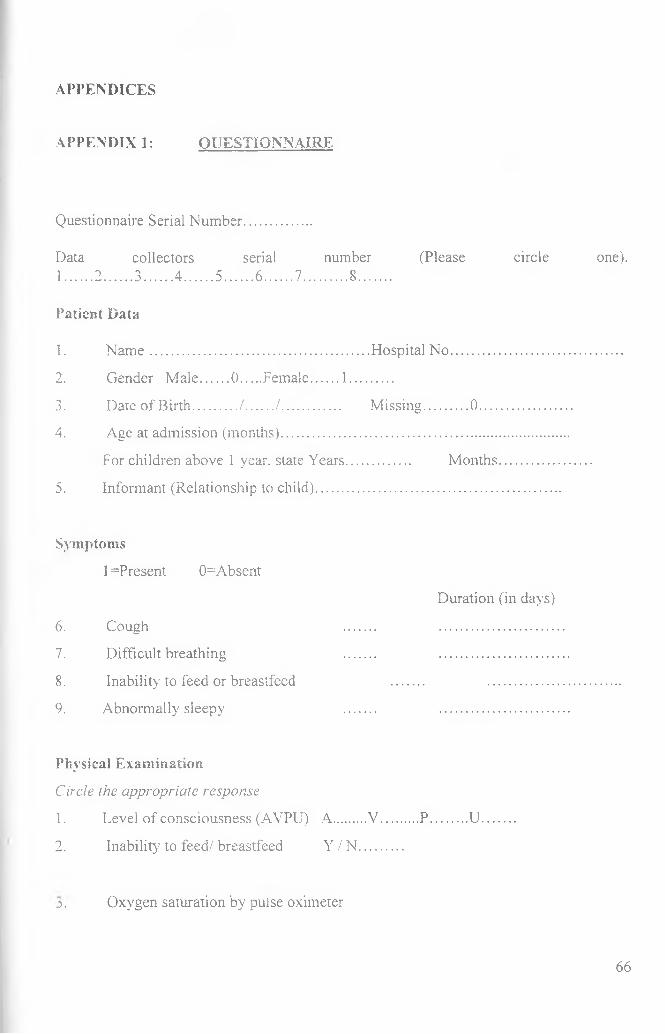
APPENDICES
APPENDIX 1: QUESTIONNAIRE
Questionnaire Serial Number
Data collectors serial number (Please circle one).1....... 2 ........3 ........ 4 ........5 ........ 6 ........7............ 8.........
Patient Data
1. N am e........................................................... Hospital N o...............................................
2. Gender Male....... 0 ......Female........1 ...........
3. Date of Birth............/ ......../ ................ Missing............0 ..........................
4. Age at admission (months)...........................................................
For children above 1 year, state Years................. Months
5. Informant (Relationship to child).................................................
Symptoms
I =Present 0=Absent
6. Cough
7. Difficult breathing
8. Inability' to feed or breastfeed
9. Abnormally sleepy
Duration (in days)
Physical Exam ination
Circle ihe appropriate response
1. Level of consciousness (AVPU) A........ V .... .... P .... ....U.
2. Inability to feed/ breastfeed Y / N ..........
j . Oxygen saturation by pulse oximeter
66
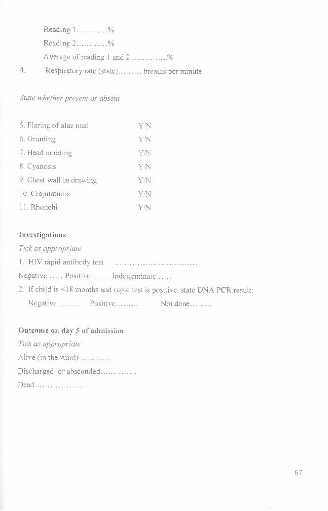
Reading 1................%
Reading 2 ................%
Average of reading 1 and 2 .................. %
4. Respiratory rate (state)............breaths per minute
State whether present or absent
5. Flaring of alae nasi Y/N
6. Grunting Y/N
7. Head nodding Y/N
8. Cyanosis Y/N
9. Chest wall in drawing Y/N
10. Crepitations Y/N
11. Rhonchi Y/N
Investigations
Tick as appropriate
1. HIV rapid antibody test ..............................................
Negative....... Positive........... Indeterminate.......
2. If child is <18 months and rapid test is positive, state DNA PCR result:
Negative............ Positive............ Not done............
Outcome on day 5 of admission
Tick as appropriate
Alive (in the w a rd )................
Discharged or absconded....................
Dead..........................
67
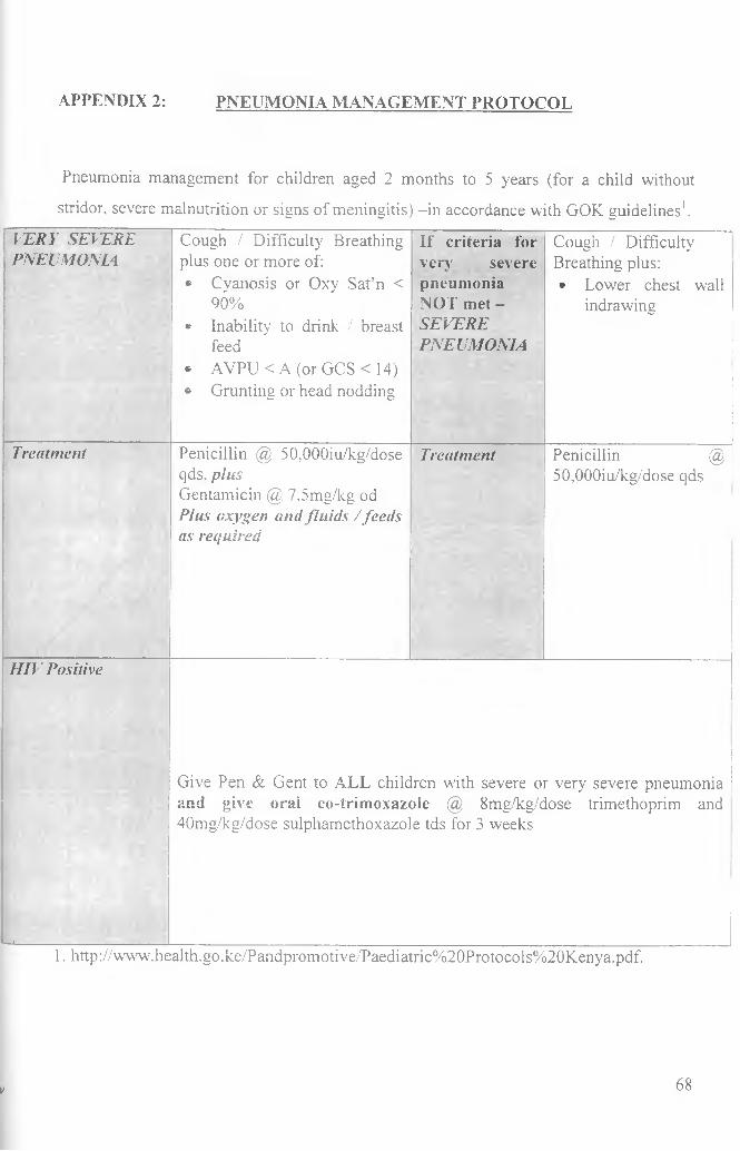
APPENDIX 2: PNEUMONIA MANAGEMENT PROTOCOL
Pneumonia management for children aged 2 months to 5 years (for a child without
stridor, severe malnutrition or signs of meningitis) -in accordance with GOK guidelines1.
VERY SEVERE PNEUMONIA
Cough / Difficulty Breathing plus one or more of:* Cyanosis or Oxy Sat’n <
90%* Inability to drink / breast
feed* AVPU < A (or GCS < 14)* Grunting or head nodding
If criteria for very severe pneumonia NOT met -SEVEREPNEUMONIA
Cough / Difficulty Breathing plus:• Lower chest wall
indrawing
Treatment Penicillin @ 50,000iu/kg/dose qds. plusGentamicin @. 7.5mg/kg od Plus oxygen and fluids / feeds as required
Treatment Penicillin @ 50,000iu/kg/dose qds
H IV Positive
■— »__________________
Give Pen & Gent to ALL children with severe or very severe pneumonia and give oral co-trimoxazole @ 8mg/kg/dose trimethoprim and 40mg/kg/dose sulphamethoxazole tds for 3 weeks
1. http://www.health.go.ke/Pandpromotive/Paediatric%20Protocols%20Kenya.pdf.
V 68
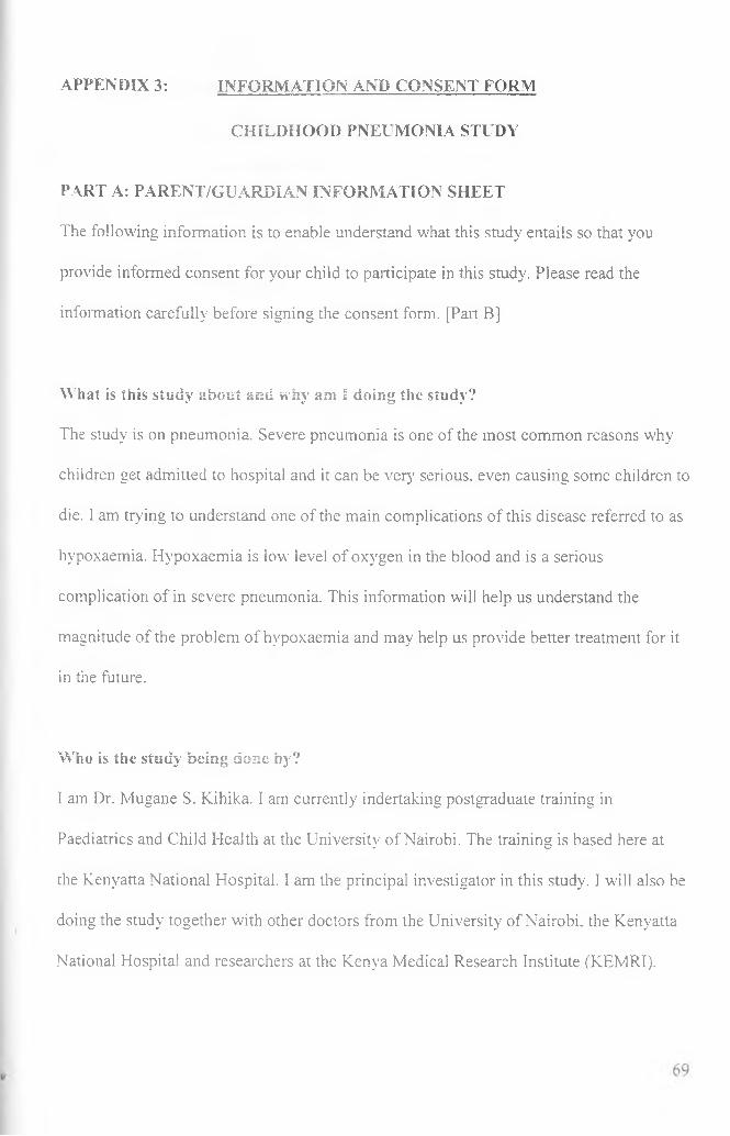
APPENDIX 3: INFORMATION AND CONSENT FORM
CHILDHOOD PNEUMONIA STUDY
PART A: PARENT/GUARDIAN INFORMATION SHEET
The following information is to enable understand what this study entails so that you
provide informed consent for your child to participate in this study. Please read the
information carefully before signing the consent form. [Part B]
What is this study about and why am I doing the study?
The study is on pneumonia. Severe pneumonia is one of the most common reasons why
children get admitted to hospital and it can be very serious, even causing some children to
die. I am trying to understand one of the main complications of this disease referred to as
hypoxaemia. Hypoxaemia is low level of oxygen in the blood and is a serious
complication of in severe pneumonia. This information will help us understand the
magnitude of the problem of hypoxaemia and may help us provide better treatment for it
in the future.
Who is the study being done by?
I am Dr. Mugane S. Kihika. I am currently indertaking postgraduate training in
Paediatrics and Child Health at the University of Nairobi. The training is based here at
the Kenyatta National Hospital. I am the principal investigator in this study. I will also be
doing the study together with other doctors from the University of Nairobi, the Kenyatta
National Hospital and researchers at the Kenya Medical Research Institute (KEMRI).
Why are we requesting to indude your child?
The study is on severe forms of pneumonia. Your child has features suggesting he/she
has pneumonia severe enough to indicate that admission is necessary.
We are doing a stud}- on such severe pneumonia and would like to explain this to you and
ask your permission to include your son / daughter {name_______________________).
We are asking if we can study ail children with these forms of severe pneumonia coming
to Kenyatta National Hospital - so your child is one of many we are asking about as
pneumonia is a common, serious disease.
What will the study involve for my child if I agree?
If you are happy for your child to be involved we would like to ask you questions about
the child's illness, examine them carefully and record the information about your child's
illness.
For this study, we will measure the level of oxygen in the blood using this device {show
oximeter) that is entirely painless and takes only 1-2 minutes. Testing for HIV is now a
routine test on admission to this hospital - it is recommended for all inpatients and should
be done for all children whether or not they are in the study. I will explain this in more
detail separately and ask if you agree to the test on your child. You are free to refuse the
HIY test after this explanation. There may be other blood tests your may need. These are
not part of any study but we will try and do all the tests needed at the same time to avoid
any extra needles.
70
After examining your child and doing the necessary tests and giving the recommended
treatment, your child will be admitted to the general paediatric ward. We will not be in
charge of the treatment all the time, your ward doctors will be in charge but we will come
and check on your child's progress from time to time and also by looking at your child's
hospital records at the time they are discharged.
Are there any risks to my child participating?
Pulse oximetry is a completely painless procedure and takes only 1-2 minutes.
For the HIV test, we will require only about 3 to 4 drops of blood. Taking blood from the
tip of the index or middle finger causes a small amount of temporary pain, but the amount
taken is too small to affect your child's health and we will take the blood test for the
study at the same time with any other routine blood tests that may be needed.
There may be some slight inconvenience to you because of the time taken to answer the
questions and get the tests but this should not cause any harm.
Are there any benefits to my child participating?
Your child will receive no major benefit. The tests being done as part of the research may
sometimes help us provide better treatment to your child. There will be no extra cost to
you. The research may help us provide better treatment to children in the future.
W hat happens i f f refuse to participate?
All participation in research is voluntary. You are free to decide if you want your child to
take part. If you do agree you can change your mind at an}' time and withdraw your child
71
from the research. This will not affect your child’s care now or in the future. If you do not
agree for your child to be included that will cause no problem.
Who will have access to information about me/my child in this research?
Information that is important to providing the right medical care for your child will be
shared with the doctors looking after your child but all of the staff at KNH will ensure
your medical records are kept confidentially. All the research records are stored securely
without the name of you or your child and only people who are closely concerned with
the research will be able to this view information.
Who has allowed this research to take place?
A committee from KNH has looked carefully at this work and has agreed that the
research is important, that it will be conducted properly and that participants' safety and
rights have been respected.
What if I have any questions?
Please feel free to ask any questions about the study. If there is any part of this form that
you do not understand, be sure to ask questions about it. You can contact me or those
who are responsible for the care of your child and this research. For any question or
clarification please contact me,
Dr. Mugane S.K.
Department o f Paediatrics and Child Health
University of Nairobi,
72
P.O. Box 19676-00200, Nairobi
Cell phone number: 0721442785
Email: [email protected]
OR
The Secretary, KNH/UON-Ethics and Research Committee
P.O.Box 20723-00202, Nairobi
Telephone 0202726300-9
Email: [email protected]
PART B: CONSENT FORM
I. being a guardian o f ........................................................................... (name of child),
Hospital Number................................ have understood the information in Part A above
on what the research entails. I have also had a chance to ask questions which have been
answered satisfactorily. I understand that I can withdraw from the research at any stage
and it will not affect me/my child in any way.
- I agree to allow my child to take part in this research and for the collection of clinical
data.
Parent/guardiairs signature: _______ ________ Date-
Parent/guardian’s nam e:__________________ Time-
V 73
I certify that I have followed all the study specific procedures in the SOP for obtaining
informed consent.
Designee/investigator’s signature:___________________________Date:
Designee/investigator’s nam e:______________________________ Time:
Only necessary i f the parent/guardian cannot read:
I* attest that the information concerning this research was accurately explained to and
apparently understood by the parent/guardian and that informed consent was freely given
by the parent/guardian.
Witness’ signature:__________________________________ Date:
Witness’ nam e:___________________________ __________ Time:
*A witness is a person who is independent from the trial or a member of staff who was
not involved in gaining the consent.
Thumbprint of the parent as named above if they cannot write:
__________________________ Investigator’s statement
74
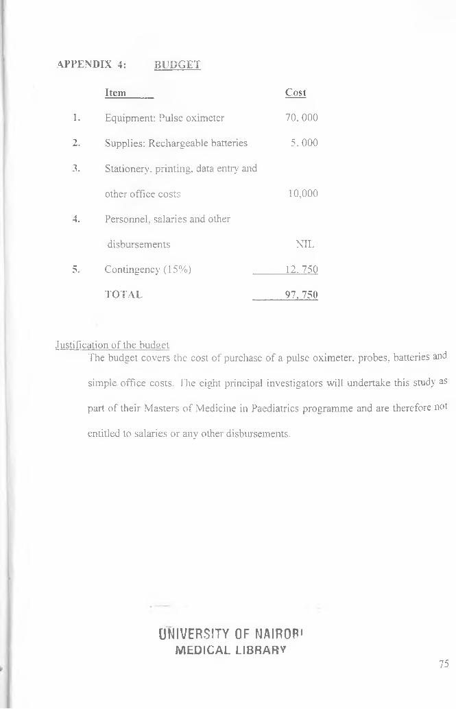
APPENDIX 4: BUDGET
Item_____ Cost
1. Equipment: Pulse oximeter 70.000
2. Supplies: Rechargeable batteries 5.000
3. Stationery, printing, data entry and
other office costs 10,000
4. Personnel, salaries and other
disbursements NIL
5. Contingency (15%) ______ 12. 750
TOTAL 97, 750
Justification of the budgetThe budget covers the cost of purchase of a pulse oximeter, probes, batteries and
simple office costs. The eight principal investigators will undertake this study as
part of their Masters of Medicine in Paediatrics programme and are therefore not
entitled to salaries or any other disbursements.
U NI V ERSI T Y OF NAIR0B»MEDICAL LIBRARY
75