Embed Size (px)
Citation preview
RESEARCH Open Access
Hypoxia-challenged MSC-derived exosomesdeliver miR-210 to attenuate post-infarctioncardiac apoptosisHao Cheng1† , Shufu Chang1†, Rende Xu1†, Lu Chen1†, Xiaoyue Song1, Jian Wu2, Juying Qian1, Yunzeng Zou2 andJianying Ma1*
Abstract
Background: Myocardial infarction (MI) is a major cause of death worldwide. Although percutaneous coronaryintervention and coronary artery bypass grafting can prolong life, cardiac damage persists. In particular,cardiomyocytes have no regenerative capacity. Mesenchymal stem cells (MSCs) are attractive candidates for thetreatment of MI. The manner by which MSCs exert a beneficial effect upon injured cells is a source of continuedstudy.
Methods: After the isolation and identification of exosomes from MSCs, the expression of miR-210 was determinedby microarray chip. Subsequently, gain- and loss-function approaches were conducted to detect the role ofexosomes and exosomal-miR-210 in cell proliferation and apoptosis of cardiomyocytes, as well as the MI in vivo.Dual-Luciferase Report Gene System was used to demonstrate the target gene of miR-210.
Results: We tested the hypothesis that MSC-derived exosomes transfer specific miRNA to protect cardiomyocytesfrom apoptotic cell death. Interestingly, direct cardiac injection of MSC exosomes reduced infarct size and improvedheart function after coronary ligation. In vitro, the MSC exosomes enhanced cardiomyocyte survival to hypoxia.Confirmation of exosome uptake in myocytes was confirmed. Dual-luciferase reporter assay implicated miR-210 as amediator of the therapeutic effect and AIFM3 as a downstream target. Treatment with miR-210 overexpressing MSCexosomes improved myocyte protection to both in vitro and in vivo stress. Furthermore, the endogenous andexogenous miR-210 had the same therapeutic effects.
Conclusion: These results demonstrated that the beneficial effects offered by MSC-exosomes transplantation afterMI are at least partially because of excreted exosome containing mainly miR-210.
Keywords: Myocardial infarction, Exosome, Mesenchymal stem cells, Hypoxia, Apoptosis
© The Author(s). 2020 Open Access This article is licensed under a Creative Commons Attribution 4.0 International License,which permits use, sharing, adaptation, distribution and reproduction in any medium or format, as long as you giveappropriate credit to the original author(s) and the source, provide a link to the Creative Commons licence, and indicate ifchanges were made. The images or other third party material in this article are included in the article's Creative Commonslicence, unless indicated otherwise in a credit line to the material. If material is not included in the article's Creative Commonslicence and your intended use is not permitted by statutory regulation or exceeds the permitted use, you will need to obtainpermission directly from the copyright holder. To view a copy of this licence, visit http://creativecommons.org/licenses/by/4.0/.The Creative Commons Public Domain Dedication waiver (http://creativecommons.org/publicdomain/zero/1.0/) applies to thedata made available in this article, unless otherwise stated in a credit line to the data.
* Correspondence: [email protected]†Hao Cheng, Shufu Chang, Rende Xu and Lu Chen contributed equally tothis work.1Department of Cardiology, Zhongshan Hospital, Fudan University, 1609Xietu Road, Shanghai 200032, ChinaFull list of author information is available at the end of the article
Cheng et al. Stem Cell Research & Therapy (2020) 11:224 https://doi.org/10.1186/s13287-020-01737-0
BackgroundIschemic heart disease is a significant cause of morbidityand mortality [1]. Percutaneous coronary thrombolytictherapy improves clinical outcomes. However, nearly25% of patients are not re-perfused in a timely manner[2]. Furthermore, cardiomyocytes have little to no regen-erative capacity. Cell-based therapies, such as stem cells,which have regenerative potential, are being applied toindividuals with cardiac disease [3]. Stem cells can secretquantities of growth, anti-apoptotic, and anti-inflammatory factors that may improve heart function.However, technical issues have shown stem cells them-selves to be inefficient [4]. Mesenchymal stem cells(MSCs) are prototypic adult stem cells [5] that produceparacrine growth factors that may increase survivabilityof cardiomyocytes and stimulate angiogenesis [6].All cell types secrete membrane vesicles, including
exosomes, that contain bioactive substances such as pro-teins, mRNAs, and miRNAs, and enzymes, among others[7]. During uptake by neighboring cells, exosome-delivered cargo has the potential to alter cell response[8]. Embryonic stem cell exosomes were reported todrive cardiac regeneration [9]. Others noted exosomesprovided remote pre-conditioning to limit kidney injury[10]. Since prior studies have noted that MSC exosomesare cardio-protective, they are being tested as an off-the-shelf therapy for MI [11].At the same time, MSC exosome treatment can alter
miRNA levels [7]. miRNAs activated pro-survival kinasesand induced a glycolytic switch to increase cellular re-sistance to hypoxic stress [12]. Post-MI individuals werefound to have increased serum and decreased infarct tis-sue levels of miR-1 and miR-133a [13]. Parenthetically,miR-210 and miR-744 were found increased in exo-somes in response to hypoxia. However, there was nomiR-744 gene of rat in NCBI Gene. We chose miR-210as our key role in myocardial protection. Furthermore, anumber of studies have linked exosomal-miR-210 toprotection from ischemic injury [14–16]. We hypothe-sized that MSCs secrete miRNA-210-enriched exosomesthat, in a paracrine manner, protect cells and organsfrom injury. Herein, we show that exosome miR-210limits hypoxia-driven myocyte apoptosis and tissuedeath following coronary ligation.
MethodsAnimalsAnimal experiments were approved by the Ethics Com-mittee of Fudan University (reference number: 20140226-095). Wild type, male SD rats aged 10–12 weeks and new-born male SD rats were purchased from Shanghai Sippr-BK Laboratory Animal Co. Ltd. (Shanghai, China) andmaintained under pathogen-free conditions.
Rat model of myocardium infarction and assessment ofheart functionsMale SD rats (~ 250 g) were anesthetized with 10%chloral hydrate (w/v; Acros, Japan) by intraperitoneal in-jection. The heart was exposed and the left anterior de-scending artery ligated. Sham-ligated rats served ascontrols. Animals underwent injection of the cardiacmuscle close to the area of arterial ligation with the fol-lowing agents: PBS, exosomes derived from MSCs, exo-somes derived from MSCs cultured in 10 μM GW4869that could inhibit exosome production sufficiently with-out toxic effects on cells [17], exosomes derived fromMSCs infected with anti-miR-210, and exosomes fromMSCs infected with empty lentivirus. The exosomes ac-quired from 1 × 10⁶ MSCs were delivered in 20 μl ofPBS. Echocardiography was performed on days 0 and 28after coronary ligation using a Vevo 2100 system(VisualSonics Inc., Toronto, ON, Canada) with an 80-MHz probe. Upon study completion, animals were eu-thanized, and tissues were harvested.
Cell isolation and cultureBone mesenchymal stem cells were isolated from the fe-murs of SD rats and seeded onto culture dishes. Thenthey were cultured in DMEM/F12 supplemented with10% fetal bovine serum (FBS; Invitrogen, Carlsbad, CA,USA) at 37 °C and 5% CO2 until confluent. Medium wasreplaced every 2 days. At passage 3, the phenotype ofMSCs was confirmed by flow cytometry using antibodiesagainst rat CD90-APC and CD45-PEcy7 (#553080, BDBioscience, San Diego, CA, USA). The SD Rat MSCOsteogenic Differentiation Basal Medium and Adipo-genic Differentiation Basal Medium (Cyagen Bioscience,Guangzhou, China) were used to promote the differenti-ation of MSCs to further confirm differentiationcapacity.
Exosome isolationExosomes from culture supernatants were isolated byultracentrifugation. Collected cell culture supernatantwas subjected to several centrifugations (300, 2000, and10,000g for 15, 15, and 40min, respectively). After eachcentrifugation, the supernatant was filtered through0.22 μm filters and the resultant was collected. Then theresultant was subjected to centrifugation at 110,000g for75 min to yield a pellet that was suspended in PBS andthen centrifuged again at 110,000g for 75 min. The pelletobtained with the final centrifugation was considered theexosomes.A BCA assay kit (Beyotime, China) was used to
analyze the protein level of lysed exosomes (50μl RIPAlysis buffer, Beyotime, China). CD63 and TSG101 pro-tein levels were detected by Western blot. A mirVanamiRNA isolation kit (Invitrogen, Austin, TX, USA) was
Cheng et al. Stem Cell Research & Therapy (2020) 11:224 Page 2 of 14
used to isolate exosome miRNA, and relative expressionlevels of miR-210 were determined by q-PCR.
Transmission electron microscopyFor electron microscopy analysis, exosome suspensionswere absorbed onto formvar carbon-coated EM grids.Three grids were prepared for each exosome sample. Anabsorbing page was used to gently remove excess liquid.Then, the exosome suspension was subjected to 2.5% ur-anyl acetate staining for 7 min. Grids were washed threetimes with PBS and maintained in a semi-dry state. Sam-ples were observed using a Hitachi-8100IV transmissionelectron microscope (Hitachi, Tokyo, Japan) at 100 kV.
Quantitative real-time PCR analysisTotal RNA was extracted from cells using TRIzol re-agent (Invitrogen, Austin, TX, USA) following the man-ufacturer’s instructions. Reverse-transcript reactionswere conducted using the PrimeScript RT reagent kit(Takara, Japan). qPCR primers were purchased fromTiangen Biotech Co. Ltd. (Beijing, China). The has-miR-210 primers were CTGTGCGTGTGACAGCGGCTGA.qPCR was conducted using a standard SYBR Green PCRkit (Toyobo, Osaka, Japan) protocol on an Applied Bio-systems 7500 Real-Time PCR System (Applied Biosys-tems, Foster City, CA, USA). The relative mRNAexpression level was analyzed by the 2(−ΔΔCT) method.
Co-culture of cardiomyocytes and exosomesCardiomyocytes were isolated from newborn male SDrats with 1 mg/mL collagenase II (Invitrogen, Austin,TX, USA). After 3 days, the isolated cardiomyocyteswere co-cultured with exosomes derived from MSCs,MSCs treated with GW4869, and MSCs transfected withmiR-210 agomir, miR-210 antagomir, or negative ve-hicle. After 48 h, cardiomyocytes were collected for sub-sequent analyses.
Viability assayCell viability was evaluated by LDH-release assay (Beyo-time, China) and CCK8 assay (Beyotime, China). Cardio-myocytes in 6-well plates were challenged with hypoxia± the indicated treatment. Culture supernatants werealiqouted to fresh 96-well plates with LDH-release assaybuffer. Absorbance at 492 nm and 630 nm was measuredwith a Multi-Mode Microplate Reader (BioTek, Winoo-ski, VT, USA) controlling for background signal. Inother experiments, cells were treated as above andCCK8 reagent was added and absorbance at 450 nmmeasured.
Colocalization of miR210 and exosomesRat BMSCs P3 generation cells in good condition weredigested with trypsin then centrifuged. The cells were
resuspended in complete medium and were spread in 4wells of 6-well plate. The cell density will reach 80% nextday. The cells were transfected with miR210 mimics.The transfection systems were (a) 125 μl Opti-MEM +7 μl Lipofectamine3000; (b) 125 μl Opti-MEM+ 50 nMmiR-210 mimics + 10 μl P3000 Reagent. Twenty-fourafter transfection, the medium was replaced with low-glucose DMEM without FBS. The cells in 2 wells wereplaced in a 5% CO2 incubator at 37 °C for 24 h; the cellsin the other 2 wells were placed in an anoxic incubatorfor 24 h. The next day, the exosomes were isolated fromthe cell culture medium by ultracentrifugation. Then theexosomes were incubated with CD63 (1:100) and CD81(1:100) at 4 °C overnight. After 24 h, the secondary anti-bodies were added and incubated in room temperaturefor 1 h in the dark. The secondary antibody correspond-ing to CD63 was goat anti-rabbit (1;100), blue fluores-cence (Alexa Fluor 350); the secondary antibodycorresponding to CD81 was donkey anti-mouse(1;100),red fluorescence (Alexa Fluor 555); and miR210 mimicscome with green fluorescence (5’FAM). Finally, we usedlaser confocal microscope to observe and take pictures.
Exosomes endocytosis into cardiomyocytesRat Bone MSCs (BMSCs) P3 generation cells in goodcondition, were digested with trypsin then centrifuged.The cells were resuspended in complete medium andwere spread in 2 wells of 6-well plate. The cell densitywill reach 80% the next day. The cells were transfectedwith miR210 mimics. The transfection systems and exo-somes isolation procedures have been mentioned above.The exosomes were resuspended with high-glucoseDMEM to make conditioned medium for cardiomyo-cytes. The cardiomyocytes were divided into 0 and 24 hculture groups. After reaching the time, the cell slideswere removed and rinsed twice with PBS. It was treatedwith 4% paraformaldehyde under room temperature for10 min and then rinsed with PBS three times, each 5min. Then the 0.5% Txiton X-100 was added for 5 minunder room temperature and rinsed with TBST threetimes, each 5 min. Next, the DAPI was treated for 10min away from light and then rinsed with PBS threetimes, each 5min. Finally, we used mount slides andtook pictures under laser confocal microscope.
Dual-luciferase reporter assayIn order to construct the overexpression vector PGL3-AIFM3 promoter region, PCR primers were designedand synthesized based on the sequence information ofthe AIFM3 (NM_001013977.3) promoter region in theNCBI (see Supplemental Table 1). The AIFM3 promoterregion was used for the PCR target gene, plus Kpn I/Xho I restriction sites at the 5′ UTR of the AIFM3 pro-moter region-F and AIFM3 promoter region-R primers
Cheng et al. Stem Cell Research & Therapy (2020) 11:224 Page 3 of 14
to ligate to the vector PGL3. For analysis of luciferase ac-tivity, human embryonic kidney cells (HEK293T, ATCC)were cultured in 6-well plates and transfected. The trans-fection systems were (a) 150 μl Opti-MEM+ 4 μl Lipofec-tamine2000 and (b) 150 μl Opti-MEM+ 3 μl plasmid/50nM miR-210/0.3 μl RL-TK. The four groups were PGL3 +RL-TK, miR-210 + PGL3 + RL-TK, miR-210 + PGL3-AIFM3 + RL-TK, and miR-210 control + PGL3-AIFM3 +RL-TK. After growing 24 h, the cells were collected for ap-plication in the Dual-Luciferase Reporter Assay System(Beytime) using a GloMax 20/20 Luminometer (Promega,Winooski) under recommended condition. After lysed for15min at room temperature, renilla luciferase activity wasemployed as internal control, and the ratios of firefly lucif-erase luminescence relative to control were measured.
Western blotAll proteins from cells or myocardial tissue were extractedand quantified. Micro BCA™ Protein Assay Kits (ThermoFisher Scientific, Waltham, MA) were used to quantify pro-tein levels. Equivalent amounts of protein was electropho-resed through 12% SDS-PAGE (stacking gel, 70 V;separating gel, 110 V) and transferred to nitrocellulosemembranes (200mA, 50min). Blots were incubated for 1 hat room temperature with the indicated antibodies (CD63:ab108950, 1:1000, Abcam, CA, USA; TSG101: ab125011, 1:5000, Abcam; Cleaved-Caspase-3: ab2302, 1:500, Abcam;Bcl-2: ab 196,495, 1:1000, Abcam; Bad: ab32445, 1:5000,Abcam; Bax: ab32503, 1:5000, Abcam), and then incubated
with goat-anti-rabbit/mouse HRP-linked secondary anti-body (Abcam, Cambridge, MA, USA). Chemiluminescencesubstrate (Pierce Chemical, Rockford, IL, USA) was used tovisualize protein signals. The intensity of the protein bandswas analyzed by ImageJ software (NIH, USA).
Statistical analysisAll experiments were repeated at least three times. Datais shown as the mean ± standard error. Statistical ana-lysis was carried out using GraphPad Prism software(version 5.01; San Diego, CA, USA). The unpaired t testwas used to compare data between groups. A P value <0.05 was regarded as statistically significant.
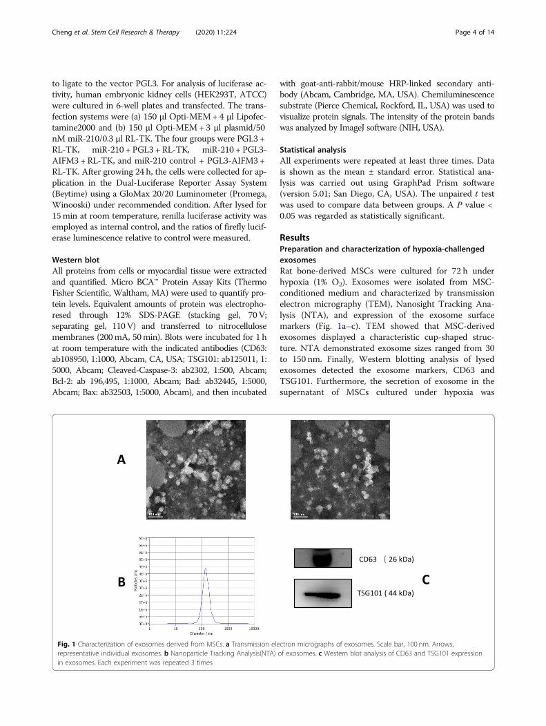
ResultsPreparation and characterization of hypoxia-challengedexosomesRat bone-derived MSCs were cultured for 72 h underhypoxia (1% O2). Exosomes were isolated from MSC-conditioned medium and characterized by transmissionelectron micrography (TEM), Nanosight Tracking Ana-lysis (NTA), and expression of the exosome surfacemarkers (Fig. 1a–c). TEM showed that MSC-derivedexosomes displayed a characteristic cup-shaped struc-ture. NTA demonstrated exosome sizes ranged from 30to 150 nm. Finally, Western blotting analysis of lysedexosomes detected the exosome markers, CD63 andTSG101. Furthermore, the secretion of exosome in thesupernatant of MSCs cultured under hypoxia was
Fig. 1 Characterization of exosomes derived from MSCs. a Transmission electron micrographs of exosomes. Scale bar, 100 nm. Arrows,representative individual exosomes. b Nanoparticle Tracking Analysis(NTA) of exosomes. c Western blot analysis of CD63 and TSG101 expressionin exosomes. Each experiment was repeated 3 times
Cheng et al. Stem Cell Research & Therapy (2020) 11:224 Page 4 of 14
significantly increased compared with that in normoxicculture (Supplemental Fig. 1A).
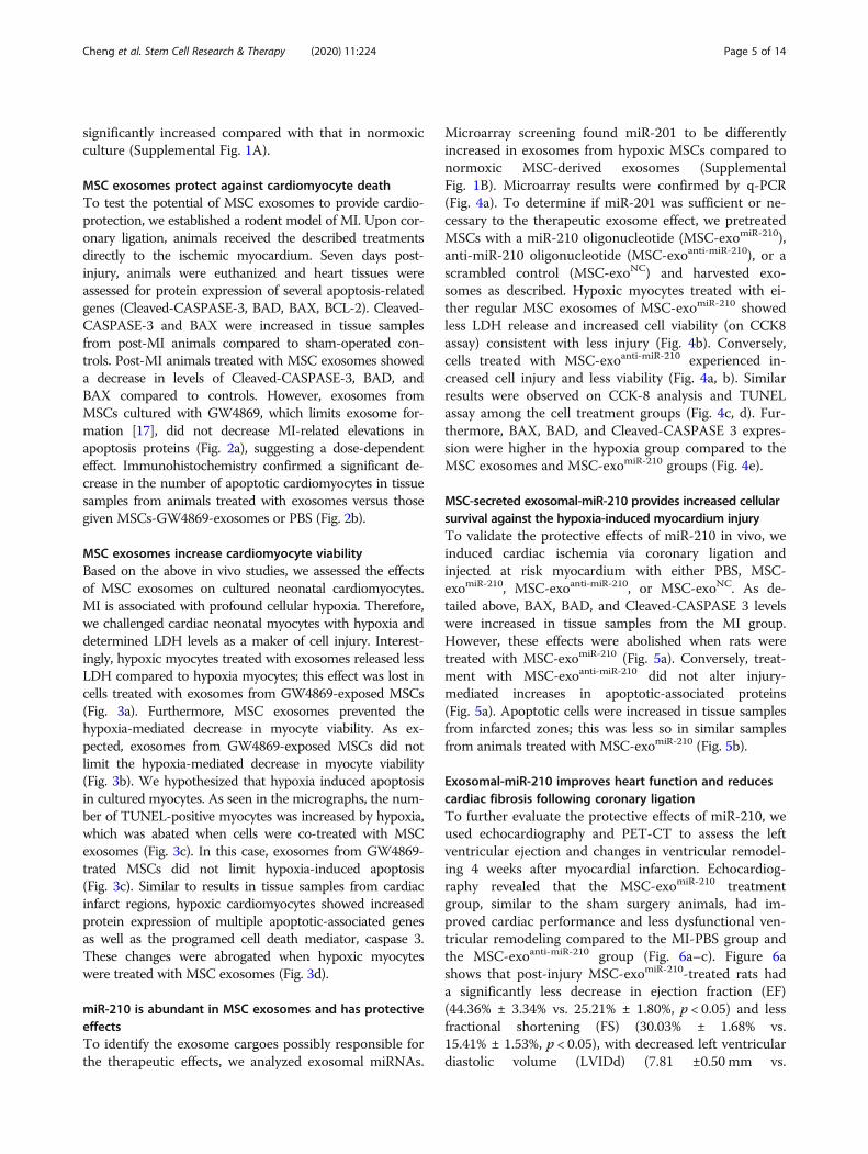
MSC exosomes protect against cardiomyocyte deathTo test the potential of MSC exosomes to provide cardio-protection, we established a rodent model of MI. Upon cor-onary ligation, animals received the described treatmentsdirectly to the ischemic myocardium. Seven days post-injury, animals were euthanized and heart tissues wereassessed for protein expression of several apoptosis-relatedgenes (Cleaved-CASPASE-3, BAD, BAX, BCL-2). Cleaved-CASPASE-3 and BAX were increased in tissue samplesfrom post-MI animals compared to sham-operated con-trols. Post-MI animals treated with MSC exosomes showeda decrease in levels of Cleaved-CASPASE-3, BAD, andBAX compared to controls. However, exosomes fromMSCs cultured with GW4869, which limits exosome for-mation [17], did not decrease MI-related elevations inapoptosis proteins (Fig. 2a), suggesting a dose-dependenteffect. Immunohistochemistry confirmed a significant de-crease in the number of apoptotic cardiomyocytes in tissuesamples from animals treated with exosomes versus thosegiven MSCs-GW4869-exosomes or PBS (Fig. 2b).
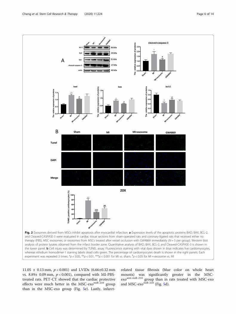
MSC exosomes increase cardiomyocyte viabilityBased on the above in vivo studies, we assessed the effectsof MSC exosomes on cultured neonatal cardiomyocytes.MI is associated with profound cellular hypoxia. Therefore,we challenged cardiac neonatal myocytes with hypoxia anddetermined LDH levels as a maker of cell injury. Interest-ingly, hypoxic myocytes treated with exosomes released lessLDH compared to hypoxia myocytes; this effect was lost incells treated with exosomes from GW4869-exposed MSCs(Fig. 3a). Furthermore, MSC exosomes prevented thehypoxia-mediated decrease in myocyte viability. As ex-pected, exosomes from GW4869-exposed MSCs did notlimit the hypoxia-mediated decrease in myocyte viability(Fig. 3b). We hypothesized that hypoxia induced apoptosisin cultured myocytes. As seen in the micrographs, the num-ber of TUNEL-positive myocytes was increased by hypoxia,which was abated when cells were co-treated with MSCexosomes (Fig. 3c). In this case, exosomes from GW4869-trated MSCs did not limit hypoxia-induced apoptosis(Fig. 3c). Similar to results in tissue samples from cardiacinfarct regions, hypoxic cardiomyocytes showed increasedprotein expression of multiple apoptotic-associated genesas well as the programed cell death mediator, caspase 3.These changes were abrogated when hypoxic myocyteswere treated with MSC exosomes (Fig. 3d).
miR-210 is abundant in MSC exosomes and has protectiveeffectsTo identify the exosome cargoes possibly responsible forthe therapeutic effects, we analyzed exosomal miRNAs.
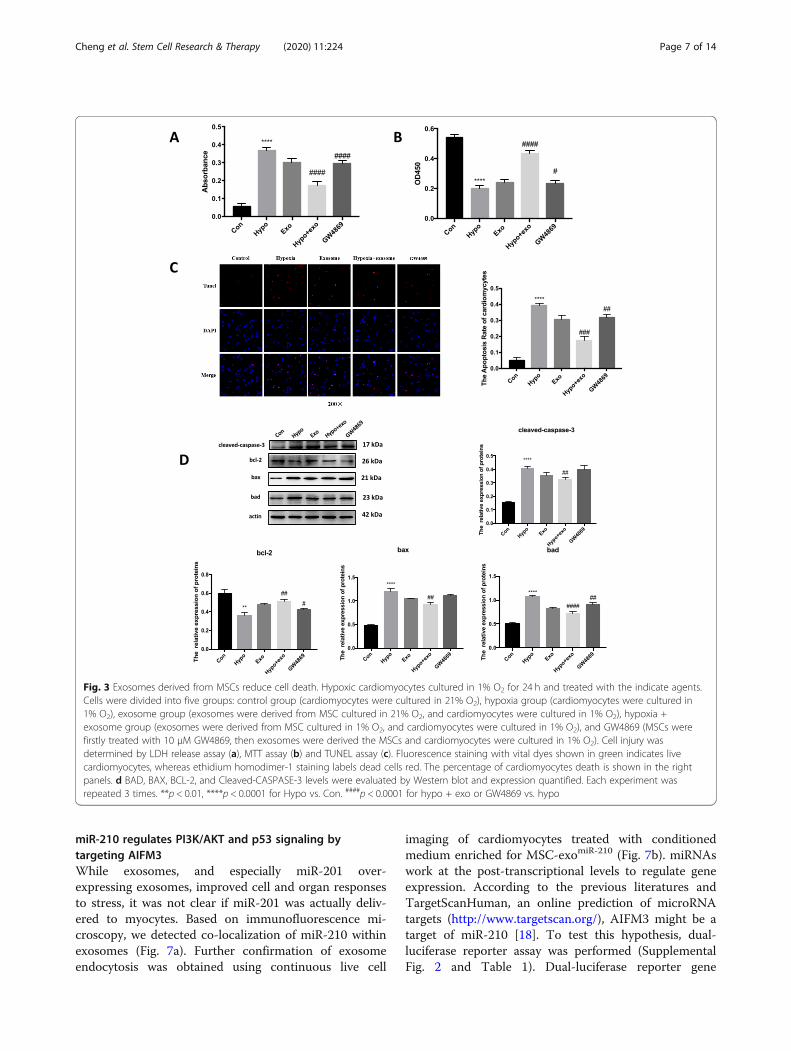
Microarray screening found miR-201 to be differentlyincreased in exosomes from hypoxic MSCs compared tonormoxic MSC-derived exosomes (SupplementalFig. 1B). Microarray results were confirmed by q-PCR(Fig. 4a). To determine if miR-201 was sufficient or ne-cessary to the therapeutic exosome effect, we pretreatedMSCs with a miR-210 oligonucleotide (MSC-exomiR-210),anti-miR-210 oligonucleotide (MSC-exoanti-miR-210), or ascrambled control (MSC-exoNC) and harvested exo-somes as described. Hypoxic myocytes treated with ei-ther regular MSC exosomes of MSC-exomiR-210 showedless LDH release and increased cell viability (on CCK8assay) consistent with less injury (Fig. 4b). Conversely,cells treated with MSC-exoanti-miR-210 experienced in-creased cell injury and less viability (Fig. 4a, b). Similarresults were observed on CCK-8 analysis and TUNELassay among the cell treatment groups (Fig. 4c, d). Fur-thermore, BAX, BAD, and Cleaved-CASPASE 3 expres-sion were higher in the hypoxia group compared to theMSC exosomes and MSC-exomiR-210 groups (Fig. 4e).
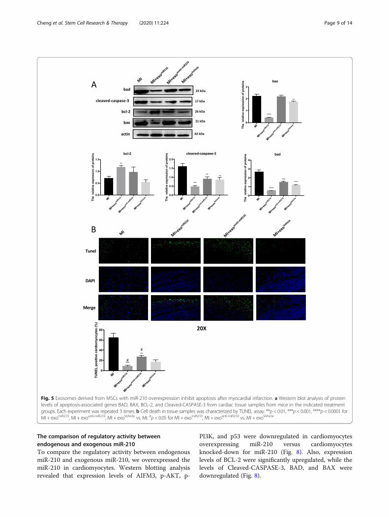
MSC-secreted exosomal-miR-210 provides increased cellularsurvival against the hypoxia-induced myocardium injuryTo validate the protective effects of miR-210 in vivo, weinduced cardiac ischemia via coronary ligation andinjected at risk myocardium with either PBS, MSC-exomiR-210, MSC-exoanti-miR-210, or MSC-exoNC. As de-tailed above, BAX, BAD, and Cleaved-CASPASE 3 levelswere increased in tissue samples from the MI group.However, these effects were abolished when rats weretreated with MSC-exomiR-210 (Fig. 5a). Conversely, treat-ment with MSC-exoanti-miR-210 did not alter injury-mediated increases in apoptotic-associated proteins(Fig. 5a). Apoptotic cells were increased in tissue samplesfrom infarcted zones; this was less so in similar samplesfrom animals treated with MSC-exomiR-210 (Fig. 5b).
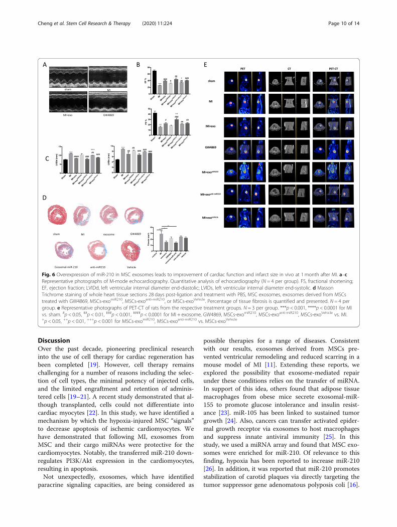
Exosomal-miR-210 improves heart function and reducescardiac fibrosis following coronary ligationTo further evaluate the protective effects of miR-210, weused echocardiography and PET-CT to assess the leftventricular ejection and changes in ventricular remodel-ing 4 weeks after myocardial infarction. Echocardiog-raphy revealed that the MSC-exomiR-210 treatmentgroup, similar to the sham surgery animals, had im-proved cardiac performance and less dysfunctional ven-tricular remodeling compared to the MI-PBS group andthe MSC-exoanti-miR-210 group (Fig. 6a–c). Figure 6ashows that post-injury MSC-exomiR-210-treated rats hada significantly less decrease in ejection fraction (EF)(44.36% ± 3.34% vs. 25.21% ± 1.80%, p < 0.05) and lessfractional shortening (FS) (30.03% ± 1.68% vs.15.41% ± 1.53%, p < 0.05), with decreased left ventriculardiastolic volume (LVIDd) (7.81 ±0.50 mm vs.
Cheng et al. Stem Cell Research & Therapy (2020) 11:224 Page 5 of 14
11.05 ± 0.13 mm, p < 0.001) and LVIDs (6.66±0.32 mmvs. 8.89± 0.09 mm, p < 0.001), compared with MI-PBS-treated rats. PET-CT showed that the cardiac protectiveeffects were much better in the MSC-exomiR-210 groupthan in the MSC-exo group (Fig. 5e). Lastly, infarct-
related tissue fibrosis (blue color on whole heartmounts) was significantly greater in the MSC-exoanti-miR-210 group than in rats treated with MSC-exoand MSC-exomiR-210 (Fig. 5d).
Fig. 2 Exosomes derived from MSCs inhibit apoptosis after myocardial infarction. a Expression levels of the apoptotic proteins BAD, BAX, BCL-2,and Cleaved-CASPASE-3 were evaluated in cardiac tissue sections from sham-operated rats and coronary-ligated rats that received either notherapy (PBS), MSC exosomes, or exosomes from MSCs treated after vessel occlusion with GW4869 immediately (N = 5 per group). Western blotanalysis of protein lysates obtained from the infarct border zone. Quantitative analysis of BAD, BAX, BCL-2, and Cleaved-CASPASE-3 is shown inthe lower panel. b Cell injury was determined by TUNEL assay. Fluorescence staining with vital dyes shown in blue indicates live cardiomyocytes,whereas ethidium homodimer-1 staining labels dead cells green. The percentage of cardiomyocytes death is shown in the right panels. Eachexperiment was repeated 3 times. *p < 0.05, **p < 0.01, ***p < 0.001 for MI vs. sham. #p < 0.05 for MI + exosome vs. MI
Cheng et al. Stem Cell Research & Therapy (2020) 11:224 Page 6 of 14
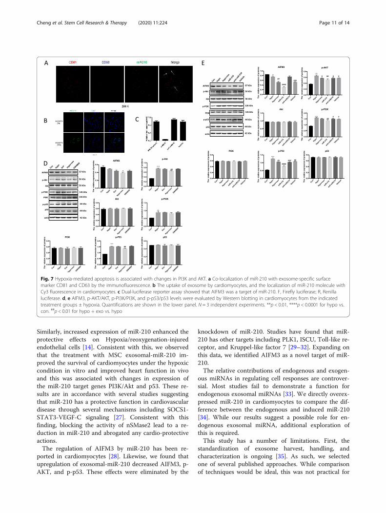
miR-210 regulates PI3K/AKT and p53 signaling bytargeting AIFM3While exosomes, and especially miR-201 over-expressing exosomes, improved cell and organ responsesto stress, it was not clear if miR-201 was actually deliv-ered to myocytes. Based on immunofluorescence mi-croscopy, we detected co-localization of miR-210 withinexosomes (Fig. 7a). Further confirmation of exosomeendocytosis was obtained using continuous live cell
imaging of cardiomyocytes treated with conditionedmedium enriched for MSC-exomiR-210 (Fig. 7b). miRNAswork at the post-transcriptional levels to regulate geneexpression. According to the previous literatures andTargetScanHuman, an online prediction of microRNAtargets (http://www.targetscan.org/), AIFM3 might be atarget of miR-210 [18]. To test this hypothesis, dual-luciferase reporter assay was performed (SupplementalFig. 2 and Table 1). Dual-luciferase reporter gene
Fig. 3 Exosomes derived from MSCs reduce cell death. Hypoxic cardiomyocytes cultured in 1% O2 for 24 h and treated with the indicate agents.Cells were divided into five groups: control group (cardiomyocytes were cultured in 21% O2), hypoxia group (cardiomyocytes were cultured in1% O2), exosome group (exosomes were derived from MSC cultured in 21% O2, and cardiomyocytes were cultured in 1% O2), hypoxia +exosome group (exosomes were derived from MSC cultured in 1% O2, and cardiomyocytes were cultured in 1% O2), and GW4869 (MSCs werefirstly treated with 10 μM GW4869, then exosomes were derived the MSCs and cardiomyocytes were cultured in 1% O2). Cell injury wasdetermined by LDH release assay (a), MTT assay (b) and TUNEL assay (c). Fluorescence staining with vital dyes shown in green indicates livecardiomyocytes, whereas ethidium homodimer-1 staining labels dead cells red. The percentage of cardiomyocytes death is shown in the rightpanels. d BAD, BAX, BCL-2, and Cleaved-CASPASE-3 levels were evaluated by Western blot and expression quantified. Each experiment wasrepeated 3 times. **p < 0.01, ****p < 0.0001 for Hypo vs. Con. ####p < 0.0001 for hypo + exo or GW4869 vs. hypo
Cheng et al. Stem Cell Research & Therapy (2020) 11:224 Page 7 of 14
analysis of myocytes treated with MSC-exomiR-210 re-vealed that firefly luciferase activity was significantlyinhibited when co-transfected with miR-210, indicatingthat AIFM3 as a down-stream target of miR-210 (Fig. 7c).We confirmed this result via Western blotting and q-PCR in miR-210 mimic-transfected cardiomyocytes.RNA levels of AIFM3 were significantly reduced in car-diomyocytes transfected with the miR-210 mimic
(Fig. 7d). Studies have linked AIFM3/p53 and PI3K/Aktsignaling pathways in the setting of MI [18]. Therefore,we investigated the crosstalk between these two signal-ing pathways. Western blotting analysis suggested thatlevels AIFM3 were upregulated and levels of p-AKT, p-PI3K, and p-p53 were downregulated (Fig. 7c). Aftertransfection with the miR-210 mimic, similar resultswere obtained (Fig. 7d).
Fig. 4 Overexpression of miR210 in MSC exosomes strengthens their cardioprotective function in vitro. a Polymerase chain reaction quantificationof miR210 in the exosomes obtained from MSCs. Hypoxic cardiomyocytes were treated with exosomes obtained from MSCs that were pre-treated with miR210 (exomiR210), anti-miR210 (exoanti-miR210), or scramble (exoVehicle) for the indicted time and cell injury characterized by LDHrelease (b), viability by MTT assay (c), and apoptotic programmed cell death by TUNEL assay (d). Red, TUNEL-positive nuclei; blue, DAPI-stainednuclei; green, troponin-positive cardiomyocytes. Scar bar × 200. The percentage of TUNEL-positive cells is shown in the right panels. e Westernblot identification of BAD, BAX, BCL-2, and Cleaved-CASPASE-3 in cardiomyocyte lysates after cell incubation with the indicated exosomes ±hypoxia. Quantitative analysis of BAD, BAX, BCL-2, and Cleaved-CASPASE-3 is shown in the lower panel. Each experiment was repeated 3 times.**p < 0.01, ***p < 0.001, ****p < 0.0001 for hypo vs. con. #p < 0.05, ##p < 0.01, ###p < 0.001, ####p < 0.0001 for hypo + exo, hypo + exomiR210, hypo +exoanti-miR210
, or hypo + exoVehicle vs. hypo. +p < 0.05, ++p < 0.01, +++p < 0.001, ++++p < 0.0001 for hypo + exomiR210 vs. hypo + exo
Cheng et al. Stem Cell Research & Therapy (2020) 11:224 Page 8 of 14
The comparison of regulatory activity betweenendogenous and exogenous miR-210To compare the regulatory activity between endogenousmiR-210 and exogenous miR-210, we overexpressed themiR-210 in cardiomyocytes. Western blotting analysisrevealed that expression levels of AIFM3, p-AKT, p-
PI3K, and p53 were downregulated in cardiomyocytesoverexpressing miR-210 versus cardiomyocytesknocked-down for miR-210 (Fig. 8). Also, expressionlevels of BCL-2 were significantly upregulated, while thelevels of Cleaved-CASPASE-3, BAD, and BAX weredownregulated (Fig. 8).
Fig. 5 Exosomes derived from MSCs with miR-210 overexpression inhibit apoptosis after myocardial infarction. a Western blot analysis of proteinlevels of apoptosis-associated genes BAD, BAX, BCL-2, and Cleaved-CASPASE-3 from cardiac tissue samples from mice in the indicated treatmentgroups. Each experiment was repeated 3 times. b Cell death in tissue samples was characterized by TUNEL assay. **p< 0.01, ***p< 0.001, ****p< 0.0001 forMI + exomiR210, MI + exoanti-miR210, MI + exoVehicle vs. MI. #p< 0.05 for MI + exomiR210, MI + exoanti-miR210 vs. MI + exoVehicle
Cheng et al. Stem Cell Research & Therapy (2020) 11:224 Page 9 of 14
DiscussionOver the past decade, pioneering preclinical researchinto the use of cell therapy for cardiac regeneration hasbeen completed [19]. However, cell therapy remainschallenging for a number of reasons including the selec-tion of cell types, the minimal potency of injected cells,and the limited engraftment and retention of adminis-tered cells [19–21]. A recent study demonstrated that al-though transplanted, cells could not differentiate intocardiac myocytes [22]. In this study, we have identified amechanism by which the hypoxia-injured MSC “signals”to decrease apoptosis of ischemic cardiomyocytes. Wehave demonstrated that following MI, exosomes fromMSC and their cargo miRNAs were protective for thecardiomyocytes. Notably, the transferred miR-210 down-regulates PI3K/Akt expression in the cardiomyocytes,resulting in apoptosis.Not unexpectedly, exosomes, which have identified
paracrine signaling capacities, are being considered as
possible therapies for a range of diseases. Consistentwith our results, exosomes derived from MSCs pre-vented ventricular remodeling and reduced scarring in amouse model of MI [11]. Extending these reports, weexplored the possibility that exosome-mediated repairunder these conditions relies on the transfer of miRNA.In support of this idea, others found that adipose tissuemacrophages from obese mice secrete exosomal-miR-155 to promote glucose intolerance and insulin resist-ance [23]. miR-105 has been linked to sustained tumorgrowth [24]. Also, cancers can transfer activated epider-mal growth receptor via exosomes to host macrophagesand suppress innate antiviral immunity [25]. In thisstudy, we used a miRNA array and found that MSC exo-somes were enriched for miR-210. Of relevance to thisfinding, hypoxia has been reported to increase miR-210[26]. In addition, it was reported that miR-210 promotesstabilization of carotid plaques via directly targeting thetumor suppressor gene adenomatous polyposis coli [16].
Fig. 6 Overexpression of miR-210 in MSC exosomes leads to improvement of cardiac function and infarct size in vivo at 1 month after MI. a–cRepresentative photographs of M-mode echocardiography. Quantitative analysis of echocardiography (N = 4 per group). FS, fractional shortening;EF, ejection fraction; LVIDd, left ventricular internal diameter end-diastolic; LVIDs, left ventricular internal diameter end-systolic. d MassonTrichrome staining of whole heart tissue sections 28 days post-ligation and treatment with PBS, MSC exosomes, exosomes derived from MSCstreated with GW4869, MSCs-exomiR210, MSCs-exoanti-miR210, or MSCs-exoVehicle. Percentage of tissue fibrosis is quantified and presented. N = 4 pergroup. e Representative photographs of PET-CT of rats from the respective treatment groups. N = 3 per group. ***p < 0.001, ****p < 0.0001 for MIvs. sham. #p < 0.05, ##p < 0.01, ###p < 0.001, ####p < 0.0001 for MI + exosome, GW4869, MSCs-exomiR210, MSCs-exoanti-miR210, MSCs-exoVehicle vs. MI.+p < 0.05, ++p < 0.01, +++p < 0.001 for MSCs-exomiR210, MSCs-exoanti-miR210 vs. MSCs-exoVehicle
Cheng et al. Stem Cell Research & Therapy (2020) 11:224 Page 10 of 14
Similarly, increased expression of miR-210 enhanced theprotective effects on Hypoxia/reoxygenation-injuredendothelial cells [14]. Consistent with this, we observedthat the treatment with MSC exosomal-miR-210 im-proved the survival of cardiomyocytes under the hypoxiccondition in vitro and improved heart function in vivoand this was associated with changes in expression ofthe miR-210 target genes PI3K/Akt and p53. These re-sults are in accordance with several studies suggestingthat miR-210 has a protective function in cardiovasculardisease through several mechanisms including SOCS1-STAT3-VEGF-C signaling [27]. Consistent with thisfinding, blocking the activity of nSMase2 lead to a re-duction in miR-210 and abrogated any cardio-protectiveactions.The regulation of AIFM3 by miR-210 has been re-
ported in cardiomyocytes [28]. Likewise, we found thatupregulation of exosomal-miR-210 decreased AIFM3, p-AKT, and p-p53. These effects were eliminated by the
knockdown of miR-210. Studies have found that miR-210 has other targets including PLK1, ISCU, Toll-like re-ceptor, and Kruppel-like factor 7 [29–32]. Expanding onthis data, we identified AIFM3 as a novel target of miR-210.The relative contributions of endogenous and exogen-
ous miRNAs in regulating cell responses are controver-sial. Most studies fail to demonstrate a function forendogenous exosomal miRNAs [33]. We directly overex-pressed miR-210 in cardiomyocytes to compare the dif-ference between the endogenous and induced miR-210[34]. While our results suggest a possible role for en-dogenous exosomal miRNA, additional exploration ofthis is required.This study has a number of limitations. First, the
standardization of exosome harvest, handling, andcharacterization is ongoing [35]. As such, we selectedone of several published approaches. While comparisonof techniques would be ideal, this was not practical for
Fig. 7 Hypoxia-mediated apoptosis is associated with changes in PI3K and AKT. a Co-localization of miR-210 with exosome-specific surfacemarker CD81 and CD63 by the immunofluorescence. b The uptake of exosome by cardiomyocytes, and the localization of miR-210 molecule withCy3 fluorescence in cardiomyocytes. c Dual-luciferase reporter assay showed that AIFM3 was a target of miR-210. F, Firefly luciferase; R, Renillaluciferase. d, e AIFM3, p-AKT/AKT, p-PI3K/PI3K, and p-p53/p53 levels were evaluated by Western blotting in cardiomyocytes from the indicatedtreatment groups ± hypoxia. Quantifications are shown in the lower panel. N = 3 independent experiments. **p < 0.01, ****p < 0.0001 for hypo vs.con. ##p < 0.01 for hypo + exo vs. hypo
Cheng et al. Stem Cell Research & Therapy (2020) 11:224 Page 11 of 14
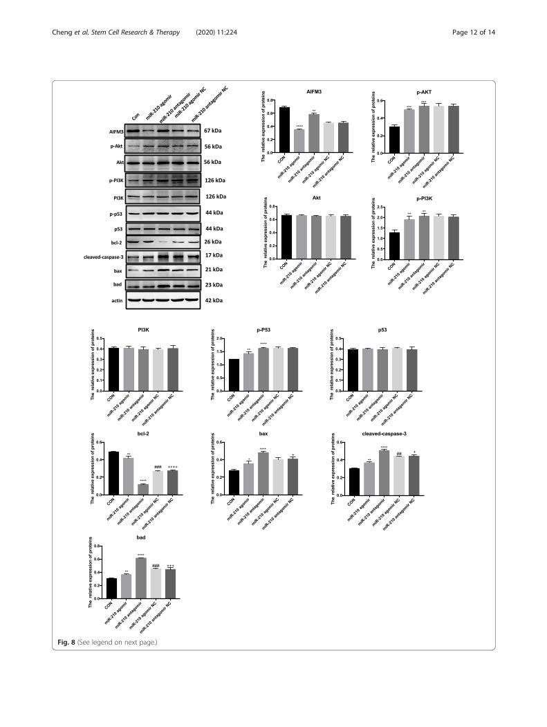
Fig. 8 (See legend on next page.)
Cheng et al. Stem Cell Research & Therapy (2020) 11:224 Page 12 of 14
this study. Second, confocal imaging is not able to dis-tinguish molecular proximity from interaction. Finally,exosomes express cell surface proteins, such as anti-angiogenic thrombospondin-1, that have profound sin-gling effects in the cardiovascular system. Thus, we can-not exclude the possible role of exosome membraneexpressed molecules in our assays.
ConclusionsIn conclusion, our study demonstrated the benefits asso-ciated with MSC exosomes after MI as well as a novelmechanism responsible for their anti-apoptotic effect viamiR-210. Collectively, these results provide new insightsinto the mechanism of cell therapy and might be lever-aged for the treatment of ischemic heart diseases.
Supplementary informationSupplementary information accompanies this paper at https://doi.org/10.1186/s13287-020-01737-0.
Additional file 1: Figure S1. Exosomes densities and miR-210 levels indifferent conditions.
Additional file 2: Figure S2. Successful construction of AIMF3promoter report vector.
Additional file 3: Table S1. Primer used for PCR.
AbbreviationsMI: Myocardial infarction; MSC: Mesenchymal stem cell; BMSC: Bonemesenchymal stem cell; TEM: Transmission electron micrography;NTA: Nanosight Tracking Analysis; FBS: Fetal bovine serum; PBS: Phosphate-buffered saline; RT-PCR: Reverse transcription-polymerase chain reaction;EF: L Ejection fraction; FS: Fractional shortening; LVIDd: Left ventricular end-diastolic internal diameters; LVIDs: Left ventricular end-systolic internaldiameters
AcknowledgementsThe authors acknowledge the Department of Cardiology, ZhongshanHospital, Fudan University and Shanghai Institute of Cardiovascular Diseases.The manuscript has not been submitted elsewhere and is not underconsideration for publication. All authors read and agreed with thesubmission of the manuscript. We give our sincere appreciation to thereviewers for their helpful comments on this article.
Authors’ contributionsHao Cheng, Shufu Chang, Rende Xu, and Lu Chen contributed equally tothis work. Hao Cheng and Shufu Chang performed the experiments,analyzed the data, and wrote and revised manuscript. Rende Xu, Lu Chen,and Xiaoyue Song performed the experiments and analyzed the data.JianWu did the animal models and echocardiography and PET-CT. YunzengZou and Juying Qian provided some suggestions. Jianying Ma designed theexperiments and wrote and revised manuscript. The authors read andapproved the final manuscript.
FundingThis work was supported by the grants from National Basic ResearchProgram of China (No. 81470467).
Availability of data and materialsAll data generated or analyzed in this study are included in this article (andits supplementary information files).
Ethics approval and consent to participateThis study was conducted under the approval of the Department ofCardiology, Zhongshan Hospital, Fudan University and Shanghai Institute ofCardiovascular Diseases. Animal experiments were conducted under theapproval of the Animal Ethics Committee of Zhongshan Hospital, FudanUniversity. Treatment of experimental animals followed the internationallyrecognized 3R principle, which is the replacement, reduction, and refinementof experimental animals. Moreover, the protocols involved experimentalanimals followed the welfare ethics of the experimental animals accordingto the requirements of the guidelines for ethical review of experimentalanimal welfare (GB/T 35892-2018) of China.
Consent for publicationNot applicable.
Competing interestsThe authors declare that they have no completing interests.
Author details1Department of Cardiology, Zhongshan Hospital, Fudan University, 1609Xietu Road, Shanghai 200032, China. 2Shanghai Institute of CardiovascularDiseases, Zhongshan Hospital and Institute of Biomedical Sciences, FudanUniversity, 180 Feng Lin Road, Shanghai 20032, China.
Received: 18 March 2020 Revised: 14 April 2020Accepted: 19 May 2020
References1. Benjamin EJ, Muntner P, Alonso A, Bittencourt MS, Callaway CW, Carson AP,
Chamberlain AM, Chang AR, Cheng S, Das SR, Delling FN, Djousse L, ElkindMSV, Ferguson JF, Fornage M, Jordan LC, Khan SS, Kissela BM, Knutson KL,Kwan TW, Lackland DT, Lewis TT, Lichtman JH, Longenecker CT, Loop MS,Lutsey PL, Martin SS, Matsushita K, Moran AE, Mussolino ME, O'Flaherty M,Pandey A, Perak AM, Rosamond WD, Roth GA, Sampson UKA, Satou GM,Schroeder EB, Shah SH, Spartano NL, Stokes A, Tirschwell DL, Tsao CW,Turakhia MP, VanWagner LB, Wilkins JT, Wong SS, Virani SS, Epid AHAC,Comm PS, Subcomm SS. Heart Disease and Stroke Statistics-2019 Update AReport From the American Heart Association. Circulation. 2019;139:E56–E528.
2. Cohen M, Boiangiu C, Abidi M. Therapy for ST-segment elevationmyocardial infarction patients who present late or are ineligible forreperfusion therapy. J Am Coll Cardiol. 2010;55:1895–906.
3. Dias LD, Casali KR, Ghem C, da Silva MK, Sausen G, Palma PB, Covas DT, KalilRAK, Schaan BD, Nardi NB, Markoski MM. Mesenchymal stem cells fromsternum: the type of heart disease, ischemic or valvular, does not influencethe cell culture establishment and growth kinetics. J Transl Med. 2017;15:161.
4. Vrijsen KR, Maring JA, Chamuleau SA, Verhage V, Mol EA, Deddens JC, MetzCH, Lodder K, van Eeuwijk EC, van Dommelen SM, Doevendans PA, SmitsAM, Goumans MJ, Sluijter JP. Exosomes from cardiomyocyte progenitor cellsand mesenchymal stem cells stimulate angiogenesis via EMMPRIN. AdvHealthc Mater. 2016;5:2555–65.
5. Williams AR, Hare JM. Mesenchymal stem cells biology, pathophysiology,translational findings, and therapeutic implications for cardiac disease. CircRes. 2011;109:923–40.
(See figure on previous page.)Fig. 8 Comparison of the uptake of exosomal miR-210 with the regulatory activity of endogenous miR-210. The protein levels of AIFM3, p-AKT/AKT, p-PI3K/PI3K, p-p53/p53, BAD, BAX, BCL-2, and Cleaved-CASPASE-3 were evaluated by Western blotting in cardiomyocytes treated withoverexpressed miR-210. Quantifications are shown in the lower panel. N = 3 independent experiments. **p < 0.01, ***p < 0.001, ****p < 0.0001 formiR-210 agomir and miR-210 antagomir vs. con. ##p < 0.01, ###p < 0.001 for miR-210 agomir NC vs. miR-210 agomir. +p < 0.05, +++p < 0.001,++++p < 0.0001 for miR-210 antagomir NC vs. miR-210 antagomir
Cheng et al. Stem Cell Research & Therapy (2020) 11:224 Page 13 of 14
6. Shafei AE, Ali MA, Ghanem HG, Shehata AI, Abdelgawad AA, Handal HR,Talaat KA, Ashaal AE, El-Shal AS. Mesenchymal stem cell therapy: apromising cell-based therapy for treatment of myocardial infarction. J GeneMed. 2017;19:e2995.
7. Zhang ZW, Yang JJ, Yan WY, Li YX, Shen ZY, Asahara T. Pretreatment ofcardiac stem cells with exosomes derived from mesenchymal stem cellsenhances myocardial repair. J Am Heart Assoc. 2016;5:e002856.
8. Suzuki E, Fujita D, Takahashi M, Oba S, Nishimatsu H. Stem cell-derivedexosomes as a therapeutic tool for cardiovascular disease. World J StemCells. 2016;8:297–305.
9. Khan M, Nickoloff E, Abramova T, Johnson J, Verma SK, Krishnamurthy P,Mackie AR, Vaughan E, Garikipati VN, Benedict C, Ramirez V, Lambers E, ItoA, Gao E, Misener S, Luongo T, Elrod J, Qin G, Houser SR, Koch WJ, KishoreR. Embryonic stem cell-derived exosomes promote endogenous repairmechanisms and enhance cardiac function following myocardial infarction.Circ Res. 2015;117:52–64.
10. Pan T, Jia P, Chen N, Fang Y, Liang Y, Guo M, Ding X. Delayed remoteischemic preconditioning ConfersRenoprotection against septic acutekidney injury via exosomal miR-21. Theranostics. 2019;9:405–23.
11. Arslan F, Lai RC, Smeets MB, Akeroyd L, Choo A, Aguor ENE, Timmers L, vanRijen HV, Doevendans PA, Pasterkamp G, Lim SK, de Kleijn DP. Mesenchymalstem cell-derived exosomes increase ATP levels, decrease oxidative stressand activate PI3K/Akt pathway to enhance myocardial viability and preventadverse remodeling after myocardial ischemia/reperfusion injury. Stem CellRes. 2013;10:301–12.
12. Ong SG, Lee WH, Huang M, Dey D, Kodo K, Sanchez-Freire V, Gold JD, WuJC. Cross talk of combined gene and cell therapy in ischemic heart diseaserole of exosomal microrna transfer. Circulation. 2014;130:S60.
13. Kuwabara Y, Ono K, Horie T, Nishi H, Nagao K, Kinoshita M, Watanabe S,Baba O, Kojima Y, Shizuta S, Imai M, Tamura T, Kita T, Kimura T. IncreasedmicroRNA-1 and microRNA-133a levels in serum of patients withcardiovascular disease indicate myocardial damage. Circ-Cardiovasc Gene.2011;4:446–U287.
14. Ma XT, Wang JJ, Li J, Ma CL, Chen SZ, Lei W, Yang Y, Liu SM, Bihl J, Chen C.Loading MiR-210 in endothelial progenitor cells derived exosomes booststheir beneficial effects on hypoxia/eeoxygeneation-injured humanendothelial cells via protecting mitochondrial function. Cell PhysiolBiochem. 2018;46:664–75.
15. Zaccagnini G, Maimone B, Fuschi P, Maselli D, Spinetti G, Gaetano C, MartelliF. Overexpression of miR-210 and its significance in ischemic tissue damage.Sci Rep-Uk. 2017;7:9563.
16. Eken SM, Jin H, Chernogubova E, Li YH, Simon N, Sun CY, Korzunowicz G,Busch A, Backlund A, Osterholm C, Razuvaev A, Renne T, Eckstein HH,Pelisek J, Eriksson P, Diez MG, Matic LP, Schellinger IN, Raaz U, Leeper NJ,Hansson GK, Paulsson-Berne G, Hedin U, Maegdefessel L. MicroRNA-210enhances fibrous cap stability in advanced atherosclerotic lesions. Circ Res.2017;120:633.
17. Essandoh K, Yang L, Wang X, Huang W, Qin D, Hao J, Wang Y, Zingarelli B,Peng T, Fan GC. Blockade of exosome generation with GW4869 dampensthe sepsis-induced inflammation and cardiac dysfunction. Biochim BiophysActa. 2015;1852:2362–71.
18. Mutharasan RK, Nagpal V, Ichikawa Y, Ardehali H. microRNA-210 isupregulated in hypoxic cardiomyocytes through Akt- and p53-dependentpathways and exerts cytoprotective effects. Am J Physiol Heart Circ Physiol.2011;301:H1519–30.
19. Braunwald E. Cell-based therapy in cardiac regeneration: an overview. CircRes. 2018;123:132–7.
20. Broughton KM, Sussman MA. Enhancement strategies for cardiacregenerative cell therapy: focus on adult stem cells. Circ Res. 2018;123:177–87.
21. Banerjee MN, Bolli R, Hare JM. Clinical studies of cell therapy incardiovascular medicine: recent developments and future directions. CircRes. 2018;123:266–87.
22. Tang XL, Li QH, Rokosh G, Sanganalmath SK, Chen N, Ou QH, Stowers H,Hunt G, Bolli R. Long-term outcome of administration of c-kit(POS) cardiacprogenitor cells after acute myocardial infarction transplanted cells do notbecome cardiomyocytes, but structural and functional improvement andproliferation of endogenous cells persist for at least one year. Circ Res. 2016;118:1091–105.
23. Ying W, Riopel M, Bandyopadhyay G, Dong Y, Birmingham A, Seo JB,Ofrecio JM, Wollam J, Hernandez-Carretero A, Fu WX, Li PP, Olefsky JM.
Adipose tissue macrophage-derived exosomal miRNAs can modulatein vivo and in vitro insulin sensitivity. Cell. 2017;171:372.
24. Yan W, Wu X, Zhou W, Fong MY, Cao M, Liu J, Liu X, Chen CH, Fadare O,Pizzo DP, Wu J, Liu L, Liu X, Chin AR, Ren X, Chen Y, Locasale JW, Wang SE.Cancer-cell-secreted exosomal miR-105 promotes tumour growth throughthe MYC-dependent metabolic reprogramming of stromal cells. Nat CellBiol. 2018;20:597–609.
25. Gao L, Wang L, Dai T, Jin K, Zhang Z, Wang S, Xie F, Fang P, Yang B, HuangH, van Dam H, Zhou F, Zhang L. Tumor-derived exosomes antagonizeinnate antiviral immunity. Nat Immunol. 2018;19:233–45.
26. Wang H, Flach H, Onizawa M, Wei L, McManus MT, Weiss A. Negativeregulation of Hif1a expression and TH17 differentiation by the hypoxia-regulated microRNA miR-210. Nat Immunol. 2014;15:393–401.
27. Meng ZY, Kang HL, Duan W, Zheng J, Li QN, Zhou ZJ. MicroRNA-210promotes accumulation of neural precursor cells around ischemic foci aftercerebral ischemia by regulating the SOCS1-STAT3-VEGF-C pathway. J AmHeart Assoc. 2018;7:e005052.
28. Ke X, Yan R, Sun Z, Cheng Y, Meltzer A, Lu N, Shu X, Wang Z, Huang B, LiuX, Wang Z, Song JH, Ng CK, Ibrahim S, Abraham JM, Shin EJ, He S, MeltzerSJ. Esophageal adenocarcinoma-derived extracellular vesicle microRNAsinduce a neoplastic phenotype in gastric organoids. Neoplasia. 2017;19:941–9.
29. Li C, Zhou X, Wang Y, Jing S, Yang C, Sun G, Liu Q, Cheng Y, Wang L.miR210 regulates esophageal cancer cell proliferation by inducing G2/Mphase cell cycle arrest through targeting PLK1. Mol Med Rep. 2014;10:2099–104.
30. Lu Y, Huang J, Geng S, Chen H, Song C, Zhu S, Zhao S, Yuan M, Li X, Hu H.MitoKATP regulating HIF/miR210/ISCU signaling axis and formation of apositive feedback loop in chronic hypoxia-induced PAH rat model. Exp TherMed. 2017;13:1697–701.
31. Li C, Zhao M, Zhang C, Zhang W, Zhao X, Duan X, Xu W. miR210 modulatesrespiratory burst in Apostichopus japonicus coelomocytes via targeting toll-like receptor. Dev Comp Immunol. 2016;65:377–81.
32. Lv JX, Zhou J, Tong RQ, Wang B, Chen XL, Zhuang YY, Xia F, Wei XD.Hypoxiainduced miR210 contributes to apoptosis of mouse spermatocyteGC2 cells by targeting Kruppellike factor 7. Mol Med Rep. 2019;19:271–9.
33. Xiao C, Wang K, Xu Y, Hu H, Zhang N, Wang Y, Zhong Z, Zhao J, Li Q, ZhuD, Ke C, Zhong S, Wu X, Yu H, Zhu W, Chen J, Zhang J, Wang J, Hu X.Transplanted mesenchymal stem cells reduce autophagic flux in infarctedhearts via the exosomal transfer of miR-125b. Circ Res. 2018;123:564–78.
34. Paul P, Chakraborty A, Sarkar D, Langthasa M, Rahman M, Singha RKS,Malakar AK, Chakraborty S. Interplay between miRNAs and human diseases.J Cell Physiol. 2018;233:2007–18.
35. Witwer KW, Van Balkom BWM, Bruno S, Choo A, Dominici M, Gimona M, HillAF, De Kleijn D, Koh M, Lai RC, Mitsialis SA, Ortiz LA, Rohde E, Asada T, TohWS, Weiss DJ, Zheng L, Giebel B, Lim SK. Defining mesenchymal stromal cell(MSC)-derived small extracellular vesicles for therapeutic applications. JExtracell Vesicles. 2019;8:1609206.
Publisher’s NoteSpringer Nature remains neutral with regard to jurisdictional claims inpublished maps and institutional affiliations.
Cheng et al. Stem Cell Research & Therapy (2020) 11:224 Page 14 of 14