Embed Size (px)
Citation preview
06 I
I special _ orthodontics
_Introduction
_Orthodontic treat-ment generally follows
aesthetic, functional, andprophylactic objectives,where individual aspectsof isolated cases are ac-
corded varying importanceas they arise. Increasing aesthetic
expectations and awareness of modern dental treat-ment options disseminated by the media have resultedin increased interest and greater willingness of adults toconsider orthodontic treatment. Aesthetic orthodon-tics is thus primarily adult orthodontics.
A peculiarity of orthodontic treatment in adultscompared with paediatric or adolescent orthodontics isthe age-associated involution of the connective tissues
that leads to a reduction in cell density, thickening of thefibre bundles, delayed fibroblast proliferation, and re-duced vascularisation. These are the causes of slowerdental movement and delayed tissue and bone reac-tions. Absent sutural growth, the age of the periodon-tium, specific periodontal diagnoses, and tissue atrophyalso make treatment in adults particularly challenging.As a rule, aesthetically oriented adult orthodonticstherefore has an interdisciplinary inclination.
Occlusion, function and aesthetics are considered tobe equivalent parameters in modern orthodontics andparticularly here in combined orthodontic-maxillofa-cial surgical treatment.32,33 This was achieved throughoptimisation of diagnostic tools and further develop-ment and increasing experience in orthopaedic sur-gery.4
Nowadays, treatment of adult patients with dentalmalposition and mastication impairment is one of thestandard tasks of the orthodontist. If the discrepanciesin spatial allocations of the upper and lower dentitionare particularly pronounced and where the cause is pri-marily skeletal and not only dentoalveolar, conventionalorthodontic therapy is limited and combined orthodon-tic-surgical therapy is indicated for remodelling of thejaw bases.
Treatment for a skeletal dysgnathia (Class III) usingcombined orthodontic-maxillofacial surgical correc-tion is discussed in this article.
_Chronological development of maxillofacial surgery of the mandible
The first orthodontic-maxillofacial surgical proce-dure on the mandible described in the literature was that of the American surgeon Hullihen in 1848.13 This procedure was a segmental osteotomy of the anterior
cosmeticdentistry 2_2009
Orthodontic surgeryand aestheticsAuthor_ Prof Nezar Watted, Prof Josip Bill, Prof Ori Blanc & Prof B. Schlomi, Germany & Israel
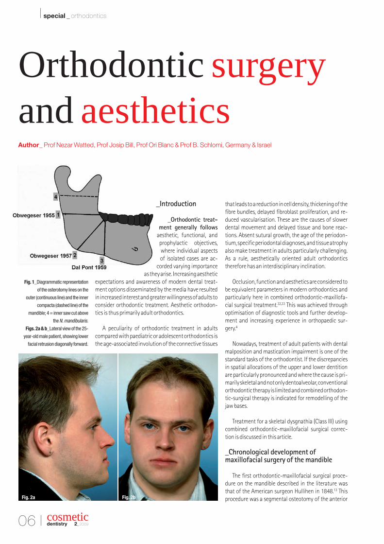
Fig. 1_Diagrammatic representation
of the osterotomy lines on the
outer (continuous line) and the inner
compacta (dashed line) of the
mandible; 4 = inner saw cut above
the N. mandibularis.
Figs. 2a & b_Lateral view of the 25-
year-old male patient, showing lower
facial retrusion diagonally forward.
Fig. 2a Fig. 2b
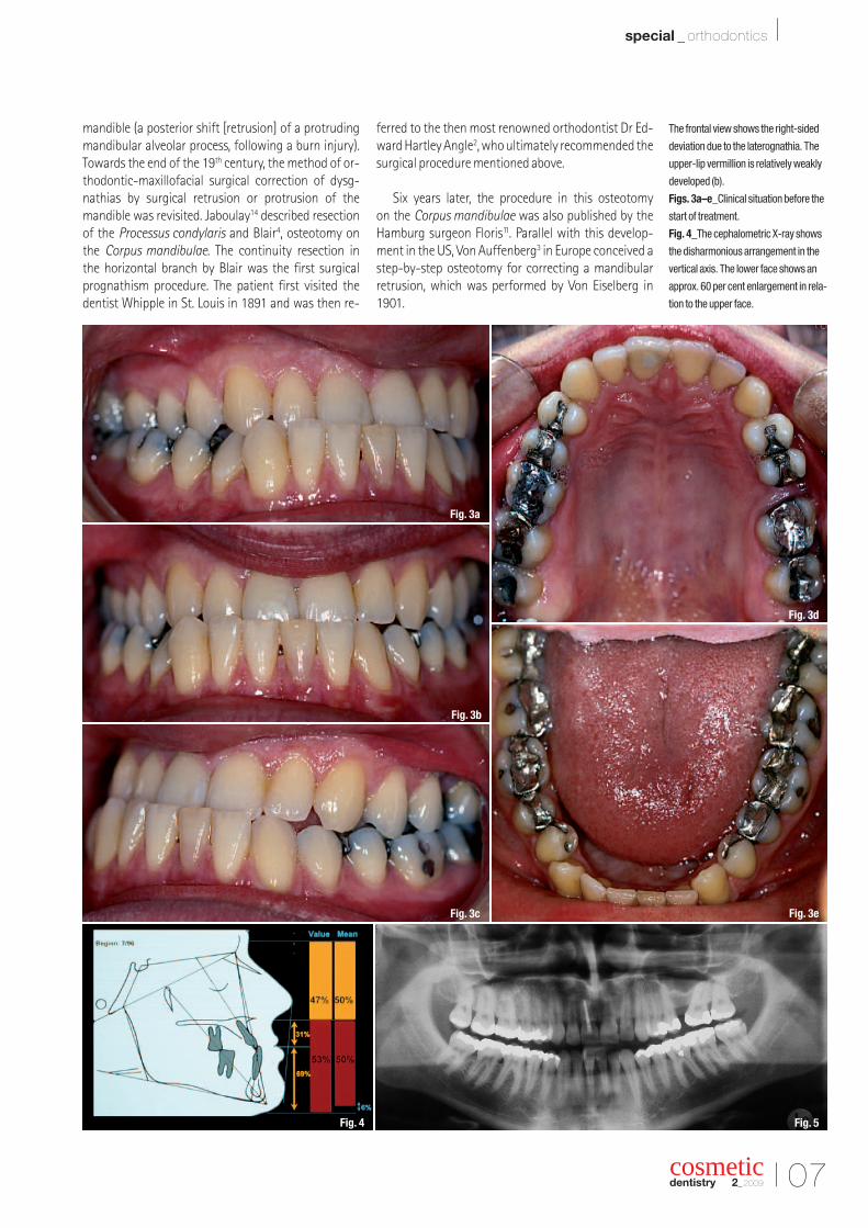
The frontal view shows the right-sided
deviation due to the laterognathia. The
upper-lip vermillion is relatively weakly
developed (b).
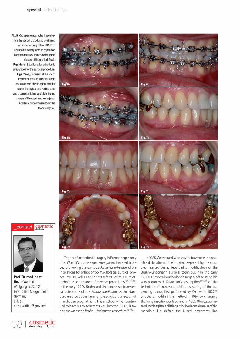
Figs. 3a–e_Clinical situation before the
start of treatment.
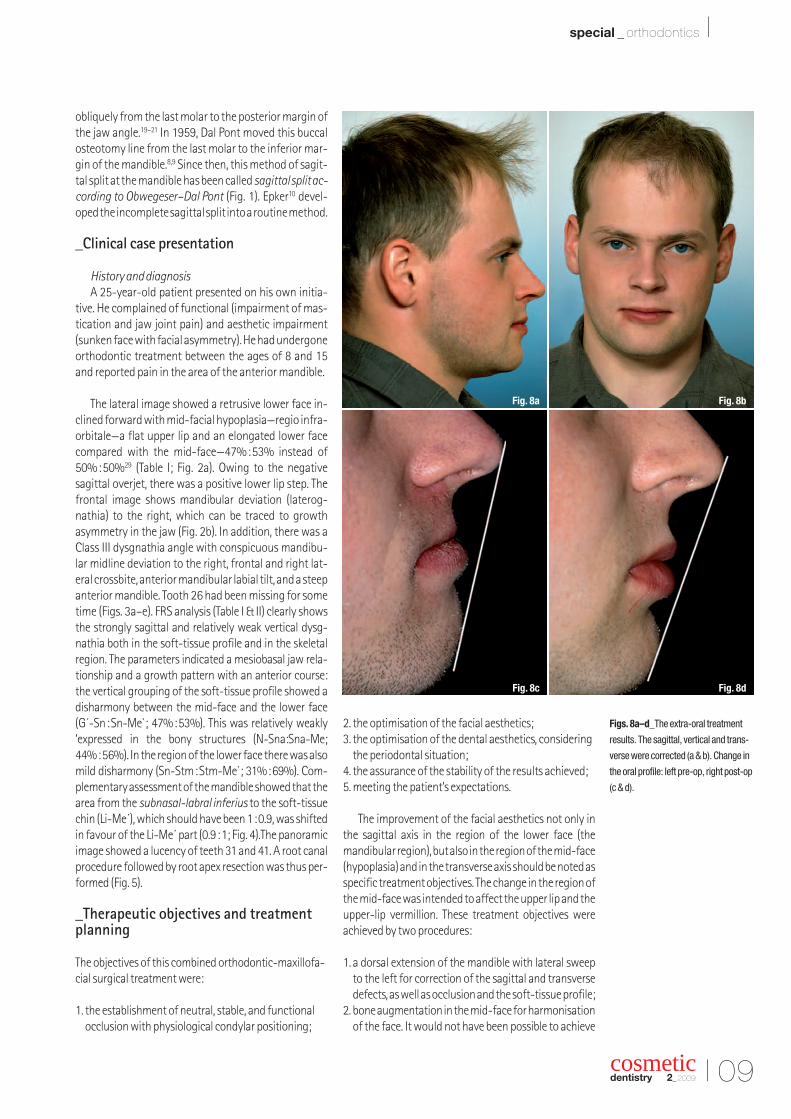
Fig. 4_The cephalometric X-ray shows
the disharmonious arrangement in the
vertical axis. The lower face shows an
approx. 60 per cent enlargement in rela-
tion to the upper face.
I 07
special _ orthodontics I
cosmeticdentistry 2_2009
mandible (a posterior shift [retrusion] of a protrudingmandibular alveolar process, following a burn injury).Towards the end of the 19th century, the method of or-thodontic-maxillofacial surgical correction of dysg-nathias by surgical retrusion or protrusion of themandible was revisited. Jaboulay14 described resectionof the Processus condylaris and Blair4, osteotomy on the Corpus mandibulae. The continuity resection in the horizontal branch by Blair was the first surgicalprognathism procedure. The patient first visited thedentist Whipple in St. Louis in 1891 and was then re-
ferred to the then most renowned orthodontist Dr Ed-ward Hartley Angle2, who ultimately recommended thesurgical procedure mentioned above.
Six years later, the procedure in this osteotomy on the Corpus mandibulae was also published by theHamburg surgeon Floris11. Parallel with this develop-ment in the US, Von Auffenberg3 in Europe conceived astep-by-step osteotomy for correcting a mandibularretrusion, which was performed by Von Eiselberg in1901.
Fig. 3a
Fig. 3b
Fig. 3c
Fig. 3d
Fig. 3e
Fig. 4 Fig. 5
08 I
I special _ orthodontics
The era of orthodontic surgery in Europe began onlyafter World War I. The experience gained there led in theyears following the war to a substantial extension of theindications for orthodontic-maxillofacial surgical pro-cedures, as well as to the transferral of this surgical technique to the area of elective procedures.5,6,16–18,24
In the early 1920s, Bruhn and Lindemann set transver-sal osteotomy of the Ramus madibulae as the stan-dard method at the time for the surgical correction ofmandibular prognathism. This method, which contin-ued to have many adherents well into the 1960s, is to-day known as the Bruhn–Lindemann procedure.1,6,25,45
In 1935, Wassmund, who saw its drawbacks in a pos-sible dislocation of the proximal segment by the mus-cles inserted there, described a modification of theBruhn–Lindemann surgical technique.26 In the early1950s, a new era in orthodontic surgery of the mandiblewas begun with Kazanjian’s resumption12,15,23 of thetechnique of transverse, oblique severing of the as-cending ramus, first performed by Perthes in 192222.Shuchard modified this method in 1954 by enlargingthe bony insertion surface, and in 1955 Obwegeser in-troduced sagittal splitting at the horizontal ramus of themandible. He shifted the buccal osteotomy line
Fig. 5_Orthopantomographic image be-
fore the start of orthodontic treatment.
An apical lucency at tooth 31. Pro-
nounced maxillary-antrum expansion
between teeth 25 and 27. Orthodontic
closure of the gap is difficult.
Figs. 6a–c_Situation after orthodontic
preparation for the surgical procedure.
Figs. 7a–e_Occlusion at the end of
treatment; there is a neutral stable
occlusion with physiological anterior
bite in the sagittal and vertical axes
and a correct midline (a–c). Monitoring
images of the upper and lower jaws.
A ceramic bridge was made in the
lower jaw (d, e).
cosmeticdentistry 2_2009
Prof. Dr. med. dent.Nezar WattedWolfgangstraße 1297980 Bad MergentheimGermanyE-Mail:[email protected]
cosmeticdentistry_contact
Fig. 6a Fig. 6b
Fig. 6c Fig. 7a
Fig. 7b Fig. 7c
Fig. 7d Fig. 7e
I 09
special _ orthodontics I
cosmeticdentistry 2_2009
obliquely from the last molar to the posterior margin ofthe jaw angle.19–21 In 1959, Dal Pont moved this buccalosteotomy line from the last molar to the inferior mar-gin of the mandible.8,9 Since then, this method of sagit-tal split at the mandible has been called sagittal split ac-cording to Obwegeser–Dal Pont (Fig. 1). Epker10 devel-oped the incomplete sagittal split into a routine method.
_Clinical case presentation
History and diagnosisA 25-year-old patient presented on his own initia-
tive. He complained of functional (impairment of mas-tication and jaw joint pain) and aesthetic impairment(sunken face with facial asymmetry). He had undergoneorthodontic treatment between the ages of 8 and 15and reported pain in the area of the anterior mandible.
The lateral image showed a retrusive lower face in-clined forward with mid-facial hypoplasia—regio infra-orbitale—a flat upper lip and an elongated lower facecompared with the mid-face—47%:53% instead of50%:50%29 (Table I; Fig. 2a). Owing to the negativesagittal overjet, there was a positive lower lip step. Thefrontal image shows mandibular deviation (laterog-nathia) to the right, which can be traced to growthasymmetry in the jaw (Fig. 2b). In addition, there was aClass III dysgnathia angle with conspicuous mandibu-lar midline deviation to the right, frontal and right lat-eral crossbite, anterior mandibular labial tilt, and a steepanterior mandible. Tooth 26 had been missing for sometime (Figs. 3a–e). FRS analysis (Table I & II) clearly showsthe strongly sagittal and relatively weak vertical dysg-nathia both in the soft-tissue profile and in the skeletalregion. The parameters indicated a mesiobasal jaw rela-tionship and a growth pattern with an anterior course:the vertical grouping of the soft-tissue profile showed adisharmony between the mid-face and the lower face (G´-Sn:Sn-Me`; 47%:53%). This was relatively weakly‘expressed in the bony structures (N-Sna:Sna-Me;44%:56%). In the region of the lower face there was alsomild disharmony (Sn-Stm:Stm-Me`; 31%:69%). Com-plementary assessment of the mandible showed that thearea from the subnasal-labral inferius to the soft-tissuechin (Li-Me´), which should have been 1:0.9, was shiftedin favour of the Li-Me´ part (0.9:1; Fig. 4).The panoramicimage showed a lucency of teeth 31 and 41. A root canalprocedure followed by root apex resection was thus per-formed (Fig. 5).
_Therapeutic objectives and treatmentplanning
The objectives of this combined orthodontic-maxillofa-cial surgical treatment were:
1. the establishment of neutral, stable, and functionalocclusion with physiological condylar positioning;
2. the optimisation of the facial aesthetics;3. the optimisation of the dental aesthetics, considering
the periodontal situation;4. the assurance of the stability of the results achieved; 5. meeting the patient’s expectations.
The improvement of the facial aesthetics not only inthe sagittal axis in the region of the lower face (themandibular region), but also in the region of the mid-face(hypoplasia) and in the transverse axis should be noted asspecific treatment objectives. The change in the region ofthe mid-face was intended to affect the upper lip and theupper-lip vermillion. These treatment objectives wereachieved by two procedures:
1. a dorsal extension of the mandible with lateral sweepto the left for correction of the sagittal and transversedefects, as well as occlusion and the soft-tissue profile;
2. bone augmentation in the mid-face for harmonisationof the face. It would not have been possible to achieve
Figs. 8a–d_The extra-oral treatment
results. The sagittal, vertical and trans-
verse were corrected (a & b). Change in
the oral profile: left pre-op, right post-op
(c & d).
Fig. 8a Fig. 8b
Fig. 8c Fig. 8d
10 I
I special _ orthodontics
the desired treatment objectives with respect to func-tion and aesthetics using orthodontic proceduresalone.27
Therapeutic procedureCorrection of the pronounced dysgnathia was done
in six phases:28,30–33
1. Splint therapy: a flat bite guard splint was installed forsix weeks in order to determine the physiologicalcondylar position or centrics before the final treat-ment planning. By doing this, the forced bite could bedemonstrated to its full extent.
2. Orthodontics for forming and adjusting the dentalarches relative to each other and decompensation ofthe skeletal dysgnathia (Figs. 6a–c).
3. Splint therapy for determining the condylar position.This was performed in the 4 to 6 weeks prior to the surgical procedure. The objective was registration ofthe jaw joint in a physiological position (centrics).
4. Oral surgery for correction of the skeletal dysgnathia:after model operation, determination of the transpo-sition path and production of the splint in the targetocclusion, the surgical mandibular translocation using sagittal split according to Obwegeser–Dal Pontwas done. Augmentation in the mid-facial region wasdone using autologous bone.
5. Orthodontics for fine adjustment of occlusion.6. Retention: 3-3 retainers were cemented in the
mandible.
Mandibular and maxillary plates were used as the retention appliance. Prosthetic care was provided aftersix months.
_Results
Figures 7a–e show the situation after the conclusionof treatment and after extraction of tooth 31 and subse-quent prosthetic treatment, neutral occlusion, and correct midline with physiological sagittal and verticalbite. The extra-oral images show a harmonious profile inthe vertical as well as in the sagittal axis (Figs. 8a & b). Theoral profile is harmonious. The upper-lip vermillion isdistinctly visible in comparison to the original situation(Fig. 8c & d).
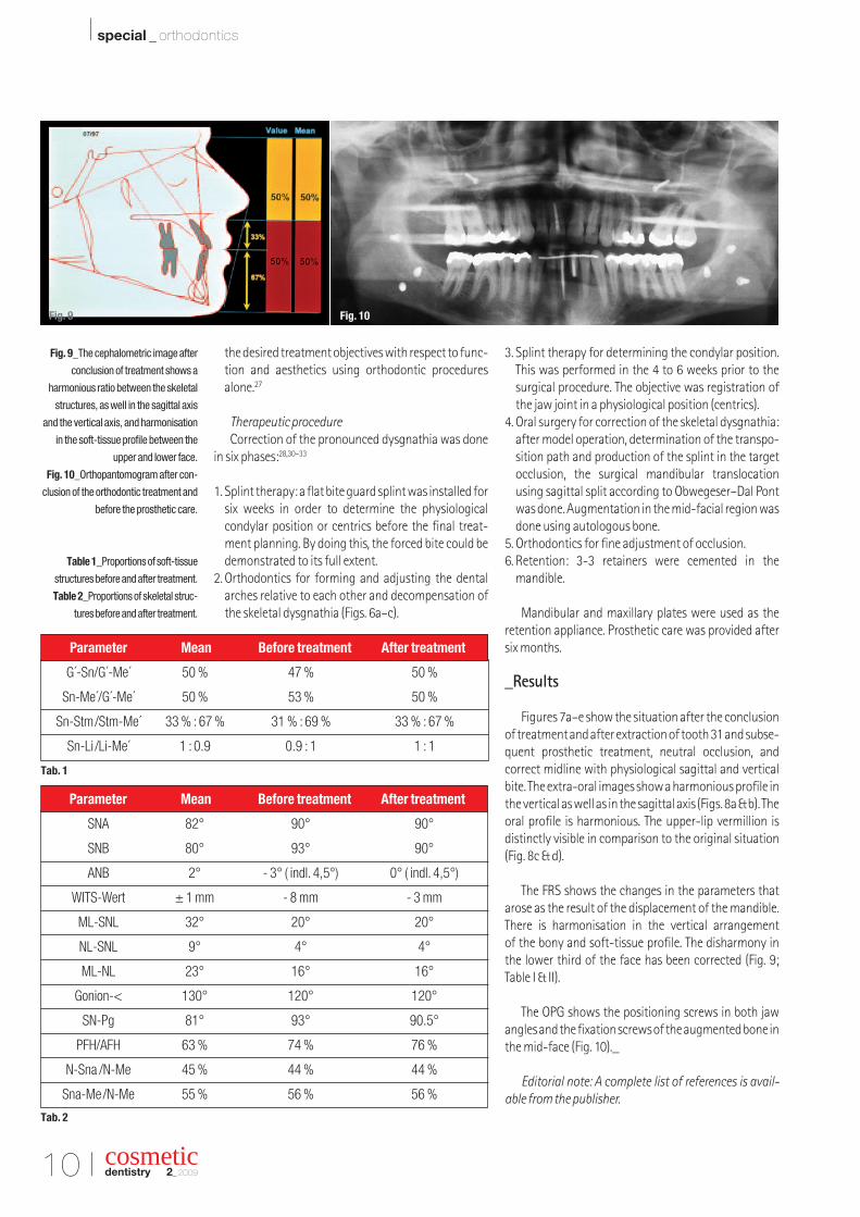
The FRS shows the changes in the parameters thatarose as the result of the displacement of the mandible.There is harmonisation in the vertical arrangement of the bony and soft-tissue profile. The disharmony inthe lower third of the face has been corrected (Fig. 9;Table I & II).
The OPG shows the positioning screws in both jawangles and the fixation screws of the augmented bone inthe mid-face (Fig. 10)._
Editorial note: A complete list of references is avail-able from the publisher.
Fig. 9_The cephalometric image after
conclusion of treatment shows a
harmonious ratio between the skeletal
structures, as well in the sagittal axis
and the vertical axis, and harmonisation
in the soft-tissue profile between the
upper and lower face.
Fig. 10_Orthopantomogram after con-
clusion of the orthodontic treatment and
before the prosthetic care.
Table 1_Proportions of soft-tissue
structures before and after treatment.
Table 2_Proportions of skeletal struc-
tures before and after treatment.
cosmeticdentistry 2_2009
Parameter Mean Before treatment After treatment
G´-Sn/G´-Me´ 50 % 47 % 50 %
Sn-Me´/G´-Me´ 50 % 53 % 50 %
Sn-Stm/Stm-Me´ 33 % : 67 % 31 % : 69 % 33 % : 67 %
Sn-Li /Li-Me´ 1 : 0.9 0.9 : 1 1 : 1
Parameter Mean Before treatment After treatment
SNA 82° 90° 90°
SNB 80° 93° 90°
ANB 2° - 3° ( indl. 4,5°) 0° ( indl. 4,5°)
WITS-Wert ± 1 mm - 8 mm - 3 mm
ML-SNL 32° 20° 20°
NL-SNL 9° 4° 4°
ML-NL 23° 16° 16°
Gonion-< 130° 120° 120°
SN-Pg 81° 93° 90.5°
PFH/AFH 63 % 74 % 76 %
N-Sna /N-Me 45 % 44 % 44 %
Sna-Me/N-Me 55 % 56 % 56 %
Fig. 9
Tab. 1
Tab. 2
Fig. 10


![Cell signaling -_introduction[1]](https://img.pdfslide.net/doc/110x75/58ed407f1a28ab28158b45f5/cell-signaling-introduction1.jpg)


![]_Introduction... · Created Date: 10/8/2013 2:57:50 PM](https://img.pdfslide.net/doc/110x75/5f99ea75020a9d4f117e7884/-introduction-created-date-1082013-25750-pm.jpg)
![Urban landscape -_introduction[1]](https://img.pdfslide.net/doc/110x75/55544c59b4c905b2428b4d16/urban-landscape-introduction1-5584a066c304a.jpg)