Embed Size (px)
Citation preview
INTERNATIONAL ASSOCIATION FOR THE STUDY OF LUNG CANCER
EDITED BY MING SOUND TSAO, MD, FRCPCKEITH M. KERR, MB CHB, FRCPATH, FRCPESANJA DACIC, MD, PHDYASUSHI YATABE, MD, PHDFRED R. HIRSCH, MD, PHD
IASLC ATLAS OF PD-L1 IMMUNOHISTOCHEMISTRY TESTING IN LUNG CANCER
Conquering Thoracic Cancers Worldwide

IASLC ATLAS OF PD-L1 IMMUNOHISTOCHEMISTRY TESTING IN LUNG CANCER
International Association for the Study of Lung Cancer, Aurora, CO, USA
Editors:Ming Sound Tsao, MD, FRCPCKeith M. Kerr, MB ChB, FRCPath, FRCPESanja Dacic, MD, PhDYasushi Yatabe, MD, PhDFred R. Hirsch, MD, PhD
An IASLC publication published by Editorial Rx Press
Cover and interior design by Amy Boches, Biographics
IASLC Office: IASLC, 13100 East Colfax Ave., Unit 10, Aurora, Colorado 80011, USAwww.iaslc.org
April 2017, October 201710 9 8 7 6 5 4 3 2
ISBN: 978-0-9832958-7-7
Copyright © 2017 International Association for the Study of Lung CancerAll rights reserved
Without limiting the rights under copyright reserved above, no part of this publication may be reproduced, stored in or introduced into a retrieval system, or transmitted in any form, or by any means without prior written permission.
While the information in this book is believed to be true and accurate as of the publication date, neither the IASLC nor the editors nor the publisher can accept any legal responsibility for any errors or omissions that may be made. The publisher makes no warranty, express or implied, with response to the material contained therein.
Erratum: The images in Figures 8 and 10 in Chapter 3, “Immunohistochemistry for PD-L1” (page 47) were reversed in the original publication of this PD-L1 Atlas. These images are correct in this printing of the publication.
AN INTERNATIONAL ASSOCIATION FOR THE STUDY OF LUNG CANCER PUBLICATION
Editorial Rx Press
North Fort Myers, FL
EDITED BY MING SOUND TSAO, MD, FRCPCKEITH M. KERR, MB CHB, FRCPATH, FRCPESANJA DACIC, MD, PHDYASUSHI YATABE, MD, PHDFRED R. HIRSCH, MD, PHD
IASLC ATLAS OF PD-L1 IMMUNOHISTOCHEMISTRY TESTING IN LUNG CANCER
IASLC acknowledges the generous funding and support provided by AstraZeneca, Bristol-Myers Squibb, and Merck for the IASLC Atlas of PD-L1 Immunohistochemistry Testing in Lung Cancer. The coeditors and contributors also acknowledge the assistance of Dr. Murry Wynes, PhD, Scientific Affairs Director, IASLC, for coordinating the project; the editorial assistance of Joy Curzio and Lori Alexander, MTPW, ELS, MWC; and, the publishing support of Deb Whippen, Editor and Publisher, Editorial Rx Press, for the publication of this text.
Acknowledgments
Contents
Contributors . . . . . . . . . . . . . . . . . . . . . . . . . . . . . . . . . . . . . . . . . . . . . . . . . . . . . . . . . . . . . . . . . . . . . . . . . . . . . . . . . . . . . . . . . . . . . . . . . . . . 6
Abbreviations . . . . . . . . . . . . . . . . . . . . . . . . . . . . . . . . . . . . . . . . . . . . . . . . . . . . . . . . . . . . . . . . . . . . . . . . . . . . . . . . . . . . . . . . . . . . . . . . . . 8
Manufacturers . . . . . . . . . . . . . . . . . . . . . . . . . . . . . . . . . . . . . . . . . . . . . . . . . . . . . . . . . . . . . . . . . . . . . . . . . . . . . . . . . . . . . . . . . . . . . . . . . 9
Introduction . . . . . . . . . . . . . . . . . . . . . . . . . . . . . . . . . . . . . . . . . . . . . . . . . . . . . . . . . . . . . . . . . . . . . . . . . . . . . . . . . . . . . . . . . . . . . . . . . . .11
1 Tumor Immunology . . . . . . . . . . . . . . . . . . . . . . . . . . . . . . . . . . . . . . . . . . . . . . . . . . . . . . . . . . . . . . . . . . . . . . . . . . . . . . . . . . . . . . . . .13
2 Cancer Immunotherapy for Lung Cancer . . . . . . . . . . . . . . . . . . . . . . . . . . . . . . . . . . . . . . . . . . . . . . . . . . . . . . . . . . . . . .21
3 Immunohistochemistry for PD-L1 . . . . . . . . . . . . . . . . . . . . . . . . . . . . . . . . . . . . . . . . . . . . . . . . . . . . . . . . . . . . . . . . . . . . . . .35
4 PD-L1 28-8 pharmDx Assay. . . . . . . . . . . . . . . . . . . . . . . . . . . . . . . . . . . . . . . . . . . . . . . . . . . . . . . . . . . . . . . . . . . . . . . . . . . . . . . .49
5 PD-L1 22C3 pharmDx Assay. . . . . . . . . . . . . . . . . . . . . . . . . . . . . . . . . . . . . . . . . . . . . . . . . . . . . . . . . . . . . . . . . . . . . . . . . . . . . . .55
6 PD-L1 SP142 Assay. . . . . . . . . . . . . . . . . . . . . . . . . . . . . . . . . . . . . . . . . . . . . . . . . . . . . . . . . . . . . . . . . . . . . . . . . . . . . . . . . . . . . . . . . . .63
7 PD-L1 SP263 Assay . . . . . . . . . . . . . . . . . . . . . . . . . . . . . . . . . . . . . . . . . . . . . . . . . . . . . . . . . . . . . . . . . . . . . . . . . . . . . . . . . . . . . . . . . .73
8 PD-L1 73-10 Assay . . . . . . . . . . . . . . . . . . . . . . . . . . . . . . . . . . . . . . . . . . . . . . . . . . . . . . . . . . . . . . . . . . . . . . . . . . . . . . . . . . . . . . . . . . .79
9 Other Anti–PD-L1 Clones: Alternative Assays and Laboratory-Developed Tests . . . . . . . . . . . .83
10 Complementary and Companion Diagnostics . . . . . . . . . . . . . . . . . . . . . . . . . . . . . . . . . . . . . . . . . . . . . . . . . . . . . . .93
11 Assay Harmonization: Is It Possible? . . . . . . . . . . . . . . . . . . . . . . . . . . . . . . . . . . . . . . . . . . . . . . . . . . . . . . . . . . . . . . . . . . . .97
12 Implementation of PD-L1 Testing for Personalized Therapy for Lung Cancer . . . . . . . . . . . . . . 109
13 Summary and Future Perspectives . . . . . . . . . . . . . . . . . . . . . . . . . . . . . . . . . . . . . . . . . . . . . . . . . . . . . . . . . . . . . . . . . . . . 115
References . . . . . . . . . . . . . . . . . . . . . . . . . . . . . . . . . . . . . . . . . . . . . . . . . . . . . . . . . . . . . . . . . . . . . . . . . . . . . . . . . . . . . . . . . . . . . . . . . . . 119
6 IASLC ATLAS OF EGFR TESTING IN LUNG CANCER
Contributors
EditorsMing Sound Tsao, MD, FRCPCPathologist, Senior Scientist, and ProfessorM. Qasim Choksi Chair in Lung CancerTranslational ResearchPrincess Margaret Cancer Centre, UniversityHealth NetworkDepartment of Laboratory Medicine andPathobiologyUniversity of TorontoToronto, Canada
Keith M. Kerr, FRCPathProfessorDepartment of PathologyAberdeen University Medical School,Aberdeen Royal InfirmaryAberdeen, Scotland, United Kingdom
Sanja Dacic, MD, PhDProfessorDirector of the FISH and Developmental LaboratoryDepartment of PathologyUniversity of Pittsburgh Medical CenterPennsylvania, U.S.A.
Yasushi Yatabe, MD, PhDChiefDepartment of Pathology and Molecular DiagnosticsAichi Cancer CenterNagoya, Japan
Fred R. Hirsch, MD, PhDProfessorDepartment of MedicineDepartment of PathologyPia and Fred R. Hirsch Endowed ChairUniversity of Colorado at DenverCEO, International Association for the Studyof Lung Cancer (IASLC)Colorado, U.S.A.
Contributing AuthorsBernadette Reyna Asuncion, MDResearch FellowCancer Science Institute of SingaporeNational University of SingaporeSingapore, Singapore
Elisabeth Brambilla, MD, PhDProfessorDepartment of PathologyCentre Hospitalier Universitaire de GrenobleGrenoble, France
Lukas Bubendorf, MD Professor and HeadDivision of CytopathologyInstitute for PathologyUniversity Hospital BaselBasel, Switzerland
Reinhard Buettner, MDProfessor and ChairmanInstitute for PathologyUniversity Hospital Medical SchoolUniversity of CologneCologne, Germany
Teh-Ying Chou, MD, PhDProfessorInstitute of Clinical Medicine National Yang-Ming University Department of Pathology and Laboratory MedicineTaipei Veterans General HospitalTaipei, Taiwan
Wendy A. Cooper, MBBS, Bsc(Med), FRCPA, PhDAssociate ProfessorTissue Pathology and Diagnostic Oncology, Royal Prince Alfred HospitalSydney Medical School, University of SydneySchool of Medicine, Western Sydney UniversitySydney, Australia

7CONTRIBUTORS
Sylvie Lantuéjoul, MD, PhDDepartment of PathologyPôle de Biologie, Institut de Biologie et de PathologieCHU A Michallon, CS 10217Université Joseph Fourier- INSERM U 823Institut Albert BonniotGrenoble, France
Mari Mino-Kenudson, MDDirector, Pulmonary Pathology ServiceMassachusetts General HospitalAssociate Professor of PathologyHarvard Medical SchoolMassachusetts, U.S.A.
Andrew G. Nicholson, DMProfessorDepartment of HistopathologyRoyal Brompton and Harefield NHSFoundation Trust and Imperial CollegeLondon, United Kingdom
Lynette M. Sholl, MDAssistant ProfessorDepartment of PathologyBrigham and Women’s HospitalHarvard Medical SchoolMassachusetts, U.S.A.
Ross A. Soo, MBBSPrincipal InvestigatorCancer Science Institute of SingaporeNational University of SingaporeSenior ConsultantNational University Health SystemSingapore
Erik Thunnissen, MD, PhDConsultant PathologistVU University Medical centerAmsterdam, The Netherlands
Arne Warth, MD, PhDInstitute of PathologyHeidelberg University Hospital Heidelberg, Germany
Ignacio Wistuba, MDProfessor and ChairJay and Lori Eisenberg Endowed ProfessorDepartment of Translational MolecularPathologyThe University of Texas MD AndersonCancer CenterTexas, U.S.A.
Murry W. Wynes, PhDDirector of Scientific AffairsInternational Association for the Study of Lung Cancer Colorado, U.S.A.
Left to right: Teh-Ying Chou, Yasushi Yatabe, Lukas Bubendorf, Elisabeth Brambilla, Ming Sound Tsao, Mari Mino-Kenudson, Sylvie Lantuéjoul, Erik Thunnissen, Sanja Dacic, Keith M. Kerr, Pia Hirsch, Ross A. Soo, Fred R. Hirsch, and Murry Wynes. Not present: Bernadette Reyna Asuncion, Reinhard Buettner, Wendy A. Cooper, Andrew G. Nicholson, Lynette M. Sholl, Arne Warth, and Ignacio Wistuba.
8 IASLC ATLAS OF EGFR TESTING IN LUNG CANCER
Abbreviations
The following abbreviations are used in the text.
AEC: 3-amino-9-ethylcarbazol
ALK: anaplastic lymphoma kinase (gene)
APC: antigen presenting cell
CTLA-4: cytotoxic T-lymphocyte-associated antigen 4
DAB: 3, 3’ diaminobenzidine
DC: dendritic cell
EDTA: ethylenediaminetetraacetic acid
EGFR: epidermal growth factor receptor (gene)
ER: estrogen receptor
FDA: US Food and Drug Administration
FFPE: formalin-fixed paraffin-embedded
H&E: hematoxylin & eosin
HER2: human epidermal growth factor receptor-2 (gene)
HLA: human leukocyte antigen
HRP: horseradish peroxidase
ICC: immunocytochemistry
IFN-g: interferon gamma
IgG: immunoglobulin G
IHC: immunohistochemistry
IL-2: interleukin-2
KIF5B: kinesin family member 5B (gene)
KIR: killer cell immunoglobulin-like receptor
LAG3: lymphocyte-activation gene 3
LDT: laboratory-developed test
MAGE-A3: melanoma-associated antigen-A3
MHC: major histocompatibility complex
MUC1: mucinous glycoprotein-1
NSCLC: non-small cell lung cancer
PD-1: programmed cell death protein-1
PD-L1: programmed cell death ligand-1
PR: progesterone receptor
ROS1: c-ros oncogene 1 (gene)
SCLC: small cell lung cancer
TGF-b2: transforming growth factor beta 2
TIL: tumor infiltrating lymphocyte
TNF-α: tumor-necrosis factor alpha
9MANUFACTURERS
Manufacturers
The following manufacturers and their PD-L1 testing-related products are noted in this Atlas. The location given for each manufacturer is not the only location; most manufacturers have offices worldwide.
AbcamCambridge, UKAntibody clone 28-8, rabbit polyclonal anti PD-L1 ab58810
Agilent Technologies/DakoCarpineteria, California, USAPD-L1 IHC 22C3 pharmDx; PD-L1 IHC 28-8 pharmDx; Autostainer Link 48; PT Link Pre-Treatment Module; EnVision FLEX Target Retrieval Solution; EnVision FLEX+ Polymer Reagents; EnVision FLEX+ Wash Buffer; FLEX IHC microscope slides
Anatech LTDBattle Creek, Michigan, USA Prefer fixative
BioCare MedicalPacheco, California, USA DaVinci Green Diluent
Bio SBSanta Barbara, California, USA PD-L1/CD274 clone: RBT-PDL1 rabbit monoclonal
Cell Signaling Technology, Inc.Danvers, Massachusetts, USAE1L3N rabbit monoclonal antibody; SignalStain Boost IHC Detection Reagent
Enzo Life Sciences Inc.Farmingdale, New York, USAEDTA pH 8
Leica BiosystemsBuffalo Grove, Illinois, USABOND-MAX Automated IHC/ISH Stainer; Novolink Polymer Detection System
Proteintech Group, Inc.Rosemont, Illinois, USAPD-L1 rabbit polyclonal CD274 antibody
R&D Systems Inc.Minneapolis, Minnesota, USAMouse monoclonal MAB1561
Sigma-AldrichSt. Louis, Missouri, USA Anti-CD274 rabbit antibody
Thermo Fisher ScientificWaltham, Massachusetts, USAFisherbrand Superfrost Plus Microscope Slides; mouse monoclonal MIH1; Tris-EDTA buffer solution; UltraVision Quanto Detection System HRP DAB
Ventana Medical Systems, Inc.Tucson, Arizona, USAPD-L1 (SP263) Assay; PD-L1 (SP142) Assay; Benchmark ULTRA platform, OptiView DAB IHC Detection Kit, OptiView Amplification Kit, Rabbit Monoclonal Negative Control Ig; ChromoMAP DAB; Benchmark XT Autostainer; Cell Conditioning 1 (CC1); ultraView Universal DAB Detection Kit; DISCOVERY ChromoMap DAB Kit

Despite very encouraging progress in the development and use of immunotherapy for patients with non-small cell lung cancer, much confusion remains regarding patient selection for each therapy. Programmed cell death ligand-1 (PD-L1) protein expression, as detected by immunohistochemistry (IHC) testing, has been widely used as a predictive biomarker assay for anti–PD-1/PD-L1 therapies. In fact, an assay for determination of PD-L1 expres-sion is approved by the US Food and Drug Administration for both first-line and second-line therapy with pembrolizumab. There is no clear understanding among physicians, health care personnel, or patients, however, regarding which assay to use for PD-L1 testing and whether the various assays are interchangeable because each assay was co-developed with a therapy. This complex biomarker scenario—the likes of which we have not faced before in lung cancer diagnostics—poses many challenges for pathologists, oncologists, and patients. The International Association for the Study of Lung Cancer (IASLC) has recognized the importance and timeliness of this topic and has convened an expert panel of authors to pres-ent current information about the emerging PD-L1 IHC assays, as well as to highlight both areas of clarity and debate. The authors have approached this topic with a wider lens, looking at the changing landscape of laboratory testing in general, as well as with a detailed focus on the specifics of each assay and on the current controversies regarding PD-L1 expression testing in lung cancer. Although this Atlas primarily aims to be a guide or resource for phy-sicians and others involved in lung cancer diagnosis and treatment, it is our hope that this text eventually also may give patients a more comprehensive understanding of the current biomarker scenario. Ultimately, we hope that through the creation of this Atlas, patients with lung cancer will receive the most contemporary and well-suited treatment options, based on up-to-date evidence, and will feel more confident and knowledgeable regarding their therapy. The authors acknowledge that updates to this Atlas will almost certainly be needed, sooner rather than later, due to the rapidly evolving nature of the field. Other biomarkers relating to the immune response itself or to tumor mutational burden are being investigated. Whether these will prove to be superior to PD-L1 IHC testing as a guide for therapeutic
Introduction By Ming S. Tsao, Keith M. Kerr, Sanja Dacic, Yasushi Yatabe, and Fred R. Hirsch
12 IASLC ATLAS OF EGFR TESTING IN LUNG CANCER
selection remains to be seen. In the meantime, PD-L1 IHC is the validated biomarker of choice. There are numerous ongoing trials investigating this biomarker and its associated analytic tools, and it seems likely that it will be at least part of the biomarker profile required for administration of anti–PD-1/PD-L1 drugs for the foreseeable future.
1It is widely accepted that cancer develops because of the accumulation of various alterations, including genetic and epigenetic changes, that make cancer cells genotypically and pheno-typically different from normal cells. One such example is the expression of cancer-testis antigens in several solid tumors, including lung cancer (Rousseaux 2013). These antigens are normally expressed in early embryonic and germ cells but silenced in adult somatic cells. Disrupted DNA methylation patterns of promoter CpG islands in cancer cells lead to aberrant expression; thus, expression of the cancer-testis antigens is restricted to cancer cells. More than 100 gene families with such an expression pattern have been identified. The antigens can be recognized by the host immune system and induce an immune response, although the testis is protected from immune attack by a lack of major histocompatibility complex (MHC) class I and II molecule expression. Furthermore, gene mutations and amplification may change the protein structure and expression level, which are also capable of being immu-nogenic. The number of genetic mutations in a tumor, the mutation burden, is associated with neoantigen burden (Rizvi 2015). The host immune system recognizes and responds to these antigens to a certain extent. However, cancer cells can find ways to survive through the acquisition of tolerance mechanisms, thus escaping from immune recognition. Under the current hypothesis, the immune system initially recognizes cancer cells and induces an immune response. After the equilibrium between cancer cell elimination by so-called immune surveillance and cancer cell evolution by genetic instability, the tumor cell clone is either eliminated or cancer cells survive but remain dormant. This dormancy is due to a decreased immunogenic state with adaptation within the cancer microenvironment, known as immunoediting (Schreiber 2011). Immune escape must then occur for a clinically evident tumor to develop. In the following sections, mechanisms of escape from immune surveil-lance in cancer are discussed, with reference to immunotherapeutic approaches (Box 1).
Cancer and T-CellsEven within the same species, organ transplantation causes immune responses. This fact implies that the immune system distinguishes self-antigens from non-self-antigens.
Tumor ImmunologyBy Yasushi Yatabe, Elisabeth Brambilla, and Keith M. Kerr

14 IASLC ATLAS OF PD-L1 IMMUNOHISTOCHEMISTRY TESTING IN LUNG CANCER
T cells recognize an antigen, which is presented with MHC by antigen-pre-senting cells (APCs), such as dendritic cells (DCs; Figure 1). Phagocytosed antigens are processed to the peptides and presented onto MHC in the sur-face of the APCs. As a result of this presentation, T cells that have T-cell receptors specific to the antigens rec-ognize the antigens and are activated in coordination with costimulatory receptors (CD28 and 4-1BB). In the early studies of tumor challenge after immunization in mice, CD8+ cytotoxic T-cells were a major player in mediation of tumor rejection. Using this function of T cells, researchers have attempted adaptive T-cell immunotherapy. The therapy is based on ex vivo expansion of patient-derived tumor-specific T cells and reinfusion to the patients. Peripheral mononuclear cells in the blood are isolated and stimulated with DCs that have been exposed to peptides of tumor-associated antigens. Through repeated stimu-lation and expansion, specific T-cell clones are collected and reinfused into the patients. This approach has some advantages related to cancer-specific activity and irrelevant immu-nosuppressive tumor microenvironment. Similarly, tumor infiltrating lymphocytes (TILs) are used with adaptive immunotherapy because lymphocytes with anticancer activity are likely to be enriched. It has been reported that complete remission was achieved with this method in 20 of 93 patients with metastatic melanoma, and 19 patients have experienced ongoing complete regression for 3 years (Rosenberg 2011). In this study, TILs were collected from resected melanoma, and T-cell clones with optimal anticancer activity were isolated,
Adaptive immunotherapy• Passive transfer of the immune cells with anticancer
activity, such as tumor-associated antigen (TAA)-specific T-cell clones and tumor-infiltrating lymphocytes
• Genetically engineered immune cells
Cancer vaccine• Immunization to enhance antitumor reactions
Nonspecific stimulation of immune responses• Stimulation of effector cells
• Inhibition of regulatory cells
Box 1. Major Immunotherapeutic Approaches
DC
B cell
CD8
CD4Cytokines
Antibodies
CTL
Cancer
MHC Class IIPeptide
TCR
MHC Class I
Peptide
TCR
Figure 1. Immune reaction against cancer cells is initiated by interaction between T-cell receptors (TCR) and major histo-compatibility complex (MHC) molecules, the latter presenting processed peptides of immunogens. DC = dendritic cells, and CTL = cytotoxic T-lymphocytes.
15TUMOR IMMUNOLOGY
followed by expansion with interleukin-2 (IL-2) stimulation and reinfusion immediately after lymphodepleting chemotherapy using cyclophosphamide and fludarabine, with or without total body radiation. Because obtaining TILs with anticancer activity tends to be difficult in cancers other than melanoma, genetically engineered immune cells that specifi-cally recognize cancer cells have also been examined, and the reinfusion of the lymphocytes with exogenous high affinity to cancer cells has been shown to achieve objective clinical responses (Morgan 2006).
Dendritic CellsAs with many vaccines, it would be expected that active immunization against cancer-spe-cific antigens would provoke cellular immune recognition to inhibit the growth of established cancer. DCs play a key role in the induction of T-cell responses through presentation of the target peptides on MHC molecules (Figure 1). In DCs, phagocytosed antigens are processed into peptides by the proteasome in the cytosol. The complexes are moved via the endoplas-mic reticulum through special channels, and the processed peptides are loaded onto MHC molecules. Lastly, the MHC molecules that present the peptides are expressed on the cell surface. Therefore, it should be efficient to use DCs for cancer vaccination. A common method for generating the vaccine is as follows. DC precursors are obtained from bone marrow or peripheral blood mononuclear cells and are differentiated into imma-ture DCs with stimulation of granulocyte macrophage colony-stimulating factor (GM-CSF) and/or interleukin-4 (IL-4), followed by exposure to peptides to generate mature DCs. Several methods, including fusion of the DCs with tumor cells, and co-stimulation with toll-like receptor (TLR) ligands and/or agonistic anti-CD40 antibody may be used to enhance the maturation. Sipuleucel-T was approved as a vaccine therapy for patients with castration-resistant prostatic cancer by the US Food and Drug Administration (FDA) based on the results of a phase III clinical trial (Kantoff 2010). With this treatment, peripheral blood mononuclear cells are collected and incubated with a fusion protein of prostatic acid phosphatase and GM-CSF, as a cancer-associated antigen and an antigen-presentation activator, respectively. Antigen-pulsed APCs were reinfused once a week for one month. With this vaccine therapy, the relative risk of death was reduced by 22%, although the time to disease progression was similar for the treatment and placebo arms. However, Sipuleucel-T is exceptional, as most cancer vaccine therapies have failed to show clinical effectiveness. In a summary of findings for cancer vaccine trials of 440 patients in the National Cancer Institute Center for Cancer Research Surgery Branch, the authors report that the objective response rate was 2.6%, and similar results were observed in other studies (Rosenberg 2004).
Coordination of Immune ResponsesA remarkable characteristic of the immune system is its ability to recognize and eliminate the targets specifically. The features are mediated by complex mechanisms involving T cells, DCs, and other immune cells by a balance between co-stimulatory and inhibitory signals (Figure 2). Recent developments in the understanding of cancer immunology allow the use of such immune coordination mechanisms for cancer management by means of enhancement of T-cell responses or by blockage of the negative regulation of T-cell responses.
16 IASLC ATLAS OF PD-L1 IMMUNOHISTOCHEMISTRY TESTING IN LUNG CANCER
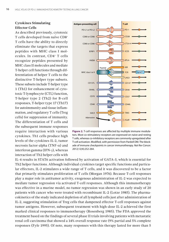
Cytokines Stimulating Effector CellsAs described previously, cytotoxic T cells developed from naïve CD8+ T cells have the ability to directly eliminate the targets that express peptides with MHC class I mol-ecules. In contrast, CD4+ T cells recognize peptides presented by MHC class II molecules and mediate T-helper cell functions through dif-ferentiation of helper T cells to the distinctive T-helper type subsets. These subsets include T-helper type 1 (Th1) for enhancement of cyto-toxic T-lymphocyte (CTL) function, T-helper type 2 (Th2) for B-cell responses, T-helper type 17 (Th17) for autoimmunity and tissue inflam-mation, and regulatory T cells (Treg cells) for suppression of immunity. The differentiation of T cells and the subsequent immune responses require interaction with various cytokines. Th1 cells produce high levels of the cytokines IL-2, tumor necrosis factor-alpha (TNF-α) and interferon gamma (IFN-γ), whereas interaction of Th2 helper cells with IL-4 results in STAT6 activation followed by activation of GATA-3, which is essential for Th2 helper functions. Although individual cytokines target specific functions and particu-lar effectors, IL-2 stimulates a wide range of T cells, and it was discovered to be a factor that primarily stimulates proliferation of T cells (Morgan 1976). Because T-cell responses play a major role in antitumor activity, exogenous administration of IL-2 was expected to mediate tumor regression via activated T-cell responses. Although this immunotherapy was effective in a murine model, no tumor regression was shown in an early study of 20 patients with cancer who were treated with recombinant IL-2 (Lotze 1985). The pharma-cokinetics of the study indicated depletion of all lymphoid cells just after administration of IL-2, suggesting stimulation of Treg cells that dampened effector T-cell responses against tumor antigens. However, subsequent treatment with high-dose IL-2 achieved the first marked clinical responses to immunotherapy (Rosenberg 1985). The FDA approved the treatment based on the findings of several phase II trials involving patients with metastatic renal cell carcinoma that showed a 14% overall response rate (9% partial and 5% complete responses (Fyfe 1995). Of note, many responses with this therapy lasted for more than 5
Figure 2. T-cell responses are affected by multiple immune modula-tors. Most co-stimulatory receptors are expressed on naive and resting T cells, whereas co-inhibitory receptors are commonly upregulated after T-cell activation. Modified, with permission from Pardoll DM. The block-ade of immune checkpoints in cancer immunotherapy. Nat Rev Cancer. 2012;12(4):252-264.
Antigen-presenting cell T cell
PD-L1 or PD-L2
CD80 or CD86
CD80 or CD86
B7RP1
HVEM
MHC class I or II
CD137L
OX40L
CD70
CD40
GAL9
Adenosine
PD-1
CD28
CTLA-4
ICOS
BTLA
KIR
TCR
LAG3
CD137
OX40
CD27
CD40L
TIM3
A2aR
–
+
–
+
–
–
–
+
+
+
–
–
Signal 1
Peptide
17TUMOR IMMUNOLOGY
years, suggesting remarkable durability of anticancer responses in contrast to other cyto-toxic chemotherapies. In addition to being approved for metastatic renal cell carcinoma, this IL-2 therapy was approved for advanced melanoma; a 16% overall response rate and flat tails in the Kaplan-Meier curve were also reported (Atkins 1999). Major toxicities of this therapy are due to the responses mediated by IL-2-induced IFN-γ and TNF-α, which result in capillary leak syndrome and decreased systemic vascular resistance. These, in turn, lead to fever, hypotension, arrhythmia, lethargy, renal failure, and systemic edema. Other immunotherapy, through stimulation of effector cells, includes treatment using IFN-α and imiquimod; however, this treatment resulted in clinical efficacy only for some cancer types (Kirkwood 1996, Motzer 2002, van Seters 2008).
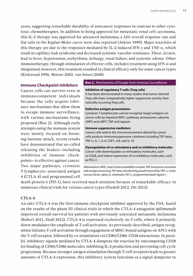
Immune Checkpoint InhibitorsCancer cells can survive even in immunocompetent individuals because the cells acquire toler-ance mechanisms that allow them to escape immune surveillance, with various mechanisms being proposed (Box 2). Although early attempts using the immune system were mostly focused on boost-ing immune attack, recent results have demonstrated that so-called releasing the brakes—including inhibition of immune check-points—is effective against cancer. Two major pathways, cytotoxic T-lymphocyte–associated antigen 4 (CTLA-4) and programmed cell death protein-1 (PD-1), have received much attention because of remarkable efficacy in numerous clinical trials for various cancer types (Pardoll 2012, Ott 2013).
CTLA-4An anti–CTLA-4 was the first immune checkpoint inhibitor approved by the FDA, based on the results of the phase III clinical trials in which the CTLA-4 antagonist ipilimumab improved overall survival for patients with previously untreated metastatic melanoma (Robert 2011, Hodi 2012). CTLA-4 is expressed exclusively on T-cells, where it primarily down-modulates the amplitude of T-cell activation. As previously described, antigen recog-nition initiates T-cell activation through engagement of MHC-bound antigens on APCs with the T-cell receptor, followed by co-stimulation via CD80/CD86-CD28 interactions. In paral-lel, inhibitory signals mediated by CTLA-4 dampens the reaction by outcompeting CD28 for binding of CD80/CD86 molecules, inhibiting IL-2 production and preventing cell-cycle progression. Because stronger antigen stimulation through T-cell receptors leads to greater amounts of CTLA-4 expression, this inhibitory system functions as a signal dampener to
Inhibition of regulatory T-cells (Treg cells)It has been demonstrated in many studies that tumor-derived Treg cells have comparatively higher suppressive activity than naturally occurring Treg cells.
Defective antigen presentationCytotoxic T lymphocytes cannot recognize target antigens on cancer cells by impaired MHC I pathway, proteasome subunits LMP2 and LMP7, TAP, and tapasin.
Immune suppressive mediatorsCancer cells and/or the microenvironment altered by cancer cells produce immunosuppressive cytokines including TGF-beta, TNF-α, IL-1, IL-6, CDF1, IL8, and IL-10.
Dysregulation of co-stimulatory and co-inhibitory moleculesCancer cells downregulate co-stimulatory molecules, such as CD28, and induce expression of co-inhibitory molecules, such as PD-L1.
Box 2. Mechanisms of Escape from Immune Surveillance
Abbreviations: MHC, major histocompatibility complex; TAP, transporter associated with antigen processing; TGF-beta, transforming growth factor beta; TNF-α , tumor necrosis factor-alpha; IL, interleukin; PD-L1, programmed death-ligand 1.
18 IASLC ATLAS OF PD-L1 IMMUNOHISTOCHEMISTRY TESTING IN LUNG CANCER
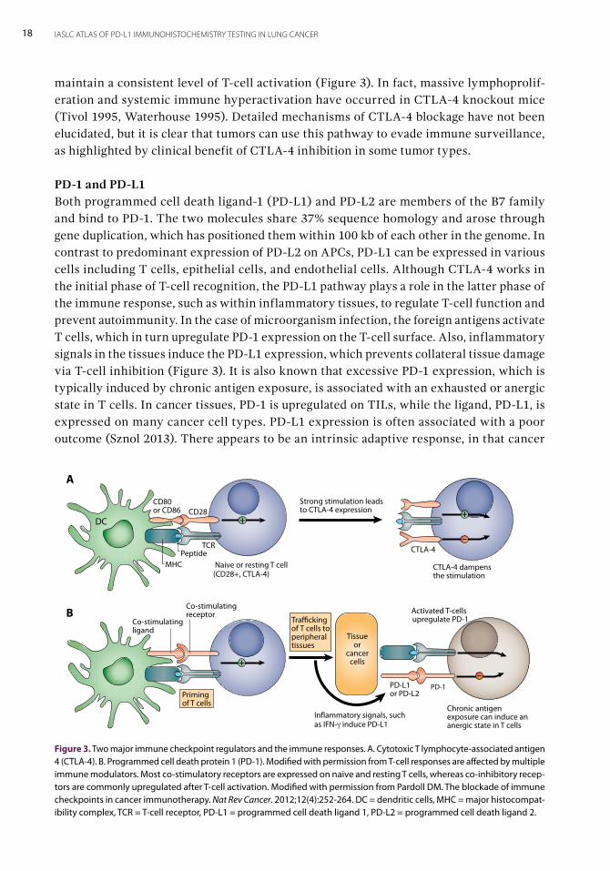
maintain a consistent level of T-cell activation (Figure 3). In fact, massive lymphoprolif-eration and systemic immune hyperactivation have occurred in CTLA-4 knockout mice (Tivol 1995, Waterhouse 1995). Detailed mechanisms of CTLA-4 blockage have not been elucidated, but it is clear that tumors can use this pathway to evade immune surveillance, as highlighted by clinical benefit of CTLA-4 inhibition in some tumor types.
PD-1 and PD-L1Both programmed cell death ligand-1 (PD-L1) and PD-L2 are members of the B7 family and bind to PD-1. The two molecules share 37% sequence homology and arose through gene duplication, which has positioned them within 100 kb of each other in the genome. In contrast to predominant expression of PD-L2 on APCs, PD-L1 can be expressed in various cells including T cells, epithelial cells, and endothelial cells. Although CTLA-4 works in the initial phase of T-cell recognition, the PD-L1 pathway plays a role in the latter phase of the immune response, such as within inflammatory tissues, to regulate T-cell function and prevent autoimmunity. In the case of microorganism infection, the foreign antigens activate T cells, which in turn upregulate PD-1 expression on the T-cell surface. Also, inflammatory signals in the tissues induce the PD-L1 expression, which prevents collateral tissue damage via T-cell inhibition (Figure 3). It is also known that excessive PD-1 expression, which is typically induced by chronic antigen exposure, is associated with an exhausted or anergic state in T cells. In cancer tissues, PD-1 is upregulated on TILs, while the ligand, PD-L1, is expressed on many cancer cell types. PD-L1 expression is often associated with a poor outcome (Sznol 2013). There appears to be an intrinsic adaptive response, in that cancer
Figure 3. Two major immune checkpoint regulators and the immune responses. A. Cytotoxic T lymphocyte-associated antigen 4 (CTLA-4). B. Programmed cell death protein 1 (PD-1). Modified with permission from T-cell responses are affected by multiple immune modulators. Most co-stimulatory receptors are expressed on naive and resting T cells, whereas co-inhibitory recep-tors are commonly upregulated after T-cell activation. Modified with permission from Pardoll DM. The blockade of immune checkpoints in cancer immunotherapy. Nat Rev Cancer. 2012;12(4):252-264. DC = dendritic cells, MHC = major histocompat-ibility complex, TCR = T-cell receptor, PD-L1 = programmed cell death ligand 1, PD-L2 = programmed cell death ligand 2.
–
+
+
+
–
CD80or CD86 CD28
MHC
TCRPeptide CTLA-4
Co-stimulatingligand
Co-stimulatingreceptor
Traffickingof T cells toperipheraltissues
Tissue or
cancer cells
PD-L1or PD-L2
PD-1
DC
A
B
Primingof T cells
Naive or resting T cell (CD28+, CTLA-4)
Strong stimulation leads to CTLA-4 expression
CTLA-4 dampens the stimulation
Inflammatory signals, such as IFN-γ induce PD-L1
Activated T-cells upregulate PD-1
Chronic antigen exposure can induce an anergic state in T cells
19TUMOR IMMUNOLOGY
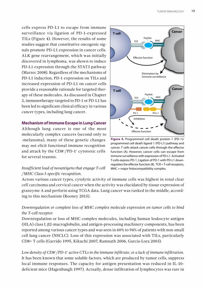
cells express PD-L1 to escape from immune surveillance via ligation of PD-1-expressed TILs (Figure 4). However, the results of some studies suggest that constitutive oncogenic sig-nals promote PD-L1 expression in cancer cells. ALK gene rearrangement, which was initially discovered in lymphoma, was shown to induce PD-L1 expression through the STAT3 pathway (Marzec 2008). Regardless of the mechanisms of PD-L1 induction, PD-1 expression on TILs and increased expression of PD-L1 on cancer cells provide a reasonable rationale for targeted ther-apy of these molecules. As discussed in Chapter 2, immunotherapy-targeted to PD-1 or PD-L1 has been led to significant clinical efficacy in various cancer types, including lung cancer.
Mechanism of Immune Escape in Lung CancerAlthough lung cancer is one of the most molecularly complex cancers (second only to -melanoma), many of these genetic changes may not elicit functional immune recognition and attack by the CD8+/PD-1+ cytotoxic cells for several reasons.
Insufficient load of neoantigens that engage T-cell /MHC Class I-specific recognition. Across various cancer types, cytolytic activity of immune cells was highest in renal clear cell carcinoma and cervical cancer when the activity was elucidated by tissue expression of granzyme A and perforin using TCGA data. Lung cancer was ranked in the middle, accord-ing to this mechanism (Rooney 2015).
Downregulation or complete loss of MHC complex molecule expression on tumor cells to bind the T-cell receptor. Downregulation or loss of MHC complex molecules, including human leukocyte antigen (HLA) class I, β2-macroglobulin, and antigen-processing machinery components, has been reported among various cancer types and was seen in 60% to 94% of patients with non-small cell lung cancer (NSCLC). Loss of this expression was associated with TILs, particularly CD8+ T cells (Garrido 1995, Kikuchi 2007, Ramnath 2006, Garcia-Lora 2003).
Low density of CD8+/PD-1+ active CTLs in the immune infiltrate, or a lack of immune infiltration. It has been known that some soluble factors, which are produced by tumor cells, suppress local immune responses. The capacity for antigen presentation was reduced in IL-10-deficient mice (Hagenbaugh 1997). Actually, dense infiltration of lymphocytes was rare in
Figure 4. Programmed cell death protein 1 (PD-1)/programmed cell death ligand 1 (PD-L1) pathway and cancer. T cells attack cancer cells through the effector function (A). However, cancer cells can escape from immune surveillance with expression of PD-L1. Activated T-cells express PD-1. Ligation of PD-1 with PD-L1 down-regulates the effector function (B). TCR = T-cell receptors, MHC = major histocompatibility complex.
T cell Cancer
MHCPeptide
TCR
E�ector function
Elimination by Immune surveillance
T cell Cancer
MHCPeptide
TCR
Inhibition
E�ector function
PD-L1PD-1
20 IASLC ATLAS OF PD-L1 IMMUNOHISTOCHEMISTRY TESTING IN LUNG CANCER
lung cancer (Brambilla 2016), and low density of active CD8+/PD-1+ CTLs has been reported (Tumeh 2014, Kim 2015).
Anergy of CD8 cytotoxicity. In contrast to some reports of low density of CD8+/PD-1+ cytotoxic T cells, some tumors have high numbers of infiltrating CD8+ cytotoxic T cells. This finding is explained by anergy, which results from T-cell inactivation, with a lack of granzyme B, a lack of proper cytokines for achieving CD8+ T-cell maturation and activation (IFN-γ, IL-2, IL-21), or the presence of inhibitory cytokines, such as IL-10 and transforming growth factor beta (Zaretsky 2015).
Counter-regulation by excessive CD25+FOXP3+CD4+ on CD8+PD-1+ Treg cells in the immune infiltrate. A high ratio of intratumoral Tregs to effector T cells is generally associated with poor out-comes across many cancer types, including lung cancer (Fridman 2012, Petersen 2006).
Unavailability of functional apoptotic pathways in tumor cells by lack of functional Fas-FasL receptor- complex. Approximately 20% to 25% of lung cancers lack Fas expression and overexpress FasL, sug-gesting impaired cytokilling via the Fas/FasL pathway (Li 2015, Viard-Leveugle 2003).
Immune checkpoints with increased PD-L1 or CTLA-4 on tumor cells and or immune cells. As discussed previously, expression of PD-L1 and CTLA-4 on cancer cells allows immune cells to fail to react to tumor-cell antigens.
SummaryEven from this brief and short review, it is clear that the interactions of tumor cells and the immune system are extremely complex and not entirely understood. These interactions have a pivotal role in allowing tumors to develop and progress. Primarily, tumors, by one or several mechanisms, must develop the ability to avoid or negate an immune response in those cases in which a specific immune response to tumor neoantigens has been developed. Among those mechanisms are the interaction of membrane-bound ligands and receptors that act as immune checkpoints, regulating the immune system. This important physiologic mechanism—which prevents uncontrolled immune responses and, thus, autoimmunity—appears to be adopted by some tumors as a means of switching off an otherwise primed and available cellular immune response to that tumor. These receptor–ligand interactions are, therefore, important therapeutic targets, as evidenced by the successes seen with the use of anti-CTLA-4 therapies in melanoma and anti-PD-1 or PD-L1 agents in a number of tumor types, such as NSCLC. In some ways, it is remarkable, in such a complex, multifaceted, and closely regulated system, that inhibiting only one regulatory mechanism can achieve such results. This atlas will discuss the inhibition of the PD-1/PD-L1 axis in lung cancer, the clinical evidence to date for such therapies, and the challenges posed by a very complex biomarker backdrop to this exciting new therapeutic approach.
Cancer Immunotherapy for Lung CancerBy Ross A. Soo, Murry W. Wynes, and Fred R. Hirsch 2Lung cancer is the leading cause of cancer death worldwide, with 1.6 million attributed deaths annually (World Health Organization International Agency for Research on Cancer, 2017). Non-small cell lung cancer (NSCLC) accounts for the majority of lung cancer diag-noses, and the disease is metastatic at the time of diagnosis for most patients (National Cancer Institute Surveillance, Epidemiology, and End Results Program, 2017). Despite an improvement in overall survival with platinum-based chemotherapy (NSCLC Meta-Analyses Collaborative Group, 2008), prognosis remains poor for patients with advanced-stage NSCLC, with a median survival of 8 to 12 months (Schiller 2002, Sandler 2006). Advances in the molecular characterization of NSCLC, especially in adenocarcinoma histologic subtypes, have enabled the identification of key genetic aberrations in NSCLC that can be exploited with molecularly targeted therapy (Pao 2011). Genetic aberrations in EGFR, ALK, ROS1, RET, BRAF, and NTRK predict for sensitivity to receptor tyrosine-kinase inhibitors (Mok 2009, Solomon 2014, Shaw 2014, Planchard 2016). Despite the success of molecularly targeted treatment, acquired resistance and disease progression inevitably occur (Camidge 2014; Hirsch 2016). Treatment options for patients with small cell lung cancer (SCLC) in whom disease has progressed after platinum-based chemotherapy are even more limited. Novel therapeutic approaches are needed for patients with NSCLC and SCLC. Cancer immunotherapy has been described as any therapy that interacts with the immune system to treat cancer. As an option for cancer, cancer immunotherapy predates even cyto-toxic chemotherapy. Cancer immunotherapy can be categorized into passive and active types (Figure 1). Passive immunotherapy has been described as administration of an immunologi-cally active agent manufactured or generated outside of the patient’s body. Theoretically, such an approach is not dependent on the host’s own immune system to have an effect. Examples of passive immunotherapy include the use of monoclonal antibodies, such as trastuzumab or rituximab (Slamon 2001, Coiffier 2002), and adoptive cellular therapy, such as tumor-infiltrating lymphocyte infusion, T-cell receptor (TCR) engineering, and chimeric antigen receptor T-cell therapy (Morgan 2006, Maude 2014). Active cancer immunotherapy involves the stimulation or priming of the host’s immune system to recognize a tumor as
22 IASLC ATLAS OF PD-L1 IMMUNOHISTOCHEMISTRY TESTING IN LUNG CANCER
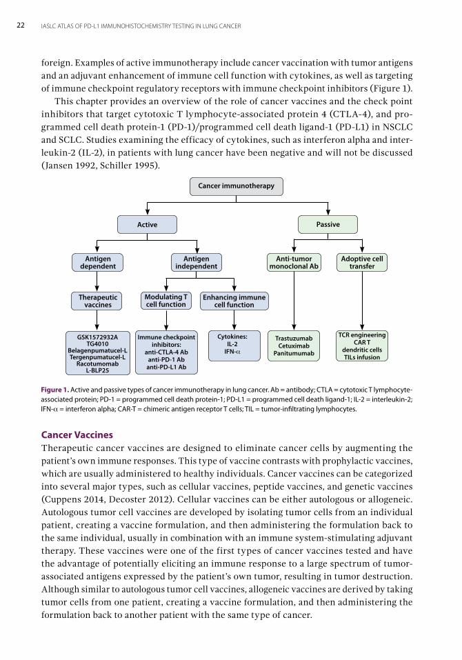
foreign. Examples of active immunotherapy include cancer vaccination with tumor antigens and an adjuvant enhancement of immune cell function with cytokines, as well as targeting of immune checkpoint regulatory receptors with immune checkpoint inhibitors (Figure 1). This chapter provides an overview of the role of cancer vaccines and the check point inhibitors that target cytotoxic T lymphocyte-associated protein 4 (CTLA-4), and pro-grammed cell death protein-1 (PD-1)/programmed cell death ligand-1 (PD-L1) in NSCLC and SCLC. Studies examining the efficacy of cytokines, such as interferon alpha and inter-leukin-2 (IL-2), in patients with lung cancer have been negative and will not be discussed (Jansen 1992, Schiller 1995).
Cancer VaccinesTherapeutic cancer vaccines are designed to eliminate cancer cells by augmenting the patient’s own immune responses. This type of vaccine contrasts with prophylactic vaccines, which are usually administered to healthy individuals. Cancer vaccines can be categorized into several major types, such as cellular vaccines, peptide vaccines, and genetic vaccines (Cuppens 2014, Decoster 2012). Cellular vaccines can be either autologous or allogeneic. Autologous tumor cell vaccines are developed by isolating tumor cells from an individual patient, creating a vaccine formulation, and then administering the formulation back to the same individual, usually in combination with an immune system-stimulating adjuvant therapy. These vaccines were one of the first types of cancer vaccines tested and have the advantage of potentially eliciting an immune response to a large spectrum of tumor-associated antigens expressed by the patient’s own tumor, resulting in tumor destruction. Although similar to autologous tumor cell vaccines, allogeneic vaccines are derived by taking tumor cells from one patient, creating a vaccine formulation, and then administering the formulation back to another patient with the same type of cancer.
Cancer immunotherapy
Active Passive
Antigen dependent
Antigen independent
Adoptive cell transfer
Anti-tumor monoclonal Ab
Therapeutic vaccines
Modulating T cell function
Enhancing immune cell function
GSK1572932A TG4010
Belagenpumatucel-L Tergenpumatucel-L
Racotumomab L-BLP25
Immune checkpoint inhibitors:
anti-CTLA-4 Ab anti-PD-1 Ab
anti-PD-L1 Ab
Cytokines: IL-2
IFN-a
Trastuzumab Cetuximab
Panitumumab
TCR engineering CAR T
dendritic cells TILs infusion
Figure 1. Active and passive types of cancer immunotherapy in lung cancer. Ab = antibody; CTLA = cytotoxic T lymphocyte-associated protein; PD-1 = programmed cell death protein-1; PD-L1 = programmed cell death ligand-1; IL-2 = interleukin-2; IFN-a = interferon alpha; CAR-T = chimeric antigen receptor T cells; TIL = tumor-infiltrating lymphocytes.
23CANCER IMMUNOTHERAPY FOR LUNG CANCER
Unlike cellular vaccines, which are made directly from patients’ tumors, peptide vac-cines are often synthesized in vitro to mimic tumor-associated proteins, with the goal of eliciting an immune response against tumor cells that express that specific tumor-associated protein. Genetic vaccines are composed of synthetic DNA or RNA molecules that encode for tumor-associated proteins and are administered either alone or packaged within a non-pathogenic virus. The genetic material is taken up by cells within the recipient, translated in the encoded proteins, processed, and presented to the immune system to provoke an immune response against tumor-associated proteins. Early studies of vaccine therapy with bacillus Calmette-Guerin in the adjuvant and neo-adjuvant setting were negative (Bakker 1986, Miller 1982, Matthay 1986). In the modern era, multiple vaccine studies have been conducted in early, locally advanced, and advanced-stage NSCLC. The melanoma-associated antigen (MAGE)-A3 recombinant protein vaccine has been extensively studied in the adjuvant setting after complete resection. A randomized phase II study showed that, for patients with completely resected stage IB-II, MAGE-A3–positive NSCLC who received no adjuvant chemotherapy, there was a trend toward superior disease-free survival with MAGE-A3 vaccine compared with placebo after a median follow up of 70 months (HR: 0.75; 95% CI: 0.46-1.23; p=0.254) (Vansteenkiste 2016). However, no clinical benefit was found in the subsequent randomized, double-blind, placebo-controlled large phase III study (MAGRIT) of completely resected stage IB-IIIA MAGE-A3 positive NSCLC, with or without adjuvant chemotherapy. For the overall population in this later study, the median disease-free survival was 60.5 months for the MAGE-A3 vaccine group and 57.9 months for the placebo group (HR: 1.02, 95% CI: 0.89-1.18; p=0.74). In the subgroup that did not receive adjuvant chemotherapy, the median disease-free survival was 58.0 months for the vaccine group and 56.9 months for placebo group (HR: 0.97; 95% CI: 0.80-1.18; p=0.76) (Vansteenkiste 2016). Based on these results, the clinical development of the MAGE-A3 vaccine has been terminated. Tecemotide (L-BLP25) is a peptide vaccine based on a 25-amino acid sequence from the mucinous glycoprotein-1 (MUC1) protein that demonstrated promising activity in the setting of locally advanced NSCLC in a phase II study (Butts 2005), subsequently result-ing in the initiation of two randomized studies. One was a global phase III trial, START, in which tecemotide was compared with placebo for patients with stage III NSCLC without disease progression after chemoradiation therapy (Butts 2014). The second trial, INSPIRE, was a randomized phase II trial of Asian patients (Wu 2011). The START trial showed no difference in median overall survival between the tecemotide arm and placebo arms (25.6 months vs. 22.3 months; adjusted HR: 0.88; 95% CI: 0.75-1.03; p=0.123). However, follow-ing a prespecified subgroup analysis, the median overall survival did differ between the vaccine and placebo arms for patients who received concurrent chemoradiation therapy (30.8 months vs. 20.6 months; HR: 0.78; 95% CI: 0.64-0.95; p=0.016) compared with patients who received sequential chemoradiation therapy (19.4 months vs. 24.6 months; HR: 1.12; 95% CI: 0.87-1.44; p=0.38). INSPIRE was terminated in 2014 after Merck announced that it planned to discontinue the clinical development of tecemotide as monotherapy for patients with stage III NSCLC because of disappointing results from the Japanese phase I/II EMR 63325-009 study (Merck KGaA 2014).
24 IASLC ATLAS OF PD-L1 IMMUNOHISTOCHEMISTRY TESTING IN LUNG CANCER
In the advanced-disease setting, TG4010, another MUC1-targeting vaccine that uses a viral vector to express both the full-length MUC1 and IL-2 (a T-cell stimulant), showed promising clinical activity in the TG4010 immunotherapy and first-line chemotherapy for advanced NSCLC (TIME) study. Results from the phase IIb part of the randomized, double-blind, placebo-controlled, phase IIb/III trial showed that, in the overall population, progression-free survival was 5.9 months for the TG4010 group and 5.1 months for the placebo group (HR: 0.74; 95% CI: 0.55–0.98; p=0.019) (Quoix 2016). The phase III portion of the trial is continuing. Belagenpumatucel-L is an allogeneic whole tumor-cell vaccine derived from four radiated NSCLC cell lines of varying histologies that also express an antisense transgene for trans-forming growth factor beta2, which downregulates the immunosuppressant transforming growth factor beta2. The findings of a phase II study suggested clinical efficacy in advanced NSCLC (Nemunaitis 2006), and a phase III study (STOP) was initiated to randomly assign patients with stage III/IV NSCLC in whom disease did not progress after platinum-based chemotherapy to either belagenpumatucel-L or placebo (Giaccone 2015). There was no significant difference in overall survival between the two treatment arms (20.3 months vs. 17.8 months; HR: 0.94; p=0.594); likewise, there was no difference in progression-free survival between the two groups (4.3 months vs. 4.0 months; HR: 0.99; p=0.947). Epidermal growth factor receptor (EGFR) is an important signalling pathway in NSCLC, and a vaccine has been developed against its cognate ligand EGF using recombinant human EGF coupled to a carrier protein. In a randomized phase II study, patients with stage IIIB/IV NSCLC were randomly assigned to receive either best supportive care or EGF vaccinations after first-line chemotherapy (Neninger 2008). In the overall population, there was a trend for improved overall survival, and a significant survival advantage for patients who had a good antibody response to EGF. A later phase III trial included patients with stage IIIB/IV NSCLC who were randomly assigned after first-line chemotherapy to either vaccine or best supportive care. In the safety population, overall survival was 10.83 months for the vaccine arm and 8.86 months for the control arm (Rodriguez 2016). This difference was not significant according to the standard log rank (HR: 0.82; p=0.100), but was significant accord-ing to a weighted log rank (p=0.04) that was applied once the nonproportionality of the hazard ratio was verified. In the per-protocol setting (patients who received at least four vaccine doses), overall survival differed significantly between the vaccine and best sup-portive care arms (12.43 months vs. 9.43 months; HR: 0.77; p=0.036). In addition, overall survival was longer (14.66 months) for vaccinated patients with high EGF concentrations at baseline.
Immune Checkpoint Inhibitors More recently, a deeper understanding of the interaction between the immune system and tumors has led to the identification of CTLA-4 and PD-1/ PD-L1 as key factors by which tumors evade host immune response (Pardoll 2012). This discovery has led to the devel-opment of a new generation of immunotherapy agents that target these molecules. The immune checkpoint inhibitors represent an important breakthrough in the treatment of cancer. Multiple studies have shown immune checkpoint inhibitors to be highly active and durable in a variety of solid tumors, including NSCLC. The immune checkpoint inhibitors
25CANCER IMMUNOTHERAPY FOR LUNG CANCER
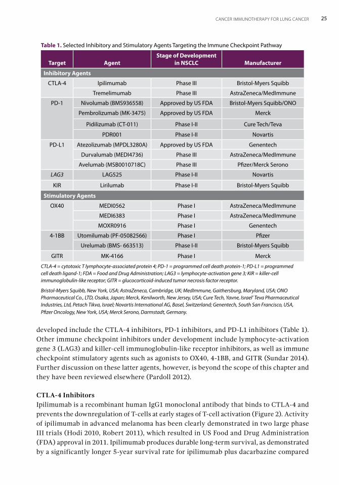
developed include the CTLA-4 inhibitors, PD-1 inhibitors, and PD-L1 inhibitors (Table 1). Other immune checkpoint inhibitors under development include lymphocyte-activation gene 3 (LAG3) and killer-cell immunoglobulin-like receptor inhibitors, as well as immune checkpoint stimulatory agents such as agonists to OX40, 4-1BB, and GITR (Sundar 2014). Further discussion on these latter agents, however, is beyond the scope of this chapter and they have been reviewed elsewhere (Pardoll 2012).
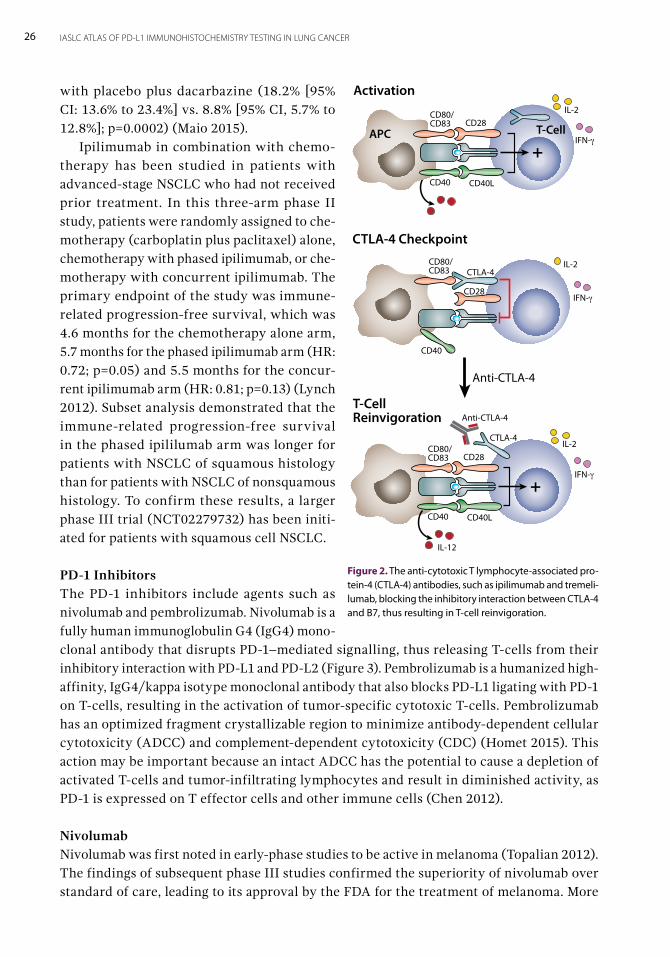
CTLA-4 InhibitorsIpilimumab is a recombinant human IgG1 monoclonal antibody that binds to CTLA-4 and prevents the downregulation of T-cells at early stages of T-cell activation (Figure 2). Activity of ipilimumab in advanced melanoma has been clearly demonstrated in two large phase III trials (Hodi 2010, Robert 2011), which resulted in US Food and Drug Administration (FDA) approval in 2011. Ipilimumab produces durable long-term survival, as demonstrated by a significantly longer 5-year survival rate for ipilimumab plus dacarbazine compared
Table 1. Selected Inhibitory and Stimulatory Agents Targeting the Immune Checkpoint Pathway
Target AgentStage of Development
in NSCLC Manufacturer
Inhibitory Agents
CTLA-4 Ipilimumab Phase III Bristol-Myers Squibb
Tremelimumab Phase III AstraZeneca/MedImmune
PD-1 Nivolumab (BMS936558) Approved by US FDA Bristol-Myers Squibb/ONO
Pembrolizumab (MK-3475) Approved by US FDA Merck
Pidilizumab (CT-011) Phase I-II Cure Tech/Teva
PDR001 Phase I-II Novartis
PD-L1 Atezolizumab (MPDL3280A) Approved by US FDA Genentech
Durvalumab (MEDI4736) Phase III AstraZeneca/MedImmune
Avelumab (MSB0010718C) Phase III Pfizer/Merck Serono
LAG3 LAG525 Phase I-II Novartis
KIR Lirilumab Phase I-II Bristol-Myers Squibb
Stimulatory Agents
OX40 MEDI0562 Phase I AstraZeneca/MedImmune
MEDI6383 Phase I AstraZeneca/MedImmune
MOXR0916 Phase I Genentech
4-1BB Utomilumab (PF-05082566) Phase I Pfizer
Urelumab (BMS- 663513) Phase I-II Bristol-Myers Squibb
GITR MK-4166 Phase I Merck
CTLA-4 = cytotoxic T lymphocyte-associated protein 4; PD-1 = programmed cell death protein-1; PD-L1 = programmed cell death ligand-1; FDA = Food and Drug Administration; LAG3 = lymphocyte-activation gene 3; KIR = killer-cell immunoglobulin-like receptor; GITR = glucocorticoid-induced tumor necrosis factor receptor.
Bristol-Myers Squibb, New York, USA; AstraZeneca, Cambridge, UK; MedImmune, Gaithersburg, Maryland, USA; ONO Pharmaceutical Co., LTD, Osaka, Japan; Merck, Kenilworth, New Jersey, USA; Cure Tech, Yavne, Israel’ Teva Pharmaceutical Industries, Ltd, Petach Tikva, Israel; Novartis International AG, Basel, Switzerland; Genentech, South San Francisco, USA, Pfizer Oncology, New York, USA; Merck Serono, Darmstadt, Germany.
26 IASLC ATLAS OF PD-L1 IMMUNOHISTOCHEMISTRY TESTING IN LUNG CANCER
with placebo plus dacarbazine (18.2% [95% CI: 13.6% to 23.4%] vs. 8.8% [95% CI, 5.7% to 12.8%]; p=0.0002) (Maio 2015). Ipilimumab in combination with chemo-therapy has been studied in patients with advanced-stage NSCLC who had not received prior treatment. In this three-arm phase II study, patients were randomly assigned to che-motherapy (carboplatin plus paclitaxel) alone, chemotherapy with phased ipilimumab, or che-motherapy with concurrent ipilimumab. The primary endpoint of the study was immune-related progression-free survival, which was 4.6 months for the chemotherapy alone arm, 5.7 months for the phased ipilimumab arm (HR: 0.72; p=0.05) and 5.5 months for the concur-rent ipilimumab arm (HR: 0.81; p=0.13) (Lynch 2012). Subset analysis demonstrated that the immune-related progression-free survival in the phased ipililumab arm was longer for patients with NSCLC of squamous histology than for patients with NSCLC of nonsquamous histology. To confirm these results, a larger phase III trial (NCT02279732) has been initi-ated for patients with squamous cell NSCLC.
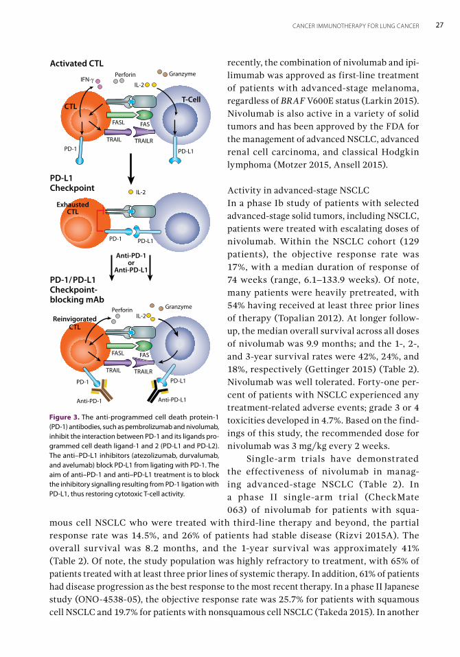
PD-1 InhibitorsThe PD-1 inhibitors include agents such as nivolumab and pembrolizumab. Nivolumab is a fully human immunoglobulin G4 (IgG4) mono-clonal antibody that disrupts PD-1–mediated signalling, thus releasing T-cells from their inhibitory interaction with PD-L1 and PD-L2 (Figure 3). Pembrolizumab is a humanized high-affinity, IgG4/kappa isotype monoclonal antibody that also blocks PD-L1 ligating with PD-1 on T-cells, resulting in the activation of tumor-specific cytotoxic T-cells. Pembrolizumab has an optimized fragment crystallizable region to minimize antibody-dependent cellular cytotoxicity (ADCC) and complement-dependent cytotoxicity (CDC) (Homet 2015). This action may be important because an intact ADCC has the potential to cause a depletion of activated T-cells and tumor-infiltrating lymphocytes and result in diminished activity, as PD-1 is expressed on T effector cells and other immune cells (Chen 2012).
NivolumabNivolumab was first noted in early-phase studies to be active in melanoma (Topalian 2012). The findings of subsequent phase III studies confirmed the superiority of nivolumab over standard of care, leading to its approval by the FDA for the treatment of melanoma. More
Anti-CTLA-4
T-CellReinvigoration
+
CTLA-4
Anti-CTLA-4
CD80/CD83 CD28
CD40LCD40
IL-12
IL-2
IFN-γ
+
CD80/CD83
CD28
CTLA-4 Checkpoint
CD40
IL-2
IFN-γ
CTLA-4
CD80/CD83 CD28
APC T-Cell
Activation
CD40LCD40
IL-2
IFN-γ
Figure 2. The anti-cytotoxic T lymphocyte-associated pro-tein-4 (CTLA-4) antibodies, such as ipilimumab and tremeli-lumab, blocking the inhibitory interaction between CTLA-4 and B7, thus resulting in T-cell reinvigoration.
27CANCER IMMUNOTHERAPY FOR LUNG CANCER
recently, the combination of nivolumab and ipi-limumab was approved as first-line treatment of patients with advanced-stage melanoma, regardless of BRAF V600E status (Larkin 2015). Nivolumab is also active in a variety of solid tumors and has been approved by the FDA for the management of advanced NSCLC, advanced renal cell carcinoma, and classical Hodgkin lymphoma (Motzer 2015, Ansell 2015). Activity in advanced-stage NSCLCIn a phase Ib study of patients with selected advanced-stage solid tumors, including NSCLC, patients were treated with escalating doses of nivolumab. Within the NSCLC cohort (129 patients), the objective response rate was 17%, with a median duration of response of 74 weeks (range, 6.1–133.9 weeks). Of note, many patients were heavily pretreated, with 54% having received at least three prior lines of therapy (Topalian 2012). At longer follow-up, the median overall survival across all doses of nivolumab was 9.9 months; and the 1-, 2-, and 3-year survival rates were 42%, 24%, and 18%, respectively (Gettinger 2015) (Table 2). Nivolumab was well tolerated. Forty-one per-cent of patients with NSCLC experienced any treatment-related adverse events; grade 3 or 4 toxicities developed in 4.7%. Based on the find-ings of this study, the recommended dose for nivolumab was 3 mg/kg every 2 weeks. Single-arm trials have demonstrated the effectiveness of nivolumab in manag-ing advanced-stage NSCLC (Table 2). In a phase II single-arm trial (CheckMate 063) of nivolumab for patients with squa-
mous cell NSCLC who were treated with third-line therapy and beyond, the partial response rate was 14.5%, and 26% of patients had stable disease (Rizvi 2015A). The overall survival was 8.2 months, and the 1-year survival was approximately 41% (Table 2). Of note, the study population was highly refractory to treatment, with 65% of patients treated with at least three prior lines of systemic therapy. In addition, 61% of patients had disease progression as the best response to the most recent therapy. In a phase II Japanese study (ONO-4538-05), the objective response rate was 25.7% for patients with squamous cell NSCLC and 19.7% for patients with nonsquamous cell NSCLC (Takeda 2015). In another
Figure 3. The anti-programmed cell death protein-1 (PD-1) antibodies, such as pembrolizumab and nivolumab, inhibit the interaction between PD-1 and its ligands pro-grammed cell death ligand-1 and 2 (PD-L1 and PD-L2). The anti–PD-L1 inhibitors (atezolizumab, durvalumab, and avelumab) block PD-L1 from ligating with PD-1. The aim of anti–PD-1 and anti–PD-L1 treatment is to block the inhibitory signalling resulting from PD-1 ligation with PD-L1, thus restoring cytotoxic T-cell activity.
PD-1/PD-L1 Checkpoint-blocking mAb
ExhaustedCTL
PD-1 PD-L1
IL-2Perforin Granzyme
FASFASL
PD-1 PD-L1TRAILRTRAIL
ReinvigoratedCTL
Anti-PD-1 Anti-PD-L1
Anti-PD-1or
Anti-PD-L1
PD-L1 Checkpoint
T-Cell
Activated CTL
IL-2IFN-γ
CTL
Perforin Granzyme
FASFASL
PD-1 PD-L1
TRAILRTRAIL
IL-2
28 IASLC ATLAS OF PD-L1 IMMUNOHISTOCHEMISTRY TESTING IN LUNG CANCER
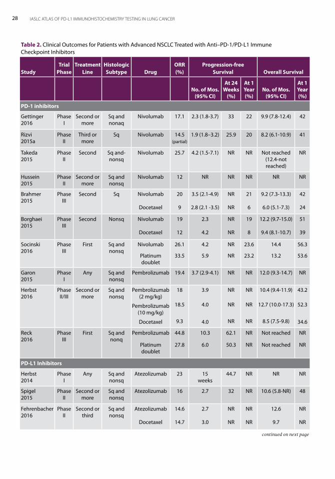
Table 2. Clinical Outcomes for Patients with Advanced NSCLC Treated with Anti–PD-1/PD-L1 Immune Checkpoint Inhibitors
StudyTrial
PhaseTreatment
LineHistologic Subtype Drug
ORR (%)
Progression-free Survival Overall Survival
No. of Mos. (95% CI)
At 24 Weeks
(%)
At 1 Year (%)
No. of Mos.(95% CI)
At 1 Year (%)
PD-1 inhibitors
Gettinger 2016
Phase I
Second or more
Sq and nonaq
Nivolumab 17.1 2.3 (1.8-3.7) 33 22 9.9 (7.8-12.4) 42
Rizvi 2015a
Phase II
Third or more
Sq Nivolumab 14.5 (partial)
1.9 (1.8–3.2) 25.9 20 8.2 (6.1-10.9) 41
Takeda 2015
Phase II
Second Sq and-nonsq
Nivolumab 25.7 4.2 (1.5-7.1) NR NR Not reached (12.4-not reached)
NR
Hussein 2015
Phase II
Second or more
Sq and nonsq
Nivolumab 12 NR NR NR NR NR
Brahmer 2015
Phase III
Second Sq Nivolumab
Docetaxel
20
9
3.5 (2.1-4.9)
2.8 (2.1 -3.5)
NR
NR
21
6
9.2 (7.3-13.3)
6.0 (5.1-7.3)
42
24
Borghaei 2015
Phase III
Second Nonsq Nivolumab
Docetaxel
19
12
2.3
4.2
NR
NR
19
8
12.2 (9.7-15.0)
9.4 (8.1-10.7)
51
39
Socinski 2016
Phase III
First Sq and nonsq
Nivolumab
Platinum doublet
26.1
33.5
4.2
5.9
NR
NR
23.6
23.2
14.4
13.2
56.3
53.6
Garon 2015
Phase I
Any Sq and nonsq
Pembrolizumab 19.4 3.7 (2.9-4.1) NR NR 12.0 (9.3-14.7) NR
Herbst 2016
Phase II/III
Second or more
Sq and nonsq
Pembrolizumab
(2 mg/kg)
Pembrolizumab
(10 mg/kg)
Docetaxel
18
18.5
9.3
3.9
4.0
4.0
NR
NR
NR
NR
NR
NR
10.4 (9.4-11.9)
12.7 (10.0-17.3)
8.5 (7.5-9.8)
43.2
52.3
34.6
Reck 2016
Phase III
First Sq and nonq
Pembrolizumab
Platinum doublet
44.8
27.8
10.3
6.0
62.1
50.3
NR
NR
Not reached
Not reached
NR
NR
PD-L1 Inhibitors
Herbst 2014
Phase I
Any Sq and nonsq
Atezolizumab 23 15 weeks
44.7 NR NR NR
Spigel 2015
Phase II
Second or more
Sq and nonsq
Atezolizumab 16 2.7 32 NR 10.6 (5.8-NR) 48
Fehrenbacher 2016
Phase II
Second or third
Sq and nonsq
Atezolizumab
Docetaxel
14.6
14.7
2.7
3.0
NR
NR
NR
NR
12.6
9.7
NR
NR
continued on next page
29CANCER IMMUNOTHERAPY FOR LUNG CANCER
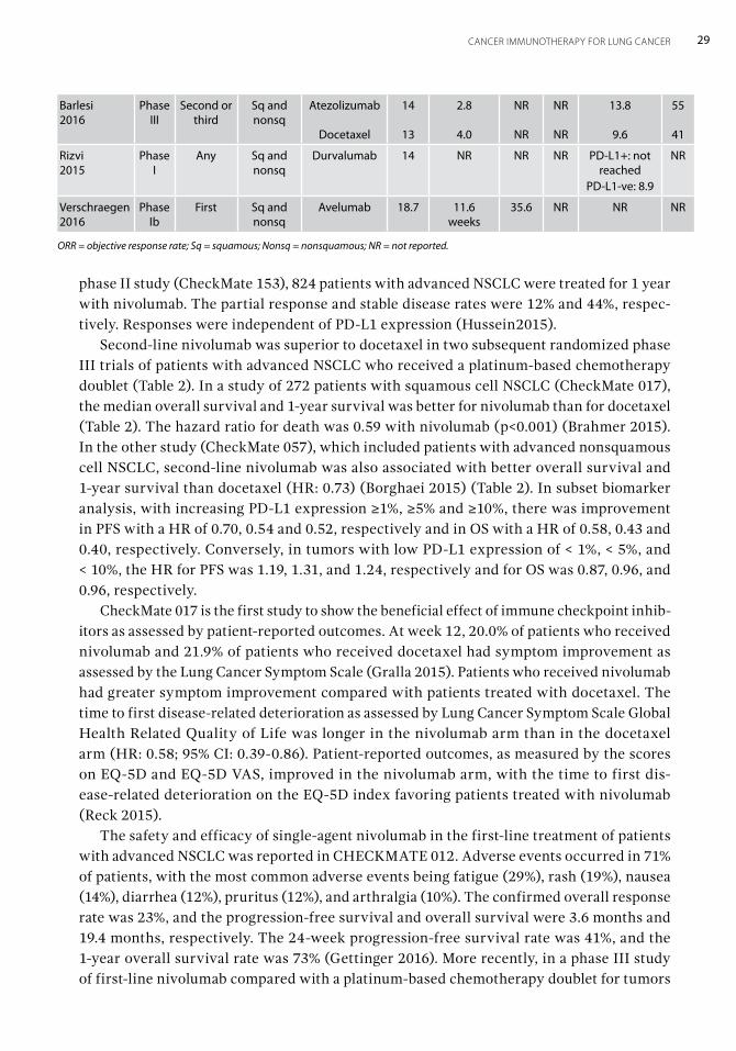
phase II study (CheckMate 153), 824 patients with advanced NSCLC were treated for 1 year with nivolumab. The partial response and stable disease rates were 12% and 44%, respec-tively. Responses were independent of PD-L1 expression (Hussein2015). Second-line nivolumab was superior to docetaxel in two subsequent randomized phase III trials of patients with advanced NSCLC who received a platinum-based chemotherapy doublet (Table 2). In a study of 272 patients with squamous cell NSCLC (CheckMate 017), the median overall survival and 1-year survival was better for nivolumab than for docetaxel (Table 2). The hazard ratio for death was 0.59 with nivolumab (p<0.001) (Brahmer 2015). In the other study (CheckMate 057), which included patients with advanced nonsquamous cell NSCLC, second-line nivolumab was also associated with better overall survival and 1-year survival than docetaxel (HR: 0.73) (Borghaei 2015) (Table 2). In subset biomarker analysis, with increasing PD-L1 expression ≥1%, ≥5% and ≥10%, there was improvement in PFS with a HR of 0.70, 0.54 and 0.52, respectively and in OS with a HR of 0.58, 0.43 and 0.40, respectively. Conversely, in tumors with low PD-L1 expression of < 1%, < 5%, and < 10%, the HR for PFS was 1.19, 1.31, and 1.24, respectively and for OS was 0.87, 0.96, and 0.96, respectively. CheckMate 017 is the first study to show the beneficial effect of immune checkpoint inhib-itors as assessed by patient-reported outcomes. At week 12, 20.0% of patients who received nivolumab and 21.9% of patients who received docetaxel had symptom improvement as assessed by the Lung Cancer Symptom Scale (Gralla 2015). Patients who received nivolumab had greater symptom improvement compared with patients treated with docetaxel. The time to first disease-related deterioration as assessed by Lung Cancer Symptom Scale Global Health Related Quality of Life was longer in the nivolumab arm than in the docetaxel arm (HR: 0.58; 95% CI: 0.39-0.86). Patient-reported outcomes, as measured by the scores on EQ-5D and EQ-5D VAS, improved in the nivolumab arm, with the time to first dis-ease-related deterioration on the EQ-5D index favoring patients treated with nivolumab (Reck 2015). The safety and efficacy of single-agent nivolumab in the first-line treatment of patients with advanced NSCLC was reported in CHECKMATE 012. Adverse events occurred in 71% of patients, with the most common adverse events being fatigue (29%), rash (19%), nausea (14%), diarrhea (12%), pruritus (12%), and arthralgia (10%). The confirmed overall response rate was 23%, and the progression-free survival and overall survival were 3.6 months and 19.4 months, respectively. The 24-week progression-free survival rate was 41%, and the 1-year overall survival rate was 73% (Gettinger 2016). More recently, in a phase III study of first-line nivolumab compared with a platinum-based chemotherapy doublet for tumors
Barlesi 2016
Phase III
Second or third
Sq and nonsq
Atezolizumab
Docetaxel
14
13
2.8
4.0
NR
NR
NR
NR
13.8
9.6
55
41
Rizvi 2015
Phase I
Any Sq and nonsq
Durvalumab 14 NR NR NR PD-L1+: not reached
PD-L1-ve: 8.9
NR
Verschraegen 2016
Phase Ib
First Sq and nonsq
Avelumab 18.7 11.6 weeks
35.6 NR NR NR
ORR = objective response rate; Sq = squamous; Nonsq = nonsquamous; NR = not reported.
30 IASLC ATLAS OF PD-L1 IMMUNOHISTOCHEMISTRY TESTING IN LUNG CANCER
with PD-L1 expression of 5% or greater (CheckMate 026), the progression-free survival was longer for the chemotherapy arm but overall survival was better for the nivolumab arm (Socinski 2016). The objective response rate was lower for the nivolumab arm (Table 2).
Activity in SCLCSCLC is most often extensive-stage disease at the time of diagnosis. Although first-line platinum-based chemotherapy doublets have activity, disease inevitably progresses, and response rates in the second-line setting are low and not durable. The activity and safety of nivolumab with or without ipilimumab in previously treated SCLC were evaluated in CheckMate 032. The objective response rate was 10% with 3 mg/kg of nivolumab alone, 23% with 1 mg/kg of nivolumab in combination with 3 mg/kg of ipilimumab, and 19% with 3 mg/kg of nivolumab in combination with 1 mg/kg of ipilimumab (Antonia 2016A). PD-L1 expression was not associated with responses.
PembrolizumabPembrolizumab is active in a variety of solid tumors including melanoma, mismatch repair-deficient colorectal cancer, NSCLC, gastric cancer, and urothelial cancer, as well as in Merkel cell and Hodgkin lymphoma (Robert 2015, Le 2015, Muro 2016, Seiwert 2016, Nghiem 2016, Armand 2016). The agent has been approved by the FDA for the treatment of metastatic melanoma, advanced-stage NSCLC, and recurrent or metastatic head and neck squamous cell carcinoma.
Activity in NSCLCThe efficacy and safety of pembrolizumab at two different doses in patients with untreated or previously treated advanced-stage NSCLC was reported in KEYNOTE-001, a large phase I study. Among all patients, the objective response rate was 19.4%, and the median duration of response was 12.5 months. The progression-free survival was 3.7 months, and overall survival was 12.0 months (Garon 2015). The objective response rate was 18% among pre-viously treated patients and 24.8% among untreated patients. For patients with a tumor proportion score of at least 50%, the objective response rate was 45.2%, and progression-free survival was 6.3 months. The objective response rate was similar regardless of dose, sched-ule, and histologic subtype; the response rate was higher among smokers than nonsmokers. Treatment-related adverse events of any grade occurred in 70.9% of patients; 9.5% had an adverse event of grade 3 or higher. Pembrolizumab was evaluated in a phase II/III study of patients with previously treated advanced NSCLC (KEYNOTE-010). A total of 1,034 patients were randomly assigned to either 2 mg/kg or 10 mg/kg of pembrolizumab or to 75 mg/m2 of docetaxel every 3 weeks (Herbst 2016). All patients had at least 1% of tumor cells that stained positively for PD-L1 protein expression on immunohistochemistry (IHC). The overall survival was improved with both doses of pembrolizumab compared with docetaxel (Table 2). Among patients with at least 50% of tumor cells expressing PD-L1, the overall survival was 14.9 and 17.3 months with pembrolizumab at doses of 2 mg/kg and 10 mg/kg, respectively, compared with 8.2 months with docetaxel. Any grade of treatment-related adverse events occurred in 63% of patients who received 2 mg/kg of pembrolizumab and in 66% of patients who
31CANCER IMMUNOTHERAPY FOR LUNG CANCER
received the 10 mg/kg dose. Treatment-related toxicity was higher (81%) in the docetaxel arm. Grade 3 to 5 treatment-related adverse events were less common among patients treated with pembrolizumab (2mg/ kg, 13%; 10 mg/kg, 16%) compared with docetaxel (35%). The safety and efficacy of first-line pembrolizumab in patients with advanced NSCLC was evaluated in KEYNOTE-001. The progression-free and overall survival were 6.2 months and 22.1 months, respectively. Increased PD-L1 expression was associated with longer survival; in patients with PD-L1 expression of 50% or greater, progression-free and overall survival were 12.5 months and not reached, respectively. In contrast, for tumors with PD-L1 expression of 1% to 49%, the progression-free and overall survival were 4.2 months and 14.7 months, respectively (Hui 2016). In the phase III study for first-line therapy of advanced NSCLC, KEYNOTE-024, patients with tumor PD-L1 expression of 50% or greater were randomly assigned to pembrolizumab or a platinum-based chemotherapy doublet, and progression-free survival was significantly better for pembrolizumab (HR: 0.50; 95% CI: 0.37–0.68; p<0.001) (Reck 2016). The hazard ratio for overall survival was 0.60 (95% CI: 0.41–0.89; p=0.005). In addition, the response rate was higher for pembrolizumab than for chemotherapy (Table 2), and fewer adverse events were associated with pembrolizumab. These results are groundbreaking because this study is the first to demonstrate the superiority of anti–PD-1 therapy over platinum-based combination chemotherapy in the first-line setting for advanced NSCLC. Patients had no sensitizing EGFR mutations or ALK translocations and had high PD-L1 expression.
Activity in SCLCPreliminary data from a phase Ib multicohort study of pembrolizumab in patients with previously treated PD-L1-positive SCLC include an objective response rate of 25% and a disease-control rate of 31% (Ott 2015).
PD-L1 InhibitorsPD-L1 inhibitors also obstruct PD-1/PD-L1 interactions but leave the PD-1/PD-L2 pathway intact (Figure 3). The PD-L1 inhibitors include atezolizumab, durvalumab, and avelumab (Table 1). Atezolizumab and durvalumab are human IgG1 anti–PD-L1 antibodies engineered with mutations in their Fc domains to remove both ADCC and CDC activity. Avelumab is a fully human IgG1 anti–PD-L1 monoclonal antibody and, unlike the other PD-1/PD-L1 inhibitors, it has appeared to have retained its ADCC and CDC activity in preclinical stud-ies (Boyerinas 2015). Several PD-L1 inhibitors have reported shown promising activity in Merkel cell carci-noma, urothelial cancer, and NSCLC (Rosenberg 2016, Kaufman 2016, Massard 2016). Phase III studies confirming the activity of these agents in various solid tumors are ongoing.
AtezolizumabAtezolizumab was reported to be active in urothelial cancer in a phase I study (Powles 2014) and was subsequently approved by the FDA for the treatment of advanced-stage urothelial carcinoma. In a single-arm phase II study (IMvigor 210 trial) the objective response rate was 16%, irrespective of immune cell PD-L1 expression, and was 28% for patients with 5% or greater PD-L1 expression (Rosenberg 2016).
32 IASLC ATLAS OF PD-L1 IMMUNOHISTOCHEMISTRY TESTING IN LUNG CANCER
Atezolizumab was reported to be active in a phase I study of advanced-stage NSCLC (Herbst 2014). In a dose-escalation and expansion study, the objective response rate was 23%, progression-free survival was 4 months, and overall survival was 16 months for patients who received 20 mg/kg intravenously every 3 weeks (Horn 2015). In a randomized phase II study (POPLAR) of patients who had received platinum-based chemotherapy, atezoli-zumab was associated with superior overall survival (HR: 0.73; 95% CI: 0•53–0•99; p=0.04) (Fehrenbacher 2016) (Table 2). In another phase II study (BIRCH), patients with advanced NSCLC who were selected for PD-L1 expression received atezolizumab as first-line or sub-sequent therapy. The objective response rates ranged from 17% to 27% (Besse 2016), and the median overall survival was 14 months for patients who received atezolizumab as first-line therapy. Overall survival has not yet been reached for patients who received atezolizumab as subsequent therapy (Broderick 2016). The overall response rates ranged from 16% to 26% in a phase II study of a population with advanced NSCLC enriched for PD-L1 expres-sion in tumor and immune cells (Spigel 2015). In the OAK trial, a phase III study of patients with advanced, previously treated NSCLC who were randomly assigned to atezolizumab or docetaxel, the overall survival was significantly better for atezolizumab (13.8 months vs. 9.6 months; HR: 0.73; 95% CI: 0.62-0.87; p=0.0003) (Rittmeyer 2017). The OAK study led to FDA approval of atezolizumab for second-line therapy of advanced NSCLC.
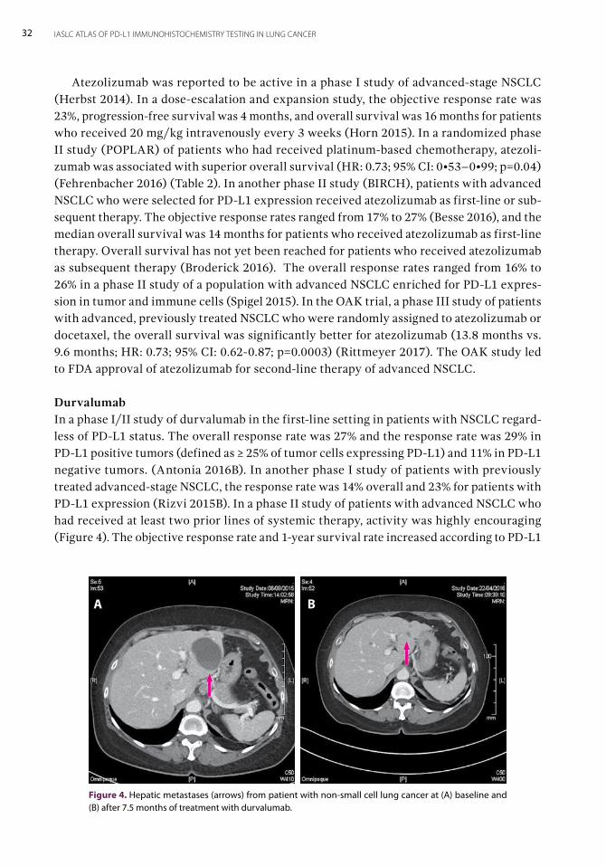
DurvalumabIn a phase I/II study of durvalumab in the first-line setting in patients with NSCLC regard-less of PD-L1 status. The overall response rate was 27% and the response rate was 29% in PD-L1 positive tumors (defined as ≥ 25% of tumor cells expressing PD-L1) and 11% in PD-L1 negative tumors. (Antonia 2016B). In another phase I study of patients with previously treated advanced-stage NSCLC, the response rate was 14% overall and 23% for patients with PD-L1 expression (Rizvi 2015B). In a phase II study of patients with advanced NSCLC who had received at least two prior lines of systemic therapy, activity was highly encouraging (Figure 4). The objective response rate and 1-year survival rate increased according to PD-L1
Figure 4. Hepatic metastases (arrows) from patient with non-small cell lung cancer at (A) baseline and (B) after 7.5 months of treatment with durvalumab.
A B
33CANCER IMMUNOTHERAPY FOR LUNG CANCER
expression: 7.5% (less than 25% PD-L1 expression), 16.4% [(more than 25% expression)], and 30.9% (more than 90% expression); the corresponding 1-year survival rates were 34.5%, 47.7%, and 50.8% (Garassino 2016).
AvelumabThe findings of early studies of avelumab in NSCLC have been promising, with an over-all response rate of 12% for patients who had disease progression after platinum-based chemotherapy. There was a trend toward greater activity in patients with PD-L1-positive tumors (Gulley 2015). Among patients treated with avelumab in the first-line setting, the objective response rate and disease-control rates were 18.7% and 64.0%, respectively (Verschraegen 2016).
Combined CTLA-4 and PD-1/PD-L1 InhibitorsCTLA-4 inhibitors are also being studied in combination with PD-1 and PD-L1 inhibitors. The results of preclinical studies indicate that the combination may work synergistically to produce enhanced antitumor activity (Curran 2010). The combination of ipilimumab with nivolumab in advanced melanoma resulted in improved antitumor activity compared with single-agent therapy; however, toxicities were increased with the combination therapy (Larkin 2015). Nivolumab has been combined with ipilimumab for advanced NSCLC in the first-line setting in a phase I study (CheckMate 12). Results have included objective response rates ranging from 13% to 39% (Hellman 2016). A randomized phase III trial (CheckMate 227) is currently ongoing to compare nivolumab plus ipilimumab with nivolumab alone, nivolumab plus platinum-based chemotherapy, and platinum-based chemotherapy alone in PD-L1-defined untreated NSCLC. Additionally, durvalumab has been combined with the CTLA-4 inhibitor tremelimumab in a phase Ib trial of patients with advanced NSCLC. Although numerous adverse events occurred during the dose-escalation phase of the study, antitumor activity was evident (objective response rate of 23%), regardless of PD-L1 status in the evaluable patients in the dose-expansion phase of the study (Antonia 2016A).
Detection of PD-L1 Expression Benefit is found in only a subset of patients with NSCLC treated with PD-1/PD-L1 inhibi-tors. As the PD-1/PD-L1 pathway is involved in immune escape in NSCLC, tumor PD-L1 expression with IHC has been used to identify patients who may benefit from PD-1/PD-L1 inhibitors. Studies of pembrolizumab have used PD-L1 IHC 22C3 PharmDx (Dako) for detec-tion of tumor PD-L1 expression (Garon 2015, Herbst 2016). This test has been approved as a companion diagnostic test by the FDA. Studies of nivolumab have used the anti-PD-L1 antibody [28-8] (Abcam) (Borghaei 2016, Brahmer 2015), which has been approved as a complementary diagnostic test. (The differences between companion and complementary diagnostic tests are discussed in Chapter 10). The antibody clones SP142 and SP263 have been used to develop tumor PD-L1 expression in studies of atezolizumab (Fehrenbacher 2016) and durvalumab (Rebelatto 2016), respectively. The use of PD-L1 as a biomarker
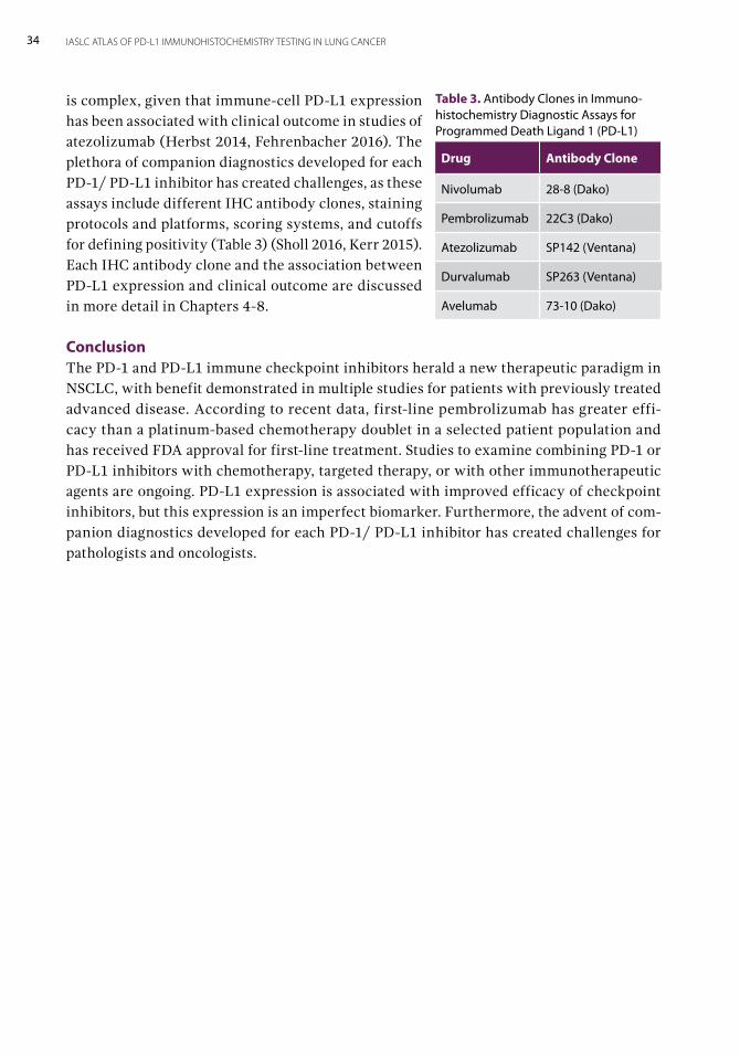
34 IASLC ATLAS OF PD-L1 IMMUNOHISTOCHEMISTRY TESTING IN LUNG CANCER
is complex, given that immune-cell PD-L1 expression has been associated with clinical outcome in studies of atezolizumab (Herbst 2014, Fehrenbacher 2016). The plethora of companion diagnostics developed for each PD-1/ PD-L1 inhibitor has created challenges, as these assays include different IHC antibody clones, staining protocols and platforms, scoring systems, and cutoffs for defining positivity (Table 3) (Sholl 2016, Kerr 2015). Each IHC antibody clone and the association between PD-L1 expression and clinical outcome are discussed in more detail in Chapters 4-8.
ConclusionThe PD-1 and PD-L1 immune checkpoint inhibitors herald a new therapeutic paradigm in NSCLC, with benefit demonstrated in multiple studies for patients with previously treated advanced disease. According to recent data, first-line pembrolizumab has greater effi-cacy than a platinum-based chemotherapy doublet in a selected patient population and has received FDA approval for first-line treatment. Studies to examine combining PD-1 or PD-L1 inhibitors with chemotherapy, targeted therapy, or with other immunotherapeutic agents are ongoing. PD-L1 expression is associated with improved efficacy of checkpoint inhibitors, but this expression is an imperfect biomarker. Furthermore, the advent of com-panion diagnostics developed for each PD-1/ PD-L1 inhibitor has created challenges for pathologists and oncologists.
Table 3. Antibody Clones in Immuno-histochemistry Diagnostic Assays for Programmed Death Ligand 1 (PD-L1)
Drug Antibody Clone
Nivolumab 28-8 (Dako)
Pembrolizumab 22C3 (Dako)
Atezolizumab SP142 (Ventana)
Durvalumab SP263 (Ventana)
Avelumab 73-10 (Dako)
Immunohistochemistry for PD-L1By Erik Thunnissen, Yasushi Yatabe, Sylvie Lantuéjoul, and Lukas Bubendorf 3Immunohistochemistry (IHC) is a technique that allows visualization of proteins in histologic sections, and a similar approach on cells in cytologic specimens is called immu-nocytochemistry. With IHC, the variable domain of the primary antibody recognizes and binds to the three-dimensional structure of a protein, an epitope, present in the section. A second antibody that binds to the primary antibody and subsequent chemical reactions are used to visualize the localization of the epitope, a process known as signal enhancement. The location of the IHC staining is detected in the tissue context with use of a microscope. IHC staining may be located on or in one or more subcellular areas, such as on the cell membrane, in the cytoplasm, or in the nucleus. IHC is a rapid and relatively inexpensive method that is preferred by most pathologists primarily because it allows for the evaluation of tissue architecture and tumor cells. Potentially, IHC can detect rare positive cells more easily than fluorescence in situ hybrid-ization, even at a low magnification, because of the high contrast of IHC-positive tumor cells in an IHC-negative background. IHC can be performed successfully on a variety of tumor specimens—formalin -fixed paraffin-embedded (FFPE) tissue blocks, fluid, and fine-needle aspiration cytology cell blocks or smears--as long as at least a few clusters of viable tumor cells are present in the specimen. In addition, a validated and robust programmed cell death ligand-1 (PD-L1) IHC assay provides a cost-effective platform. Several methodologic aspects may influence the outcome of IHC, which are discussed here, in general and specifically for PD-L1.
Preanalytic PhaseGeneralFFPE tissue is the global standard material for IHC. The cold ischemia time—the time between sampling of the tissue and the start of fixation—should be kept to a minimum to avoid cold ischemia effects. Regardless of origin, diagnostic biopsy or surgical resection specimens should immediately be fixed in an adequate amount (ratio of at least 10 times the volume of the tissue specimen) of 10% neutral buffered formalin (ie, 4% formaldehyde),
36 IASLC ATLAS OF PD-L1 IMMUNOHISTOCHEMISTRY TESTING IN LUNG CANCER
processed, and embedded in paraffin (FFPE tissue). Fixation times of fewer than 6 hours are not recommended because conventional H&E staining, as well as IHC, can be adversely affected. For practical purposes, a fixation time of 6 to 48 hours is recommended for all specimens (Thunnissen 2012, Kerr 2014). During gross handling of resection tissue, samples should be cut to slices 3 or 4 mm thick. The cassettes containing the tissue samples should also be placed in buffered formalin, then dehydrated and cleared in a series of alcohols and xylene, followed by infiltration with melted paraffin. The paraffin temperature should not exceed 60°C. The FFPE samples are assumed to be stable and preserved against oxidation. FFPE tissue specimens are cut to create sec-tions of 3 to 4 μm, which are mounted and dried onto glass slides, ready for staining. After sectioning, the tissues are mounted on special coated slides suitable for IHC. If not used within a few days, the tissue sections should be stored in the dark, in a closed box at 2°C to 8°C to preserve antigenicity, and stained within 3 months of sectioning to avoid possible false-negative results. These measures aim to prevent photo-oxidation and drying, which lead to loss of antigenicity (Blind 2008). An alternative procedure involves dipping glass slides with mounted sections in paraffin wax to avoid oxidative damage. When such slides are subsequently used for IHC, care must be taken to use enough solvent to dissolve all the excess paraffin wax. It is possible for inadequate volumes of solvent to become saturated, leading to incomplete dewaxing. Incomplete wax removal hampers epitope retrieval and IHC staining. In addition, sections may detach from the glass slides during dewaxing. Decalcification can destroy antigenicity, especially when highly acidic agents, such as hydrochloric acid and nitric acid, are used. Weaker acids, such as EDTA, an effective decal-cifying agent, have become more popular because they do not seem to interfere with IHC for breast cancer testing (Schrijver 2016). For other organs and proteins, some generalizable effects are known. Regarding breast and colorectal cancers, a rapid change in phosphoepitope-specific protein staining was found to be related to delay in fixation (Bonnas 2012, Meric-Bernstam 2014, Theiss 2014, Vassilakopoulou 2015). Frequently, staining signals—for example, for expression of phos-phorylated Akt (pAkt)—were substantially reduced or lost in resection specimens compared with small biopsy specimens (Vassilakopoulou 2015). Thus, for phosphoproteins, which are usually less stable than other proteins, a delay in fixation of 30 minutes to 1 hour can negatively affect measurement outcome. In the cases of HER2, estrogen and/or progesterone receptors, and Ki67 IHC in breast cancer, cold ischemia of up to 3 to 4 hours was shown to have no deleterious effect (Neumeister 2012, Portier 2013, Li 2013).
PD-L1There are several recommendations regarding preanalytic conditions for preparation and storage of tissues for future PD-L1 staining (Table 1). For PD-L1 there is, to date, no infor-mation about possible effect of fixation delay. Proper tissue collection and processing may reduce the failure rate. Currently, an ischemia time from excision to formalin fixation start time of fewer than 30 minutes followed by immersion in neutral buffered formalin for 6 to 48 hours is recommended. Biopsy specimens should be immersed immediately in buff-ered formalin. According to the interpretation manuals for the 22C3 and 28-8 pharmDx kits (Agilent Technologies/Dako), the only critical step for PD-L1 IHC is a fixation of at
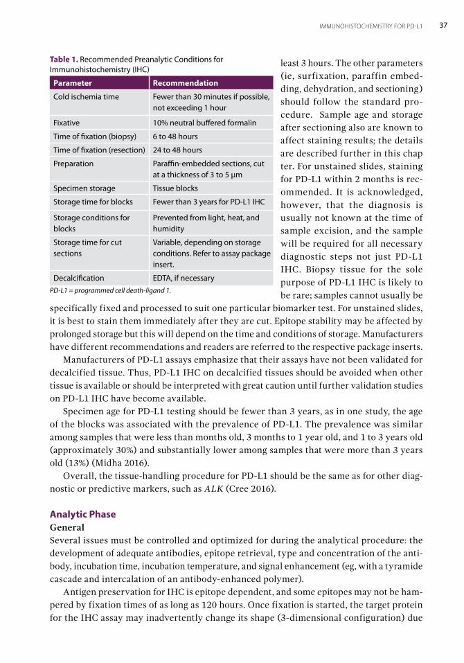
37IMMUNOHISTOCHEMISTRY FOR PD-L1
least 3 hours. The other parameters (ie, surfixation, paraffin embed-ding, dehydration, and sectioning) should follow the standard pro-cedure. Sample age and storage after sectioning also are known to affect staining results; the details are described further in this chap ter. For unstained slides, staining for PD-L1 within 2 months is rec-ommended. It is acknowledged, however, that the diagnosis is usually not known at the time of sample excision, and the sample will be required for all necessary diagnostic steps not just PD-L1 IHC. Biopsy tissue for the sole purpose of PD-L1 IHC is likely to be rare; samples cannot usually be
specifically fixed and processed to suit one particular biomarker test. For unstained slides, it is best to stain them immediately after they are cut. Epitope stability may be affected by prolonged storage but this will depend on the time and conditions of storage. Manufacturers have different recommendations and readers are referred to the respective package inserts. Manufacturers of PD-L1 assays emphasize that their assays have not been validated for decalcified tissue. Thus, PD-L1 IHC on decalcified tissues should be avoided when other tissue is available or should be interpreted with great caution until further validation studies on PD-L1 IHC have become available. Specimen age for PD-L1 testing should be fewer than 3 years, as in one study, the age of the blocks was associated with the prevalence of PD-L1. The prevalence was similar among samples that were less than months old, 3 months to 1 year old, and 1 to 3 years old (approximately 30%) and substantially lower among samples that were more than 3 years old (13%) (Midha 2016). Overall, the tissue-handling procedure for PD-L1 should be the same as for other diag-nostic or predictive markers, such as ALK (Cree 2016).
Analytic PhaseGeneralSeveral issues must be controlled and optimized for during the analytical procedure: the development of adequate antibodies, epitope retrieval, type and concentration of the anti-body, incubation time, incubation temperature, and signal enhancement (eg, with a tyramide cascade and intercalation of an antibody-enhanced polymer). Antigen preservation for IHC is epitope dependent, and some epitopes may not be ham-pered by fixation times of as long as 120 hours. Once fixation is started, the target protein for the IHC assay may inadvertently change its shape (3-dimensional configuration) due
Table 1. Recommended Preanalytic Conditions for Immunohistochemistry (IHC)
Parameter Recommendation
Cold ischemia time Fewer than 30 minutes if possible, not exceeding 1 hour
Fixative 10% neutral buffered formalin
Time of fixation (biopsy) 6 to 48 hours
Time of fixation (resection) 24 to 48 hours
Preparation Paraffin-embedded sections, cut at a thickness of 3 to 5 μm
Specimen storage Tissue blocks
Storage time for blocks Fewer than 3 years for PD-L1 IHC
Storage conditions for blocks
Prevented from light, heat, and humidity
Storage time for cut sections
Variable, depending on storage conditions. Refer to assay package insert.
Decalcification EDTA, if necessaryPD-L1 = programmed cell death-ligand 1.
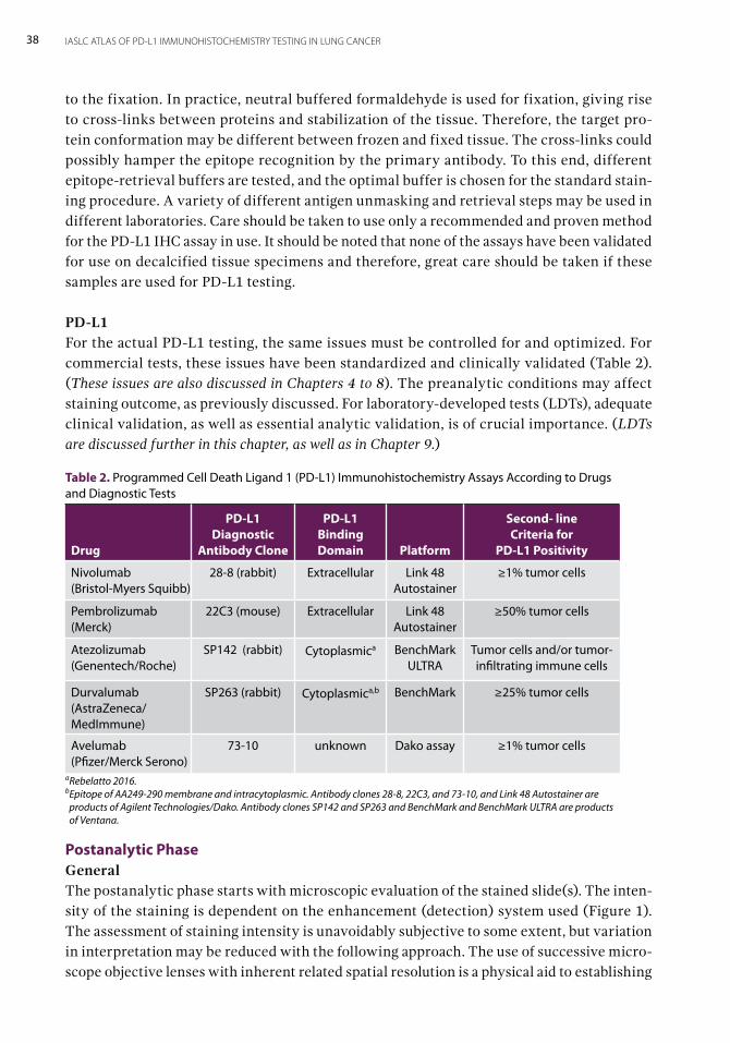
38 IASLC ATLAS OF PD-L1 IMMUNOHISTOCHEMISTRY TESTING IN LUNG CANCER
to the fixation. In practice, neutral buffered formaldehyde is used for fixation, giving rise to cross-links between proteins and stabilization of the tissue. Therefore, the target pro-tein conformation may be different between frozen and fixed tissue. The cross-links could possibly hamper the epitope recognition by the primary antibody. To this end, different epitope-retrieval buffers are tested, and the optimal buffer is chosen for the standard stain-ing procedure. A variety of different antigen unmasking and retrieval steps may be used in different laboratories. Care should be taken to use only a recommended and proven method for the PD-L1 IHC assay in use. It should be noted that none of the assays have been validated for use on decalcified tissue specimens and therefore, great care should be taken if these samples are used for PD-L1 testing.
PD-L1For the actual PD-L1 testing, the same issues must be controlled for and optimized. For commercial tests, these issues have been standardized and clinically validated (Table 2). (These issues are also discussed in Chapters 4 to 8). The preanalytic conditions may affect staining outcome, as previously discussed. For laboratory-developed tests (LDTs), adequate clinical validation, as well as essential analytic validation, is of crucial importance. (LDTs are discussed further in this chapter, as well as in Chapter 9.)
Postanalytic PhaseGeneralThe postanalytic phase starts with microscopic evaluation of the stained slide(s). The inten-sity of the staining is dependent on the enhancement (detection) system used (Figure 1). The assessment of staining intensity is unavoidably subjective to some extent, but variation in interpretation may be reduced with the following approach. The use of successive micro-scope objective lenses with inherent related spatial resolution is a physical aid to establishing
Table 2. Programmed Cell Death Ligand 1 (PD-L1) Immunohistochemistry Assays According to Drugs and Diagnostic Tests
Drug
PD-L1 Diagnostic
Antibody Clone
PD-L1 Binding Domain Platform
Second- line Criteria for
PD-L1 Positivity
Nivolumab (Bristol-Myers Squibb)
28-8 (rabbit) Extracellular Link 48 Autostainer
≥1% tumor cells
Pembrolizumab (Merck)
22C3 (mouse) Extracellular Link 48 Autostainer
≥50% tumor cells
Atezolizumab (Genentech/Roche)
SP142 (rabbit) Cytoplasmica BenchMark ULTRA
Tumor cells and/or tumor-infiltrating immune cells
Durvalumab (AstraZeneca/ MedImmune)
SP263 (rabbit) Cytoplasmica,b BenchMark ≥25% tumor cells
Avelumab (Pfizer/Merck Serono)
73-10 unknown Dako assay ≥1% tumor cells
aRebelatto 2016. bEpitope of AA249-290 membrane and intracytoplasmic. Antibody clones 28-8, 22C3, and 73-10, and Link 48 Autostainer are products of Agilent Technologies/Dako. Antibody clones SP142 and SP263 and BenchMark and BenchMark ULTRA are products of Ventana.
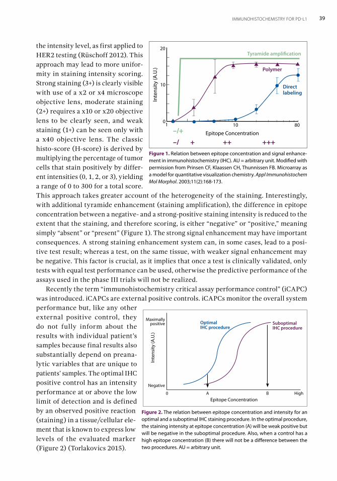
39IMMUNOHISTOCHEMISTRY FOR PD-L1
the intensity level, as first applied to HER2 testing (Rüschoff 2012). This approach may lead to more unifor-mity in staining intensity scoring. Strong staining (3+) is clearly visible with use of a x2 or x4 microscope objective lens, moderate staining (2+) requires a x10 or x20 objective lens to be clearly seen, and weak staining (1+) can be seen only with a x40 objective lens. The classic histo-score (H-score) is derived by multiplying the percentage of tumor cells that stain positively by differ-ent intensities (0, 1, 2, or 3), yielding a range of 0 to 300 for a total score. This approach takes greater account of the heterogeneity of the staining. Interestingly, with additional tyramide enhancement (staining amplification), the difference in epitope concentration between a negative- and a strong-positive staining intensity is reduced to the extent that the staining, and therefore scoring, is either “negative” or “positive,” meaning simply “absent” or “present” (Figure 1). The strong signal enhancement may have important consequences. A strong staining enhancement system can, in some cases, lead to a posi-tive test result; whereas a test, on the same tissue, with weaker signal enhancement may be negative. This factor is crucial, as it implies that once a test is clinically validated, only tests with equal test performance can be used, otherwise the predictive performance of the assays used in the phase III trials will not be realized. Recently the term “immunohistochemistry critical assay performance control” (iCAPC) was introduced. iCAPCs are external positive controls. iCAPCs monitor the overall system performance but, like any other external positive control, they do not fully inform about the results with individual patient’s samples because final results also substantially depend on preana-lytic variables that are unique to patients’ samples. The optimal IHC positive control has an intensity performance at or above the low limit of detection and is defined by an observed positive reaction (staining) in a tissue/cellular ele-ment that is known to express low levels of the evaluated marker (Figure 2) (Torlakovics 2015).
801010
20
10
Inte
nsity
(A.U
.)
Epitope Concentration
Tyramide amplification
Polymer
Direct labeling
–/+
–/ + ++ +++Figure 1. Relation between epitope concentration and signal enhance-ment in immunohistochemistry (IHC). AU = arbitrary unit. Modified with permission from Prinsen CF, Klaassen CH, Thunnissen FB. Microarray as a model for quantitative visualization chemistry. Appl Immunohistochem Mol Morphol. 2003;11(2):168-173.
0 A B HighEpitope Concentration
Negative
Maximally positive
Inte
nsity
(A.U
.)
Optimal IHC procedure
Suboptimal IHC procedure
Figure 2. The relation between epitope concentration and intensity for an optimal and a suboptimal IHC staining procedure. In the optimal procedure, the staining intensity at epitope concentration (A) will be weak positive but will be negative in the suboptimal procedure. Also, when a control has a high epitope concentration (B) there will not be a difference between the two procedures. AU = arbitrary unit.
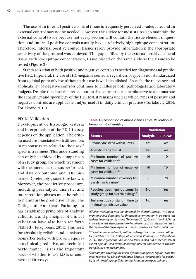
40 IASLC ATLAS OF PD-L1 IMMUNOHISTOCHEMISTRY TESTING IN LUNG CANCER
The use of an internal positive control tissue is frequently perceived as adequate, and an external control may not be needed. However, the advice for most stains is to maintain the external control tissue because not every section will contain the tissue element in ques-tion, and internal positive controls usually have a relatively high epitope concentration. Therefore, internal positive control tissues rarely provide information if the appropriate sensitivity of the protocol was achieved. This gap is filled by the external positive control tissue with low epitope concentration, tissue placed on the same slide as the tissue to be tested (Figure 2). Standardization of both positive and negative controls is needed for diagnostic and predic-tive IHC. In general, the use of IHC-negative controls, regardless of type, is not standardized from a global point of view, although this use is well established. As such, the relevance and applicability of negative controls continues to challenge both pathologists and laboratory budgets. Despite the clear theoretical notion that appropriate controls serve to demonstrate the sensitivity and specificity of the IHC test, it remains unclear which types of positive and negative controls are applicable and/or useful in daily clinical practice (Torlakovic 2014, Torlakovic 2015).
PD-L1 ValidationDevelopment of histologic criteria and interpretation of the PD-L1 assay depends on the application. The crite-ria used are associated with differences in response rates related to the use of specific treatment. This understanding can only be achieved by comparison of a study group, for which treatment with the intended drug was performed, and data on outcome and IHC bio-marker (preferably graded) are known. Moreover, the predictive procedure, including preanalytic, analytic, and interpretation phases must be robust to maintain the predictive value. The College of American Pathologists has established principles of analytic validation, and principles of clinical validation have also been set forth (Table 3) (Fitzgibbons 2014). This need for absolutely reliable and consistent biomarker tests, with proven, equiva-lent clinical, predictive, and technical performance, raises the important issue of whether to use LDTs or com-mercial kit assays.
Table 3. Comparison of Analytic and Clinical Validation in Immunohistochemistry
Validation
Factors Analytic Clinical a
Preanalytic steps within limits Yes Yes
Analytic steps robust Yes Yes
Minimum number of positive cases for validationb
10 10
Minimum number of negative cases for validationb
10 10
Minimum number covering lin-ear dynamic rangec
– 20
Requires treatment outcome in study group for a certain druga 3
No Yes
Test must be constant in time to maintain predictive value
No Yes
aClinical validation may be obtained by clinical samples with treat-ment response data used for threshold determination in a certain test with its linear dynamic range (Rebelatto 2016). Once a threshold is set in a certain test, demonstration of equivalence of an alternative test in the region of the linear dynamic range is needed for clinical validation. bThe minimum number of positive and negative cases are according to guidelines of the College of American Pathologists (Fitzgibbons 2014). These guidelines are not evidence-based but rather represent expert opinion, and every laboratory director can decide to validate using fewer or more samples.cThe samples covering the linear dynamic range (see Figure 1) are the most relevant for clinical validation because the threshold for positiv-ity is within this group. This number is based on expert opinion.
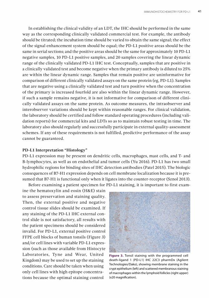
41IMMUNOHISTOCHEMISTRY FOR PD-L1
In establishing the clinical validity of an LDT, the IHC should be performed in the same way as the corresponding clinically validated commercial test. For example, the antibody should be titrated; the incubation time should be varied to obtain the same signal; the effect of the signal enhancement system should be equal; the PD-L1 positive areas should be the same in serial sections; and the positive areas should be the same for approximately 10 PD-L1 negative samples, 10 PD-L1 positive samples, and 20 samples covering the linear dynamic range of the clinically validated PD-L1 IHC test. Conceptually, samples that are positive in a clinically validated test and become negative when the primary antibody is diluted to 25% are within the linear dynamic range. Samples that remain positive are uninformative for comparison of different clinically validated assays on the same protein (eg, PD-L1). Samples that are negative using a clinically validated test and turn positive when the concentration of the primary is increased fourfold are also within the linear dynamic range. However, if such a sample remains negative, it is not informative for comparison of different clini-cally validated assays on the same protein. As outcome measures, the intraobserver and interobserver variations should be kept within reasonable ranges. For clinical validation, the laboratory should be certified and follow standard operating procedures (including vali-dation reports) for commercial kits and LDTs so as to maintain robust testing in time. The laboratory also should regularly and successfully participate in external quality-assessment schemes. If any of these requirements is not fulfilled, predictive performance of the assay cannot be guaranteed.
PD-L1 Interpretation “Histology” PD-L1 expression may be present on dendritic cells, macrophages, mast cells, and T- and B-lymphocytes, as well as on endothelial and tumor cells (Yu 2016). PD-L1 has two small hydrophilic regions for binding sites of IHC detection antibodies (Patel 2015). The biologic consequences of B7-H1 expression depends on cell membrane localization because it is pre-sumed that B7-H1 is functional only when it ligates into the counter-receptor (Sznol 2013). Before examining a patient specimen for PD-L1 staining, it is important to first exam-ine the hematoxylin and eosin (H&E) stain to assess preservation and staining quality. Then, the external positive and negative control tissue slides should be examined. If any staining of the PD-L1 IHC external con-trol slide is not satisfactory, all results with the patient specimens should be considered invalid. For PD-L1, external positive control FFPE cell blocks of human tonsils (Figure 3) and/or cell lines with variable PD-L1 expres-sion (such as those available from Histocyte Laboratories, Tyne and Wear, United Kingdom) may be used to set up the staining conditions. Care should be taken when using only cell lines with high epitope concentra-tions because the optimal staining control
Figure 3. Tonsil staining with the programmed cell death-ligand 1 (PD-L1) IHC 22C3 pharmDx (Agilent Technologies/Dako), showing membrane staining in the crypt epithelium (left) and scattered membranous staining of macrophages within the lymphoid follicles (right upper) (x20 magnification).
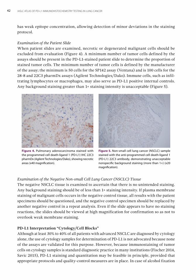
42 IASLC ATLAS OF PD-L1 IMMUNOHISTOCHEMISTRY TESTING IN LUNG CANCER
has weak epitope concentration, allowing detection of minor deviations in the staining protocol.
Examination of the Patient Slide When patient slides are examined, necrotic or degenerated malignant cells should be excluded from evaluation (Figure 4). A minimum number of tumor cells defined by the assays should be present in the PD-L1-stained patient slide to determine the proportion of stained tumor cells. The minimum number of tumor cells is defined by the manufacturer of the assay; the minimum is 50 cells for the SP142 assay (Ventana) and is 100 cells for the 28-8 and 22C3 pharmDx assays (Agilent Technologies/Dako). Immune cells, such as infil-trating lymphocytes or macrophages, may also serve as PD-L1 positive internal controls. Any background staining greater than 1+ staining intensity is unacceptable (Figure 5).
Examination of the Negative Non-small Cell Lung Cancer (NSCLC) Tissue The negative NSCLC tissue is examined to ascertain that there is no unintended staining. Any background staining should be of less than 1+ staining intensity. If plasma membrane staining of malignant cells occurs in the negative control tissue, all results with the patient specimens should be questioned, and the negative control specimen should be replaced by another negative control in a repeat analysis. Even if the slide appears to have no staining reactions, the slides should be viewed at high magnification for confirmation so as not to overlook weak membrane staining.
PD-L1 Interpretation “Cytology/Cell Blocks”Although at least 30% to 40% of all patients with advanced NSCLC are diagnosed by cytology alone, the use of cytology samples for determination of PD-L1 is not advocated because none of the assays are validated for this purpose. However, because immunostaining of tumor cells on cytology samples is standard diagnostic practice in many institutions (Fischer 2014, Savic 2015), PD-L1 staining and quantitation may be feasible in principle, provided that appropriate protocols and quality-control measures are in place. In case of alcohol fixation
Figure 4. Pulmonary adenocarcinoma stained with the programmed cell death-ligand 1 (PD-L1) IHC 22C3 pharmDx (Agilent Technologies/Dako), showing necrotic areas (x40 magnification).
Figure 5. Non-small cell lung cancer (NSCLC) sample stained with the anti-programmed cell death-ligand 1 (PD-L1) 22C3 antibody, demonstrating unacceptable nonspecific background staining (more than 1+) (x20 magnification).
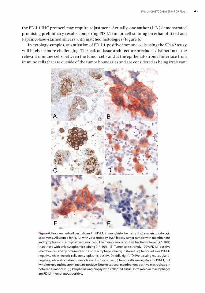
43IMMUNOHISTOCHEMISTRY FOR PD-L1
Figure 6. Programmed cell death-ligand 1 (PD-L1) immunohistochemistry (IHC) analysis of cytologic specimens. All stained for PD-L1 with 28-8 antibody. (A) A biopsy tumor sample with membranous and cytoplasmic PD-L1-positive tumor cells. The membranous positive fraction is lower (+/- 10%) than those with only cytoplasmic staining (+/- 60%). (B) Tumor cells strongly 100% PD-L1-positive (membranous and cytoplasmic) with also macrophage staining in stroma. (C) Tumor cells are PD-L1-negative, while necrotic cells are cytoplasmic-positive (middle right). (D) Pre-existing mucus gland-negative, while stromal immune cells are PD-L1-positive. (E) Tumor cells are negative for PD-L1, but lymphocytes and macrophages are positive. Note occasional membranous positive macrophage in between tumor cells. (F) Peripheral lung biopsy with collapsed tissue. Intra-avleolar macrophages are PD-L1-membranous positive.
the PD-L1 IHC protocol may require adjustment. Actually, one author (L.B.) demonstrated promising preliminary results comparing PD-L1 tumor cell staining on ethanol-fixed and Papanicolaou-stained smears with matched histologies (Figure 6). In cytology samples, quantitation of PD-L1-positive immune cells using the SP142 assay will likely be more challenging. The lack of tissue architecture precludes distinction of the relevant immune cells between the tumor cells and at the epithelial-stromal interface from immune cells that are outside of the tumor boundaries and are considered as being irrelevant
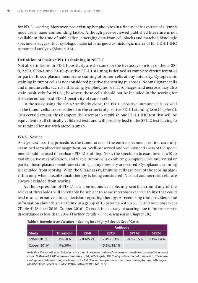
44 IASLC ATLAS OF PD-L1 IMMUNOHISTOCHEMISTRY TESTING IN LUNG CANCER
for PD-L1 scoring. Moreover, pre-existing lymphocytes in a fine-needle aspirate of a lymph node are a major confounding factor. Although peer-reviewed published literature is not available at the time of publication, emerging data from cell blocks and matched histologic specimens suggest that cytologic material is as good as histologic material for PD-L1 IHC tumor cell analysis (Skov 2016).
Definition of Positive PD-L1 Staining in NSCLCNot all definitions for PD-L1 positivity are the same for the five assays. In four of them–28-8, 22C3, SP263, and 73-10–positive PD-L1 staining is defined as complete circumferential or partial linear plasma membrane staining of tumor cells at any intensity. Cytoplasmic staining in tumor cells is not considered positive for scoring purposes. Nonmalignant cells and immune cells, such as infiltrating lymphocytes or macrophages, and necrosis may also stain positively for PD-L1; however, these cells should not be included in the scoring for the determination of PD-L1 positivity of tumor cells. In the assay using the SP142 antibody clone, the PD-L1-positive immune cells, as well as the tumor cells, are considered in the criteria of positive PD-L1 staining (See Chapter 6). To a certain extent, this hampers the attempt to establish one PD-L1 IHC test that will be equivalent to all clinically validated tests and will possibly lead to the SP142 test having to be retained for use with atezolizumab.
PD-L1 Scoring As a general scoring procedure, the tumor areas of the entire specimen are first carefully examined at x4 objective magnification. Well-preserved and well-stained areas of the speci-men should be used to evaluate PD-L1 staining. Next, the specimen is examined at x10 to x40 objective magnification, and viable tumor cells exhibiting complete circumferential or partial linear plasma membrane staining at any intensity are scored. Cytoplasmic staining is excluded from scoring. With the SP142 assay, immune cells are part of the scoring algo-rithm only when atezolizumab therapy is being considered. Normal and necrotic cells are always excluded from scoring. As the expression of PD-L1 is a continuous variable, any scoring around any of the relevant thresholds will inevitably be subject to some interobserver variability that could lead to an alternative clinical decision regarding therapy. A recent ring trial provides some information about this variability in a group of 15 patients with NSCLC and nine observers (Table 4) (Scheel 2016; Cooper 2016). Overall, inaccuracy of scoring due to interobserver discordance is less than 10%. (Further details will be discussed in Chapter 10.)
Table 4. Interobserver Variation in Scoring for a Highly Selected Set of CasesAntibody
Antibody
Study Threshold 28-8 22C3 SP142 SP263
Scheel 20161 1%/50% 2.8%/5.2% 7.4%/8.3% 9.6%/8.5% 6.3%/7.4%
Cooper 20162 1%/50% 15.8%/18.1%
Note that the variation in clinical practice is not known yet and needs to be determined on a consecutive series of cases. 2) Mean of 2,700 pairwise comparisons, 10 pathologists, 108 (highly selected set of samples). 1) These per-centages are obtained using a selection of 15 NSCLC resection specimens after some training by nine pathologists. Modified from Scheel et al. Mod Pathol. 2016;29(10):1165-1172.
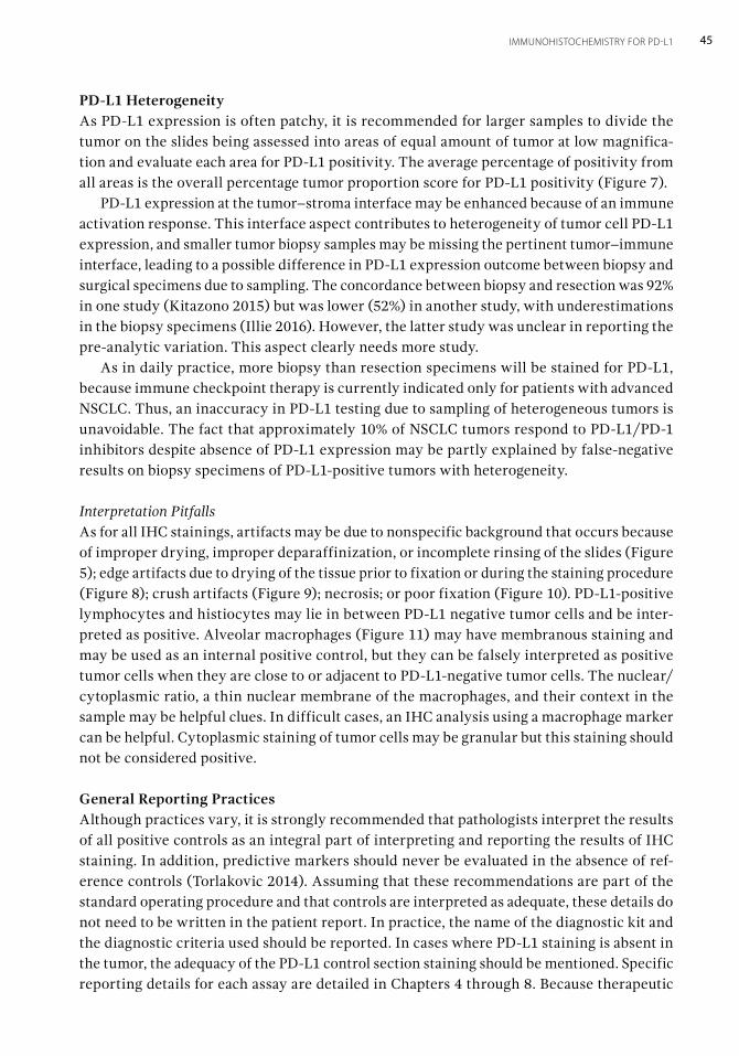
45IMMUNOHISTOCHEMISTRY FOR PD-L1
PD-L1 Heterogeneity As PD-L1 expression is often patchy, it is recommended for larger samples to divide the tumor on the slides being assessed into areas of equal amount of tumor at low magnifica-tion and evaluate each area for PD-L1 positivity. The average percentage of positivity from all areas is the overall percentage tumor proportion score for PD-L1 positivity (Figure 7). PD-L1 expression at the tumor–stroma interface may be enhanced because of an immune activation response. This interface aspect contributes to heterogeneity of tumor cell PD-L1 expression, and smaller tumor biopsy samples may be missing the pertinent tumor–immune interface, leading to a possible difference in PD-L1 expression outcome between biopsy and surgical specimens due to sampling. The concordance between biopsy and resection was 92% in one study (Kitazono 2015) but was lower (52%) in another study, with underestimations in the biopsy specimens (Illie 2016). However, the latter study was unclear in reporting the pre-analytic variation. This aspect clearly needs more study. As in daily practice, more biopsy than resection specimens will be stained for PD-L1, because immune checkpoint therapy is currently indicated only for patients with advanced NSCLC. Thus, an inaccuracy in PD-L1 testing due to sampling of heterogeneous tumors is unavoidable. The fact that approximately 10% of NSCLC tumors respond to PD-L1/PD-1 inhibitors despite absence of PD-L1 expression may be partly explained by false-negative results on biopsy specimens of PD-L1-positive tumors with heterogeneity.
Interpretation Pitfalls As for all IHC stainings, artifacts may be due to nonspecific background that occurs because of improper drying, improper deparaffinization, or incomplete rinsing of the slides (Figure 5); edge artifacts due to drying of the tissue prior to fixation or during the staining procedure (Figure 8); crush artifacts (Figure 9); necrosis; or poor fixation (Figure 10). PD-L1-positive lymphocytes and histiocytes may lie in between PD-L1 negative tumor cells and be inter-preted as positive. Alveolar macrophages (Figure 11) may have membranous staining and may be used as an internal positive control, but they can be falsely interpreted as positive tumor cells when they are close to or adjacent to PD-L1-negative tumor cells. The nuclear/cytoplasmic ratio, a thin nuclear membrane of the macrophages, and their context in the sample may be helpful clues. In difficult cases, an IHC analysis using a macrophage marker can be helpful. Cytoplasmic staining of tumor cells may be granular but this staining should not be considered positive.
General Reporting Practices Although practices vary, it is strongly recommended that pathologists interpret the results of all positive controls as an integral part of interpreting and reporting the results of IHC staining. In addition, predictive markers should never be evaluated in the absence of ref-erence controls (Torlakovic 2014). Assuming that these recommendations are part of the standard operating procedure and that controls are interpreted as adequate, these details do not need to be written in the patient report. In practice, the name of the diagnostic kit and the diagnostic criteria used should be reported. In cases where PD-L1 staining is absent in the tumor, the adequacy of the PD-L1 control section staining should be mentioned. Specific reporting details for each assay are detailed in Chapters 4 through 8. Because therapeutic
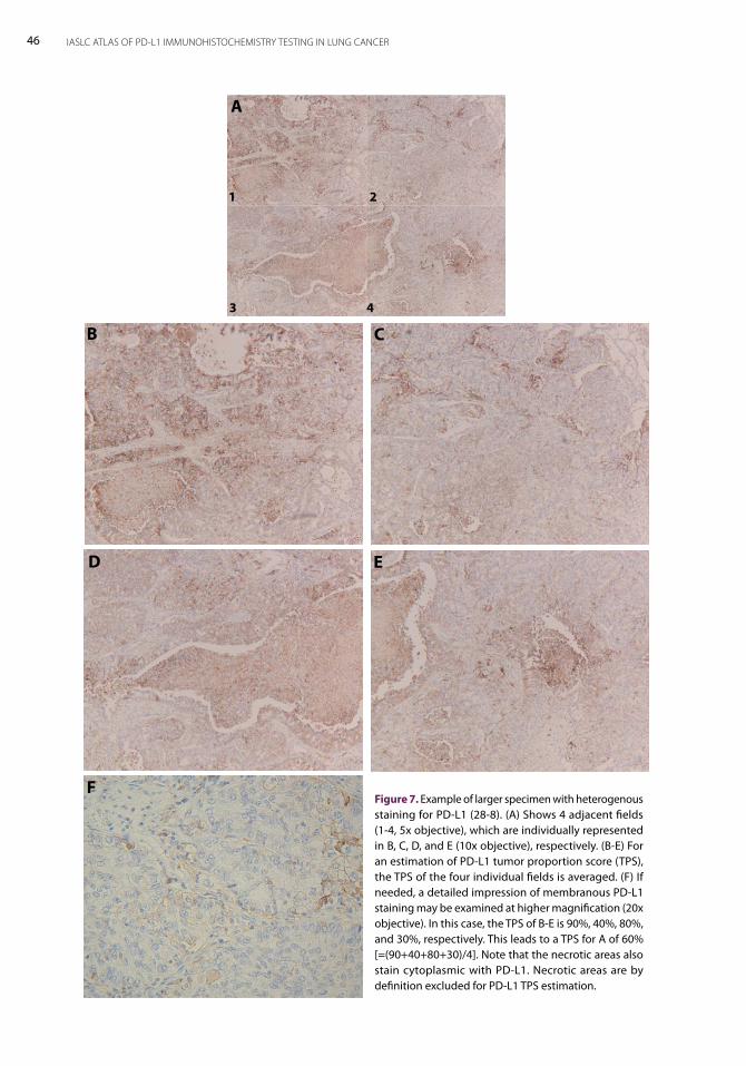
46 IASLC ATLAS OF PD-L1 IMMUNOHISTOCHEMISTRY TESTING IN LUNG CANCER
Figure 7. Example of larger specimen with heterogenous staining for PD-L1 (28-8). (A) Shows 4 adjacent fields (1-4, 5x objective), which are individually represented in B, C, D, and E (10x objective), respectively. (B-E) For an estimation of PD-L1 tumor proportion score (TPS), the TPS of the four individual fields is averaged. (F) If needed, a detailed impression of membranous PD-L1 staining may be examined at higher magnification (20x objective). In this case, the TPS of B-E is 90%, 40%, 80%, and 30%, respectively. This leads to a TPS for A of 60% [=(90+40+80+30)/4]. Note that the necrotic areas also stain cytoplasmic with PD-L1. Necrotic areas are by definition excluded for PD-L1 TPS estimation.
A
B C
D E
F
1 2
3 4
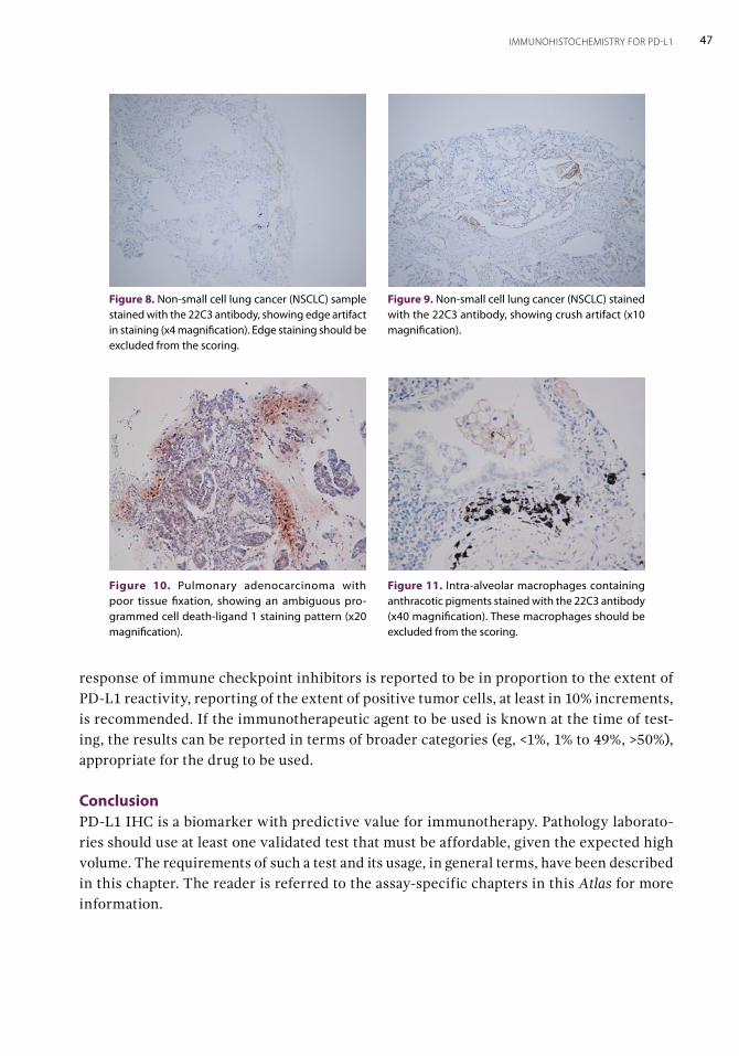
47IMMUNOHISTOCHEMISTRY FOR PD-L1
response of immune checkpoint inhibitors is reported to be in proportion to the extent of PD-L1 reactivity, reporting of the extent of positive tumor cells, at least in 10% increments, is recommended. If the immunotherapeutic agent to be used is known at the time of test-ing, the results can be reported in terms of broader categories (eg, <1%, 1% to 49%, >50%), appropriate for the drug to be used.
ConclusionPD-L1 IHC is a biomarker with predictive value for immunotherapy. Pathology laborato-ries should use at least one validated test that must be affordable, given the expected high volume. The requirements of such a test and its usage, in general terms, have been described in this chapter. The reader is referred to the assay-specific chapters in this Atlas for more information.
Figure 8. Non-small cell lung cancer (NSCLC) sample stained with the 22C3 antibody, showing edge artifact in staining (x4 magnification). Edge staining should be excluded from the scoring.
Figure 9. Non-small cell lung cancer (NSCLC) stained with the 22C3 antibody, showing crush artifact (x10 magnification).
Figure 10. Pulmonary adenocarcinoma with poor tissue fixation, showing an ambiguous pro-grammed cell death-ligand 1 staining pattern (x20 magnification).
Figure 11. Intra-alveolar macrophages containing anthracotic pigments stained with the 22C3 antibody (x40 magnification). These macrophages should be excluded from the scoring.

PD-L1 28-8 pharmDx AssayBy Sylvie Lantuéjoul and Erik Thunnissen 4The PD-L1 IHC 28-8 pharmDx (Agilent Technologies/Dako) is a laboratory test that measures programmed cell death ligand-1 (PD-L1) expression in formalin-fixed, paraffin-embedded (FFPE) tissue samples on the Autostainer Link 48 platform (Agilent Technologies/Dako) (Phillips 2015). This assay is considered a complementary diagnostic tool for the treatment of patients with advanced non-small cell lung cancer (NSCLC) with nivolumab (see Chapter 10 for details). However, PD-L1 testing is not required as a selection biomarker to treat patients with either squamous or non-squamous lung cancer with nivolumab (Brahmer 2015, Borghaei 2015). Nivolumab is a human immunoglobulin G4 (IgG4) monoclonal antibody that binds to the programmed cell death protein-1 (PD-1) receptor and blocks its interaction with PD-L1 and PD-L2, restoring the antitumor immune response (Wang 2014). It has been approved by the US Food and Drug Administration (FDA) and the European Commission to treat patients with advanced (metastatic) NSCLC whose disease progressed during or after platinum-based chemotherapy (second-line therapy), because of enhanced survival.
Antibody Characteristics and Immunostaining Conditions Clone 28-8 (ab205921; Abcam) is an IgG4 isotype rabbit monoclonal anti–(human) PD-L1 antibody. Its immunogen is a recombinant full-length protein that corresponds to the extra-cellular domain (Phe19-Thr239) of human PD-L1 (Phillips 2015). Clone 28-8 detects PD-L1 protein on FFPE specimens. Human tonsil is a recommended positive tissue control, with highest expression of PD-L1 in the crypt epithelium, macrophages homing the germinal centers, and interfollicular mononuclear leukocytes. Cell lines, such as B-CPAP (a human papillary thyroid cancer cell line with high expression), ES-2 (an ovarian clear cell carcinoma cell line with intermediate expression), and HCC70 (a ductal carcinoma cell line with low expression) can also be used as external positive controls. This PD-L1 primary antibody showed no cross-reactivity for PD-L2 exogenously expressed in Chinese hamster ovary (CHO) cells (an epithelial cell line). The PD-L1 IHC 28-8 pharmDx is an FDA-approved and European Conformity (CE) In Vitro Device (IVD)-marked, qualitative, IHC assay that contains ready-to-use optimized
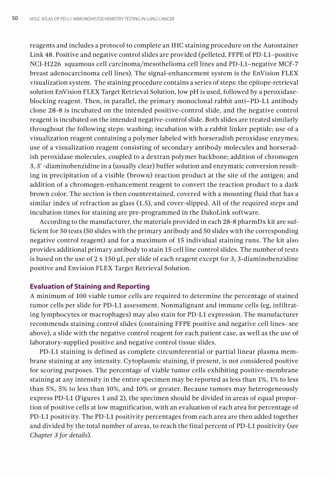
50 IASLC ATLAS OF PD-L1 IMMUNOHISTOCHEMISTRY TESTING IN LUNG CANCER
reagents and includes a protocol to complete an IHC staining procedure on the Autostainer Link 48. Positive and negative control slides are provided (pelleted, FFPE of PD-L1–positive NCI-H226 squamous cell carcinoma/mesothelioma cell lines and PD-L1–negative MCF-7 breast adenocarcinoma cell lines). The signal-enhancement system is the EnVision FLEX visualization system. The staining procedure contains a series of steps: the epitope-retrieval solution EnVision FLEX Target Retrieval Solution, low pH is used, followed by a peroxidase-blocking reagent. Then, in parallel, the primary monoclonal rabbit anti–PD-L1 antibody clone 28-8 is incubated on the intended positive-control slide, and the negative control reagent is incubated on the intended negative-control slide. Both slides are treated similarly throughout the following steps: washing; incubation with a rabbit linker peptide; use of a visualization reagent containing a polymer labeled with horseradish peroxidase enzymes; use of a visualization reagent consisting of secondary antibody molecules and horserad-ish peroxidase molecules, coupled to a dextran polymer backbone; addition of chromogen 3, 3’ -diaminobenzidine in a (usually clear) buffer solution and enzymatic conversion result-ing in precipitation of a visible (brown) reaction product at the site of the antigen; and addition of a chromogen-enhancement reagent to convert the reaction product to a dark brown color. The section is then counterstained, covered with a mounting fluid that has a similar index of refraction as glass (1.5), and cover-slipped. All of the required steps and incubation times for staining are pre-programmed in the DakoLink software. According to the manufacturer, the materials provided in each 28-8 pharmDx kit are suf-ficient for 50 tests (50 slides with the primary antibody and 50 slides with the corresponding negative control reagent) and for a maximum of 15 individual staining runs. The kit also provides additional primary antibody to stain 15 cell line control slides. The number of tests is based on the use of 2 x 150 μL per slide of each reagent except for 3, 3-diaminobenzidine positive and Envision FLEX Target Retrieval Solution.
Evaluation of Staining and ReportingA minimum of 100 viable tumor cells are required to determine the percentage of stained tumor cells per slide for PD-L1 assessment. Nonmalignant and immune cells (eg, infiltrat-ing lymphocytes or macrophages) may also stain for PD-L1 expression. The manufacturer recommends staining control slides (containing FFPE positive and negative cell lines- see above), a slide with the negative control reagent for each patient case, as well as the use of laboratory-supplied positive and negative control tissue slides. PD-L1 staining is defined as complete circumferential or partial linear plasma mem-brane staining at any intensity. Cytoplasmic staining, if present, is not considered positive for scoring purposes. The percentage of viable tumor cells exhibiting positive-membrane staining at any intensity in the entire specimen may be reported as less than 1%, 1% to less than 5%, 5% to less than 10%, and 10% or greater. Because tumors may heterogeneously express PD-L1 (Figures 1 and 2), the specimen should be divided in areas of equal propor-tion of positive cells at low magnification, with an evaluation of each area for percentage of PD-L1 positivity. The PD-L1 positivity percentages from each area are then added together and divided by the total number of areas, to reach the final percent of PD-L1 positivity (see Chapter 3 for details).
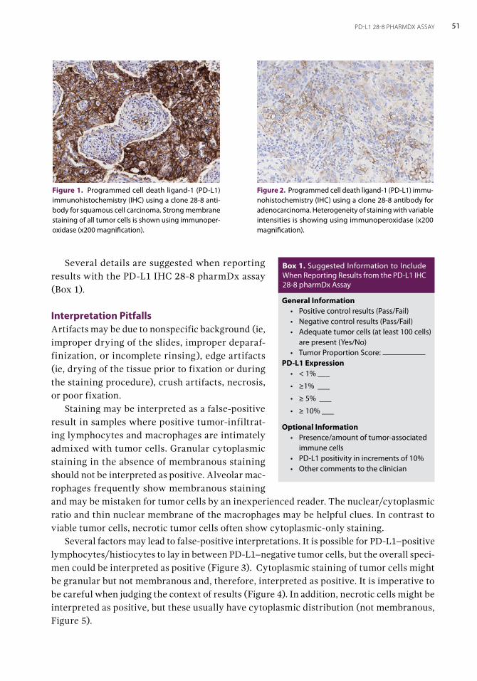
51PD-L1 28-8 PHARMDX ASSAY
Several details are suggested when reporting results with the PD-L1 IHC 28-8 pharmDx assay (Box 1).
Interpretation PitfallsArtifacts may be due to nonspecific background (ie, improper drying of the slides, improper deparaf-finization, or incomplete rinsing), edge artifacts (ie, drying of the tissue prior to fixation or during the staining procedure), crush artifacts, necrosis, or poor fixation. Staining may be interpreted as a false-positive result in samples where positive tumor-infiltrat-ing lymphocytes and macrophages are intimately admixed with tumor cells. Granular cytoplasmic staining in the absence of membranous staining should not be interpreted as positive. Alveolar mac-rophages frequently show membranous staining and may be mistaken for tumor cells by an inexperienced reader. The nuclear/cytoplasmic ratio and thin nuclear membrane of the macrophages may be helpful clues. In contrast to viable tumor cells, necrotic tumor cells often show cytoplasmic-only staining. Several factors may lead to false-positive interpretations. It is possible for PD-L1–positive lymphocytes/histiocytes to lay in between PD-L1–negative tumor cells, but the overall speci-men could be interpreted as positive (Figure 3). Cytoplasmic staining of tumor cells might be granular but not membranous and, therefore, interpreted as positive. It is imperative to be careful when judging the context of results (Figure 4). In addition, necrotic cells might be interpreted as positive, but these usually have cytoplasmic distribution (not membranous, Figure 5).
Figure 1. Programmed cell death ligand-1 (PD-L1) immunohistochemistry (IHC) using a clone 28-8 anti-body for squamous cell carcinoma. Strong membrane staining of all tumor cells is shown using immunoper-oxidase (x200 magnification).
Figure 2. Programmed cell death ligand-1 (PD-L1) immu-nohistochemistry (IHC) using a clone 28-8 antibody for adenocarcinoma. Heterogeneity of staining with variable intensities is showing using immunoperoxidase (x200 magnification).
General Information• Positive control results (Pass/Fail)• Negative control results (Pass/Fail)• Adequate tumor cells (at least 100 cells)
are present (Yes/No)• Tumor Proportion Score:
PD-L1 Expression • < 1% ___
• ≥1% ___
• ≥ 5% ___
• ≥ 10% ___
Optional Information • Presence/amount of tumor-associated
immune cells• PD-L1 positivity in increments of 10%• Other comments to the clinician
Box 1. Suggested Information to Include When Reporting Results from the PD-L1 IHC 28-8 pharmDx Assay
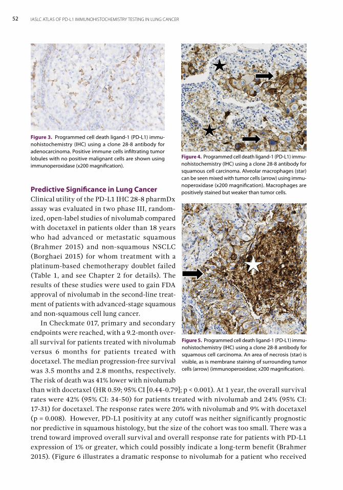
52 IASLC ATLAS OF PD-L1 IMMUNOHISTOCHEMISTRY TESTING IN LUNG CANCER
Predictive Significance in Lung CancerClinical utility of the PD-L1 IHC 28-8 pharmDx assay was evaluated in two phase III, random-ized, open-label studies of nivolumab compared with docetaxel in patients older than 18 years who had advanced or metastatic squamous (Brahmer 2015) and non-squamous NSCLC (Borghaei 2015) for whom treatment with a platinum-based chemotherapy doublet failed (Table 1, and see Chapter 2 for details). The results of these studies were used to gain FDA approval of nivolumab in the second-line treat-ment of patients with advanced-stage squamous and non-squamous cell lung cancer. In Checkmate 017, primary and secondary endpoints were reached, with a 9.2-month over-all survival for patients treated with nivolumab versus 6 months for patients treated with docetaxel. The median progression-free survival was 3.5 months and 2.8 months, respectively. The risk of death was 41% lower with nivolumab than with docetaxel (HR 0.59; 95% CI [0.44-0.79]; p < 0.001). At 1 year, the overall survival rates were 42% (95% CI: 34-50) for patients treated with nivolumab and 24% (95% CI: 17-31) for docetaxel. The response rates were 20% with nivolumab and 9% with docetaxel (p = 0.008). However, PD-L1 positivity at any cutoff was neither significantly prognostic nor predictive in squamous histology, but the size of the cohort was too small. There was a trend toward improved overall survival and overall response rate for patients with PD-L1 expression of 1% or greater, which could possibly indicate a long-term benefit (Brahmer 2015). (Figure 6 illustrates a dramatic response to nivolumab for a patient who received
Figure 3. Programmed cell death ligand-1 (PD-L1) immu-nohistochemistry (IHC) using a clone 28-8 antibody for adenocarcinoma. Positive immune cells infiltrating tumor lobules with no positive malignant cells are shown using immunoperoxidase (x200 magnification).
Figure 4. Programmed cell death ligand-1 (PD-L1) immu-nohistochemistry (IHC) using a clone 28-8 antibody for squamous cell carcinoma. Alveolar macrophages (star) can be seen mixed with tumor cells (arrow) using immu-noperoxidase (x200 magnification). Macrophages are positively stained but weaker than tumor cells.
Figure 5. Programmed cell death ligand-1 (PD-L1) immu-nohistochemistry (IHC) using a clone 28-8 antibody for squamous cell carcinoma. An area of necrosis (star) is visible, as is membrane staining of surrounding tumor cells (arrow) (immunoperoxidase; x200 magnification).
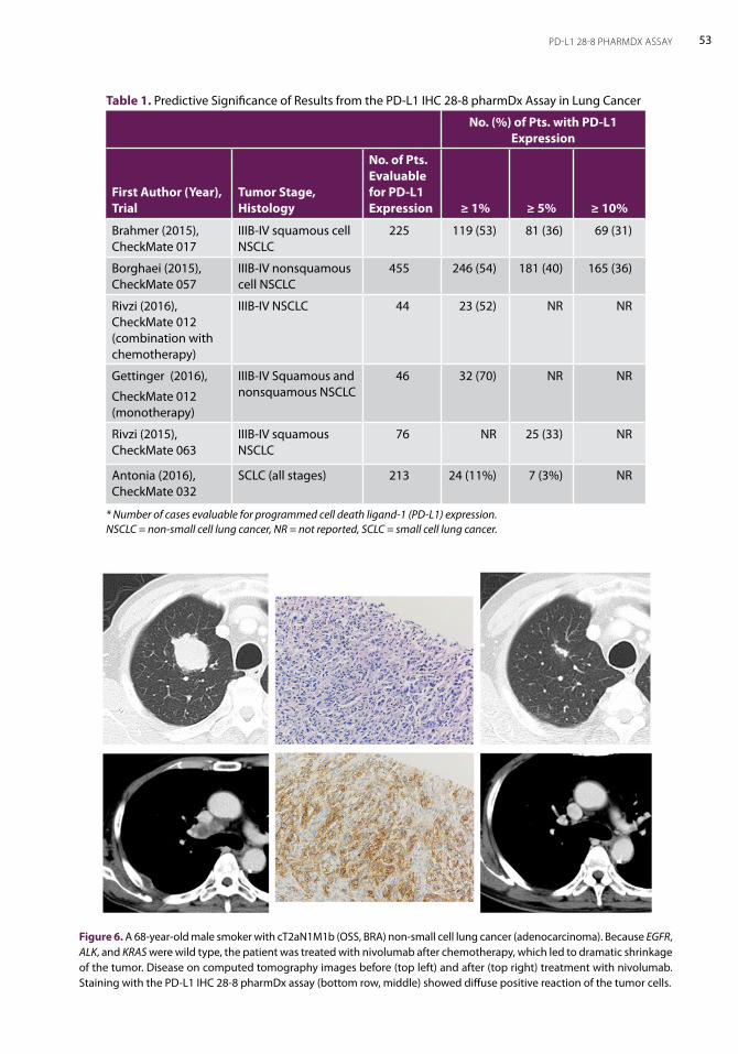
53PD-L1 28-8 PHARMDX ASSAY
Figure 6. A 68-year-old male smoker with cT2aN1M1b (OSS, BRA) non-small cell lung cancer (adenocarcinoma). Because EGFR, ALK, and KRAS were wild type, the patient was treated with nivolumab after chemotherapy, which led to dramatic shrinkage of the tumor. Disease on computed tomography images before (top left) and after (top right) treatment with nivolumab. Staining with the PD-L1 IHC 28-8 pharmDx assay (bottom row, middle) showed diffuse positive reaction of the tumor cells.
Table 1. Predictive Significance of Results from the PD-L1 IHC 28-8 pharmDx Assay in Lung Cancer
No. (%) of Pts. with PD-L1 Expression
First Author (Year), Trial
Tumor Stage, Histology
No. of Pts. Evaluable for PD-L1 Expression ≥ 1% ≥ 5% ≥ 10%
Brahmer (2015), CheckMate 017
IIIB-IV squamous cell NSCLC
225 119 (53) 81 (36) 69 (31)
Borghaei (2015), CheckMate 057
IIIB-IV nonsquamous cell NSCLC
455 246 (54) 181 (40) 165 (36)
Rivzi (2016), CheckMate 012 (combination with chemotherapy)
IIIB-IV NSCLC 44 23 (52) NR NR
Gettinger (2016),
CheckMate 012 (monotherapy)
IIIB-IV Squamous and nonsquamous NSCLC
46 32 (70) NR NR
Rivzi (2015), CheckMate 063
IIIB-IV squamous NSCLC
76 NR 25 (33) NR
Antonia (2016), CheckMate 032
SCLC (all stages) 213 24 (11%) 7 (3%) NR
* Number of cases evaluable for programmed cell death ligand-1 (PD-L1) expression. NSCLC = non-small cell lung cancer, NR = not reported, SCLC = small cell lung cancer.
54 IASLC ATLAS OF PD-L1 IMMUNOHISTOCHEMISTRY TESTING IN LUNG CANCER
prior chemotherapy and was subsequently treated with nivolumab.) Eventually, because a significant proportion of patients with PD-L1–negative tumors benefitted from treatment with nivolumab, the FDA did not require PD-L1 testing before treatment. The FDA has approved the PD-L1 IHC 28-8 pharmDx assay, however, to detect PD-L1 expression levels and to help physicians determine which patients may benefit most from treatment with nivolumab. In Checkmate 057, the median overall survival was 12.2 months (95% CI: 9.7-15.0) for patients treated with nivolumab and 9.4 months (95% CI: 8.1-10.7) for docetaxel. At 1 year, the overall survival rate was 51% (95% CI: 45-56) with nivolumab versus 39% (95% CI: 33-45) with docetaxel. The response rate was 19% with nivolumab versus 12% with docetaxel (p = 0.02). Regarding PD-L1 expression, nivolumab was associated with greater efficacy than docetaxel in subgroups, which were defined according to prespecified levels of tumor-membrane expression of PD-L1 (1% or greater, 5% or greater, and 10% or greater). The median overall survival for patients in these subgroups was 17.1, 18.2, and 19.4 months, respectively, with nivolumab compared with 9.0, 8.1, and 8.0 months, respectively, with docetaxel (Borghaei 2015). Nivolumab was investigated as monotherapy for first-line management of advanced NSCLC in the phase I multicohort CheckMate 012 trial. Overall response rates were 28% for patients with any degree of tumor PD-L1 expression and 14% for patients with PD-L1–negative tumors. The median progression-free survival was 3.6 months, the median overall survival was 19.4 months, and the 1-year and 18-month overall survival rates, respectively, were 73% (95% CI, 59-83) and 57% (95% CI, 42-70) (Rizvi 2016, Gettinger 2016). Recently, the CheckMate 026 trial, a phase III, open-label, randomized study of nivolumab as monotherapy versus the investigators’ choice of chemotherapy for patients with advanced NSCLC did not meet its primary endpoint of progression-free survival in patients with previously untreated advanced NSCLC whose tumors expressed PD-L1 at 5% or greater (Socinski 2016, see Chapter 2 for details).
ConclusionThe PD-L1 IHC 28-8 pharmDx assay is a complementary diagnostic tool for the management of non-squamous NSCLC with PD-L1 expression of 1% or greater using nivolumab. This test has been validated for FFPE tissue samples and is used on the Autostainer Link 48 platform. PD-L1 IHC testing is not required to treat patients with squamous NSCLC with nivolumab.
5PD-L1 22C3 pharmDx AssayBy Teh-Ying Chou, Wendy A. Cooper, and Keith M. Kerr
Introduction of the Platform PD-L1 IHC 22C3 pharmDx assay (Dako, Glostrup, Denmark) is an in vitro diagnostic (IVD) immunohistochemistry (IHC) assay for detection of PD-L1 protein expression in non-small cell lung cancer (NSCLC) tissue (Dako 2016). This assay is performed on the Dako Autostainer Link 48 platform with an automated staining protocol using a mouse monoclonal anti-PD-L1 antibody, clone 22C3. The assay is indicated as an aid in identifying advanced-stage NSCLC patients who would be eligible for treatment with pembrolizumab (KEYTRUDA®, Merck Sharp & Dohme Corp.), a humanized monoclonal IgG4 kappa isotype antibody against PD-1. In October 2015, the PD-L1 IHC 22C3 pharmDx assay was approved by the US Food and Drug Administration (FDA) as a companion diagnostic test for treat-ment with pembrolizumab in patients with advanced (metastatic) NSCLC (FDA, 2016). This assay assesses PD-L1 protein expression by evaluating “tumor proportion score” (TPS), which is the percentage of viable tumor cells showing either partial or complete membrane staining (Dako 2016). Increased PD-L1 expression (higher TPS) is generally associated with higher objective response rate (ORR) and favorable outcome in patients treated with pembrolizumab (Baas 2016).
Antibody Characteristics and Immunostaining ConditionsPD-L1 IHC 22C3 pharmDx uses a mouse monoclonal antibody clone 22C3 that recognizes the extracellular domain of PD-L1 to assess the PD-L1 expression in formalin-fixed, paraffin-embedded (FFPE) tissue with use of IHC. The IHC staining procedure is performed on Dako Autostainer Link 48 platform with a validated staining protocol. Briefly, slides are baked at 60°C for 30 minutes and then subjected to deparaffinization, rehydration, and target retrieval on the Dako PT Link Pre-Treatment Module (Dako No. PT100) using the Dako EnVision FLEX Target Retrieval Solution, Low pH. The following staining procedure is performed on Dako Automated Link 48 platform. First, the slides are incubated with anti-PD-L1 22C3 antibody or a negative control reagent (mouse IgG isotype) for 20 minutes. Subsequently, Dako EnVision FLEX+ Polymer Reagents, including a mouse linker, horseradish peroxidase
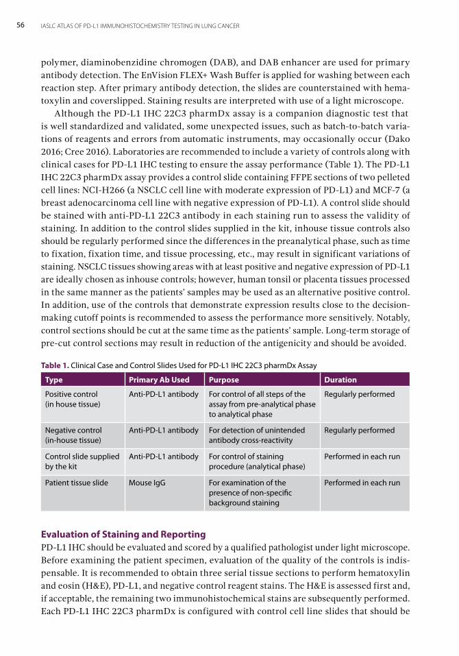
56 IASLC ATLAS OF PD-L1 IMMUNOHISTOCHEMISTRY TESTING IN LUNG CANCER
polymer, diaminobenzidine chromogen (DAB), and DAB enhancer are used for primary antibody detection. The EnVision FLEX+ Wash Buffer is applied for washing between each reaction step. After primary antibody detection, the slides are counterstained with hema-toxylin and coverslipped. Staining results are interpreted with use of a light microscope. Although the PD-L1 IHC 22C3 pharmDx assay is a companion diagnostic test that is well standardized and validated, some unexpected issues, such as batch-to-batch varia-tions of reagents and errors from automatic instruments, may occasionally occur (Dako 2016; Cree 2016). Laboratories are recommended to include a variety of controls along with clinical cases for PD-L1 IHC testing to ensure the assay performance (Table 1). The PD-L1 IHC 22C3 pharmDx assay provides a control slide containing FFPE sections of two pelleted cell lines: NCI-H266 (a NSCLC cell line with moderate expression of PD-L1) and MCF-7 (a breast adenocarcinoma cell line with negative expression of PD-L1). A control slide should be stained with anti-PD-L1 22C3 antibody in each staining run to assess the validity of staining. In addition to the control slides supplied in the kit, inhouse tissue controls also should be regularly performed since the differences in the preanalytical phase, such as time to fixation, fixation time, and tissue processing, etc., may result in significant variations of staining. NSCLC tissues showing areas with at least positive and negative expression of PD-L1 are ideally chosen as inhouse controls; however, human tonsil or placenta tissues processed in the same manner as the patients’ samples may be used as an alternative positive control. In addition, use of the controls that demonstrate expression results close to the decision-making cutoff points is recommended to assess the performance more sensitively. Notably, control sections should be cut at the same time as the patients’ sample. Long-term storage of pre-cut control sections may result in reduction of the antigenicity and should be avoided.
Evaluation of Staining and ReportingPD-L1 IHC should be evaluated and scored by a qualified pathologist under light microscope. Before examining the patient specimen, evaluation of the quality of the controls is indis-pensable. It is recommended to obtain three serial tissue sections to perform hematoxylin and eosin (H&E), PD-L1, and negative control reagent stains. The H&E is assessed first and, if acceptable, the remaining two immunohistochemical stains are subsequently performed. Each PD-L1 IHC 22C3 pharmDx is configured with control cell line slides that should be
Table 1. Clinical Case and Control Slides Used for PD-L1 IHC 22C3 pharmDx Assay
Type Primary Ab Used Purpose Duration
Positive control (in house tissue)
Anti-PD-L1 antibody For control of all steps of the assay from pre-analytical phase to analytical phase
Regularly performed
Negative control (in-house tissue)
Anti-PD-L1 antibody For detection of unintended antibody cross-reactivity
Regularly performed
Control slide supplied by the kit
Anti-PD-L1 antibody For control of staining procedure (analytical phase)
Performed in each run
Patient tissue slide Mouse IgG For examination of the presence of non-specific background staining
Performed in each run
57PD-L1 22C3 PHARMDX ASSAY
included in each IHC run. Both the control cell line slide and patient-tissue control slide for non-specific background staining should be assessed with every IHC run (Dako 2016). At least 100 viable tumor cells are required for a valid interpretation of PD-L1 staining, as well as for evaluation of positive control and negative control reagent stains. Therefore, the evaluation of serial sections from the same paraffin block of the patient specimen is impor-tant. If the patient specimen sections harbor fewer than 100 viable tumor cells, a deeper level of sections (if judged likely to be helpful) or another block of choice (if available) are suggested to obtain a sufficient number of viable tumor cells. Examination of the control cell line slide is essential for determining whether the reagents are functioning properly. Each control cell line slide contains both positive and negative cell pellets. If the staining of the control cell line slide is unsatisfactory, the result for the patient specimen should be considered invalid. In the positive control cell pellet, at least 70% of the cells containing cell membrane staining with at least 2+ intensity, and any background stain-ing less than 1+ intensity are considered acceptable. In the negative cell pellet, the majority of cells should demonstrate no staining, and any background staining should be less than 1+ intensity. The ideal positive inhouse NSCLC control tissue should provide the spectrum of staining intensity from weak-to-moderate cell membrane staining, whereas the ideal negative inhouse control should demonstrate no staining on the tumor cells except on the tumor associated immune cells. All results for the patient specimen should be considered invalid if the staining of control tissue is inappropriate. Formalin-fixed paraffin embedded (FFPE) tonsil tissue can be used as an optional control with PD-L1 staining on the crypt epithelium and follicular macrophages in the germinal centers, but not on the surface epi-thelium. FFPE placental tissue is another control option, with PD-L1 staining observed in syncitiotrophoblastic cells (Dolled-Filhart 2016). The PD-L1 expression is evaluated by tumor proportion score (TPS), which is defined as the percentage of viable tumor cells with at least partial membrane staining relative to all viable tumor cells in the examined section (Garon 2015). The evaluation of the scores includes partial or complete membrane staining (at least 1+ intensity) that is perceived distinct from cytoplasmic staining. Exclusive cytoplasmic stain-ing should be excluded from the scoring; cytoplasmic staining is seen with membranous staining in most instances. Only viable tumor cells are included in the scoring. All other (stained) cells, such as tumor-associated immune cells, normal/non-neoplastic cells, and necrotic cells, should be excluded from evaluation. The scoring is interpreted as:
1. no PD-L1 expression (TPS<1%) (Figure 1);2. PD-L1 expression (TPS 1-49%) (Figure 2); and,3. high PD-L1 expression (TPS ≥ 50%) (Figure 3).
The tumor should be considered PD-L1 positive, and the patient eligible for KEYTRUDA®
(pembrolizumab) first-line therapy (Garon 2015) if the specimen shows high PD-L1 expres-sion (TPS ≥ 50%), while at least PD-L1 expression (1-49% TPS) is required for treatment in second-line or later. The PD-L1 scoring is best evaluated on a representative tumor block from the surgical resection specimen. Alternatively, staining can be undertaken on small biopsy specimens,
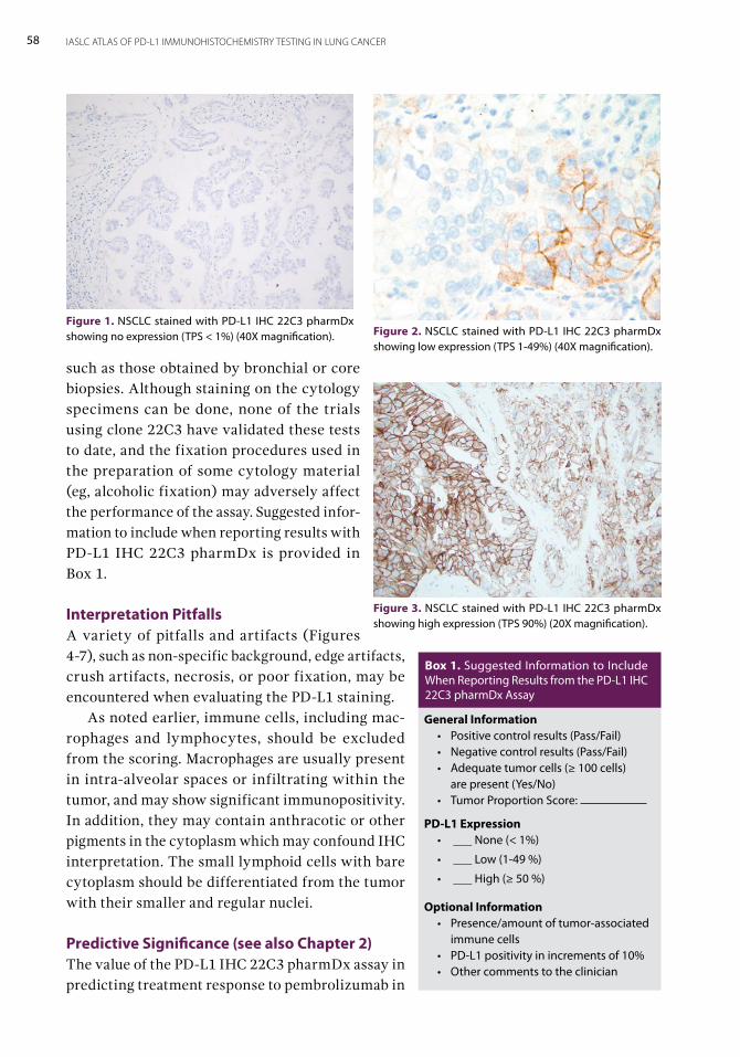
58 IASLC ATLAS OF PD-L1 IMMUNOHISTOCHEMISTRY TESTING IN LUNG CANCER
such as those obtained by bronchial or core biopsies. Although staining on the cytology specimens can be done, none of the trials using clone 22C3 have validated these tests to date, and the fixation procedures used in the preparation of some cytology material (eg, alcoholic fixation) may adversely affect the performance of the assay. Suggested infor-mation to include when reporting results with PD-L1 IHC 22C3 pharmDx is provided in Box 1.
Interpretation Pitfalls A variety of pitfalls and artifacts (Figures 4-7), such as non-specific background, edge artifacts, crush artifacts, necrosis, or poor fixation, may be encountered when evaluating the PD-L1 staining. As noted earlier, immune cells, including mac-rophages and lymphocytes, should be excluded from the scoring. Macrophages are usually present in intra-alveolar spaces or infiltrating within the tumor, and may show significant immunopositivity. In addition, they may contain anthracotic or other pigments in the cytoplasm which may confound IHC interpretation. The small lymphoid cells with bare cytoplasm should be differentiated from the tumor with their smaller and regular nuclei.
Predictive Significance (see also Chapter 2)The value of the PD-L1 IHC 22C3 pharmDx assay in predicting treatment response to pembrolizumab in
Figure 1. NSCLC stained with PD-L1 IHC 22C3 pharmDx showing no expression (TPS < 1%) (40X magnification). Figure 2. NSCLC stained with PD-L1 IHC 22C3 pharmDx
showing low expression (TPS 1-49%) (40X magnification).
Figure 3. NSCLC stained with PD-L1 IHC 22C3 pharmDx showing high expression (TPS 90%) (20X magnification).
General Information• Positive control results (Pass/Fail)• Negative control results (Pass/Fail)• Adequate tumor cells (≥ 100 cells)
are present (Yes/No)• Tumor Proportion Score:
PD-L1 Expression • ___ None (< 1%)
• ___ Low (1-49 %)
• ___ High (≥ 50 %)
Optional Information • Presence/amount of tumor-associated
immune cells• PD-L1 positivity in increments of 10%• Other comments to the clinician
Box 1. Suggested Information to Include When Reporting Results from the PD-L1 IHC 22C3 pharmDx Assay
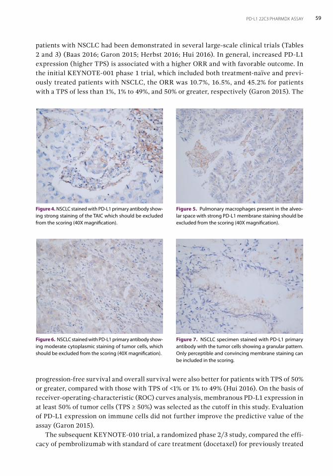
59PD-L1 22C3 PHARMDX ASSAY
patients with NSCLC had been demonstrated in several large-scale clinical trials (Tables 2 and 3) (Baas 2016; Garon 2015; Herbst 2016; Hui 2016). In general, increased PD-L1 expression (higher TPS) is associated with a higher ORR and with favorable outcome. In the initial KEYNOTE-001 phase 1 trial, which included both treatment-naïve and previ-ously treated patients with NSCLC, the ORR was 10.7%, 16.5%, and 45.2% for patients with a TPS of less than 1%, 1% to 49%, and 50% or greater, respectively (Garon 2015). The
Figure 4. NSCLC stained with PD-L1 primary antibody show-ing strong staining of the TAIC which should be excluded from the scoring (40X magnification).
Figure 5. Pulmonary macrophages present in the alveo-lar space with strong PD-L1 membrane staining should be excluded from the scoring (40X magnification).
Figure 6. NSCLC stained with PD-L1 primary antibody show-ing moderate cytoplasmic staining of tumor cells, which should be excluded from the scoring (40X magnification).
Figure 7. NSCLC specimen stained with PD-L1 primary antibody with the tumor cells showing a granular pattern. Only perceptible and convincing membrane staining can be included in the scoring.
progression-free survival and overall survival were also better for patients with TPS of 50% or greater, compared with those with TPS of <1% or 1% to 49% (Hui 2016). On the basis of receiver-operating-characteristic (ROC) curves analysis, membranous PD-L1 expression in at least 50% of tumor cells (TPS ≥ 50%) was selected as the cutoff in this study. Evaluation of PD-L1 expression on immune cells did not further improve the predictive value of the assay (Garon 2015). The subsequent KEYNOTE-010 trial, a randomized phase 2/3 study, compared the effi-cacy of pembrolizumab with standard of care treatment (docetaxel) for previously treated
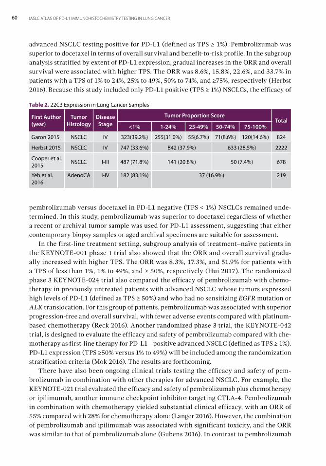
60 IASLC ATLAS OF PD-L1 IMMUNOHISTOCHEMISTRY TESTING IN LUNG CANCER
advanced NSCLC testing positive for PD-L1 (defined as TPS ≥ 1%). Pembrolizumab was superior to docetaxel in terms of overall survival and benefit-to-risk profile. In the subgroup analysis stratified by extent of PD-L1 expression, gradual increases in the ORR and overall survival were associated with higher TPS. The ORR was 8.6%, 15.8%, 22.6%, and 33.7% in patients with a TPS of 1% to 24%, 25% to 49%, 50% to 74%, and ≥75%, respectively (Herbst 2016). Because this study included only PD-L1 positive (TPS ≥ 1%) NSCLCs, the efficacy of
pembrolizumab versus docetaxel in PD-L1 negative (TPS < 1%) NSCLCs remained unde-termined. In this study, pembrolizumab was superior to docetaxel regardless of whether a recent or archival tumor sample was used for PD-L1 assessment, suggesting that either contemporary biopsy samples or aged archival specimens are suitable for assessment. In the first-line treatment setting, subgroup analysis of treatment–naïve patients in the KEYNOTE-001 phase 1 trial also showed that the ORR and overall survival gradu-ally increased with higher TPS. The ORR was 8.3%, 17.3%, and 51.9% for patients with a TPS of less than 1%, 1% to 49%, and ≥ 50%, respectively (Hui 2017). The randomized phase 3 KEYNOTE-024 trial also compared the efficacy of pembrolizumab with chemo-therapy in previously untreated patients with advanced NSCLC whose tumors expressed high levels of PD-L1 (defined as TPS ≥ 50%) and who had no sensitizing EGFR mutation or ALK translocation. For this group of patients, pembrolizumab was associated with superior progression-free and overall survival, with fewer adverse events compared with platinum-based chemotherapy (Reck 2016). Another randomized phase 3 trial, the KEYNOTE-042 trial, is designed to evaluate the efficacy and safety of pembrolizumab compared with che-motherapy as first-line therapy for PD-L1—positive advanced NSCLC (defined as TPS ≥ 1%). PD-L1 expression (TPS ≥50% versus 1% to 49%) will be included among the randomization stratification criteria (Mok 2016). The results are forthcoming. There have also been ongoing clinical trials testing the efficacy and safety of pem-brolizumab in combination with other therapies for advanced NSCLC. For example, the KEYNOTE-021 trial evaluated the efficacy and safety of pembrolizumab plus chemotherapy or ipilimumab, another immune checkpoint inhibitor targeting CTLA-4. Pembrolizumab in combination with chemotherapy yielded substantial clinical efficacy, with an ORR of 55% compared with 28% for chemotherapy alone (Langer 2016). However, the combination of pembrolizumab and ipilimumab was associated with significant toxicity, and the ORR was similar to that of pembrolizumab alone (Gubens 2016). In contrast to pembrolizumab
Table 2. 22C3 Expression in Lung Cancer Samples
First Author (year)
Tumor Histology
Disease Stage
Tumor Proportion ScoreTotal
<1% 1-24% 25-49% 50-74% 75-100%
Garon 2015 NSCLC IV 323(39.2%) 255(31.0%) 55(6.7%) 71(8.6%) 120(14.6%) 824
Herbst 2015 NSCLC IV 747 (33.6%) 842 (37.9%) 633 (28.5%) 2222
Cooper et al. 2015
NSCLC I-III 487 (71.8%) 141 (20.8%) 50 (7.4%) 678
Yeh et al. 2016
AdenoCA I-IV 182 (83.1%) 37 (16.9%) 219
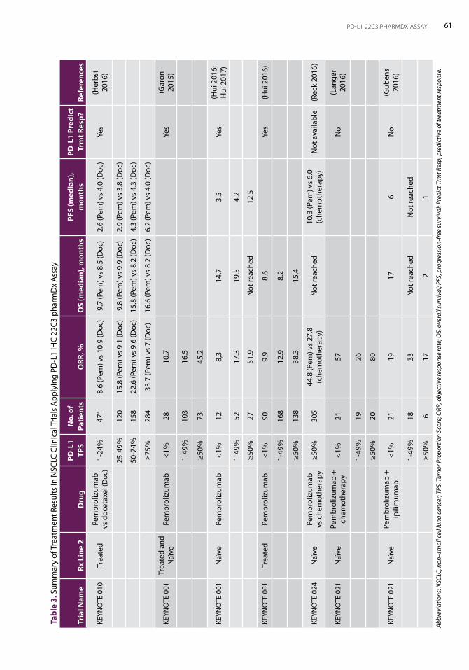
61PD-L1 22C3 PHARMDX ASSAY
Tabl
e 3.
Sum
mar
y of
Tre
atm
ent R
esul
ts in
NSC
LC C
linic
al T
rials
App
lyin
g PD
-L1
IHC
22C3
pha
rmD
x A
ssay
Tria
l Nam
eRx
Lin
e 2
Dru
gPD
-L1
TPS
No.
of
Pati
ents
ORR
, %O
S (m
edia
n), m
onth
sPF
S (m
edia
n),
mon
ths
PD-L
1 Pr
edic
t Tr
mt R
esp?
Refe
renc
es
KEYN
OTE
010
Trea
ted
Pem
brol
izum
ab
vs d
ocet
axel
(Doc
)1-
24%
471
8.6
(Pem
) vs
10.9
(Doc
)9.
7 (P
em) v
s 8.
5 (D
oc)
2.6
(Pem
) vs
4.0
(Doc
)Ye
s(H
erbs
t 20
16)
25-4
9%12
015
.8 (P
em) v
s 9.
1 (D
oc)
9.8
(Pem
) vs
9.9
(Doc
)2.
9 (P
em) v
s 3.
8 (D
oc)
50-7
4%15
822
.6 (P
em) v
s 9.
6 (D
oc)
15.8
(Pem
) vs
8.2
(Doc
)4.
3 (P
em) v
s 4.
3 (D
oc)
≥75%
284
33.7
(Pem
) vs
7 (D
oc)
16.6
(Pem
) vs
8.2
(Doc
)6.
2 (P
em) v
s 4.
0 (D
oc)
KEYN
OTE
001
Trea
ted
and
Naï
ve
Pem
brol
izum
ab<1
%28
10.7
Yes
(Gar
on
2015
)
1-49
%10
316
.5
≥50%
7345
.2
KEYN
OTE
001
Naï
vePe
mbr
oliz
umab
<1%
128,
314
.73.
5Ye
s(H
ui 2
016;
H
ui 2
017)
1-49
%52
17.3
19.5
4.2
≥50%
2751
.9N
ot re
ache
d12
.5
KEYN
OTE
001
Trea
ted
Pem
brol
izum
ab<1
%90
9.9
8.6
Yes
(Hui
201
6)
1-49
%16
812
.98.
2
≥50%
138
38.3
15.4
KEYN
OTE
024
Naï
vePe
mbr
oliz
umab
vs
che
mot
hera
py
≥50%
305
44.8
(Pem
) vs
27.8
(c
hem
othe
rapy
)N
ot re
ache
d10
.3 (P
em) v
s 6.
0 (c
hem
othe
rapy
)N
ot a
vaila
ble
(Rec
k 20
16)
KEYN
OTE
021
Naï
vePe
mbr
oliz
umab
+
chem
othe
rapy
<1%
2157
No
(Lan
ger
2016
)
1-49
%19
26
≥50%
2080
KEYN
OTE
021
Naï
vePe
mbr
oliz
umab
+
ipili
mum
ab<1
%21
1917
6N
o(G
uben
s 20
16)
1-49
%18
33N
ot re
ache
dN
ot re
ache
d
≥50%
617
21
Abbr
evia
tions
: NSC
LC, n
on–s
mal
l cel
l lun
g ca
ncer
; TPS
, Tum
or P
ropo
rtio
n Sc
ore;
ORR
, obj
ectiv
e re
spon
se ra
te; O
S, o
vera
ll su
rviv
al; P
FS, p
rogr
essi
on-fr
ee su
rviv
al; P
redi
ct T
rmt R
esp,
pre
dict
ive
of tr
eatm
ent r
espo
nse.

62 IASLC ATLAS OF PD-L1 IMMUNOHISTOCHEMISTRY TESTING IN LUNG CANCER
monotherapy, the treatment response of these combination regimens seemed to be indepen-dent of PD-L1 expression. Further studies are warranted to confirm this finding (Langer 2016; Gubens 2016).
ConclusionThe in vitro diagnostic PD-L1 IHC 22C3 pharmDx assay performed on the Dako Autostainer Link 48 platform is an immunohistochemical assay for detection of PD-L1 protein expression in advanced-stage NSCLC and for determination of eligibility for treatment with pembro-lizumab, a humanized monoclonal IgG4 kappa isotype antibody against PD-1. The assay assesses PD-L1 protein expression by evaluating TPS, which is the percentage of viable tumor cells showing either partial or complete membrane staining. Increasing PD-L1 expression (higher TPS) is generally associated with higher objective response rates and favorable outcome in patients treated with pembrolizumab and the assay is approved as a companion diagnostic assay for pembrolizumab.
AcknowledgementThe authors thank Dr. Yi-Chen Yeh, Dr. Shiou-Fu Lin, and Dr. Hsiang-Ling Ho for preparing the text and micrographs of the chapter.
PD-L1 SP142 AssayBy Ross A. Soo, Bernadette Reyna Asuncion, and Reinhard Buettner 6The Ventana PD-L1 (SP142) Assay (Ventana Medical Systems Inc.) is used to detect PD-L1 expression in tumor cells and immune cells in formalin-fixed, paraffin-embedded tissue. The SP142 antibody clone has been used in clinical trials of patients with advanced-stage non-small cell lung cancer (NSCLC), with scoring conducted using both tumor and immune cells (Fehrenbacher 2016). It has been approved by the US Food and Drug Administration as a complementary diagnostic tool to select patients with advanced urothelial carcinoma or advanced NSCLC for atezolizumab therapy. The approval in urothelial cancer was based on a phase II study in which programmed cell death ligand-1 (PD-L1) expression in 5% or greater of immune cells was associated with increased objective response for patients treated with atezolizumab (Rosenberg 2016). Regarding NSCLC, PD-L1 expression in at least 50% of viable tumor cells or in at least 10% of viable immune cells has been associated with enhanced overall survival with atezolizumab based on two trials, the phase III OAK trial (Rittmeyer 2017) and the phase II POPLAR trial (Fehrenbacher 2016). The Ventana PD-L1 (SP142) Assay is a rabbit monoclonal anti–PD-L1 antibody, that recognizes the intracellular domain of the PD-L1 protein ligand. In the United States, the Ventana PD-L1 SP142 assay is approved only for use on the BenchMark ULTRA (Ventana Medical Systems Inc.) platform, with the OptiView DAB IHC Detection Kit (Ventana Medical Systems Inc.) and the OptiView Amplification Kit (Ventana Medical Systems Inc.). Outside the US, the assay is approved for use on the Ventana Ultra, GX and XT platforms’. Three serial sections for testing are required from each case: the first for hematoxylin and eosin staining, a second for negative reagent control staining, and a third section for SP142 assay staining. The recommended control for use with this assay is tonsil tissue, which should be used as both a positive and a negative control for each staining run to monitor the performance of processed samples, as well as to test reagents and instruments. Control tissue should be fixed as soon as possible and processed in the same way as patient tumor samples. Tonsil tissue contains positive and negative staining epithelial and immune cells, which are used to confirm if the assay performed appropriately. A matched negative reagent control slide using the Rabbit Monoclonal Negative Control Ig (Ventana Medical Systems
64 IASLC ATLAS OF PD-L1 IMMUNOHISTOCHEMISTRY TESTING IN LUNG CANCER
Inc.) antibody should be conducted for each run to assess for nonspecific staining. Use of a different negative control reagent may cause false results.
Evaluation of StainingTonsillar Tissue ControlsAcceptable tonsil staining should show moderate-to-strong PD-L1 staining in lymphocytes and macrophages in the germinal centers, whereas reticulated crypt epithelial cells should show diffuse staining. Generally, there should not be any PD-L1 expression in immune cells in the interfollicular regions and on superficial squamous epithelium, however, rare PD-L1 positive immune cells may be found. Unacceptable staining in controls includes excessive nonspecific background staining that would conceal PD-L1–positive cells. Unacceptable staining may include weak-to-none PD-L1 staining in lymphocytes and macrophages in germinal centers, as well as in reticulated crypt epithelial cells. If the tissue control does not display the appropriate staining, patient samples should not be considered for evaluation, and staining should be repeated. Caution should be exercised when interpreting the staining intensity of tumor cells because of the strong PD-L1 staining in control tissues, and controls should not be used as an aid in formulating a specific PD-L1 expression score. Further data is needed regarding use of samples that reflect the tumor entity, such as with urothelial cancer or NSCLC. If nonspecific staining is present, it often has a diffuse appearance that may be identified using the negative reagent control slide stained with Rabbit Monoclonal Negative Control Ig. Intact cells should be used for interpretation of staining results. If background staining is excessive, the interpretation of the patient samples should be considered invalid.
Staining Patterns for Tumor and Immune Cells The sample should contain at least 50 tumor cells with associated stroma for tumor-cell staining results to be valid; necrotic areas should be avoided. SP142 differs significantly from other PD-L1 assays by its distinct staining pattern, revealing both membranous and granular cytoplasmic staining in tumor cells (Scheel 2016). This contrasts with all other antibody clones, which show cell surface-defined linear membranous staining. The distinct pattern for the SP142 assay relies on detection by the Ventana OptiView system and the Ventana BenchMark Ultra platform. Linear membranous staining is shown when the anti-body from the SP142 kit is used in combination with other detection systems (Scheel et al, submitted). PD-L1–positive tumor cells usually show partial or fully circumferential membranous staining. When these tumor cells are stained with the Ventana OptiView system, the cells may show cytoplasmic granular staining of variable intensity. However, only membranous staining is considered for scoring. This cytoplasmic staining is independent from mem-branous staining, and both types of positive tumor cells should be evaluated. When using the brown 3,3'-diaminobenzidine color for visualization, it is important not to confuse immunohistochemistry (IHC) signals with anthracotic or iron pigment. Another pitfall is the intense PD-L1 staining on normal bronchiolar epithelium over lymphoid tissue. This bronchus-associated lymphoid tissue-like staining must not be confused with specific tumor-cell staining.
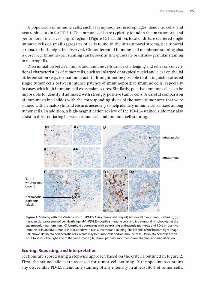
65PD-L1 SP142 ASSAY
A population of immune cells, such as lymphocytes, macrophages, dendritic cells, and neutrophils, stain for PD-L1. The immune cells are typically found in the intratumoral and peritumoral (invasive margin) regions (Figure 1). In addition, focal or diffuse scattered single immune cells or small aggregates of cells found in the intratumoral stroma, peritumoral stroma, or both might be observed. Circumferential immune-cell membrane staining also is observed. Immune-cell staining can be seen as fine-punctate or diffuse-granular staining in neutrophils. Discrimination between tumor and immune cells can be challenging and relies on conven-tional characteristics of tumor cells, such as enlarged or atypical nuclei and clear epithelial differentiation (e.g., formation of acini). It might not be possible to distinguish scattered single tumor cells between intense patches of immunopositive immune cells, especially in cases with high immune-cell expression scores. Similarly, positive immune cells can be impossible to identify if admixed with strongly positive tumor cells. A careful comparison of immunostained slides with the corresponding slides of the same tumor area that were stained with hematoxylin and eosin is necessary to help identify immune cells mixed among tumor cells. In addition, a high-magnification review of the PD-L1–stained slide may also assist in differentiating between tumor-cell and immune-cell staining.
Scoring, Reporting, and Interpretation Sections are scored using a stepwise approach based on the criteria outlined in Figure 2. First, the stained slides are assessed for tumor-cell staining. If the specimen contains any discernible PD-L1 membrane staining of any intensity in at least 50% of tumor cells,
BA1 A2
A3 A4
D1 D2C
Intravascular
Intratumoral
PD-L1+ lymphocytes(brown)
Anthracotic pigments(black)
Figure 1. Staining with the Ventana PD-L1 (SP142) Assay demonstrating: (A) tumor-cell membranous staining, (B) intravascular programmed cell death ligand-1 (PD-L1)– positive immune cells and intratumoral lymphocytes at the squamocolumnar junction, (C) lymphoid aggregates with co-existing anthracotic pigments and PD-L1– positive immune cells, and (D) tumor cells (encircled) with partial-membrane staining. The left side of the bottom right image (D1) shows darkly stained necrotic cells, which may be tumor cells and/or immune cells. Darkly stained cells are dif-ficult to assess. The right side of the same image (D2) shows partial tumor-membrane staining. 40x magnification.
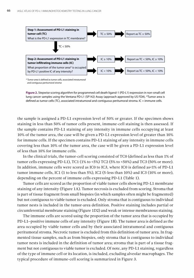
66 IASLC ATLAS OF PD-L1 IMMUNOHISTOCHEMISTRY TESTING IN LUNG CANCER
the sample is assigned a PD-L1 expression level of 50% or greater. If the specimen shows staining in less than 50% of tumor cells present, immune-cell staining is then assessed. If the sample contains PD-L1 staining of any intensity in immune cells occupying at least 10% of the tumor area, the case will be given a PD-L1 expression level of greater than 10% for immune cells. If the specimen contains PD-L1 staining of any intensity in immune cells covering less than 10% of the tumor area, the case will be given a PD-L1 expression level of less than 10% for immune cells. In the clinical trials, the tumor-cell scoring consisted of TC0 (defined as less than 1% of tumor cells expressing PD-L1), TC1 (1% to <5%) TC2 (5% to <50%) and TC3 (50% or more). In addition, immune cells were scored as IC0 to IC3, where IC0 is defined as<1% of PD-L1 tumor immune cells, IC1 (1 to less than 5%), IC2 (5-less than 10%) and IC3 (10% or more), depending on the percent of immune cells expressing PD-L1 (Table 1). Tumor cells are scored as the proportion of viable tumor cells showing PD-L1 membrane staining of any intensity (Figure 1A). Tumor necrosis is excluded from scoring. Stroma that is part of tissue fragment from small biopsies (in which samples often might be fragmented) but not contiguous to viable tumor is excluded. Only stroma that is contiguous to individual tumor nests is included in the tumor-area definition. Positive staining includes partial or circumferential membrane staining (Figure 1D2) and weak or intense membranous staining. The immune cells are scored using the proportion of the tumor area that is occupied by PD-L1–positive immune cells of any intensity (Figure 1B). The tumor area is defined as the area occupied by viable tumor cells and by their associated intratumoral and contiguous peritumoral stroma. Necrotic tumor is excluded from this definition of tumor area. In frag-mented tissue samples, such as from biopsies, only stroma that is contiguous to individual tumor nests is included in the definition of tumor area; stroma that is part of a tissue frag-ment but not contiguous to viable tumor is excluded. Of note, any PD-L1 staining, regardless of the type of immune cell or its location, is included, excluding alveolar macrophages. The typical procedure of immune-cell scoring is summarized in Figure 3.
Step 1: Assessment of PD-L1 staining in tumor cell (TC)
What is the PD-L1 expression in TC membrane?
Step 2: Assessment of PD-L1 staining in tumor infiltrating immune cells (IC)
What proportion of the tumor area* is occupied by PD-L1 positive IC of any intensity?
* Tumor area is defined as tumor cells, associated intratumoral and contiguous peritumoral stroma
TC ≥ 50% Report as TC ≥ 50%
TC < 50%
IC ≥ 10%
IC < 10%
Report as TC < 50%, IC ≥ 10%
Report as TC < 50%, IC < 10%
Figure 2. Stepwise scoring algorithm for programmed cell death ligand-1 (PD-L1) expression in non-small cell lung cancer samples using the Ventana PD-L1 (SP142) Assay (approach approved by US FDA). *Tumor area is defined as tumor cells (TC), associated intratumoral and contiguous peritumoral stroma. IC = immune cells.
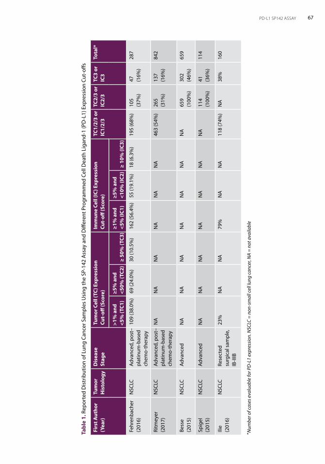
67PD-L1 SP142 ASSAY
Tabl
e 1.
Rep
orte
d D
istr
ibut
ion
of L
ung
Canc
er S
ampl
es U
sing
the
SP-1
42 A
ssay
and
Diff
eren
t Pro
gram
med
Cel
l Dea
th L
igan
d-1
(PD
-L1)
Exp
ress
ion
Cut-
offs
Firs
t Aut
hor
(Yea
r)Tu
mor
H
isto
logy
Dis
ease
St
age
Tum
or C
ell (
TC) E
xpre
ssio
n
Cut-
off (S
core
)Im
mun
e Ce
ll (IC
) Exp
ress
ion
Cu
t-off
(Sco
re)
TC1/
2/3
or
IC1/
2/3
TC2/
3 or
IC
2/3
TC3
or
IC3
Tota
l*
>1%
and
<5
% (T
C1)
≥5%
and
<5
0% (T
C2)
≥ 50
% (T
C3)
≥1%
and
<5
% (I
C1)
≥5%
and
<1
0% (I
C2)
≥ 10
% (I
C3)
Fehr
enba
cher
(2
016)
NSC
LCAd
vanc
ed, p
ost–
plat
inum
-bas
ed
chem
o-th
erap
y
109
(38.
0%)
69 (2
4.0%
)30
(10.
5%)
162
(56.
4%)
55 (1
9.1%
)18
(6.3
%)
195
(68%
)10
5 (3
7%)
47
(16%
)28
7
Ritm
eyer
(2
017)
NSC
LCAd
vanc
ed, p
ost–
plat
inum
-bas
ed
chem
o-th
erap
y
NA
NA
NA
NA
NA
NA
463
(54%
)26
5 (3
1%)
137
(16%
)84
2
Bess
e
(201
5)N
SCLC
Adva
nced
NA
NA
NA
NA
NA
NA
NA
659
(100
%)
302
(46%
)65
9
Spig
el
(201
5)N
SCLC
Adva
nced
NA
NA
NA
NA
NA
NA
NA
114
(100
%)
41
(36%
)11
4
Ilie
(201
6)N
SCLC
Rese
cted
su
rgic
al s
ampl
e,
IB-II
IB
23%
NA
NA
79%
NA
NA
118
(74%
)N
A38
%16
0
*Num
ber o
f cas
es e
valu
able
for P
D-L
1 ex
pres
sion
. NSC
LC =
non
-sm
all c
ell l
ung
canc
er, N
A =
not a
vaila
ble
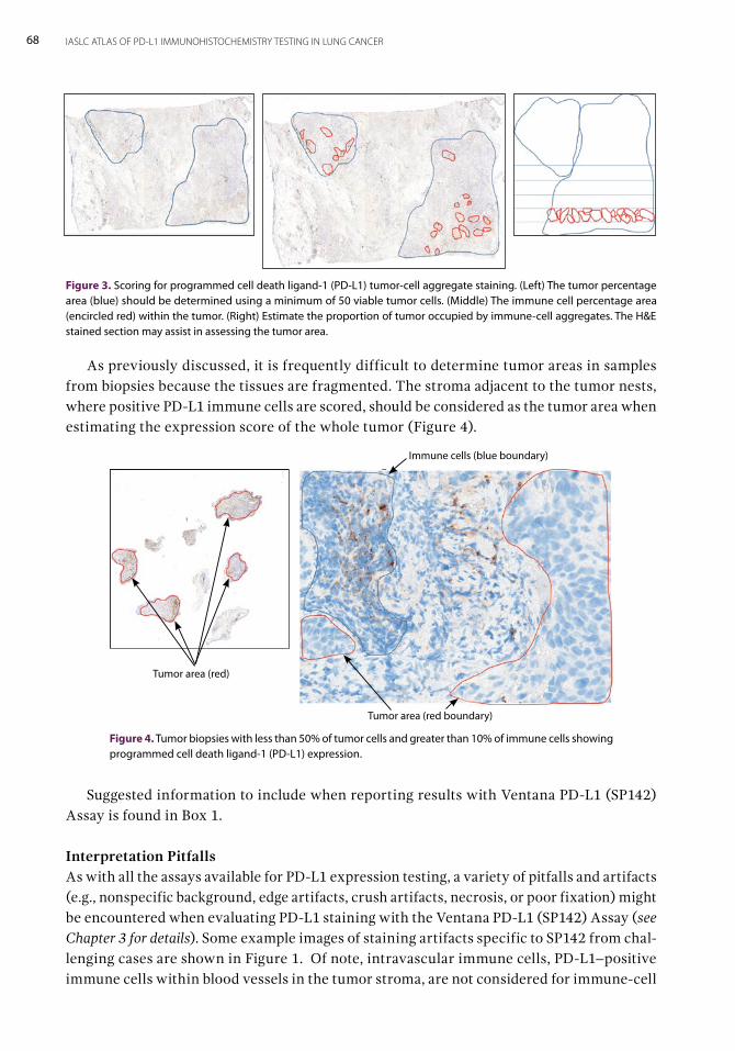
68 IASLC ATLAS OF PD-L1 IMMUNOHISTOCHEMISTRY TESTING IN LUNG CANCER
As previously discussed, it is frequently difficult to determine tumor areas in samples from biopsies because the tissues are fragmented. The stroma adjacent to the tumor nests, where positive PD-L1 immune cells are scored, should be considered as the tumor area when estimating the expression score of the whole tumor (Figure 4).
Suggested information to include when reporting results with Ventana PD-L1 (SP142) Assay is found in Box 1.
Interpretation Pitfalls As with all the assays available for PD-L1 expression testing, a variety of pitfalls and artifacts (e.g., nonspecific background, edge artifacts, crush artifacts, necrosis, or poor fixation) might be encountered when evaluating PD-L1 staining with the Ventana PD-L1 (SP142) Assay (see Chapter 3 for details). Some example images of staining artifacts specific to SP142 from chal-lenging cases are shown in Figure 1. Of note, intravascular immune cells, PD-L1–positive immune cells within blood vessels in the tumor stroma, are not considered for immune-cell
Figure 3. Scoring for programmed cell death ligand-1 (PD-L1) tumor-cell aggregate staining. (Left) The tumor percentage area (blue) should be determined using a minimum of 50 viable tumor cells. (Middle) The immune cell percentage area (encircled red) within the tumor. (Right) Estimate the proportion of tumor occupied by immune-cell aggregates. The H&E stained section may assist in assessing the tumor area.
Tumor area (red)
Tumor area (red boundary)
Immune cells (blue boundary)
Figure 4. Tumor biopsies with less than 50% of tumor cells and greater than 10% of immune cells showing programmed cell death ligand-1 (PD-L1) expression.
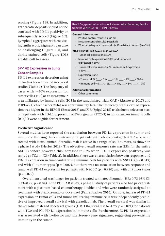
69PD-L1 SP142 ASSAY
Box 1. Suggested Information for Inclusion When Reporting Results from the VENTANA PD-L1 (SP142) Assay
scoring (Figure 1B). In addition, anthracotic deposits should not be confused with PD-L1 positivity or subsequently scored (Figure 1C). Lymphoid aggregates with coexist-ing anthracotic pigments can also be challenging (Figure 1C), and darkly stained cells (Figure 1D1) are difficult to assess.
SP-142 Expression in Lung Cancer SamplesPD-L1 expression detection using SP142 has been reported in several studies (Table 1). The frequency of cases with >=50% expression for tumor cells (TC3) or >= 10% tumour area infiltrated by immune cells (IC3 in the randomized trials OAK (Rittmeyer 2017) and POPLAR (Fehrenbacher 2016) was approximately 16%. The frequency of this level of expres-sion was higher in the BIRCH (Besse 2015) and FIR (Spigel 2015) trials due to selection bias; only patients with PD-L1 expression of 5% or greater (TC2/3) in tumor and/or immune cells (IC2/3) were eligible for treatment.
Predictive Significance Several studies have reported the association between PD-L1 expression in tumor and immune cells using clinical outcomes for patients with advanced-stage NSCLC who were treated with atezolizumab. Atezolizumab is active in a range of solid tumors, as shown in a phase I study (Herbst 2014). The objective overall response rate was 23% for the entire NSCLC cohort; however, this increased to 83% when PD-L1 expression positivity was scored as TC3 or IC3 (Table 2). In addition, there was an association between responses and PD-L1 expression in tumor-infiltrating immune cells for patients with NSCLC (p = 0.015) and with all tumor types (p = 0.007), but there was no association between response and tumor-cell PD-L1 expression for patients with NSCLC (p = 0.920) and with all tumor types (p = 0.079). Overall survival was longer for patients treated with atezolizumab (HR: 0.73 95% CI: 0.53–0.99; p = 0.04) in the POPLAR study, a phase II study of patients who had prior treat-ment with a platinum-based chemotherapy doublet and who were randomly assigned to treatment with atezolizumab or docetaxel (Fehrenbacher 2016). Of note, increased PD-L1 expression on tumor cells and tumor-infiltrating immune cells was independently predic-tive of improved overall survival with atezolizumab. The overall survival was similar in the atezolizumab and docetaxel groups (HR: 1.04; 95% CI: 0.62-1.75; p = 0.871) for patients with TC0 and IC0 PD-L1 expression in immune cells. Furthermore, IC PD-L1 expression was associated with T-effector and interferon-γ gene signature, suggesting pre-existing immunity in the tumor.
General Information• Positive control results (Pass/Fail)• Negative control results (Pass/Fail)• Whether adequate tumor cells (≥50 cells) are present (Yes/No)
PD-L1 IHC SP-142 Result to Clinician* • Tumor cell expression ≥ 50% ___
• Immune cell expression ≥10% (and tumor cell expression < 50%) ___
• Tumor cell expression < 50%, Immune cell expression < 10% ___
• Expression status
° Tumor cell % (___< 1%, ___≥ 1%, ___≥ 5%, ___≥ 50%)
° Immune cell % (___< 1%, ___> 1%, ___> 5%, ___> 10%)
Additional Information • Other comments
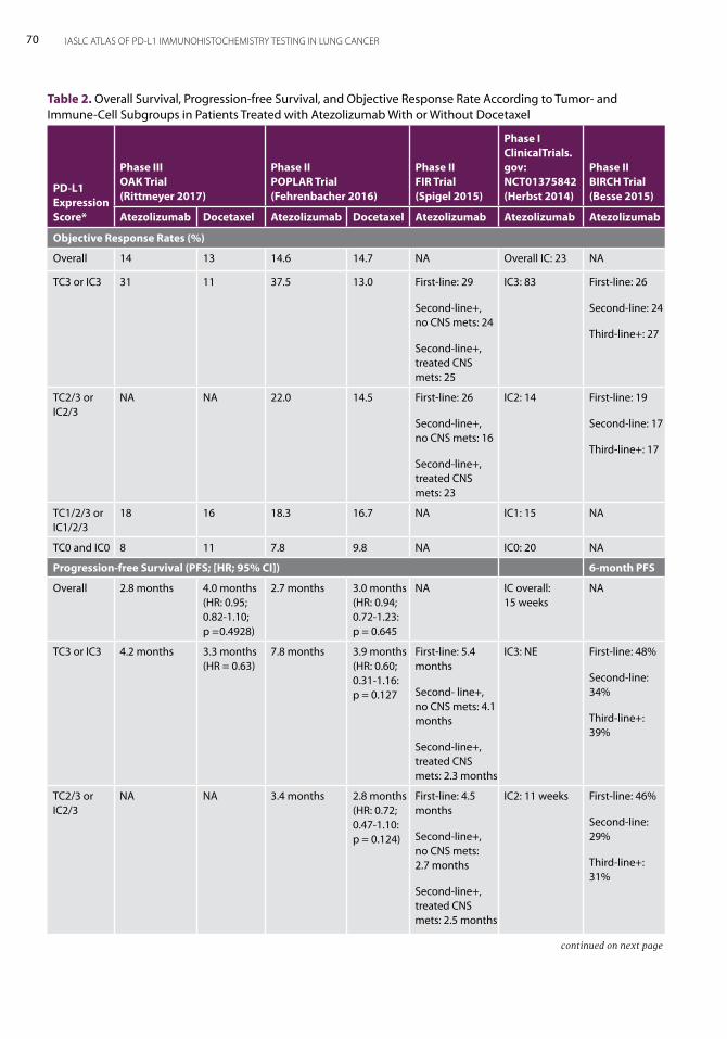
70 IASLC ATLAS OF PD-L1 IMMUNOHISTOCHEMISTRY TESTING IN LUNG CANCER
Table 2. Overall Survival, Progression-free Survival, and Objective Response Rate According to Tumor- and Immune-Cell Subgroups in Patients Treated with Atezolizumab With or Without Docetaxel
PD-L1 Expression Score*
Phase III OAK Trial (Rittmeyer 2017)
Phase II POPLAR Trial (Fehrenbacher 2016)
Phase II FIR Trial (Spigel 2015)
Phase I ClinicalTrials. gov: NCT01375842 (Herbst 2014)
Phase II BIRCH Trial (Besse 2015)
Atezolizumab Docetaxel Atezolizumab Docetaxel Atezolizumab Atezolizumab Atezolizumab
Objective Response Rates (%)
Overall 14 13 14.6 14.7 NA Overall IC: 23 NA
TC3 or IC3 31 11 37.5 13.0 First-line: 29
Second-line+, no CNS mets: 24
Second-line+, treated CNS mets: 25
IC3: 83 First-line: 26
Second-line: 24
Third-line+: 27
TC2/3 or IC2/3
NA NA 22.0 14.5 First-line: 26
Second-line+, no CNS mets: 16
Second-line+, treated CNS mets: 23
IC2: 14 First-line: 19
Second-line: 17
Third-line+: 17
TC1/2/3 or IC1/2/3
18 16 18.3 16.7 NA IC1: 15 NA
TC0 and IC0 8 11 7.8 9.8 NA IC0: 20 NA
Progression-free Survival (PFS; [HR; 95% CI]) 6-month PFS
Overall 2.8 months 4.0 months (HR: 0.95; 0.82-1.10; p =0.4928)
2.7 months 3.0 months (HR: 0.94; 0.72-1.23: p = 0.645
NA IC overall: 15 weeks
NA
TC3 or IC3 4.2 months 3.3 months (HR = 0.63)
7.8 months 3.9 months (HR: 0.60; 0.31-1.16: p = 0.127
First-line: 5.4 months
Second- line+, no CNS mets: 4.1 months
Second-line+, treated CNS mets: 2.3 months
IC3: NE First-line: 48%
Second-line: 34%
Third-line+: 39%
TC2/3 or IC2/3
NA NA 3.4 months 2.8 months (HR: 0.72; 0.47-1.10: p = 0.124)
First-line: 4.5 months
Second-line+, no CNS mets: 2.7 months
Second-line+, treated CNS mets: 2.5 months
IC2: 11 weeks First-line: 46%
Second-line: 29%
Third-line+: 31%
continued on next page
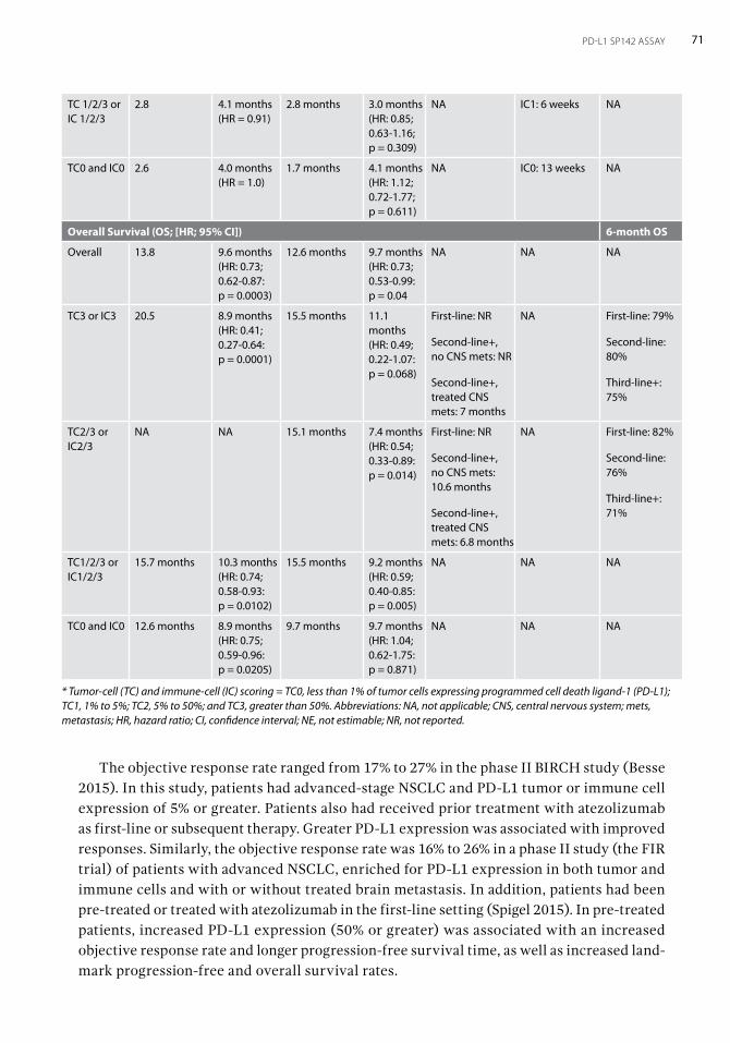
71PD-L1 SP142 ASSAY
The objective response rate ranged from 17% to 27% in the phase II BIRCH study (Besse 2015). In this study, patients had advanced-stage NSCLC and PD-L1 tumor or immune cell expression of 5% or greater. Patients also had received prior treatment with atezolizumab as first-line or subsequent therapy. Greater PD-L1 expression was associated with improved responses. Similarly, the objective response rate was 16% to 26% in a phase II study (the FIR trial) of patients with advanced NSCLC, enriched for PD-L1 expression in both tumor and immune cells and with or without treated brain metastasis. In addition, patients had been pre-treated or treated with atezolizumab in the first-line setting (Spigel 2015). In pre-treated patients, increased PD-L1 expression (50% or greater) was associated with an increased objective response rate and longer progression-free survival time, as well as increased land-mark progression-free and overall survival rates.
TC 1/2/3 or IC 1/2/3
2.8 4.1 months (HR = 0.91)
2.8 months 3.0 months (HR: 0.85; 0.63-1.16; p = 0.309)
NA IC1: 6 weeks NA
TC0 and IC0 2.6 4.0 months (HR = 1.0)
1.7 months 4.1 months (HR: 1.12; 0.72-1.77; p = 0.611)
NA IC0: 13 weeks NA
Overall Survival (OS; [HR; 95% CI]) 6-month OS
Overall 13.8 9.6 months (HR: 0.73; 0.62-0.87: p = 0.0003)
12.6 months 9.7 months (HR: 0.73; 0.53-0.99: p = 0.04
NA NA NA
TC3 or IC3 20.5 8.9 months (HR: 0.41; 0.27-0.64: p = 0.0001)
15.5 months 11.1 months (HR: 0.49; 0.22-1.07: p = 0.068)
First-line: NR
Second-line+, no CNS mets: NR
Second-line+, treated CNS mets: 7 months
NA First-line: 79%
Second-line: 80%
Third-line+: 75%
TC2/3 or IC2/3
NA NA 15.1 months 7.4 months (HR: 0.54; 0.33-0.89: p = 0.014)
First-line: NR
Second-line+, no CNS mets: 10.6 months
Second-line+, treated CNS mets: 6.8 months
NA First-line: 82%
Second-line: 76%
Third-line+: 71%
TC1/2/3 or IC1/2/3
15.7 months 10.3 months (HR: 0.74; 0.58-0.93: p = 0.0102)
15.5 months 9.2 months (HR: 0.59; 0.40-0.85: p = 0.005)
NA NA NA
TC0 and IC0 12.6 months 8.9 months (HR: 0.75; 0.59-0.96: p = 0.0205)
9.7 months 9.7 months (HR: 1.04; 0.62-1.75: p = 0.871)
NA NA NA
* Tumor-cell (TC) and immune-cell (IC) scoring = TC0, less than 1% of tumor cells expressing programmed cell death ligand-1 (PD-L1); TC1, 1% to 5%; TC2, 5% to 50%; and TC3, greater than 50%. Abbreviations: NA, not applicable; CNS, central nervous system; mets, metastasis; HR, hazard ratio; CI, confidence interval; NE, not estimable; NR, not reported.

72 IASLC ATLAS OF PD-L1 IMMUNOHISTOCHEMISTRY TESTING IN LUNG CANCER
The phase III OAK trial reported overall survival in the overall population of 13.8 months for atezolizumab compared with 9.6 months for docetaxel (HR: 0.73 95% CI: 0.62-0.87; p = 0.0003); all patients had advanced pre-treated NSCLC and were randomly assigned to either therapy (Rittmeyer 2017). Furthermore, atezolizumab conferred a survival benefit regardless of PD-L1 expression status, with extremely similar hazard ratios for PD-L1–positive (HR = 0.74) and negative (HR = 0.75) patients. Figure 5 illustrates a case of a heavily pre-treated patient with at least TC2/IC2 PD-L1 expression whose disease responded to atezolizumab.
Figure 5. Images of (A) left supraclavicular lymph nodes and (B) coeliac axis lymph nodes at (1) baseline and (2) after two cycles of atezolizumab.
A1 A2
B1 B2
ConclusionThe Ventana PD-L1 (SP142) Assay is used to detect PD-L1 expression in both tumor and immune cells as predictive marker for azetolizumab therapy. The sample should have at least 50 tumor cells with associated stroma, and PD-L1 is expressed as membranous and granular cytoplasmic staining in these cells. The SP142 assay is performed using a step-wise approach. Tumor cells are scored by determining the percentage of area covered by PD-L1–positive viable tumor cells and associated intratumoral and contiguous peritumoral stroma. Immune cells are scored by determining the proportion of the tumor area that is occupied by PD-L1–positive immune cells of any intensity. An association between clini-cal outcomes and PD-L1 expression in the tumor and immune cells have been reported in studies of patients with advanced-stage NSCLC treated with atezolizumab.
PD-L1 SP263 Assay By Sanja Dacic and Arne Warth 7The VENTANA PD-L1 (SP263) Rabbit Monoclonal Primary Antibody assay was developed by Ventana Medical Systems, Inc., a subsidiary of Roche, in collaboration with AstraZeneca for use with the VENTANA Benchmark ULTRA staining platform (Ventana Medical Systems, Inc.). This assay detects PD-L1 expression and helps determine patient eligibility for treat-ment with the immunotherapeutic drug durvalumab (Rebelatto 2016). Durvalumab is a human monoclonal antibody directed against programmed cell death ligand-1 (PD-L1). PD-L1 expression enables tumors to evade detection by the immune system through binding to the programmed cell death-1 protein (PD-1) on cytotoxic T lymphocytes (Stewart 2015). Durvalumab blocks PD-L1 interaction with both PD-1 and CD80 on T cells, countering the tumor’s immune-evading tactics. Non-small cell lung cancer (NSCLC) cells with PD-L1 expression of at least 25% were analyzed using this assay in two recent clinical trials (Stewart 2015, Garon 2015). In a multicenter phase Ib study, however, durvalumab demonstrated anti-tumor activity regardless of associated PD-L1 expression status (Antonia 2016). Recently, the SP263 assay has been commercialized for identification of patients with non-squamous cell NSCLC who are most likely to benefit from nivolumab (Chapter 4 discusses another complementary diagnostic tool for nivolumab therapy, the PD-L1 IHC 28-8 pharmDx Assay.) VENTANA PD-L1 (SP263) Rabbit Monoclonal Primary Antibody is a rabbit monoclonal primary antibody produced against PD-L1 that localizes to and stains the membranous and/or cytoplasmic regions of cells. Anti–PD-L1 SP263 binds to an epitope corresponding to amino acids 284-290 of the PD-L1 protein (Quon 2016). The SP263 assay is intended for laboratory use for the detection of the PD-L1 protein in formalin-fixed, paraffin-embedded tissue, and the marketed package includes 50 tests. Acceptable fixatives also include zinc formalin and Z-5 fixatives. Fixatives not recommended for use are: 95% alcohol; alcohol, formalin, and acetic acid (AFA); and Prefer (Anatech LTD). As previously mentioned, the assay was optimized for the automated VENTANA Benchmark ULTRA platform. Detection is optimized with the OptiView DAB IHC Detection Kit (Ventana Medical Systems, Inc.), which is an indirect, biotin-free system for detecting mouse immunoglobulins G and M, as well as rabbit primary antibodies. The slides should be stained immediately because the
74 IASLC ATLAS OF PD-L1 IMMUNOHISTOCHEMISTRY TESTING IN LUNG CANCER
antigenicity of cut tissue sections might diminish over time. Placenta is recommended as a positive control because the SP263 assay demonstrates a uniform staining of the membrane and/or cytoplasm in trophoblast lineage cells that is moderate (2+)-to strong (3+).
Evaluation of Staining and ReportingDurvalumabThe assay should be evaluated and scored by a qualified pathologist using light microscopy. The interpretation of PD-L1 staining must be complemented by the evaluation of slides stained with hematoxylin and eosin and of negative control reagent-stained slides. SP263 staining is interpreted as positive if membranous and/or cytoplasmic protein expression at any intensity greater than background staining is detected in at least 25% of tumor cells. At least 100 viable tumor cells should be scored. Data regarding the assessment of the staining intensity has not been presented. Tumor-infiltrating lymphocytes and intra-alveolar mac-rophages can also demonstrate linear membranous, diffuse cytoplasmic, and/or punctate staining. Staining of the tumor-infiltrating lymphocytes, non-neoplastic cells, and necrotic cells is not included in the scoring criteria. The staining may appear heterogenous in both small biopsy and large resection speci-mens. The staining can be very strong and homogenous (Figure 1A); however, some tumors show heterogenous expression. Variable-intensity strong and moderate staining is easily identified at low power magnification (2x or 4x), whereas weak staining is best seen at high-power magnification (20x). The “histo” (H) score, which considers both the percent-age of tumor cells that stain positively and staining intensity, has not been used in the clinical trials. Heterogeneity of staining might be, in part, caused by pre-analytical factors. Therefore, staining of freshly cut tissue on new charged (“plus”) slides is recommended to limit exposure to room humidity and to minimize plastic exposure (i.e., use of glass or metal containers and racks).
NivolumabRecently, the SP263 assay has become available in Europe for the identification of patients with non-squamous cell NSCLC who are most likely to benefit from nivolumab. This was based on high concordance between PD-L1 expression status as determined by the PD-L1 IHC 28-8 pharmDx (Agilent Technologies/Dako) and VENTANA PD-L1 (SP263) Rabbit Monoclonal Primary Antibody assays, although use of the SP263 assay tends to result in more intense staining for carcinoma and immune cells than the 28-8 and PD-L1 IHC 22C3 pharmDx (Agilent Technologies/Dako) assays (Scheel 2016, Hirsch 2017). When evaluat-ing for nivolumab therapy, PD-L1 expression status should be scored using membranous staining of tumor cells. Scoring subgroups include: less than 1%, 1% to 5%, 5% to 10%, and 10% or greater.
ReportingSuggested information to include when reporting results with the SP263 assay, for both durvalumab and nivolumab therapy consideration, is shown in Box 1.
75PD-L1 SP263 ASSAY
Interpretation PitfallsA variety of pitfalls and artifacts including nonspecific background, edge artifacts, crush artifacts, necrosis, or poor fixation might be encountered when evaluating the PD-L1 staining (see Chapter 3). There is concern that sampling error may result in discrepant results between different specimen types (i.e., biopsy vs. resection), and studies are needed to address this issue. It has been shown for other PD-L1 clones that PD-L1 expression is affected by radiation and chemotherapy, which may have been administered after a biopsy was obtained, but similar data is not available for SP263 (Sheng 2016). Many artifacts may lead to false-positive staining if staining is not carefully evaluated. Alveolar macrophages—par-ticularly smoker’s macrophages—show cytoplasmic granular staining (Figure
1B). Non-neoplastic lung parenchyma can show staining of inflammatory and interstitial stromal cells. Extracellular mucin and tumor necrosis can show granular staining (Figure 1C). The staining of papillary adenocarcinoma should be interpreted with caution because stromal cells in the fibrovascular cores may show moderate-to-strong staining that can be interpreted as false-positive tumor-cell staining (Figure 1D). Red blood cells can show a diffuse moderate staining.
Predictive, Prognostic Significance In a phase I study by Rizvi et al. the response rate to durvalumab was 27% for patients with PD-L1–positive NSCLC but only 5% for patients who were PD-L1 negative (Table 1; Rizvi 2015). In a phase II study of durvalumab in pre-treated patients with NSCLC, overall response rates were 7.5% for PD-L1 expression of less than 25%, 16.4% for PD-L1 expression of 25% to 90%, and 30.9% for PD-L1 expression of 90% or greater. Progression-free survival for the same three groupings was 1.9, 3.3, and 2.4 months, respectively. One-year survival rates were 34.5%, 47.7%, and 50.8%, respectively (Garassino 2017). Response was noted in 22% of PD-L1–positive and 29% of PD-L1–negative tumors in a multicenter, nonrandomized, open-label, phase Ib study of durvalumab plus tremelimumab in NSCLC (Table 1; Antonia 2016). Durvalumab in combination with gefitinib showed 77.8% to 80% objective response rate in patients with NSCLC and with sensitizing EGFR mutations who had not received prior therapy with a tyrosine kinase inhibitor. However, 55% of patients had grades 3 to 4 treatment-related toxicities (Gibbons 2016). The prognostic significance of PD-L1 expression as detected by the SP263 assay is uncer-tain. A recent univariate analysis demonstrated a correlation between improved overall
General Information• Positive control results (Pass/Fail)• Negative control results (Pass/Fail)• Whether adequate tumor cells (≥ 100 cells)
are present (Yes/No)• Tumor Proportion Score
PD-L1 IHC SP263 Result to Clinician • For durvalumab
< 25% Expression
≥ 25% Expression
• For nivolumab
Expression < 1%
Expression ≥ 1% to < 5%
Expression ≥ 5% to < 10%
Expression ≥ 10%
Additional Information • Information about tumor-associated immune cells • PD-L1 positivity in increments of 10%• Other comments to the clinician
Box 1. Suggested Information for Inclusion When Reporting Results from the VENTANA PD-L1 (SP263) Rabbit Monoclonal Primary Antibody Assay
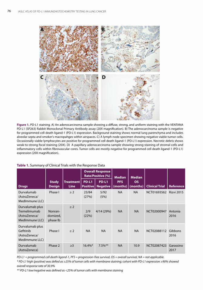
76 IASLC ATLAS OF PD-L1 IMMUNOHISTOCHEMISTRY TESTING IN LUNG CANCER
Figure 1. PD-L1 staining. A) An adenocarcinoma sample showing a diffuse, strong, and uniform staining with the VENTANA PD-L1 (SP263) Rabbit Monoclonal Primary Antibody assay (20X magnification). B) The adenocarcinoma sample is negative for programmed cell death ligand-1 (PD-L1) expression. Background staining shows normal lung parenchyma and includes alveolar septa and smoker’s macropahges within airspaces. C) A lymph node specimen showing negative viable tumor cells. Occasionally viable lymphocytes are positive for programmed cell death ligand-1 (PD-L1) expression. Necrotic debris shows weak-to-strong focal staining (20X). D) A papillary adenocarcinoma sample showing strong staining of stromal cells and inflammatory cells within fibrovascular cores. Tumor cells are mostly negative for programmed cell death ligand-1 (PD-L1) expression (20X magnification).
A B
C D
Table 1. Summary of Clinical Trials with the Response Data
DrugsStudy
DesignTreatment
Line
Overall Response Rate/Positive (%) Median
PFS (months)
Median OS
(months) Clinical Trial ReferencePD-L1
PositivePD-L1
Negative
Durvalumab(AstraZeneca/MedImmune LLC)
Phase I ≥ 2 23/84(27%)
5/92(5%)
NA NA NCT01693562 Rizvi 2015
Durvalumab plusTremelimumab (AstraZeneca/ MedImmune LLC)
Nonran-domized, phase Ib
≥ 22/9
(22%)4/14 (29%) NA NA NCT02000947 Antonia
2016
Durvalumab plusGefitinib (AstraZeneca/MedImmune LLC)
Phase I ≥ 2 NA NA NA NA NCT02088112 Gibbons 2016
Durvalumab (AstraZeneca)
Phase 2 ≥3 16.4%* 7.5%** NA 10.9 NCT02087423 Garassino 2017
PD-L1 = programmed cell death ligand-1, PFS = progression-free survival, OS = overall survival, NA = not applicable. * PD-L1 high (positive) was defied as ≥25% of tumor cells with membrane staining; cohort with PD-L1 expression ≥90% showed overall response rate of 30.9%** PD-L1 low/negative was defined as <25% of tumor cells with membrane staining
77PD-L1 SP263 ASSAY
survival and SP263 results in basaloid squamous cell carcinoma. However, this correlation was not confirmed in the multivariate analysis (Ilei 2016).
ConclusionThe VENTANA PD-L1 (SP263) Rabbit Monoclonal Primary Antibody assay is intended to be used on the Ventana BenchMark ULTRA immunohistochemical stainer for detection of PD-L1 expression in patients with NSCLC and other tumor types for treatment with durvalumab. PD-L1 tumor cell expression of at least 25% was the standard requirement in associated clinical trials. Recently, a Ventana SP263 assay has been commercialized for identification of patients with non-squamous cell NSCLC who are most likely to benefit from treatment with nivolumab.

8The immunohistochemistry (IHC) method is being developed by Dako (Agilent Technologies), to detect programmed cell death ligand-1 (PD-L1) expression as a clinical decision-making tool regarding use of the immunotherapeutic drug avelumab. The Dako PD-L1 IHC 73-10 Assay (previously known as PD-L1 IHC MSB0010718C assay) includes a primary recombi-nant rabbit monoclonal antibody clone 73-10 that is a proprietary antibody of Merck KGaA and is used by Dako under license. Avelumab is a fully human anti-PD-L1 immunoglobin G1 monoclonal antibody. By inhibiting PD-L1 interactions, avelumab is thought to enable activa-tion of T cells and the adaptive immune system. By retaining a native fragment crystallizable (Fc) region, avelumab also is thought to engage the innate immune system and may induce antibody-dependent cell-mediated cytotoxicity. In clinical trials, patients with non-small cell lung cancer (NSCLC) exhibiting PD-L1 expression of at least 1% of tumor cells as confirmed by this platform appear to have improved progression-free survival and/or overall survival (Gulley 2015, Verschraegen 2016).
Antibody Characteristics and IHC Procedure The Dako PD-L1 IHC 73-10 assay uses the antibody clone 73-10 that was produced by immunizing rabbits with a C-terminal cytoplasmic peptide of PD-L1. The test kits were optimized to detect PD-L1 expression in formalin-fixed, paraffin-embedded human tissue biopsy and surgical resection specimens as follows. The effects of different fixatives, such as alcohol, on specificity and sensitivity of staining is unknown. A histologic section on a positively charged slide prepared from the tumor specimen is first incubated with the primary antibody clone 73-10 and then with a horseradish peroxidase (HRP) visualization reagent. Subsequently the chromogen diaminobenzidine is added, which is oxidized by HRP to a visible reaction product at the antigen site. Expression of PD-L1 is detected as brown deposits on the membranes of and/or within the cytoplasm of positive cells. According to a Merck KGaA representative, the PD-L1 prototype assay has been sub-jected to extensive testing of key performance characteristics. Analytical performance of the PD-L1 IHC 73-10 assay demonstrated sensitivity and specificity for both the antibody
PD-L1 73-10 AssayBy Mari Mino-Kenudson, Arne Warth, and Yasushi Yatabe
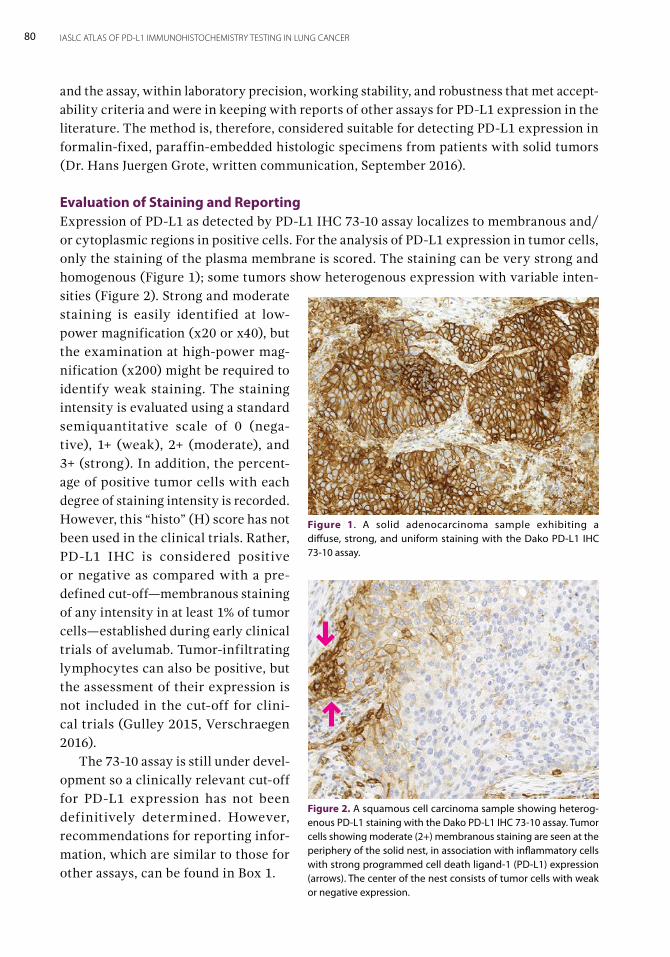
80 IASLC ATLAS OF PD-L1 IMMUNOHISTOCHEMISTRY TESTING IN LUNG CANCER
and the assay, within laboratory precision, working stability, and robustness that met accept-ability criteria and were in keeping with reports of other assays for PD-L1 expression in the literature. The method is, therefore, considered suitable for detecting PD-L1 expression in formalin-fixed, paraffin-embedded histologic specimens from patients with solid tumors (Dr. Hans Juergen Grote, written communication, September 2016).
Evaluation of Staining and ReportingExpression of PD-L1 as detected by PD-L1 IHC 73-10 assay localizes to membranous and/or cytoplasmic regions in positive cells. For the analysis of PD-L1 expression in tumor cells, only the staining of the plasma membrane is scored. The staining can be very strong and homogenous (Figure 1); some tumors show heterogenous expression with variable inten-sities (Figure 2). Strong and moderate staining is easily identified at low-power magnification (x20 or x40), but the examination at high-power mag-nification (x200) might be required to identify weak staining. The staining intensity is evaluated using a standard semiquantitative scale of 0 (nega-tive), 1+ (weak), 2+ (moderate), and 3+ (strong). In addition, the percent-age of positive tumor cells with each degree of staining intensity is recorded. However, this “histo” (H) score has not been used in the clinical trials. Rather, PD-L1 IHC is considered positive or negative as compared with a pre-defined cut-off—membranous staining of any intensity in at least 1% of tumor cells—established during early clinical trials of avelumab. Tumor-infiltrating lymphocytes can also be positive, but the assessment of their expression is not included in the cut-off for clini-cal trials (Gulley 2015, Verschraegen 2016). The 73-10 assay is still under devel-opment so a clinically relevant cut-off for PD-L1 expression has not been definitively determined. However, recommendations for reporting infor-mation, which are similar to those for other assays, can be found in Box 1.
Figure 1. A solid adenocarcinoma sample exhibiting a diffuse, strong, and uniform staining with the Dako PD-L1 IHC 73-10 assay.
Figure 2. A squamous cell carcinoma sample showing heterog-enous PD-L1 staining with the Dako PD-L1 IHC 73-10 assay. Tumor cells showing moderate (2+) membranous staining are seen at the periphery of the solid nest, in association with inflammatory cells with strong programmed cell death ligand-1 (PD-L1) expression (arrows). The center of the nest consists of tumor cells with weak or negative expression.
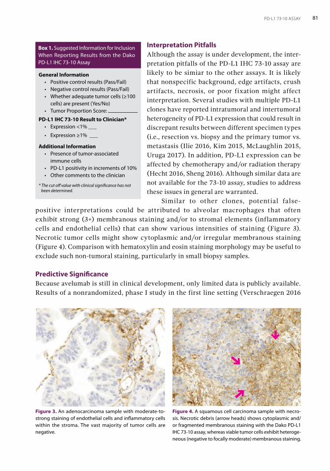
81PD-L1 73-10 ASSAY
Interpretation PitfallsAlthough the assay is under development, the inter-pretation pitfalls of the PD-L1 IHC 73-10 assay are likely to be simiar to the other assays. It is likely that nonspecific background, edge artifacts, crush artifacts, necrosis, or poor fixation might affect interpretation. Several studies with multiple PD-L1 clones have reported intratumoral and intertumoral heterogeneity of PD-L1 expression that could result in discrepant results between different specimen types (i.e., resection vs. biopsy and the primary tumor vs. metastasis (Ilie 2016, Kim 2015, McLaughlin 2015, Uruga 2017). In addition, PD-L1 expression can be affected by chemotherapy and/or radiation therapy (Hecht 2016, Sheng 2016). Although similar data are not available for the 73-10 assay, studies to address these issues in general are warranted. Similar to other clones, potential false-
positive interpretations could be attributed to alveolar macrophages that often exhibit strong (3+) membranous staining and/or to stromal elements (inflammatory cells and endothelial cells) that can show various intensities of staining (Figure 3). Necrotic tumor cells might show cytoplasmic and/or irregular membranous staining (Figure 4). Comparison with hematoxylin and eosin staining morphology may be useful to exclude such non-tumoral staining, particularly in small biopsy samples.
Predictive Significance Because avelumab is still in clinical development, only limited data is publicly available. Results of a nonrandomized, phase I study in the first line setting (Verschraegen 2016
General Information• Positive control results (Pass/Fail)• Negative control results (Pass/Fail)• Whether adequate tumor cells (≥100
cells) are present (Yes/No)• Tumor Proportion Score:
PD-L1 IHC 73-10 Result to Clinician* • Expression <1% ___
• Expression ≥1% ___
Additional Information • Presence of tumor-associated
immune cells• PD-L1 positivity in increments of 10%• Other comments to the clinician
* The cut off value with clinical significance has not been determined.
Box 1. Suggested Information for Inclusion When Reporting Results from the Dako PD-L1 IHC 73-10 Assay
Figure 4. A squamous cell carcinoma sample with necro-sis. Necrotic debris (arrow heads) shows cytoplasmic and/or fragmented membranous staining with the Dako PD-L1 IHC 73-10 assay, whereas viable tumor cells exhibit heteroge-neous (negative to focally moderate) membranous staining.
Figure 3. An adenocarcinoma sample with moderate-to-strong staining of endothelial cells and inflammatory cells within the stroma. The vast majority of tumor cells are negative.
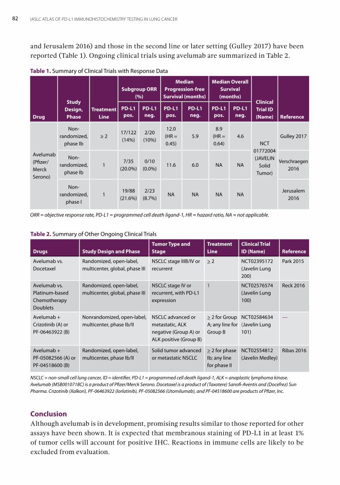
82 IASLC ATLAS OF PD-L1 IMMUNOHISTOCHEMISTRY TESTING IN LUNG CANCER
and Jerusalem 2016) and those in the second line or later setting (Gulley 2017) have been reported (Table 1). Ongoing clinical trials using avelumab are summarized in Table 2.
Conclusion Although avelumab is in development, promising results similar to those reported for other assays have been shown. It is expected that membranous staining of PD-L1 in at least 1% of tumor cells will account for positive IHC. Reactions in immune cells are likely to be excluded from evaluation.
Table 1. Summary of Clinical Trials with Response Data
Drug
StudyDesign, Phase
Treatment Line
Subgroup ORR (%)
Median Progression-free
Survival (months)
Median Overall Survival (months)
Clinical Trial ID (Name) Reference
PD-L1pos.
PD-L1neg.
PD-L1pos.
PD-L1neg.
PD-L1pos.
PD-L1neg.
Avelumab(Pfizer/Merck Serono)
Non- randomized,
phase Ib≥ 2
17/122(14%)
2/20(10%)
12.0(HR = 0.45)
5.98.9
(HR = 0.64)
4.6NCT
01772004(JAVELIN
Solid Tumor)
Gulley 2017
Non- randomized,
phase Ib1
7/35 (20.0%)
0/10 (0.0%)
11.6 6.0 NA NAVerschraegen
2016
Non- randomized,
phase I1
19/88 (21.6%)
2/23 (8.7%)
NA NA NA NAJerusalem
2016
ORR = objective response rate, PD-L1 = programmed cell death ligand-1, HR = hazard ratio, NA = not applicable.
Table 2. Summary of Other Ongoing Clinical Trials
Drugs Study Design and PhaseTumor Type and Stage
Treatment Line
Clinical Trial ID (Name) Reference
Avelumab vs. Docetaxel
Randomized, open-label, multicenter, global, phase III
NSCLC stage IIIB/IV or recurrent
> 2 NCT02395172(Javelin Lung 200)
Park 2015
Avelumab vs. Platinum-based Chemotherapy Doublets
Randomized, open-label, multicenter, global, phase III
NSCLC stage IV or recurrent, with PD-L1 expression
1 NCT02576574(Javelin Lung 100)
Reck 2016
Avelumab + Crizotinib (A) or PF-06463922 (B)
Nonrandomized, open-label, multicenter, phase Ib/II
NSCLC advanced or metastatic, ALK negative (Group A) or ALK positive (Group B)
> 2 for Group A; any line for Group B
NCT02584634(Javelin Lung 101)
—
Avelumab + PF-05082566 (A) or PF-04518600 (B)
Randomized, open-label, multicenter, phase Ib/II
Solid tumor advanced or metastatic NSCLC
> 2 for phase Ib; any line for phase II
NCT02554812(Javelin Medley)
Ribas 2016
NSCLC = non-small cell lung cancer, ID = identifier, PD-L1 = programmed cell death ligand-1, ALK = anaplastic lymphoma kinase. Avelumab (MSB0010718C) is a product of Pfizer/Merck Serono. Docetaxel is a product of (Taxotere) Sanofi-Aventis and (Docefrez) Sun Pharma. Crizotinib (Xalkori), PF-06463922 (lorlatinib), PF-05082566 (Utomilumab), and PF-04518600 are products of Pfizer, Inc.
Other Anti–PD-L1 Clones: Alternative Assays and Laboratory-Developed TestsBy Lynette M. Sholl, Mari Mino-Kenudson, Reinhard Buettner, and Ignacio Wistuba
9The US Food and Drug Administration (FDA) has approved a variety of companion and complementary diagnostics for programmed cell death ligand-1 (PD-L1) expression testing to help determine an appropriate PD-1/PD-L1 axis blockade therapy for a variety of cancer types. The proliferation of these diagnostic assays poses special challenges for pathology laboratories because most laboratories do not use all of the staining platforms required by the different assay manufacturers, and use of companion diagnostic kits often increases the cost of each individual test. Therefore, laboratories seeking to offer PD-L1 testing services face significant capital and operating expenditures to acquire the equipment and reagents necessary to offer a full range of companion and complementary PD-L1 diagnostics. Before the approval of pembrolizumab by the FDA for patients with advanced non-small cell lung cancer (NSCLC) in the first-line setting, many laboratories were sending out selected speci-mens to commercial pathology laboratories that offered companion and complimentary diagnostic kits, with the results incorporated in the pathology report when they became available. The National Comprehensive Cancer Network guidelines implemented a rec-ommendation post-approval that immunohistochemistry (IHC) testing for PD-L1 with a validated assay should be performed for both advanced squamous cell and non-squamous cell NSCLCs. The cost and administrative burden of sending tissue to reference laboratories for PD-L1 testing may be prohibitive for many laboratories, given the number of patients with advanced NSCLC seen in daily practice (especially in referral centers). Lower-cost, laboratory-developed tests (LDTs) have been commercially available for more than a decade, beginning with the mouse monoclonal anti-human PD-L1 antibody 29E.2A3 (Latchman 2001). However, this and other commercially available antibodies, such as rabbit polyclonal anti–PD-L1 ab58810 (Abcam) and mouse monoclonal MIH1 (Thermo Fisher Scientific), were shown to have lower specificity relative to another mouse monoclo-nal antibody, 5H1 (Dong 2002, Velcheti 2014), developed and made available through the Lieping Chen laboratory (Yale School of Medicine, New Haven, Conneticut) but not through commercial vendors. This 5H1 antibody was used in the phase I trial of nivolumab, and a correlation between tumor PD-L1 expression and response was demonstrated (Topalian
84 IASLC ATLAS OF PD-L1 IMMUNOHISTOCHEMISTRY TESTING IN LUNG CANCER
2012). In subsequent clinical trials of nivolumab, a novel and proprietary rabbit monoclonal 28-8 antibody (Agilent Technologies/Dako) replaced 5H1 and was subsequently incorpo-rated into a complementary diagnostic kit (Phillips 2015).
Biomarker PD-L1 AntibodiesPD-L1 is a transmembrane protein, most of which is extracellular (including the PD-1 bind-ing domain), with a 31 amino acid cytoplasmic domain. The 5H1, 7G11, 015, 22C3, and 28-8 antibodies bind the extracellular domain, whereas SP142, SP263, E1L3N, and 9A11 bind the intracellular domain (Table 1). Although both membranous and cytoplasmic expression can be seen in an antibody-dependent fashion, membranous staining has been correlated with response in most clinical trials of PD-1 inhibitors. The mechanism leading to cytoplasmic staining is not clear; however, it might represent accumulation of PD-L1 splice variants that are not effectively localized to the membrane (Mahoney 2015).
E1L3N AntibodyOne of the most commonly used commercially-available antibodies used in LDTs is E1L3N rabbit monoclonal antibody by Cell Signaling Technology, introduced in 2014. The preva-lence of PD-L1 positivity using the E1L3N antibody in lung cancers ranges from 22% to 66%, depending on histology, platform, detection systems, and cut-off definition (i.e., 1%, 5%, or 50%, Table 2). In lung cancers, E1L3N has the highest sensitivity for membranous expression when compared with SP142, 9A11, 015, and 7G11, with membranous expression by the latter two extracellular-domain antibodies obscured by high cytoplasmic staining
Table 1. Anti–PD-L1 Antibodies for Use in Formalin-fixed Paraffin-embedded IHC
Antibody Clone Source Epitope Host Validated for
Specificity
5H1Lieping Chen laboratory, Yale
School of MedicineExtracellular
domainMouse monoclonal
Dong 2002Velcheti 2014
7G11Freeman laboratory, Dana-
Farber Cancer Institute, Harvard Medical School
Extracellular domain IgV
Mouse IgG1 Mahoney 2015
015 Sino BiologicalExtracellular
domainRabbit IgG Mahoney 2015
22C3 DakoExtracellular
domainMouse monoclonal
Dolled-Filhart2016
28-8 AbcamExtracellular
domainRabbit monoclonal Cogswell 2017
SP142 Spring Bioscience/VentanaCytoplasmic
domainRabbit monoclonal Mahoney 2015
SP263 Spring Bioscience/VentanaCytoplasmic
domainRabbit monoclonal Smith 2016
E1L3N Cell Signaling TechnologyCytoplasmic
domainRabbit monoclonal Mahoney 2015
9A11 Cell Signaling TechnologyCytoplasmic
domainMouse IgG1 Mahoney 2015
Ig = immunoglobin.
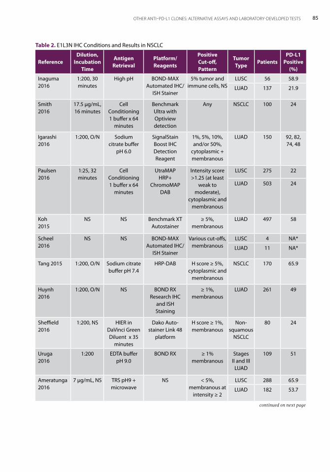
85OTHER ANTI–PD-L1 CLONES: ALTERNATIVE ASSAYS AND LABORATORY-DEVELOPED TESTS
Table 2. E1L3N IHC Conditions and Results in NSCLC
ReferenceDilution,
Incubation Time
Antigen Retrieval
Platform/ Reagents
Positive Cut-off, Pattern
Tumor Type
PatientsPD-L1
Positive (%)
Inaguma 2016
1:200, 30 minutes
High pH BOND-MAX Automated IHC/
ISH Stainer
5% tumor and immune cells, NS
LUSC 56 58.9
LUAD 137 21.9
Smith 2016
17.5 μg/mL, 16 minutes
Cell Conditioning 1 buffer x 64
minutes
Benchmark Ultra with Optiview detection
Any NSCLC 100 24
Igarashi 2016
1:200, O/N Sodium citrate buffer
pH 6.0
SignalStain Boost IHC Detection Reagent
1%, 5%, 10%, and/or 50%,
cytoplasmic + membranous
LUAD 150 92, 82, 74, 48
Paulsen 2016
1:25, 32 minutes
Cell Conditioning 1 buffer x 64
minutes
UtraMAP HRP+
ChromoMAP DAB
Intensity score >1.25 (at least
weak to moderate),
cytoplasmic and membranous
LUSC 275 22
LUAD 503 24
Koh 2015
NS NS Benchmark XT Autostainer
≥ 5%, membranous
LUAD 497 58
Scheel 2016
NS NS BOND-MAX Automated IHC/
ISH Stainer
Various cut-offs, membranous
LUSC 4 NA*
LUAD 11 NA*
Tang 2015 1:200, O/N Sodium citrate buffer pH 7.4
HRP-DAB H score ≥ 5%, cytoplasmic and
membranous
NSCLC 170 65.9
Huynh 2016
1:200, O/N NS BOND RX Research IHC
and ISH Staining
≥ 1%, membranous
LUAD 261 49
Sheffield 2016
1:200, NS HIER in DaVinci Green Diluent x 35
minutes
Dako Auto-stainer Link 48
platform
H score ≥ 1%, membranous
Non-squamous
NSCLC
80 24
Uruga 2016
1:200 EDTA buffer pH 9.0
BOND RX ≥ 1% membranous
Stages II and III
LUAD
109 51
Ameratunga 2016
7 μg/mL, NS TRS pH9 + microwave
NS < 5%, membranous at
intensity ≥ 2
LUSC 288 65.9
LUAD 182 53.7
continued on next page
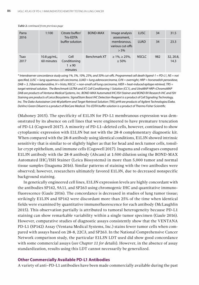
86 IASLC ATLAS OF PD-L1 IMMUNOHISTOCHEMISTRY TESTING IN LUNG CANCER
(Mahoney 2015). The specificity of E1L3N for PD-L1 membranous expression was dem-onstrated by its absence on cell lines that were engineered to have premature truncation of PD-L1 (Cogswell 2017). A minority of PD-L1–deleted cells, however, continued to show cytoplasmic expression with E1L3N but not with the 28-8 complementary diagnostic kit. When compared with the 28-8 antibody using identical conditions, E1L3N showed intrinsic sensitivity that is similar to or slightly higher as that for head and neck tumor cells, tonsil-lar crypt epithelium, and immune cells (Cogswell 2017). Inaguma and colleagues compared E1L3N antibody with the 28-8 antibody (Abcam) at 1:500 dilution using the BOND-MAX Automated IHC/ISH Stainer (Leica Biosystems) in more than 5,000 tumor and normal tissue samples (Inaguma 2016). Similar patterns of staining with the two antibodies were observed; however, researchers ultimately favored E1L3N, due to decreased nonspecific background staining. In genetically engineered cell lines, E1L3N expression levels are highly concordant with the antibodies SP142, 9A11, and SP263 using chromogenic IHC and quantitative immuno-fluorescence (Gaule 2016). The concordance is decreased in studies of lung tumor tissue; strikingly E1L3N and SP142 were discordant more than 25% of the time when identical fields were examined by quantitative immunofluorescence for each antibody (McLaughlin 2015). This observation partially is attributed to tumoral heterogeneity because PD-L1 staining can show remarkable variability within a single tumor specimen (Gaule 2016). However, comparative studies of diagnostic assays consistently show that the VENTANA PD-L1 (SP142) Assay (Ventana Medical Systems, Inc.) stains fewer tumor cells when com-pared with assays based on 28-8, 22C3, and SP263. In the National Comprehensive Cancer Network comparison study, the particular E1L3N LDT used did show good concordance with some commercial assays (see Chapter 11 for details). However, in the absence of assay standardization, results using this LDT cannot necessarily be generalized.
Other Commercially Available PD-L1 AntibodiesA variety of anti–PD-L1 antibodies have been made commercially available during the past
Parra 2016
1:100 Citrate buffer/Tris-EDTA
buffer solution
BOND-MAX Image analysis assessment,
membranous, various cut-offs
> 5%
LUSC 34 31.5
LUAD 34 23.3
Tsao 2017
10.8 μg/mL, 60 minutes
Cell Conditioning
1 x 90 minutes
Benchmark XT ≥ 1%, ≥ 25%, ≥ 50%
NSCLC 982 32, 20.8, 14.3
* Interobserver concordance study using 1%, 5%, 10%, 25%, and 50% cut-offs. Programmed cell death ligand-1 = PD-L1, NS = not specified, LUSC = lung squamous cell carcinoma, LUAD = lung adenocarcinoma, O/N = overnight, HRP = horseradish peroxidase, DAB = 3, 3’diaminobenzidine, H = histo, NSCLC = non-small cell lung carcinoma, HIER = heat-induced epitope retrieval, TRS = target retrieval solution. The Benchmark ULTRA and XT, Cell Conditioning 1 Solution (CC1), and UtraMAP HRP+ChromoMAP DAB are products of Ventana Medical Systems, Inc. BOND-MAX Automated IHC/ISH Stainer and BOND RX Research IHC and ISH Staining are products of Leica Biosystems. SignalStain Boost IHC Detection Reagent is a product of Cell Signaling Technology, Inc. The Dako Autostainer Link 48 platform and Target Retrieval Solution (TRS) pH9 are products of Agilent Technologies/Dako. DaVinci Green Diluent is a product of BioCare Medical. Tris-EDTA buffer solution is a product of Thermo Fisher Scientific.
Table 2 continued from previous page
87OTHER ANTI–PD-L1 CLONES: ALTERNATIVE ASSAYS AND LABORATORY-DEVELOPED TESTS
several years. Some, such as the PD-L1 rabbit polyclonal CD274 antibody from Proteintech Group, Inc., have been used in a variety of published studies across tumor types (Table 3, Yang 2014, Yang 2016, Song 2016). Head-to-head studies of LDTs using antibodies other than E1L3N are limited, however, Sheffield et al. demonstrated high agreement (Cohen’s kappa of 0.69) among three LDTs (Tables 2 and 3) and the 28-8 companion diagnostic in a cohort of non-squamous NSCLC samples. Correlation with RNA expression levels was very good to excellent, as shown by in situ hybridization (Sheffield 2016). In a study by Paulsen et al., E1L3N was preferred over PD-L1 antibodies MAB1561 (mouse monoclonal, R&D Systems Inc.) and ab58810 (rabbit polyclonal, Abcam) following evaluation using PD-L1–transfected cell lines, with negative (brain) and positive (placenta) tissue controls (Table 2, Paulsen 2017, Adam 2017). Studies of the 22C3 antibody using the companion diagnostic (PD-L1 IHC 22C3 pharmDx, Agilent Technologies/Dako) assay as compared with modified approaches using two different Ventana detection systems demonstrated comparable, although not identical, performance across assays (Neuman 2016). Apart from antibody performance, the success and reproducibility of IHC-based assays are heavily dependent on pre-analytic tissue handling (Gown 2016), as well as on the specific characteristics of the antigen-retrieval and detection systems used. For PD-L1, the antigen-retrieval conditions using citrate buffer pH6, citrate buffer pH 8, or EDTA buffer have been shown to significantly affect the rate of positive PD-L1 expression for E1L3N (unpublished observation). Available detection systems generate different levels of expression depending on the strength of the amplification step (Figure 1). Amplification strength has the potential to radically alter the outcome of the test and could lead to alternative PD-L1 expression scoring when the expression level is near a cut-off threshold (see Chapter 3 for details).
Future Directions in Standardization of PD-L1 IHC AssaysAll companion and complementary PD-L1 diagnostics were developed using proprietary antibodies, many of which were made commercially available only after FDA approval. This arrangement has hampered efforts to cross-compare the performance of FDA-approved in vitro diagnostics and LDTs, however, data are now emerging on the comparative perfor-mance of different antibodies and platforms in NSCLC (Chapter 11). The studies cited here, and others under way, indicate that most antibodies, including those used exclusively in the LDT setting, demonstrate comparable performance in well-controlled settings. Ultimately, the interpretation is limited by variable definitions of positivity for each companion or com-plementary diagnostic and by the lack of a clear gold-standard comparator, apart from the commercial kits themselves. The relative lack of well-controlled outcomes data for patients selected using an LDT and our limited knowledge of other factors that might modify response to PD-1/PD-L1 axis blockade confound the use of PD-L1 positive or negative expression in treatment selection, although clinical outcomes will inform this approach. Another challenge associated with the use of LDTs as predictive markers in oncology practice relates to the lack of clear oversight and dearth of standardization. In the United States, proficiency testing programs, although commonplace for molecular diagnostics laboratories and clinical pathology specialties, are not yet well established for IHC labs. The College of American Pathologists (CAP) has developed a set of recommendations and expert consensus opinions relating to the validation of IHC LDTs. The requirements are
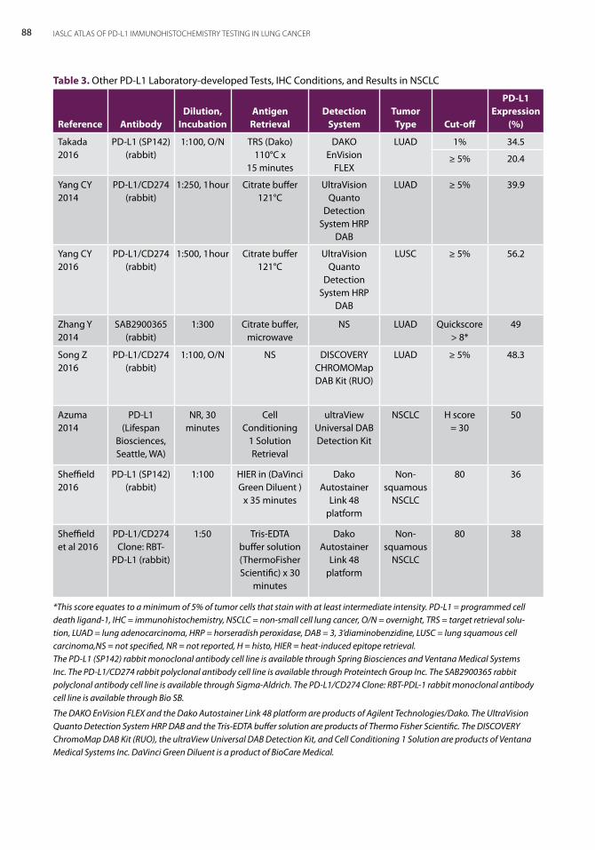
88 IASLC ATLAS OF PD-L1 IMMUNOHISTOCHEMISTRY TESTING IN LUNG CANCER
Table 3. Other PD-L1 Laboratory-developed Tests, IHC Conditions, and Results in NSCLC
Reference AntibodyDilution,
IncubationAntigen
RetrievalDetection
SystemTumor Type Cut-off
PD-L1 Expression
(%)
Takada 2016
PD-L1 (SP142) (rabbit)
1:100, O/N TRS (Dako) 110°C x
15 minutes
DAKO EnVision
FLEX
LUAD 1% 34.5
≥ 5% 20.4
Yang CY 2014
PD-L1/CD274 (rabbit)
1:250, 1 hour Citrate buffer 121°C
UltraVision Quanto
Detection System HRP
DAB
LUAD ≥ 5% 39.9
Yang CY 2016
PD-L1/CD274 (rabbit)
1:500, 1 hour Citrate buffer 121°C
UltraVision Quanto
Detection System HRP
DAB
LUSC ≥ 5% 56.2
Zhang Y 2014
SAB2900365 (rabbit)
1:300 Citrate buffer, microwave
NS LUAD Quickscore > 8*
49
Song Z 2016
PD-L1/CD274 (rabbit)
1:100, O/N NS DISCOVERY CHROMOMapDAB Kit (RUO)
LUAD ≥ 5% 48.3
Azuma 2014
PD-L1 (Lifespan
Biosciences, Seattle, WA)
NR, 30 minutes
Cell Conditioning
1 Solution Retrieval
ultraView Universal DAB Detection Kit
NSCLC H score = 30
50
Sheffield 2016
PD-L1 (SP142) (rabbit)
1:100 HIER in (DaVinci Green Diluent )
x 35 minutes
Dako Autostainer
Link 48 platform
Non-squamous
NSCLC
80 36
Sheffield et al 2016
PD-L1/CD274 Clone: RBT-
PD-L1 (rabbit)
1:50 Tris-EDTA buffer solution (ThermoFisher Scientific) x 30
minutes
Dako Autostainer
Link 48 platform
Non-squamous
NSCLC
80 38
*This score equates to a minimum of 5% of tumor cells that stain with at least intermediate intensity. PD-L1 = programmed cell death ligand-1, IHC = immunohistochemistry, NSCLC = non-small cell lung cancer, O/N = overnight, TRS = target retrieval solu-tion, LUAD = lung adenocarcinoma, HRP = horseradish peroxidase, DAB = 3, 3’diaminobenzidine, LUSC = lung squamous cell carcinoma,NS = not specified, NR = not reported, H = histo, HIER = heat-induced epitope retrieval. The PD-L1 (SP142) rabbit monoclonal antibody cell line is available through Spring Biosciences and Ventana Medical Systems Inc. The PD-L1/CD274 rabbit polyclonal antibody cell line is available through Proteintech Group Inc. The SAB2900365 rabbit polyclonal antibody cell line is available through Sigma-Aldrich. The PD-L1/CD274 Clone: RBT-PDL-1 rabbit monoclonal antibody cell line is available through Bio SB.
The DAKO EnVision FLEX and the Dako Autostainer Link 48 platform are products of Agilent Technologies/Dako. The UltraVision Quanto Detection System HRP DAB and the Tris-EDTA buffer solution are products of Thermo Fisher Scientific. The DISCOVERY ChromoMap DAB Kit (RUO), the ultraView Universal DAB Detection Kit, and Cell Conditioning 1 Solution are products of Ventana Medical Systems Inc. DaVinci Green Diluent is a product of BioCare Medical.
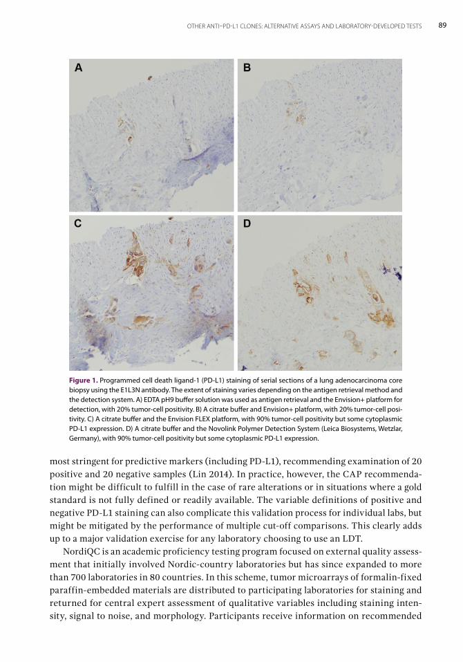
89OTHER ANTI–PD-L1 CLONES: ALTERNATIVE ASSAYS AND LABORATORY-DEVELOPED TESTS
most stringent for predictive markers (including PD-L1), recommending examination of 20 positive and 20 negative samples (Lin 2014). In practice, however, the CAP recommenda-tion might be difficult to fulfill in the case of rare alterations or in situations where a gold standard is not fully defined or readily available. The variable definitions of positive and negative PD-L1 staining can also complicate this validation process for individual labs, but might be mitigated by the performance of multiple cut-off comparisons. This clearly adds up to a major validation exercise for any laboratory choosing to use an LDT. NordiQC is an academic proficiency testing program focused on external quality assess-ment that initially involved Nordic-country laboratories but has since expanded to more than 700 laboratories in 80 countries. In this scheme, tumor microarrays of formalin-fixed paraffin-embedded materials are distributed to participating laboratories for staining and returned for central expert assessment of qualitative variables including staining inten-sity, signal to noise, and morphology. Participants receive information on recommended
Figure 1. Programmed cell death ligand-1 (PD-L1) staining of serial sections of a lung adenocarcinoma core biopsy using the E1L3N antibody. The extent of staining varies depending on the antigen retrieval method and the detection system. A) EDTA pH9 buffer solution was used as antigen retrieval and the Envision+ platform for detection, with 20% tumor-cell positivity. B) A citrate buffer and Envision+ platform, with 20% tumor-cell posi-tivity. C) A citrate buffer and the Envision FLEX platform, with 90% tumor-cell positivity but some cytoplasmic PD-L1 expression. D) A citrate buffer and the Novolink Polymer Detection System (Leica Biosystems, Wetzlar, Germany), with 90% tumor-cell positivity but some cytoplasmic PD-L1 expression.
90 IASLC ATLAS OF PD-L1 IMMUNOHISTOCHEMISTRY TESTING IN LUNG CANCER
antibodies and protocols, as well as tailored feedback for improving insufficient protocols. According to the NordiQC data, false negativity is a common reason for insufficient results and can be mitigated by improved epitope retrieval or enhanced-sensitivity detection sys-tems (Vyberg 2016). Standardization programs established for predictive IHC markers, such as for ALK expression in lung cancer, have successfully led to high inter-laboratory concordance (Cutz 2014), followed by the development of national proficiency testing in Canada for ALK IHC and fluorescence in situ hybridization testing (Cheung 2015). The UK National External Quality Assessment Service (UK NEQAS) center in the Royal Infirmary of Edinburgh, Edinburgh, United Kingdom, is in the process of developing an external quality-assessment scheme for PD-L1 IHC. It is also worth noting that, according to UK NEQAS external quality-assessment data for ALK expression IHC testing, laboratories using LDTs had a significantly greater chance of failing an external quality assessment when compared with laboratories using commercial kit assays (Ibrahim 2016). Standardization of PD-L1 LDTs should involve head-to-head comparisons of PD-L1 expression levels determined by LDTs compared with levels determined by approved com-panion diagnostics, such as the PD-L1 IHC 22C3 pharmDx in the context of first line therapy, using an adequate number of positive and negative samples (such as per CAP guidelines) and/or tissue microarrays. It is recommended that a comparison of tumor proportion scores by the two methodologies is performed to detect any systematic bias in the LDT relative to the companion diagnostic. Due to the multiple factors that could influence the outcome of any IHC test (see Chapter 3 for details) and to the lack of any clinical outcomes data using anything other than a trial-validated commercial assay, the only standard that can be used to gauge the likely clinical predictive performance of an LDT would be a commercial assay.
Recommendations for PD-L1 Testing by LDTs: Validation and PerformancePD-L1 IHC using LDTs should be carried out on approximately 4-μ sections, cut fresh when-ever possible. Immunoreactivity tends to degrade with time, and use of slides cut more than 6 months prior should be avoided, although the performance of available antibodies on archival tissues is limited. Shorter time periods for storage of cut sections might be problematic if sections are not stored in cool, dry, and dark conditions (Chapter 3). A gold standard posi-tive control has not been formally proposed, however, acceptable positive control samples include tonsil epithelium, placenta (placental trophoblasts highly express PD-L1, Inaguma 2016), and PD-L1 cell lines with high expression, such as NCI-H226 (lung large cell carci-noma), NCI-H1975 (lung adenocarcinoma), or HDLM2 (Hodgkin lymphoma). Brain tissue or known negative cell lines, such as H549 or PC3, may be used as negative controls. It is recommended to use controls with different levels of PD-L1 expression including negative, low, intermediate, and high levels. This may be facilitated by use of commercially available cell lines engineered to express membranous PD-L1. These cell lines are developed for PD-L1 IHC controls that have both high and, more importantly, low levels of epitope expression (see Chapter 3 for details). Use of tumors with known high levels of innate PD-L1 expression (typically a result of CD274 amplification), such as Hodgkin lymphoma or human papilloma virus-driven cervical squamous cell carcinoma may also serve as positive comparators in a validation set. Igarashi et al. proposed the use of alveolar macrophages as an internal
91OTHER ANTI–PD-L1 CLONES: ALTERNATIVE ASSAYS AND LABORATORY-DEVELOPED TESTS
control and intensity score reference for tumor staining in lung specimens (Igarashi 2016); however, the intensity of staining in this cell type can vary considerably among individual samples. Therefore, additional external positive control(s) showing consistent expression levels—particularly including a low expression control—is recommended in routine practice. If, however, PD-L1 expression is absent in both a tumor sample and the associated alveolar macrophages of a lung biopsy sample, an effort should be made to confirm that a sample is appropriately immunoreactive and is not falsely negative. Validation and proficiency also may be facilitated by sample exchange among diagnostic laboratories, both to confirm accept-able antibody/platform performance and to confirm good inter-pathologist agreement about established cut-off levels, such as at 1% and 50% tumor proportion scores. Inter-pathologist reproducibility for tumor cell scoring is high in multiple studies, but the same is not true for quantification of PD-L1 expression on immune cells (Rimm 2016, Scheel 2016).
ConclusionLDTs using biologically validated antibodies and clinically validated techniques may be an acceptable and economical alternative to companion or complementary diagnostics in iden-tifying tumors with PD-L1 overexpression—with caveats. In a multicenter study comparing several commercial assays, as well as several LDTs using the same antibody clones as used in the commercial assays, 50% of the LDTs did not have adequate comparative technical performance (Adam 2017). The standardization of platform and staining conditions is of critical importance for consistent and reproducible results. Ongoing studies to establish equivalence to established companion or complementary diagnostics should provide support for the use of stringently validated LDTs as predictors of response to approved immuno-therapies. However, a more robust international proficiency testing infrastructure will be essential to promote high-quality and consistent PD-L1 IHC results across antibodies and test platforms and in a variety of settings.

Complementary and Companion DiagnosticsBy Fred R. Hirsch and Sanja Dacic
10The commercial success of drugs such as tratuzumab and imatinib, which both require companion diagnostics before they can be prescribed, has advanced the co-development of therapeutic products and accompanying in vitro diagnostics. There are now numerous examples of therapeutic products with an accompanying companion diagnostic (FDA. List of Cleared or Approved Companion Diagnostic Devices). With the introduction of personalized medicine, the US Food and Drug Administration (FDA) introduced the term “companion diagnostics,” which is defined as a diagnostic assay required for the use of the associated drug based on clinical efficacy and safety data. There are several drugs approved by the FDA with companion diagnostics that assist with therapeutic selection for treatment of patients with non-small cell lung cancer (NSCLC): the Vysis ALK Break Apart FISH Probe Kit (Abbott Laboratories, Abbott Park, Illinois) or the VENTANA ALK (D5F3) CDx Assay (Ventana Medical Systems, Inc., Tucson, Arizona) for crizotinib, EGFR mutation testing with the Therascreen EGFR RGQ PCR Kit (Qiagen, Hilden, Germany) for gefitinib and afatinib, and the Cobas 4800 System (Roche Molecular Diagnostics Inc., Pleasanton, California) for erlotinib. There are several reasons why there is so much interest in the development of companion diagnostics. The main advantage of companion diagnostics is to segregate a patient popula-tion into two subsets: biomarker positive and biomarker negative in order to ensure patients have the highest chance of clinical benefit on a safety basis. This separation is based on a quantitative assay result that is translated into a qualitative result, which represents a clinical decision point, or cut-off. Furthermore, the safety and efficacy of the therapeutic product is evaluated in the population that is treated in the clinical trial. Not all of the clinical trials for drugs in development will be successful, and a companion diagnostic is one of the few tools available to drug developers that can accelerate identification of the patient population most likely to benefit from a specific therapeutic agent. In turn, this gives the therapeutic agent a higher chance for achieving regulatory approval. Furthermore, enrollment of selec-tive patients using a companion diagnostic can reduce the duration of the clinical trial. This strategy resulted in a dramatic increase in biomarker-targeted drug-development programs.
94 IASLC ATLAS OF PD-L1 IMMUNOHISTOCHEMISTRY TESTING IN LUNG CANCER
In the early 1990s, 5% of new drug approvals were for targeted therapies, whereas this per-centage increased to 45% in 2013. In 2015, the FDA introduced the term “complementary diagnostics” for personalized therapy, which is defined as a diagnostic assay that predicts a favorable outcome of the associated drug by selecting patients based on results of the assay. However, it is not harm-ful to treat patients with the associated drug in the absence of assay results or if the results are negative. In other words, use of the complementary assay is not required for treatment with the associated drug. The differences between these two types of assays have been extensively reviewed elsewhere (Milne 2015). Complimentary diagnostic test outcomes fall into the “nice to know, but not required” category, which means that results are variably interpreted and used by oncologists. Requiring use of a companion diagnostic assists with consistency of test use, although it may also be viewed as inconvenient, depending in individual perspec-tives. Laboratories have more leverage, however, if test funding is an issue but test use is a requirement. Use of the test should result in a higher probability of treatment benefit because the patient group has been better defined. However, inconsistent use of these tests and variable interpretation of results allows increased patient access to treatment groups, widening treatment populations. The flexibility may be appreciated by some, but this may make it harder for some laboratories to get funding for a test that is not viewed as necessary.
ExamplesThe PD-L1 IHC 28-8 pharmDx (Dako, Glostrop, Denmark) was given complementary diag-nostic test status based on the results of the CheckMate 017 clinical trial. In this trial, overall, patients treated with nivolumab had longer survival than those treated with docetaxel, although a subset analyses clearly suggested that the level of programmed cell death ligand-1 (PD-L1) expression in NSCLC tumors might help identify patients who are more likely to benefit from nivolumab. Furthermore, those patients who had PD-L1 expression levels less than the biomarker threshold of 1% did no worse with nivolumab when compared with docetaxel. The results, therefore, showed clinical benefit of the treatment without patient selection, but the benefit was greater for the selected patients. If use of nivolumab was based on results of a required companion diagnostic, some patients would lose the chance to receive a potentially beneficial treatment. Nivolumab has been approved by the FDA for second-line therapy of patients with advanced NSCLC, with no requirement for a companion diagnostic. The 28-8 pharmDx assay, however, serves as a complementary diagnostic using a cut-off value of PD-L1 expression of at least 1% (see Chapters 2 and 4). Pembrolizumab has been approved by the FDA and the European Medicines Agency for both first-line and second-line therapy of patients with advanced NSCLC. The PD-L1 IHC 22C3 pharmDx (Dako, Glostrop, Denmark) assay is a companion diagnostic that defines positive PD-L1 expression as expression by at least 50% of tumor cells in the first-line set-ting, and 1% or greater of tumor cell expression for second-line therapy. Some multi-industrial/academic collaborative studies, such as the “Blueprint Project” (Chapter 11), have been published (Hirsch 2017) and others are ongoing, with the goal to compare the analytical and diagnostic performance across the various assays for PD-L1 expression.
95COMPLEMENTARY AND COMPANION DIAGNOSTICS
Validation Processes and Diagnostic TestsBoth companion and complimentary diagnostic assays undergo rigorous analytical validation processes for accuracy, reproducibility, specificity, sensitivity, and stability. In addition, both types of diagnostic assays require use of particular analytical reagents and instrumentation, which makes implementation in diagnostic laboratories challenging (Kerr 2015, Kerr 2016a). Each of the five drugs that are furthest in the development process for NSCLC, have their own trial-validated assays (as discussed throughout this Atlas), which creates complexity of choice. There are several diagnostic platforms for PD-L1 IHC, all of which use different antibody clones, staining protocols, platforms, and, most importantly, different clinical deci-sion points. Most diagnostic laboratories are not able to adopt and run multiple diagnostic platforms due to cost effectiveness and to the increasingly important role of reimbursement for therapeutic tests. Health care reform in the United States is changing the model for clinical provider payment, moving to outcomes-driven reimbursement compared with testing driven. The patient-outcome payment model is based on clinical outcomes for patients as opposed to the number of clinical interventions and diagnostic tests ordered and implemented by physi-cians. The current development strategy of a single diagnostic test for a single therapeutic agent is challenged by this new payment model because the one-for-one approach is unlikely to predict outcome in a large patient population. Payers are becoming very interested in diagnostic tests that provide information about multiple potential treatment options that could be provided by multiplex assays rather than by a single assay. This approach opens the question as to whether the same diagnostic test can be used for similar therapeutic agents, such as in the case of immune check point inhibitors. This is discussed in more detail in Chapter 11.
Conclusion Use of a companion diagnostic assay is a requirement for drug eligibility and prescription to ensure the highest chance of clinical benefit on a safety basis, whereas use of the comple-mentary assay is optional, with results informing but not dictating treatment decisions. A better understanding of the different PD-L1–expression assays hopefully will lead to a more rational use of these tools in clinical practice.

Assay Harmonization: Is It Possible? By Keith M. Kerr, Fred R. Hirsch, Yasushi Yatabe, and Ming S. Tsao 11There are detailed descriptions of five different programmed cell death ligand-1 (PD-L1) immunohistochemistry (IHC) assays elsewhere in this Atlas. Each of these assays has been separately developed, or is undergoing validation, in association with a specific anti-PD-1/PD-L1 drug (Table 1). The 28-8 clone-based assay (Agilent Technologies/Dako) is now reg-istered with the US Food and Drug Administration (FDA) as a complementary diagnostic in association with nivolumab, whereas the 22C3 assay (Agilent Technologies/Dako) is a companion diagnostic for pembrolizumab. The SP142 assay (Ventana Medical Systems, Inc.) is approved for use with atezolizumab as a complementary diagnostic. At the time of this publication, the SP263 clone-based assay (Ventana Medical Systems, Inc.), associated with durvalumab, is available for use in Europe but not in the United States, and a 73-10 antibody clone-based assay for use with avelumab is in development. Recently, based upon some of the technical comparison studies discussed below, the SP263 assay has also become available as a complementary test for use with nivolumab. Unlike the trial validated 28-8 assay, however, there are no clinical validation data relating to SP263 use with nivolumab. The assays developed by Dako (Table 1) have been developed for use on a Dako automated IHC staining platform (the Dako Autostainer Link 48), whereas the two Ventana assays are being developed for use on a Ventana platform (the Benchmark XT, GX, or Ultra). The majority of the emerging evidence, reviewed elsewhere in this Atlas, indicates that the anti-PD-1/PD-L1 drugs (Table 1) all show superior response and survival rates for tumors with PD-L1 IHC expression above a given detection threshold, when compared with tumors with expression under the threshold. These tests—whether used as compan-ion or complementary diagnostic assays—will be used, either by regulatory authorities or through physician preference, to inform the prescription of these drugs. This will be the case for use in all lines of treatment. Anti–PD-L1 IHC testing will be required in non-small cell lung cancer (NSCLC) for the foreseeable future. The rapid emergence of these five drug–assay combinations poses some unique challenges for the pathology and the oncology communities (Kerr 2015; Kerr 2016a; Kerr 2016b; Sholl 2016). Some of the issues involved have been encountered before. The pathology community
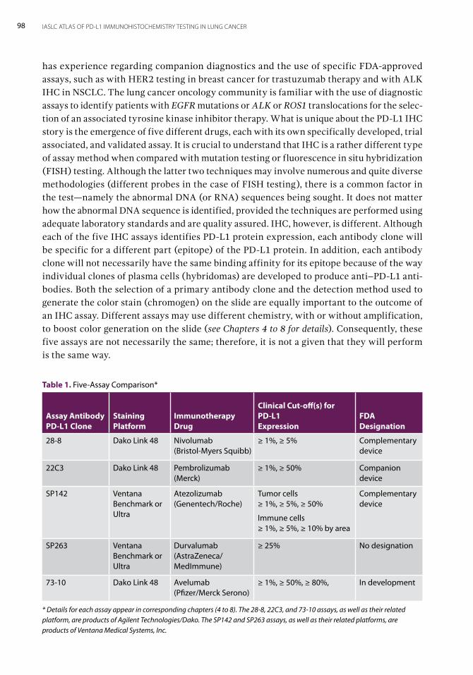
98 IASLC ATLAS OF PD-L1 IMMUNOHISTOCHEMISTRY TESTING IN LUNG CANCER
has experience regarding companion diagnostics and the use of specific FDA-approved assays, such as with HER2 testing in breast cancer for trastuzumab therapy and with ALK IHC in NSCLC. The lung cancer oncology community is familiar with the use of diagnostic assays to identify patients with EGFR mutations or ALK or ROS1 translocations for the selec-tion of an associated tyrosine kinase inhibitor therapy. What is unique about the PD-L1 IHC story is the emergence of five different drugs, each with its own specifically developed, trial associated, and validated assay. It is crucial to understand that IHC is a rather different type of assay method when compared with mutation testing or fluorescence in situ hybridization (FISH) testing. Although the latter two techniques may involve numerous and quite diverse methodologies (different probes in the case of FISH testing), there is a common factor in the test—namely the abnormal DNA (or RNA) sequences being sought. It does not matter how the abnormal DNA sequence is identified, provided the techniques are performed using adequate laboratory standards and are quality assured. IHC, however, is different. Although each of the five IHC assays identifies PD-L1 protein expression, each antibody clone will be specific for a different part (epitope) of the PD-L1 protein. In addition, each antibody clone will not necessarily have the same binding affinity for its epitope because of the way individual clones of plasma cells (hybridomas) are developed to produce anti–PD-L1 anti-bodies. Both the selection of a primary antibody clone and the detection method used to generate the color stain (chromogen) on the slide are equally important to the outcome of an IHC assay. Different assays may use different chemistry, with or without amplification, to boost color generation on the slide (see Chapters 4 to 8 for details). Consequently, these five assays are not necessarily the same; therefore, it is not a given that they will perform is the same way.
Table 1. Five-Assay Comparison*
Assay Antibody PD-L1 Clone
Staining Platform
Immunotherapy Drug
Clinical Cut-off(s) for PD-L1 Expression
FDA Designation
28-8 Dako Link 48 Nivolumab (Bristol-Myers Squibb)
≥ 1%, ≥ 5% Complementary device
22C3 Dako Link 48 Pembrolizumab (Merck)
≥ 1%, ≥ 50% Companion device
SP142 Ventana Benchmark or Ultra
Atezolizumab (Genentech/Roche)
Tumor cells ≥ 1%, ≥ 5%, ≥ 50%
Immune cells ≥ 1%, ≥ 5%, ≥ 10% by area
Complementary device
SP263 Ventana Benchmark or Ultra
Durvalumab (AstraZeneca/ MedImmune)
≥ 25% No designation
73-10 Dako Link 48 Avelumab (Pfizer/Merck Serono)
≥ 1%, ≥ 50%, ≥ 80%, In development
* Details for each assay appear in corresponding chapters (4 to 8). The 28-8, 22C3, and 73-10 assays, as well as their related platform, are products of Agilent Technologies/Dako. The SP142 and SP263 assays, as well as their related platforms, are products of Ventana Medical Systems, Inc.

99ASSAY HARMONIZATION: IS IT POSSIBLE?
Five Drugs and Five Assays: Practical ProblemsMany trials have used these agents in a range of different settings, and more trials are ongo-ing. How these drugs might be used for different indications and in clinical settings is yet to be determined. It is very likely, however, that for any given setting—stage of disease, lung cancer subtype, single-drug or combination therapy, and various lines of therapy—there will be several competing drugs available. Each commercially produced and validated assay is designed for use on a particular staining platform (Dolled-Filhart 2016; Novotny 2016), and laboratories tend to use only one type of platform. The assay reagent packaging is usually only compatible with its manufacturer’s platforms and not with competitors’ technology. Will laboratories be willing or able to invest in alternative IHC-staining platforms for the delivery of a single NSCLC diagnostic test, which might represent a very small volume of work per month? At least some of the assays are being commercially developed for alternative staining platforms, which might help laboratories better solve this problem. Biomarker testing in NSCLC involves either pathologist-ordered reflex testing at diagno-sis or custom testing, as requested by the oncologist or tumor board. If drug-specific assays must be used, reflex testing will be difficult because the pathologist will not necessarily know which drug the oncologist intends to use. Requests for custom testing would need to include the intended therapeutic agent. This could be further complicated by the fact that the treatment thresholds, or cut-offs, are different for some of the drugs; some tumors will express PD-L1 below the threshold for treatment with one drug but above the threshold for another. Pathologists will require some form of training to effectively report the results of these assays. Despite every pathologist’s best effort when reading IHC slides, experience tells us that training is essential for consistent and reproducible reporting (Rüschoff 2013). The reportable cut-offs are different for some assays and indications. For example, the SP142 assay used with atezolizumab determines PD-L1 expression on immune cells to reflect the proportional area of tumor infiltrated by those cells if the proportion PD-L1 expression on tumor cells is less than 1% (Fehrenbacher 2016; Chapter 6). Developing the expertise to reproducibly deliver consistent results from PD-L1 IHC testing using different assays will require a significant commitment by pathologists to become adequately trained in the use of each assay. Laboratory performance in IHC testing should be monitored by external quality-assur-ance (EQA) schemes. To date, such EQA schemes focus on the IHC marker (protein) being assessed. The laboratory performs the test according to its own practice, which could involve use of a commercially available assay kit or a range of available primary antibody clones that were raised against the same protein biomarker, as well as various methods of antigen retrieval and detection, known as laboratory-developed tests (LDTs) (Chapter 9). The presence of five specific assays that are individually associated with five drugs will significantly complicate the development of PD-L1 IHC EQA schemes. Furthermore, EQA schemes have traditionally measured the technical performance of the staining assay, not the ability of the pathologist to correctly read the slide.
100 IASLC ATLAS OF PD-L1 IMMUNOHISTOCHEMISTRY TESTING IN LUNG CANCER
Demonstrating a Need for a Single PD-L1 IHC Test: The Role of the Pathology LaboratoryChallenges for pathologists and oncologists regarding the availability of up to five different drug–assay combinations for a given indication in NSCLC have been described previously in this chapter and elsewhere (Kerr 2015; Kerr 2016a; Kerr 2016b; Sholl 2016). The biology of each PD-L1 IHC assay is different. The outcomes for patients, when using these drugs in a population selected for greater levels of PD-L1 expression, have only been validated in trials using the specific drug–assay combinations listed in Table 1. The practical challenges of providing up to five different PD-L1 IHC tests to support the use of five drugs in any institu-tion have, however, led to the inevitable question: Can we use only one of these assays—or any other LDT for that matter—to select patients for any anti–PD-1/PD-L1 therapy? Pathology is slowly and, to some extent, reluctantly coming to terms with commercially produced IHC kit assays. These kits are generally relatively expensive, far exceeding the cost of an LDT developed using individually sourced components. Traditionally, all diagnostic IHC tests have been developed as LDTs, as antibody clones to an ever-expanding range of biomarkers became available. Experience with EQA schemes that assess the performance of laboratories in routine diagnostic IHC performance has demonstrated a wide variation in performance (http://www.ukneqas.org.uk). Although IHC in the traditional diagnostic setting is an adjunct to morphology-based section diagnosis using hematoxylin and eosin staining, it is important that the IHC is performed to an adequate standard. When labo-ratories introduce a new IHC test to their repertoire, CAP has rigorous recommendations for the technical validation of the test based on a large number of test cases (Fitzgibbons 2014; Lin 2014). Anecdotal evidence suggests that not all laboratories adhere to these rec-ommendations, however. The stakes for the patient are higher regarding use of companion or complementary IHC assays because the IHC test outcome is no longer just an adjunct to diagnosis—it is the core, treatment-determining metric. In this scenario, consistency and accuracy are absolutely vital, not only to guarantee correct technical performance of the assay but also to ensure that the clinical outcome for the patient, as predicted from the clinical trial, can be reproduced in the treatment setting. Experience from the ALK IHC EQA, run by the UK National External Quality Assessment Service, has shown that adequate technical performance in ALK IHC testing is more likely to be achieved with a commercial ALK IHC kit compared with an LDT (Ibrahim 2016). If a single PD-L1 IHC assay is to be used for the selection of all available associated drugs, how should a laboratory decide which assay to use? Are all commercially developed, trial-validated assays the same? How technically comparable would an LDT developed from available reagents actually be? These questions can be answered by comparative studies assessing the technical performance of assays using a set of NSCLC tumor samples, all stained by the assays to be compared. These studies would allow for comparison of staining outcomes based on staining intensity and distribution in the same sample. It would also be possible to assess any variance in the allocation of expression levels above or below clinical thresholds for treatment selection. However, these studies would not tell us whether the expected probabilities of response to treatment and survival outcomes, predicted from trials for the drug-associated assays (Table 1), can be reproduced when alternative tests or assays are used to select patients for treatment. Working toward this goal, the only gold standard
101ASSAY HARMONIZATION: IS IT POSSIBLE?
we have (in the absence of validating clinical outcome data) is comparison with at least one of the trial-validated commercial assays.
Demonstrating Concordance Among Assays: Comparative StudiesThere is a growing literature base on this subject, which is not surprising because three of the commercially produced and trial-validated assays have only become available in the past year. A few studies have been published using various LDTs and methods for slide assessment (Velcheti 2014; Sheffield 2016; McLaughlin 2016), but these are of limited value because they do not involve the trial-validated assays and, as mentioned previously, there is no obvious gold standard for assessment of test performance. Unsurprisingly, these studies demonstrated that the assays being compared were not the same. In the absence of any data comparing clinical outcomes in patients selected for a treatment using alternative PD-L1 IHC assays, comparison of alternative assay performance with the performance of one or more of the trial-validated assays is the only valid method of comparison. One recent study showed that the differences observed between commercial assays and LDTs, using quan-titative IHC, may be a result of the associated chemistry used to build the assays and not a result of the differences between the individual primary antibodies SP142, E1L3N, 9A11, SP263, 22C3, and 28-8 themselves (Gaule 2016). Scheel et al. recently published data from a ring study analyzing interobserver agree-ment of PD-L1 IHC scoring and comparing assay performance (Scheel 2016). This study used surgically resected samples from 15 patients with lung cancer, stained with four of the assays listed in Table 1. Sections of each tumor were stained using the SP142 and SP263 assays at a Ventana laboratory. The 22C3 assay staining was performed at a Dako labora-tory, and the 28-8 assay staining was performed at an independent university laboratory in Germany. Each pathologist allocated each patient a score based on the percentage of tumor- and immune-cell staining, as defined by six ranges that were determined by several cut-offs that have been used in clinical trials. The six categories were: less than 1%, 1% to less than 5%, 5% to less than 10%, 10% to less than 25%, 25% to less than 50%, and 50% or greater. There was moderate concordance between observers when allocation was compared across the six different categories (K = 0.47-0.49). Within these comparisons, approximately 57% to 60% of pairs were concordant, 25% to 32% were discordant for one category, and 10% to 15% were discordant for two categories. Concordance was poor for the assessment of immune-cell staining using the six-category approach (K < 0.2). As expected, concor-dance for tumor-cell staining was better if only two categories were considered, above or below various thresholds (k = 0.59 – 0.80). There was no difference in concordance among assays. Comparison of category allocation across assays showed that use of the 28-8 and 22C3 assays resulted in similar stained populations tumor cells in 12 out of 15 cases, and a 72% concordance was seen for allocation to the six ranges. The SP142 assay stained fewer tumor cells in four cases, whereas SP263 stained more tumor cells in nine cases. This led to a 53% to 56% concordance for allocation to the six categories when comparing the SP142 or SP263 assays with the Dako assays. Only 41% concordance was demonstrated, however, when the SP142 and SP263 assays were compared with one another. Ratcliffe et al (Ratcliffe 2017) presented a comparative study of three commercially available, trial-validated assays based on 28-8, 22C3, and SP263 antibodies. The study used
102 IASLC ATLAS OF PD-L1 IMMUNOHISTOCHEMISTRY TESTING IN LUNG CANCER
500 commercially sourced NSCLC tumor specimens, which were stained in a commercial laboratory using Clinical Laboratory Improvement Amendments guidelines. The specimens were read by a single pathologist from the same laboratory. The stains were all scored for the percentage of tumor cells that showed PD-L1 expression, and pairwise comparisons were made between pairs of assays. This study showed that the technical performance of these three assays was very similar, with greater than 90% overall agreement in all comparisons across the total range of PD-L1 expression. One possible source of bias in favor of concor-dance in this study is the large proportion of specimens with completely negative staining. In conjunction with Bristol-Myers Squibb, the National Clinical Cancer Network con-ducted a comparative study using surgically resected samples from 90 patients with NSCLC. Samples were stained using three of the trial-validated assays (28-8, 22C3, and SP142) and an LDT developed using the E1L3N antibody clone (Rimm 2017). The staining was per-formed in an academic laboratory environment. Comparisons were made relating to overall expression in tumor and immune cells, and to the determination of expression levels as above and below several thresholds. The study concluded that the 28-8 and 22C3 assays and the E1L3N LDT were all very analytically similar for tumor-cell staining (concordance = 0.813), whereas the SP142 assay stained fewer tumor cells. At the IASLC World Congress on Lung Cancer in December 2016, Adam et al. presented data from a French PD-L1 IHC harmonization study (Adam J et al, WCLC 2016). This study involved 41 surgically resected tumors, which were stained using the 28-8, 22C3, SP142, and SP263 commercial assays, as well as LDTs developed in several academic laboratories and based on the same four antibody clones or on the E1L3N clone. Once again, the 28-8, 22C3, and SP263 commercial assays showed concordant technical performance. Notably, there was 95% concordance in tumor assignment using the 50% cut-off. It is also telling from this complex study that 50% of the LDTs used in this study showed poor correlation with trial-validated, commercially developed assays. There are other comparison studies worth noting. Two alternative assays using the 22C3 antibody clone and the Ventana Benchmark XT staining platform showed approximately 85% concordance with the Dako trial-validated assay (Neuman 2016). This study used the primary antibody reagents provided in the 22C3 commercial assay kit. A separate study examined 219 surgically resected adenocarcinomas in a tissue microarray that were stained using the 22C3 and SP263 commercial assays, as well as an SP142 clone-based LDT. The 22C3 assay and the SP142 clone-based LDT showed similar results, with 94% concordance; however, the SP263 assay showed greater levels of staining, which led to decreased concor-dance with the other two tests (74% to 76.3%) (Yeh YC ESMO Asia).
The IASLC BluePrint Study The BluePrint study is a collaboration among four pharmaceutical industry partners (Roche/Genentech, Astra Zeneca, Bristol-Myers Squibb Company, and Merck), two diagnostic part-ners (Ventana Medical Systems, Inc. and Agilent Technologies/Dako), and the International Association for the Study of Lung Cancer (IASLC). The purpose of this study was to compare staining for the four commercially available trial-validated assays using the same NSCLC tumor samples. In phase I, 40 commercially sourced NSCLC tumor samples were selected from a much larger cohort to reflect the full dynamic range of PD-L1 expression. Sections

103ASSAY HARMONIZATION: IS IT POSSIBLE?
of each tumor sample were stained at either a Ventana laboratory (for the SP142 and SP263 assays) or a Dako laboratory (for the 28-8 and 22C3 assays). The cases were read by in-house pathologists who were expertly trained in their company’s assays. The raw percentage of tumor and immune cells stained by each assay was determined, and cut-offs for each assay, as dictated by the manufacturers upon FDA approval, were used. PD-L1 expression cut-offs were as follows: greater than/less than 1% of tumor cell staining for the 28-8 and 22C3 assays, greater than/less than 25% of staining for the SP263 assay, tumor- and immune-cell scoring of TC0-3/IC0-3 for the SP142 assay, and tumor- and immune-cell scoring of TC1/IC1 for the SP142 assay (see Chapter 6 for tumor- and immune-cell scoring definitions) (Hirsch 2017). PD-L1 expression for each tumor sample was determined to be above or below the selected threshold for treatment determination when each assay was read using its own algorithm (Figure 1). Comparison was then made between assays, with each of the treatment-determining cut-offs applied to each of the assays (Table 2). It is important to note that this study did not involve any immunotherapy selection and treatment; it merely examined whether PD-L1 expression for individual patients would have been determined as above or below various detection thresholds. These data could potentially be used to select patients for immunotherapy.
3837363534333231302928272625242322212019181716151413121110987654321
Incr
easi
ng P
D-L
1 Ex
pres
sion
Case
Num
ber
22C31% TPS
28-81% TPS
SP142TC1/IC1
SP26325% TPS
26/38samples(60.5%)
26/38samples(60.5%)
30/38samples(78.9%)
20/38samples(52.6%)
Number of samples (%) with programmed cell death ligand-1 (PD-L1) expression above the assay specific selected threshold.
N=19 concordant above threshold
N=14 discordant above threshold
N=5 concordant below threshold
PD-L1 expression below the selected cut-off
PD-L1 expression above the selected cut-off
Figure 1. Comparison of sample allocation above or below various thresholds for clinical assays. Reproduced with permission from Hirsch FR et al, J Thorac Oncol. 2017;12(2):208-222..
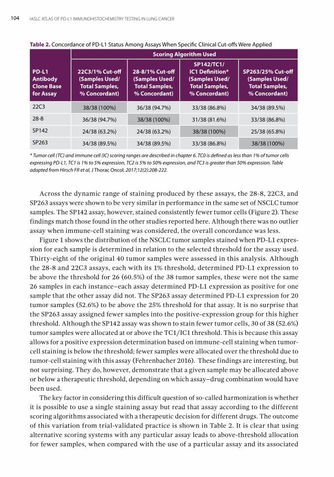
104 IASLC ATLAS OF PD-L1 IMMUNOHISTOCHEMISTRY TESTING IN LUNG CANCER
Across the dynamic range of staining produced by these assays, the 28-8, 22C3, and SP263 assays were shown to be very similar in performance in the same set of NSCLC tumor samples. The SP142 assay, however, stained consistently fewer tumor cells (Figure 2). These findings match those found in the other studies reported here. Although there was no outlier assay when immune-cell staining was considered, the overall concordance was less. Figure 1 shows the distribution of the NSCLC tumor samples stained when PD-L1 expres-sion for each sample is determined in relation to the selected threshold for the assay used. Thirty-eight of the original 40 tumor samples were assessed in this analysis. Although the 28-8 and 22C3 assays, each with its 1% threshold, determined PD-L1 expression to be above the threshold for 26 (60.5%) of the 38 tumor samples, these were not the same 26 samples in each instance–each assay determined PD-L1 expression as positive for one sample that the other assay did not. The SP263 assay determined PD-L1 expression for 20 tumor samples (52.6%) to be above the 25% threshold for that assay. It is no surprise that the SP263 assay assigned fewer samples into the positive-expression group for this higher threshold. Although the SP142 assay was shown to stain fewer tumor cells, 30 of 38 (52.6%) tumor samples were allocated at or above the TC1/IC1 threshold. This is because this assay allows for a positive expression determination based on immune-cell staining when tumor-cell staining is below the threshold; fewer samples were allocated over the threshold due to tumor-cell staining with this assay (Fehrenbacher 2016). These findings are interesting, but not surprising. They do, however, demonstrate that a given sample may be allocated above or below a therapeutic threshold, depending on which assay–drug combination would have been used. The key factor in considering this difficult question of so-called harmonization is whether it is possible to use a single staining assay but read that assay according to the different scoring algorithms associated with a therapeutic decision for different drugs. The outcome of this variation from trial-validated practice is shown in Table 2. It is clear that using alternative scoring systems with any particular assay leads to above-threshold allocation for fewer samples, when compared with the use of a particular assay and its associated
Table 2. Concordance of PD-L1 Status Among Assays When Specific Clinical Cut-offs Were Applied
PD-L1 Antibody Clone Base for Assay
Scoring Algorithm Used
22C3/1% Cut-off (Samples Used/Total Samples, % Concordant)
28-8/1% Cut-off (Samples Used/Total Samples, % Concordant)
SP142/TC1/IC1 Definition* (Samples Used/Total Samples, % Concordant)
SP263/25% Cut-off (Samples Used/ Total Samples, % Concordant)
22C3 38/38 (100%) 36/38 (94.7%) 33/38 (86.8%) 34/38 (89.5%)
28-8 36/38 (94.7%) 38/38 (100%) 31/38 (81.6%) 33/38 (86.8%)
SP142 24/38 (63.2%) 24/38 (63.2%) 38/38 (100%) 25/38 (65.8%)
SP263 34/38 (89.5%) 34/38 (89.5%) 33/38 (86.8%) 38/38 (100%)
* Tumor cell (TC) and immune cell (IC) scoring ranges are described in chapter 6. TC0 is defined as less than 1% of tumor cells expressing PD-L1, TC1 is 1% to 5% expression, TC2 is 5% to 50% expression, and TC3 is greater than 50% expression. Table adapted from Hirsch FR et al, J Thorac Oncol. 2017;12(2):208-222.
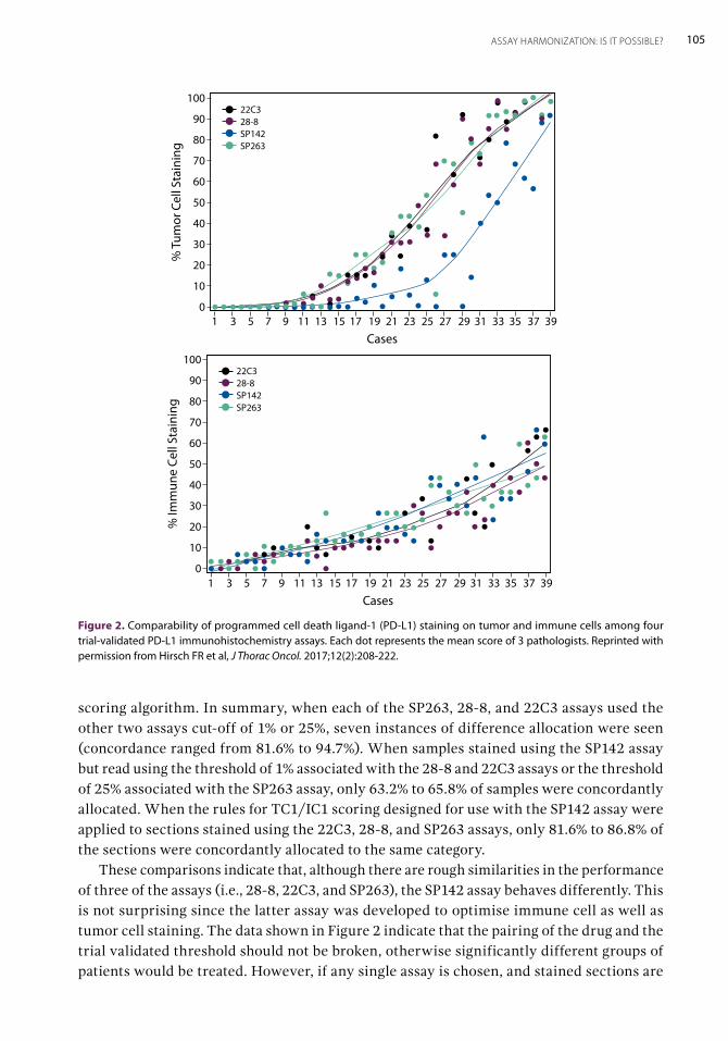
105ASSAY HARMONIZATION: IS IT POSSIBLE?
Figure 2. Comparability of programmed cell death ligand-1 (PD-L1) staining on tumor and immune cells among four trial-validated PD-L1 immunohistochemistry assays. Each dot represents the mean score of 3 pathologists. Reprinted with permission from Hirsch FR et al, J Thorac Oncol. 2017;12(2):208-222.
100
90
80
70
60
50
40
30
20
10
0
% T
umor
Cel
l Sta
inin
g
Cases1 3 5 7 9 11 13 15 17 19 21 23 25 27 29 31 33 35 37 39
22C328-8SP142SP263
100
90
80
70
60
50
40
30
20
10
0
% Im
mun
e Ce
ll St
aini
ng
Cases1 3 5 7 9 11 13 15 17 19 21 23 25 27 29 31 33 35 37 39
22C328-8SP142SP263
scoring algorithm. In summary, when each of the SP263, 28-8, and 22C3 assays used the other two assays cut-off of 1% or 25%, seven instances of difference allocation were seen (concordance ranged from 81.6% to 94.7%). When samples stained using the SP142 assay but read using the threshold of 1% associated with the 28-8 and 22C3 assays or the threshold of 25% associated with the SP263 assay, only 63.2% to 65.8% of samples were concordantly allocated. When the rules for TC1/IC1 scoring designed for use with the SP142 assay were applied to sections stained using the 22C3, 28-8, and SP263 assays, only 81.6% to 86.8% of the sections were concordantly allocated to the same category. These comparisons indicate that, although there are rough similarities in the performance of three of the assays (i.e., 28-8, 22C3, and SP263), the SP142 assay behaves differently. This is not surprising since the latter assay was developed to optimise immune cell as well as tumor cell staining. The data shown in Figure 2 indicate that the pairing of the drug and the trial validated threshold should not be broken, otherwise significantly different groups of patients would be treated. However, if any single assay is chosen, and stained sections are
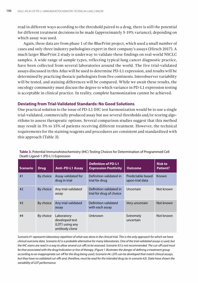
106 IASLC ATLAS OF PD-L1 IMMUNOHISTOCHEMISTRY TESTING IN LUNG CANCER
read in different ways according to the threshold paired to a drug, there is still the potential for different treatment decisions to be made (approximately 5-19% variance), depending on which assay was used. Again, these data are from phase 1 of the BluePrint project, which used a small number of cases and only three industry pathologists expert in their company’s assays (Hirsch 2017). A much larger BluePrint 2 study is underway to validate these findings on real-world NSCLC samples. A wide range of sample types, reflecting typical lung cancer diagnostic practice, have been collected from several laboratories around the world. The five trial-validated assays discussed in this Atlas will be used to determine PD-L1 expression, and results will be determined by practicing thoracic pathologists from five continents. Interobserver variability will be tested, and staining differences will be compared. While we await these results, the oncology community must discuss the degree to which variance in PD-L1 expression testing is acceptable in clinical practice. In reality, complete harmonization cannot be achieved.
Deviating from Trial-Validated Standards: No Good SolutionsOne practical solution to the issue of PD-L1 IHC test harmonization would be to use a single trial-validated, commercially produced assay but use several thresholds and/or scoring algo-rithms to assess therapeutic options. Several comparison studies suggest that this method may result in 5% to 15% of patients receiving different treatment. However, the technical requirements for the staining reagents and procedures are consistent and standardized with this approach (Table 3).
Table 3. Potential Immunohistochemistry (IHC) Testing Choices for Determination of Programmed Cell Death Ligand-1 (PD-L1) Expression
Scenario Drug Anti–PD-L1 AssayDefinition of PD-L1 Expression Positivity Outcome
Risk to Patient?
#1 By choice Assay validated for drug in trial
Definition validated in trial for drug
Predictable based upon trial data
Known
#2 By choice Any trial-validated assay
Definition validated in trial for drug of choice
Uncertain Not known
#3 By choice Any trial-validated assay
Definition validated with each assay
Very uncertain Not known
#4 By choice Laboratory-developed test (LDT) using any antibody clone
Unknown Extremely uncertain
Not known
Scenario #1 represents laboratory repetition of what was done in the clinical trial. This is the only approach for which we have clinical outcome data. Scenario #2 is a probable alternative for many laboratories. One of the trial-validated assays is used, but the IHC stains are read in a way to allow several cut-offs to be assessed. Scenario #3 is not recommended. The cut-off used must be that associated with the drug/indication or line of therapy. (Figure 1 illustrates the danger of defining a treatment group according to an inappropriate cut-off for the drug being used.) Scenario #4. LDTs can be developed that match clinical assays, but they have no validated cut-offs and, therefore, must be read for the intended drug (as in scenario #2). Data have shown the variability of LDT performance.
107ASSAY HARMONIZATION: IS IT POSSIBLE?
There is no such standardization if laboratories continue to deviate from the trial-val-idated approach to PD-L1 IHC for a given treatment plan. LDTs have no standardization regardless of whether they use a commercial assay antibody clone or other PD-L1 IHC anti-body clones. Examples of such LDTs have demonstrated differing staining results (Velcheti 2014; Sheffield 2016; McLaughlin 2016; Neuman 2016). Furthermore, approximately 50% of the LDTs developed in different laboratories and used in the French harmonization study showed significant discordance with the data generated using trial-validated assays (Adam J et al, WCLC 2016). There are generic guidelines for the development of diagnostic IHC LDTs (Fitzgibbons 2014; Lin 2014), but the technical verification process is very rigor-ous and might not always be followed precisely. Also, a development standard for LDTs—be it using a positive control tissue or an existing trial-validated assay—is undetermined. It is likely that LDTs will continue to be highly variable, and there are no data to confirm that an LDT has any predictive value if used to select patients for therapy (Table 3) (see Chapters 3 and 9 for details about LDT validation theories). The LDT route would seem to have the most potential for uncertainty for our patients. Although it is not impossible to develop an LDT that would match the performance of a trial validated assay (Rimm 2017, Adam J et al), the development process is demanding and there is no guarantee of consistent success. Participation in EQA schemes will be essential to ensure that any laboratory, regardless of how it performs PD-L1 IHC testing, provides a test that is safe and effective.
ConclusionThere are few good data on which to base any firm recommendation for harmonization of PD-L1 IHC testing. The gold-standard approach, for which there are abundant data—includ-ing clinical outcomes data—gives the oncologist and the patient an informed prediction of the likelihood of response and of progression-free and overall survivals for a therapy based on the assay and corresponding scoring used. Any other approach is less certain, less well informed, and not clinically validated, although a certain amount of analytical validation has been attempted. The possibility of assay harmonization is dependent on the amount of risk oncologists and patients are willing to accept. The use of certain alternative trial-validated assays (interchanging 28-8, 22C3, and SP263) suggests a possible 5% to 15% loss of predictive performance. It is surprising that LDTs are being widely used to make clinical decisions in an era when oncology practice is otherwise so heavily driven by evidence from clinical trials and especially when considering that PD-L1 IHC as a predictive biomarker has received considerable criticism. Much more data, including clinical treatment responses, are required before alternative practices can be determined so that optimal treatment of patients is not compromised.

Implementation of PD-L1 Testing for Personalized Therapy for Lung CancerBy Ming S. Tsao, Andrew G. Nicholson, and Fred R. Hirsch
12During the past decade, the treatment outlook for patients with advanced-stage lung cancer has transformed from nihilistic to optimistic. Many patients today receive less toxic thera-pies and experience longer disease control and survival, and there is a potential for cure for some patients. These advances partially have been made possible by the development of personalized therapy based on the molecular characteristics of individual patient’s tumor. With personalized therapy comes increased and more complex testing. The role of patholo-gists in routine diagnosis of lung cancer, therefore, has also significantly changed. Optimal processing of biopsy tissues, stratification of the cut sections, and prioritization of biomarker analysis are all essential components of both pathologic workflow and diagnosis. In the clinical practice setting, selection of targeted therapies for patients with non-small cell lung cancer (NSCLC) requires testing for EGFR mutations and rearrangements or fusion protein expression involving the ALK and ROS1 genes (Lindeman 2013). EGFR testing is conducted using DNA isolated from plasma or tumor tissue (Tan 2016). For tissue, this usually requires a large number (10 or more) of unstained tissue or cytology cell-block sections. In contrast, current ALK and ROS1 testing is mostly performed by immunohis-tochemistry (IHC) and/or fluorescence in situ hybridization and requires only one or two unstained sections (Tsao 2016). Guidelines recommend molecular testing of EGFR, ALK, and ROS1 aberrations only for patients with adenocarcinoma or non-small cell carcinoma (NSCC) when an adenocarcinoma component cannot be excluded (e.g., in biopsy samples), or for patients with squamous cell carcinoma who have high risk of an EGFR, ALK, and/or ROS1 mutation or rearrangement (e.g., never or light smokers or young women, particularly with Asian ethnicity) (Lindeman 2013). Although EGFR and ALK genomic aberrations have been reported in squamous cell carcinoma, routine testing is not recommended based on low prevalence and cost effectiveness. The previous paradigm of excluding squamous cell carcinoma for biomarker testing has changed; PD-L1 testing is also required for squamous cell carcinoma (see Chapter 2 for details).
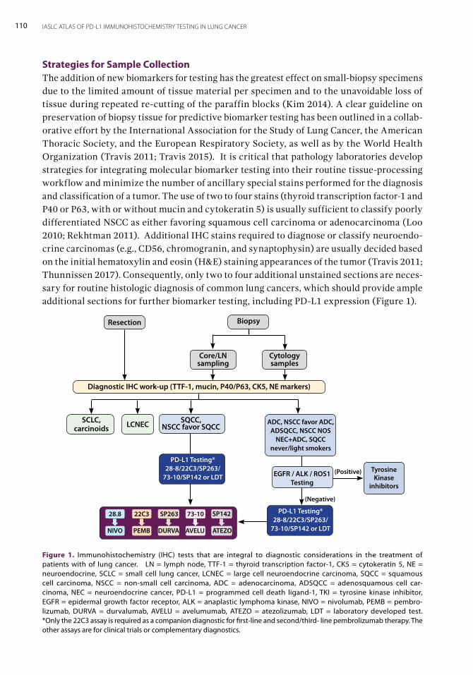
110 IASLC ATLAS OF PD-L1 IMMUNOHISTOCHEMISTRY TESTING IN LUNG CANCER
Strategies for Sample CollectionThe addition of new biomarkers for testing has the greatest effect on small-biopsy specimens due to the limited amount of tissue material per specimen and to the unavoidable loss of tissue during repeated re-cutting of the paraffin blocks (Kim 2014). A clear guideline on preservation of biopsy tissue for predictive biomarker testing has been outlined in a collab-orative effort by the International Association for the Study of Lung Cancer, the American Thoracic Society, and the European Respiratory Society, as well as by the World Health Organization (Travis 2011; Travis 2015). It is critical that pathology laboratories develop strategies for integrating molecular biomarker testing into their routine tissue-processing workflow and minimize the number of ancillary special stains performed for the diagnosis and classification of a tumor. The use of two to four stains (thyroid transcription factor-1 and P40 or P63, with or without mucin and cytokeratin 5) is usually sufficient to classify poorly differentiated NSCC as either favoring squamous cell carcinoma or adenocarcinoma (Loo 2010; Rekhtman 2011). Additional IHC stains required to diagnose or classify neuroendo-crine carcinomas (e.g., CD56, chromogranin, and synaptophysin) are usually decided based on the initial hematoxylin and eosin (H&E) staining appearances of the tumor (Travis 2011; Thunnissen 2017). Consequently, only two to four additional unstained sections are neces-sary for routine histologic diagnosis of common lung cancers, which should provide ample additional sections for further biomarker testing, including PD-L1 expression (Figure 1).
Figure 1. Immunohistochemistry (IHC) tests that are integral to diagnostic considerations in the treatment of patients with of lung cancer. LN = lymph node, TTF-1 = thyroid transcription factor-1, CK5 = cytokeratin 5, NE = neuroendocrine, SCLC = small cell lung cancer, LCNEC = large cell neuroendocrine carcinoma, SQCC = squamous cell carcinoma, NSCC = non-small cell carcinoma, ADC = adenocarcinoma, ADSQCC = adenosquamous cell car-cinoma, NEC = neuroendocrine cancer, PD-L1 = programmed cell death ligand-1, TKI = tyrosine kinase inhibitor, EGFR = epidermal growth factor receptor, ALK = anaplastic lymphoma kinase, NIVO = nivolumab, PEMB = pembro-lizumab, DURVA = durvalumab, AVELU = avelumumab, ATEZO = atezolizumab, LDT = laboratory developed test. *Only the 22C3 assay is required as a companion diagnostic for first-line and second/third- line pembrolizumab therapy. The other assays are for clinical trials or complementary diagnostics.
SQCC,NSCC favor SQCC
Diagnostic IHC work-up (TTF-1, mucin, P40/P63, CK5, NE markers)
ADC, NSCC favor ADC, ADSQCC, NSCC NOS
NEC+ADC, SQCC never/light smokers
PD-L1 Testing* 28-8/22C3/SP263/
73-10/SP142 or LDT
Biopsy
Cytology samples
Core/LN sampling
Resection
SCLC,carcinoids LCNEC
PD-L1 Testing* 28-8/22C3/SP263/
73-10/SP142 or LDT
EGFR / ALK / ROS1 Testing
(Negative)
28.8
NIVO
22C3
PEMB
SP263
DURVA
73-10
AVELU
SP142
ATEZO
(Positive) Tyrosine Kinase
inhibitors
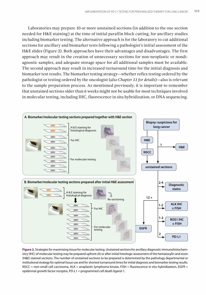
111IMPLEMENTATION OF PD-L1 TESTING FOR PERSONALIZED THERAPY FOR LUNG CANCER
Laboratories may prepare 10 or more unstained sections (in addition to the one section needed for H&E staining) at the time of initial paraffin block cutting, for ancillary studies including biomarker testing. The alternative approach is for the laboratory to cut additional sections for ancillary and biomarker tests following a pathologist’s initial assessment of the H&E slides (Figure 2). Both approaches have their advantages and disadvantages. The first approach may result in the creation of unnecessary sections for non-neoplastic or nondi-agnostic samples, and adequate storage space for all additional samples must be available. The second approach may result in increased turnaround time for the initial diagnosis and biomarker test results. The biomarker testing strategy—whether reflex testing ordered by the pathologist or testing ordered by the oncologist (also Chapter 11 for details)—also is relevant to the sample preparation process. As mentioned previously, it is important to remember that unstained sections older than 6 weeks might not be usable for most techniques involved in molecular testing, including IHC, fluorescence in situ hybridization, or DNA sequencing.
Figure 2. Strategies for maximizing tissue for molecular testing. Unstained sections for ancillary diagnostic immunohistochem-istry (IHC) of molecular testing may be prepared upfront (A) or after initial histologic assessment of the hematoxylin and eosin (H&E) stained sections. The number of unstained sections to be prepared is determined by the pathology departmental or institutional strategy for optimal tissue use and for shortest turnaround times for initial diagnosis and biomarker testing results.NSCC = non-small cell carcinoma, ALK = anaplastic lymphoma kinase, FISH = fluorescence in situ hybridization, EGFR = epidermal growth factor receptor, PD-L1 = programmed cell death ligand-1.
Biopsy: suspicious for lung cancer
Diagnostic stains
H&E
NSCCH&E
ALK IHC ± FISH
ROS1 IHC ± FISH
PD-L1
EGFR
12 +
H & E staining for histological diagnosis
For IHC
A. Biomarker/molecular testing sections prepared together with H&E section
H & E staining for histological diagnosis
For IHC
For molecular testing
B. Biomarker/molecular testing sections prepared after initial H&E assessment
For molecular testing
Re-sectioning
1
2
1-2
1-2
2-4
unstained sections
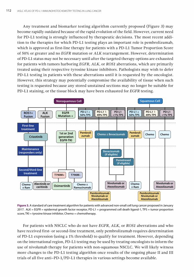
112 IASLC ATLAS OF PD-L1 IMMUNOHISTOCHEMISTRY TESTING IN LUNG CANCER
For patients with NSCLC who do not have EGFR, ALK, or ROS1 aberrations and who have received first- or second-line treatment, only pembrolizumab requires determination of PD-L1 expression (using a 1% threshold) to qualify for treatment. However, depending on the international region, PD-L1 testing may be used by treating oncologists to inform the use of nivolumab therapy for patients with non-squamous NSCLC. We will likely witness more changes to the PD-L1 testing algorithm once results of the ongoing phase II and III trials of all five anti–PD-1/PD-L1 therapies in various settings become available.
Any treatment and biomarker testing algorithm currently proposed (Figure 3) may become rapidly outdated because of the rapid evolution of the field. However, current need for PD-L1 testing is strongly influenced by therapeutic decisions. The most recent addi-tion to the therapies for which PD-L1 testing plays an important role is pembrolizumab, which is approved as first-line therapy for patients with a PD-L1 Tumor Proportion Score of 50% or greater and no EGFR mutation or ALK rearrangement. However, determination of PD-L1 status may not be necessary until after the targeted therapy options are exhausted for patients with tumors harboring EGFR, ALK, or ROS1 aberrations, which are primarily treated using their respective tyrosine kinase inhibitors. Pathologists may wish to defer PD-L1 testing in patients with these aberrations until it is requested by the oncologist. However, this strategy may potentially compromise the availability of tissue when such testing is requested because any stored unstained sections may no longer be suitable for PD-L1 staining, or the tissue block may have been exhausted for EGFR testing.
Figure 3. A standard of care treatment algorithm for patients with advanced non-small cell lung cancer proposed in January 2017. ALK = EGFR = epidermal growth factor receptor, PD-L1 = programmed cell death ligand-1, TPS = tumor proportion score, TKI = tyrosine kinase inhibitor, Chemo = chemotherapy.
First-line treatment
Maintenance (responders only)
Second/third-line treatment
Nonsquamous Cell Squamous Cell
Pembroli zumab
Pembrolizumab Nivolumab or Atezolizumab
Pembroli zumab Chemo
Chemo ± Ram
Chemo ± Ramucirumab
ChemoChemo ± Bevacizumab
Bevacizumab(if eligible)
Pemetrexed(if eligible)
EGFR Mutation +
1st or 2nd generation
EGFR-TKI
OsimertinibAlectinib or
ceritinibNivolumab or Atezolizumab
Nivolumab or Atezolizumab
ALK Fusion
ROS1+ Fusion
Crizotinib
T790M–T790M+
PD-L1 ≥ 50% TPS
PD-L1 1- 49% TPS
PD-L1 <1% TPS
PD-L1 ≥ 50% TPS
PD-L1 1- 49% TPS
PD-L1 <1% TPS
Chemo ± Ram
Pembrolizumab Nivolumab or Atezolizumab
113IMPLEMENTATION OF PD-L1 TESTING FOR PERSONALIZED THERAPY FOR LUNG CANCER
ConclusionThe current testing algorithm for PD-L1 is evolving and could change dramatically once results from ongoing trials become available. Nevertheless, pathology laboratories must be ready to update strategies regarding tissue management and unstained sample preparation to accommodate PD-L1 testing, as well as strategies on prioritization of testing for the vari-ous biomarkers encountered in routine clinical practice.

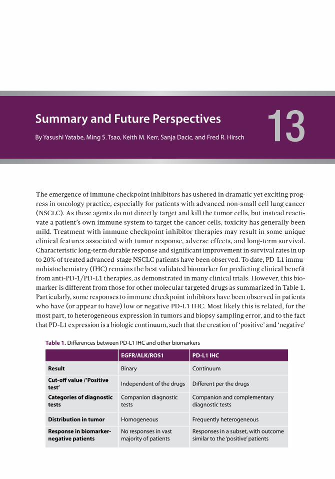
Summary and Future PerspectivesBy Yasushi Yatabe, Ming S. Tsao, Keith M. Kerr, Sanja Dacic, and Fred R. Hirsch 13The emergence of immune checkpoint inhibitors has ushered in dramatic yet exciting prog-ress in oncology practice, especially for patients with advanced non-small cell lung cancer (NSCLC). As these agents do not directly target and kill the tumor cells, but instead reacti-vate a patient’s own immune system to target the cancer cells, toxicity has generally been mild. Treatment with immune checkpoint inhibitor therapies may result in some unique clinical features associated with tumor response, adverse effects, and long-term survival. Characteristic long-term durable response and significant improvement in survival rates in up to 20% of treated advanced-stage NSCLC patients have been observed. To date, PD-L1 immu-nohistochemistry (IHC) remains the best validated biomarker for predicting clinical benefit from anti-PD-1/PD-L1 therapies, as demonstrated in many clinical trials. However, this bio-marker is different from those for other molecular targeted drugs as summarized in Table 1. Particularly, some responses to immune checkpoint inhibitors have been observed in patients who have (or appear to have) low or negative PD-L1 IHC. Most likely this is related, for the most part, to heterogeneous expression in tumors and biopsy sampling error, and to the fact that PD-L1 expression is a biologic continuum, such that the creation of ‘positive’ and ‘negative’
Table 1. Differences between PD-L1 IHC and other biomarkers
EGFR/ALK/ROS1 PD-L1 IHC
Result Binary Continuum
Cut-off value /’Positive test’ Independent of the drugs Different per the drugs
Categories of diagnostic tests
Companion diagnostic tests
Companion and complementary diagnostic tests
Distribution in tumor Homogeneous Frequently heterogeneous
Response in biomarker- negative patients
No responses in vast majority of patients
Responses in a subset, with outcome similar to the ‘positive’ patients
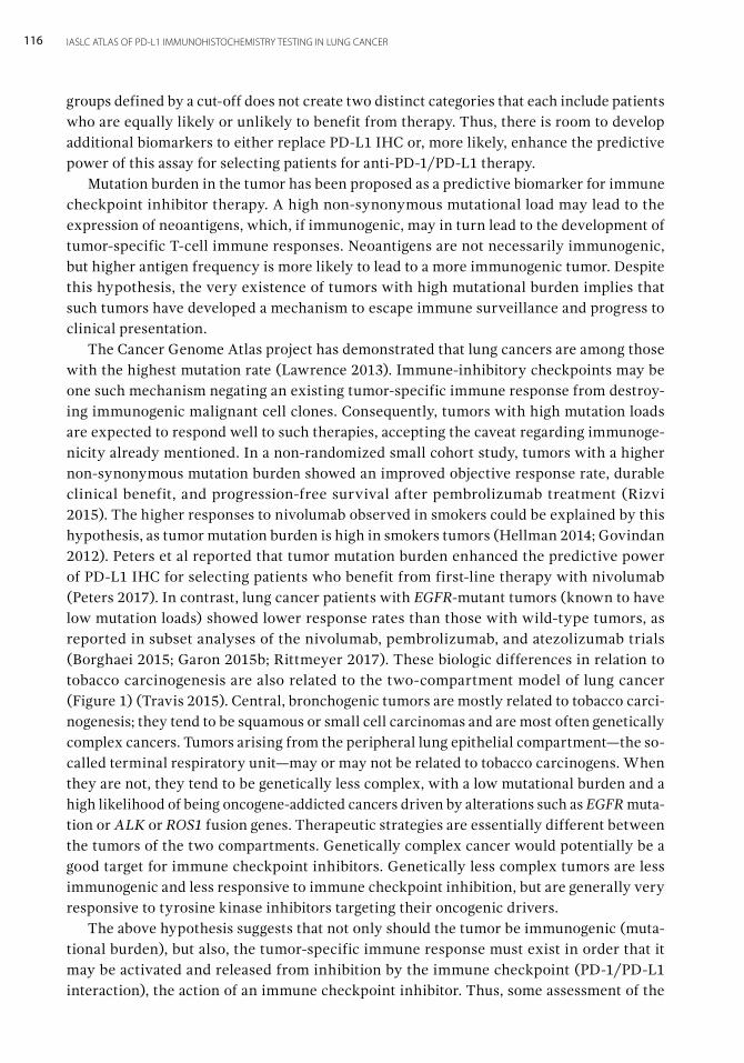
116 IASLC ATLAS OF PD-L1 IMMUNOHISTOCHEMISTRY TESTING IN LUNG CANCER
groups defined by a cut-off does not create two distinct categories that each include patients who are equally likely or unlikely to benefit from therapy. Thus, there is room to develop additional biomarkers to either replace PD-L1 IHC or, more likely, enhance the predictive power of this assay for selecting patients for anti-PD-1/PD-L1 therapy. Mutation burden in the tumor has been proposed as a predictive biomarker for immune checkpoint inhibitor therapy. A high non-synonymous mutational load may lead to the expression of neoantigens, which, if immunogenic, may in turn lead to the development of tumor-specific T-cell immune responses. Neoantigens are not necessarily immunogenic, but higher antigen frequency is more likely to lead to a more immunogenic tumor. Despite this hypothesis, the very existence of tumors with high mutational burden implies that such tumors have developed a mechanism to escape immune surveillance and progress to clinical presentation. The Cancer Genome Atlas project has demonstrated that lung cancers are among those with the highest mutation rate (Lawrence 2013). Immune-inhibitory checkpoints may be one such mechanism negating an existing tumor-specific immune response from destroy-ing immunogenic malignant cell clones. Consequently, tumors with high mutation loads are expected to respond well to such therapies, accepting the caveat regarding immunoge-nicity already mentioned. In a non-randomized small cohort study, tumors with a higher non-synonymous mutation burden showed an improved objective response rate, durable clinical benefit, and progression-free survival after pembrolizumab treatment (Rizvi 2015). The higher responses to nivolumab observed in smokers could be explained by this hypothesis, as tumor mutation burden is high in smokers tumors (Hellman 2014; Govindan 2012). Peters et al reported that tumor mutation burden enhanced the predictive power of PD-L1 IHC for selecting patients who benefit from first-line therapy with nivolumab (Peters 2017). In contrast, lung cancer patients with EGFR-mutant tumors (known to have low mutation loads) showed lower response rates than those with wild-type tumors, as reported in subset analyses of the nivolumab, pembrolizumab, and atezolizumab trials (Borghaei 2015; Garon 2015b; Rittmeyer 2017). These biologic differences in relation to tobacco carcinogenesis are also related to the two-compartment model of lung cancer (Figure 1) (Travis 2015). Central, bronchogenic tumors are mostly related to tobacco carci-nogenesis; they tend to be squamous or small cell carcinomas and are most often genetically complex cancers. Tumors arising from the peripheral lung epithelial compartment—the so-called terminal respiratory unit—may or may not be related to tobacco carcinogens. When they are not, they tend to be genetically less complex, with a low mutational burden and a high likelihood of being oncogene-addicted cancers driven by alterations such as EGFR muta-tion or ALK or ROS1 fusion genes. Therapeutic strategies are essentially different between the tumors of the two compartments. Genetically complex cancer would potentially be a good target for immune checkpoint inhibitors. Genetically less complex tumors are less immunogenic and less responsive to immune checkpoint inhibition, but are generally very responsive to tyrosine kinase inhibitors targeting their oncogenic drivers. The above hypothesis suggests that not only should the tumor be immunogenic (muta-tional burden), but also, the tumor-specific immune response must exist in order that it may be activated and released from inhibition by the immune checkpoint (PD-1/PD-L1 interaction), the action of an immune checkpoint inhibitor. Thus, some assessment of the
117SUMMARY AND FUTURE PERSPECTIVES
immune response in the tumor microenvironment (the inflamed tumor) may also act as a predictive biomarker (Blank 2016). This approach could involve assessment of actual immune cell populations in the tumor microenvironment (Hegde 2016; Teng 2015). An alternative approach has been to examine mRNA expression profiles of immune-related genes in the tumor as a measure of immunologic activity in the tumor microenvironment (Fehrenbacher 2016; Chen 2016). The consideration of immune cell activity in the tumor microenvironment mentioned above is also reflected in the way in which PD-L1 IHC has been assessed in some clinical trials. Recent results of atezolizumab (POPLAR and OAK) trials (Fehrenbacher 2016; Rittmeyer 2017) showed that PD-L1 expression in tumor-infiltrating immune cells, in the absence of tumor cell PD-L1 expression, also predicted response to therapy. Tumors showing ≥50% tumor cell PD-L1 staining (TC3) rarely show ≥10% immune cell PD-L1 staining (IC3) and vice versa. While the atezolizumab-associated SP142 assay demonstrates staining char-acteristics that may facilitate the scoring of PD-L1 expression on the immune cells (Chapter 6), PD-L1 scoring on immune cells has not been found to have predictive value using other assays and drugs. The role of immune cell PD-L1 expression levels as a predictive marker remains worthy of further studies. To date, the application of the PD-L1 IHC assays has been limited to some immune check-point monotherapies, mainly in second- or greater-line indications. The use of anti-PD-1/PD-L1 agents in first-line is now accelerating, driven by PD-L1 IHC biomarker selection as shown in the KEYNOTE 024 study [Reck 2017]. This is a practice-changing development that
Figure 1. Genetic complexity and the concept of the two-compartment model in the putative molecular pathogenesis of lung cancer. Anatomically, lung epithelial cells are divided into two compartments that are associated with lung function. The central airway compartment functions mainly for air conducting, while respiratory exchange is made in the terminal respiratory unit of the peripheral compartment. Carcinogens from smoking appear to target both central and peripheral airways, although the magnitude is weighted more on the central compartment. Long-term smoking causes mutations across whole genomes, leading to genetically complex tumors with high mutation burden. In contrast, EGFR is mutated by unidentified factors, which appear to specifically target the terminal respiratory unit. EGFR mutation occurs in the terminal respiratory unit where smoking has less effect.
Smoker
Never smoker
Mut
atio
n bu
rden
Genetically complex cancer
Genetically simple cancer
KRAS activation
EGFR activation
Other driver mutations
SCLC, SQC
ADC
Central airway
compartment
Terminalrespiratory
unit
118 IASLC ATLAS OF PD-L1 IMMUNOHISTOCHEMISTRY TESTING IN LUNG CANCER
will boost the practice of PD-L1 IHC testing, at least for the foreseeable future. Clinical trials of these inhibitors are now emerging, using combinations of immune checkpoint inhibi-tors, or immune checkpoint inhibitors with traditional cytotoxic chemotherapy, where the role of PD-L1 testing is yet to be defined. It remains to be seen whether PD-L1 IHC will be replaced by mutational burden or tumor inflammation assessment, or some other biomarker strategy. Or, perhaps more likely, the predictive power of PD-L1 IHC may be enhanced by the addition of another test. With intensive efforts to further improve the personalization of immunotherapies, there is little doubt that in the future, additional and new biomarkers for immune checkpoint inhibitors will be developed. It remains to be seen whether or not an increasingly complex, and expensive, biomarker testing strategy will provide significant improvement over the relatively simple, yet imperfect, PD-L1 IHC that is validated by clini-cal trial outcomes.
119REFERENCES
References
Adam J, Rouquette I, Damotte D, et al. PL04a.04: Multi-centric French Harmonization Study for PD-L1 IHC Test-ing in NSCLC. J Thorac Oncol. 2017;12(1 suppl):S11-S12.
Ameratunga M, Asadi K, Lin X, et al. PD-L1 and Tumor Infiltrating Lymphocytes as Prognostic Markers in Resected NSCLC. PLoS One. 2016;11(4):e0153954.
Ansell SM, Lesokhin AM, Borrello I, et al. PD-1 blockade with nivolumab in relapsed or refractory Hodgkin’s lym-phoma. N Engl J Med. 2015;372(4):311-319.
Antonia S, Goldberg SB, Balmanoukian A, et al. Safety and antitumour activity of durvalumab plus tremelim-umab in non-small cell lung cancer: a multicentre, phase 1b study. Lancet Oncol. 2016;17(3):299-308.
Antonia SJ, Kim S-W, Spira AI, et al. Safety and clini-cal activity of durvalumab (MEDI4736), an anti-PD-L1 antibody, in treatment-naïve patients with advanced non-small-cell lung cancer. J Clin Oncol. 2016;34(15_suppl):abstr 9029.
Antonia SJ, López-Martin JA, Bendell J, et al. Nivolumab alone and nivolumab plus ipilimumab in recurrent small-cell lung cancer (CheckMate 032): a multicentre, open-label, phase 1/2 trial. Lancet Oncol. 2016;17(7): 883-895.
Armand P, Shipp MA, Ribrag V, et al. Programmed death-1 blockade with pembrolizumab in patients with classical Hodgkin lymphoma after brentuximab vedotin failure. J Clin Oncol. 2016 Jun 27. [Epub ahead of print]
Atkins MB, Lotze MT, Dutcher JP, et al. High-dose recom-binant interleukin 2 therapy for patients with metastatic melanoma: analysis of 270 patients treated between 1985 and 1993. J Clin Oncol. 1999;17(7):2105-2116.
Azuma K, Ota K, Kawahara A, et al. Association of PD-L1 overexpression with activating EGFR mutations in surgically resected nonsmall-cell lung cancer. Ann Oncol. 2014;25(10):1935-1940.
Baas P, Garon EB, Herbst RS, et al. Relationship between level of PD-L1 expression and outcomes in the KEY-NOTE-010 study of pembrolizumab vs docetaxel for previously treated, PD-L1–Positive NSCLC. J Clin Oncol. 2016;34(15_suppl): abstr 9015.
Bakker W, Nijhuis-Heddes JM, van der Velde EA. Post- operative intrapleural BCG in lung cancer: a 5-year fol-low-up report. Cancer Immunol Immunother. 1986;22(2): 155-159.
Besse B, Johnson ML, Janne PA, et al. Phase II, single-arm trial (BIRCH) of atezolizumab as first-line or sub-sequent therapy for locally advanced or metastatic PD-L1–selected non-small cell lung cancer (NSCLC). Eur J Cancer. 2015;51(suppl 3):S717–S718.
Blank CU, Haanen JB, Ribas A, Schumacher TN. Cancer Immunology. The “cancer immunogram.” Science. 2016; 352(6286):658-660.
Blind C, Koepenik A, Pacyna-Gengelbach M, et al. Anti-genicity testing by immunohistochemistry after tissue oxidation. J Clin Pathol. 2008;61(1):79-83.
Bonnas C, Specht K, Spleiss O, et al. Effects of cold ischemia and inflammatory tumor microenviron-ment on detection of PI3K/AKT and MAPK pathway activation patterns in clinical cancer samples. Int J Cancer. 2012;131(7):1621-1632.
Borghaei H, Paz-Ares L, Horn L, et al. Nivolumab versus docetaxel in advanced nonsquamous non-small-cell lung cancer. N Engl J Med. 2015;373(17):1627-1639.
Boyerinas B, Jochems C, Fantini M, et al. Antibody-dependent cellular cytotoxicity activity of a novel anti-PD-L1 antibody avelumab (MSB0010718C) on human tumor cells. Cancer Immunol Res. 2015;3(10): 1148-1157.
Brahmer J, Reckamp KL, Baas P, et al. Nivolumab versus docetaxel in advanced squamous-cell non-small-cell lung cancer. N Engl J Med. 2015;373(2):123-135.
Brambilla E, Le Teuff G, Marguet S, et al. Prognostic effect of tumor lymphocytic infiltration in resectable non-small-cell lung cancer. J Clin Oncol. 2016;34(11):1223-1230.
Broderick J. Atezolizumab approaches approval for lung cancer. Cure. April 11, 2016. Available at http://www.curetoday.com/articles/atezolizumab-approaches-approval-for-lung-cancer. Accessed March 10, 2017.
Butts C, Murray N, Maksymiuk A, et al. Random-ized phase IIB trial of BLP25 liposome vaccine in stage IIIB and IV non-small-cell lung cancer. J Clin Oncol. 2005;23(27):6674-6681.
Butts C, Socinski MA, Mitchell PL, et al. Tec-emotide (L-BLP25) versus placebo after chemoradio-therapy for stage III non-small-cell lung cancer (START): a randomised, double-blind, phase 3 trial. Lancet Oncol. 2014;15(1):59-68.
Camidge DR, Pao W, Sequist LV. Acquired resistance to TKIs in solid tumours: learning from lung cancer. Nat Rev Clin Oncol. 2014;11(8):473-481.
Chen DS, Irving BA, Hodi FS. Molecular pathways: next-generation immunotherapy--inhibiting programmed death-ligand 1 and programmed death-1. Clin Cancer Res. 2012;18(24):6580-6587.
Chen PL, Roh W, Reuben A, et al. Analysis of immune signatures in longitudinal tumor samples yields insight
120 IASLC ATLAS OF PD-L1 IMMUNOHISTOCHEMISTRY TESTING IN LUNG CANCER
into biomarkers of response and mechanisms of resis-tance to immune checkpoint blockade. Cancer Discov. 2016;6(8):827-837.
Cheung CC, Garratt J, Won J, et al. Developing ALK Immunohistochemistry and In Situ Hybridization Proficiency Testing for Non-Small Cell Lung Cancer in Canada: Canadian Immunohistochemistry Quality Control Challenges and Successes. Appl Immunohistochem Mol Morphol. 2015;23(10):677-681.
Cogswell J, Inzunza HD, Wu Q, et al. An Analytical Comparison of Dako 28-8 PharmDx Assay and an E1L3N Laboratory-Developed Test in the Immunohistochemical Detection of Programmed Death-Ligand 1. Mol Diagn Ther. 2017;21(1):85-93.
Coiffier B, Lepage E, Briere J, et al. CHOP chemotherapy plus rituximab compared with CHOP alone in elderly patients with diffuse large-B-cell lymphoma. N Engl J Med. 2002;346(4):235-242.
Cooper W, Russell PA, Huot-Marhand P, et al. Intra- and interobserver reproducibility study of PD-L1 biomarker in NSCLC. The Dream study. J Thorac Oncol. 2016;12(1 suppl):S814-S815.
Cooper WA, Tran T, Vilain RE, et al. PD-L1 expression is a favorable prognostic factor in early stage non-small cell carcinoma. Lung Cancer 2015;89(2):181-188.
Cree IA, Booton R, Cane P, et al. PD-L1 testing for lung cancer in the UK: recognizing the challenges for imple-mentation. Histopathology. 2016;69(2):177-186.
Cuppens K, Vansteenkiste J. Vaccination therapy for non-small-cell lung cancer. Curr Opin Oncol. 2014;26(2): 165-170.
Curran MA, Montalvo W, Yagita H, Allison JP. PD-1 and CTLA-4 combination blockade expands infiltrat-ing T cells and reduces regulatory T and myeloid cells within B16 melanoma tumors. Proc Natl Acad Sci U S A. 2010;107(9):4275-4280.
Cutz JC, Craddock KJ, Torlakovic E, et al. Canadian ana-plastic lymphoma kinase study: a model for multicenter standardization and optimization of ALK testing in lung cancer. J Thorac Oncol. 2014;9(9):1255-1263.
Dako North America. PD-L1 IHC 22C3 pharmDx inter-pretation manual. http://www.dako.com/us/29109_pd-l1-ihc-22c3-interpretation-manual.pdf. (accessed August 13, 2016.).
Decoster L, Wauters I, Vansteenkiste JF. Vaccination therapy for non-small-cell lung cancer: review of agents in phase III development. Ann Oncol. 2012;23(6):1387-1393.
Dolled-Filhart M, Locke D, Murphy T, et al. Develop-ment of a Prototype Immunohistochemistry Assay to Measure Programmed Death Ligand-1 Expression in Tumor Tissue. Arch Pathol Lab Med. 2016;140(11): 1259-1266.
Dolled-Filhart M, Roach C, Toland G, et al. Development of a Companion Diagnostic for Pembrolizumab in Non-Small Cell Lung Cancer Using Immunohistochemistry for Programmed Death Ligand-1. Arch Pathol Lab Med. 2016 Aug 23 [Epub ahead of print].
Dong H, Strome SE, Salomao DR, et al. Tumor-associated B7-H1 promotes T-cell apoptosis: a potential mechanism of immune evasion. Nat Med. 2002;8(8):793-800.
Fehrenbacher L, Spira A, Ballinger M, et al. Atezolizumab versus docetaxel for patients with previously treated non-small-cell lung cancer (POPLAR): a multicenter, open-label, phase 2 randomised controlled trial. Lancet. 2016;387(10030):1837-1846.
Fischer AH, Schwartz MR, Moriarty AT, et al. Immu-nohistochemistry practices of cytopathology laborato-ries: a survey of participants in the College of American Pathologists Nongynecologic Cytopathology Education Program. Arch Pathol Lab Med. 2014;138(9):1167-1172.
Fitzgibbons PL, Bradley LA, Fatheree LA, et al. Principles of analytic validation of immunohistochemical assays: guideline from the College of American Pathologists Pathology and Laboratory Quality Center. Arch Pathol Lab Med. 2014;138(11):1432-1443.
Fridman WH, Pages F, Sautes-Fridman C, Galon J. The immune contexture in human tumours: impact on clini-cal outcome. Nat Rev Cancer. 2012;12(4):298-306.
Fyfe G, Fisher RI, Rosenberg SA, Sznol M, Parkinson DR, Louie AC. Results of treatment of 255 patients with metastatic renal cell carcinoma who received high-dose recombinant interleukin-2 therapy. J Clin Oncol. 1995;13(3):688-696.
Garassino M, Vansteenkiste JF, Kim J, et al. Durvalumab in ≥3rd-line locally advanced or metastatic, EGFR/ALK wild-type NSCLC: results from the phase 2 ATLANTIC study. Presented at World Conference on Lung Cancer; December 7, 2016; Vienna, Austria.
Garassino M, Vansteenkiste J, Kim JH, et al. PL04a.03: Durvalumab in ≥3rd-Line Locally Advanced or Meta-static, EGFR/ALK Wild-Type NSCLC: Results from the Phase 2 ATLANTIC Study. J Thorac Oncol. 2017;12 (1 suppl):S10-S11.
Garcia-Lora A, Martinez M, Algarra I, Gaforio JJ, Garrido F. MHC class I-deficient metastatic tumor variants immunoselected by T lymphocytes originate from the coordinated downregulation of APM components. Int J Cancer. 2003;106(4):521-527.
Garon EB. Current Perspectives in Immunotherapy for Non-Small Cell Lung Cancer. Semin Oncol. 2015;42 (suppl 2):S11-S18. (a)
Garon EB, Rizvi NA, Hui R, et al. KEYNOTE-001 Inves-tigators. Pembrolizumab for the treatment of non-small-cell lung cancer. N Engl J Med. 2015;372(21):2018- 2028. (b)
121REFERENCES
Garrido F, Cabrera T, Lopez-Nevot MA, Ruiz-Cabello F. HLA class I antigens in human tumors. Adv Cancer Res. 1995;67:155-195.
Gaule P, Smithy JW, Toki M, et al. A quantitative comparison of antibodies to programmed cell death 1 ligand 1. JAMA Oncol. 2016 Aug 18. [Epub ahead of print]
Gettinger S, Rizvi NA, Chow LQ, et al. Nivolumab monotherapy for first-line treatment of advanced non-small-cell lung cancer. J Clin Oncol. 2016;34(25):2980-2987.
Gettinger SN, Horn L, Gandhi L, et al. Overall survival and long-term safety of nivolumab (anti-programmed death 1 antibody, BMS-936558, ONO-4538) in patients with previously treated advanced non-small-cell lung cancer. J Clin Oncol. 2015;33(18):2004-2012.
Giaccone G, Bazhenova LA, Nemunaitis J, et al. A phase III study of belagenpumatucel-L, an allogeneic tumour cell vaccine, as maintenance therapy for non-small cell lung cancer. Eur J Cancer. 2015;51(16):2321-2329.
Gibbons DL, Chow LQ, Kim DW, et al. 57O Efficacy, safety and tolerability of MEDI4736 (durvalumab [D]), a human IgG1 anti-programmed cell death-ligand-1 (PD-L1) antibody, combined with gefitinib (G): A phase I expansion in TKI-naive patients (pts) with EGFR mutant NSCLC. J Thorac Oncol. 2016;11(4 suppl):S79.
Govindan R, Ding L, Griffith M, et al. Genomic landscape of non-small cell lung cancer in smokers and never-smok-ers. Cell. 2012;150(6):1121-1134.
Gown AM. Diagnostic Immunohistochemistry: What Can Go Wrong and How to Prevent It. Arch Pathol Lab Med. 2016;140(9):893-898.
Gralla RJ, Coon C, Taylor F, et al. Evaluation of disease-related symptoms in patients with advanced squamous non-small cell lung cancer treated with nivolumab or docetaxel. Presented at World Conference on Lung Cancer; September 7, 2015; Denver, CO.
Gubens MA, Sequist LV, Stevenson J, et al. Phase I/II study of pembrolizumab (pembro) plus ipilimumab (ipi) as sec-ond-line therapy for NSCLC: KEYNOTE-021 cohorts D and H. J Clin Oncol. 2016;34 (suppl; abstr 9027).
Gulley JL, Rajan A, Spigel DR, et al. Avelumab for patients with previously treated metastatic or recurrent non-small-cell lung cancer (JAVELIN Solid Tumor): Dose-expansion cohort of a multicentre, open-label, phase 1b trial. Lancet Oncol. 2017. pii: S1470-2045(17)30240-1.[Epub ahead of print]
Hagenbaugh A, Sharma S, Dubinett SM, et al. Altered immune responses in interleukin 10 transgenic mice. J Exp Med. 1997;185(12):2101-2110.
Hecht M, Büttner-Herold M, Erlenbach-Wünsch K, et al. PD-L1 is upregulated by radiochemotherapy in rectal adenocarcinoma patients and associated with a favourable prognosis. Eur J Cancer. 2016;65:52-60.
Hegde PS, Karanikas V, Evers S. The where, the when, and the how of immune monitoring for cancer immuno-therapies in the era of checkpoint inhibition. Clin Cancer Res. 2016;22:1865-1874.
Hellman MD, Creelan BC, Woo K, et al. Smoking history and response to nivolumab in patients with advanced NSCLCs. Ann Oncol. 2014;25:iv426–iv470 (suppl 4).
Hellman MD, Gettinger SN, Goldman JW, et al. Check-Mate 012: Safety and efficacy of first-line (1L) nivolumab (nivo; N) and ipilimumab (ipi; I) in advanced (adv) NSCLC. J Clin Oncol. 2016;34(suppl):abstr 3001.
Herbst RS, Baas P, Kim DW, et al. Pembrolizumab versus docetaxel for previously treated, PD-L1-pos-itive, advanced non-small-cell lung cancer (KEY-NOTE-010): a randomised controlled trial. Lancet. 2016;387(10027):1540-1550.
Herbst RS, Soria JC, Kowanetz M, et al. Predictive corre-lates of response to the anti-PD-L1 antibody MPDL3280A in cancer patients. Nature. 2014;515(7528):563-567.
Hirsch FR, McElhinny A, Stanforth D, et al. PD-L1 Immunohistochemistry assays for lung cancer: Results from phase 1 of the Blueprint PD-L1 IHC assay comparison project. J Thorac Oncol. 2017; 12(2):208-222.
Hirsch FR, Suda K, Wiens J, Bunn PA Jr. New and emerging targeted treatments in advanced non-small-cell lung cancer. Lancet. 2016;388(10048):1012-1024.
Hodi FS, O’Day SJ, McDermott DF, et al. Improved survival with ipilimumab in patients with metastatic melanoma. N Engl J Med. 2010;363(8):711-723.
Homet Moreno B, Ribas A. Anti-programmed cell death protein-1/ligand-1 therapy in different cancers. Br J Cancer. 2015;112(9):1421-1427.
Horn L, Spigel DR, Gettinger SN, et al. Clinical activ-ity, safety and predictive biomarkers of the engineered antibody MPDL3280A (anti-PDL1) in non-small cell lung cancer (NSCLC): update from a phase Ia study. J Clin Oncol 2015;33(15_suppl):abstr 8029.
Hui R, Gandhi L, Costa EC, et al. Long-term OS for patients with advanced NSCLC enrolled in the KEY-NOTE-001 study of pembrolizumab (pembro). J Clin Oncol. 2016;34 (15_suppl): abstr 9026.
Hui R, Garon EB, Goldman JW, et al. Pembrolizumab as first-line therapy for patients with PD-L1-positive advanced non-small cell lung cancer: a phase 1 trial. Ann Oncol. 2017 Feb 7. [Epub ahead of print]
Hussein M, McCleod M, Chandler J, et al. Safety and efficacy of nivolumab in an ongoing trial of a PD-L1+/- patient population with metastatic non-small cell lung cancer. Presented at World Conference on Lung Cancer; September 7, 2015; Denver, CO.
Huynh TG, Morales-Oyarvide V, Campo MJ, et al. Programmed Cell Death Ligand 1 Expression in Resected
122 IASLC ATLAS OF PD-L1 IMMUNOHISTOCHEMISTRY TESTING IN LUNG CANCER
Lung Adenocarcinomas: Association with Immune Microenvironment. J Thorac Oncol. 2016;11(11): 1869-1878.
Ibrahim M, Parry S, Wilkinson D, et al. ALK Immu-nohistochemistry in NSCLC: Discordant Staining Can Impact Patient Treatment Regimen. J Thorac Oncol. 2016;11(12):2241-2247.
Igarashi T, Teramoto K, Ishida M, Hanaoka J, Daigo Y. Scoring of PD-L1 expression intensity on pulmonary adenocarcinomas and the correlations with clinico-pathological factors. ESMO Open. 2016;1(4):e000083.
Ilie M, Falk AT, Butori C, et al. PD-L1 expression in basa-loid squamous cell lung carcinoma: Relationship to PD-1+ and CD8+ tumor-infiltrating T cells and outcome. Mod Pathol. 2016; 29(12):1552-1564.
Ilie M, Long-Mira E, Bence C, et al. Comparative study of the PD-L1 status between surgically resected specimens and matched biopsies of NSCLC patients reveal major discordances: a potential issue for anti-PD-L1 therapeutic strategies. Ann Oncol. 2016;27(1):147-153.
Inaguma S, Wang Z, Lasota J, et al. Comprehensive Immunohistochemical Study of Programmed Cell Death Ligand 1 (PD-L1): Analysis in 5536 Cases Revealed Consistent Expression in Trophoblastic Tumors. Am J Surg Pathol. 2016;40(8):1133-1142.
Jansen RL, Slingerland R, Goey SH, Franks CR, Bolhuis RL, Stoter G. Interleukin-2 and interferon-alpha in the treatment of patients with advanced non-small-cell lung cancer. J Immunother (1991). 1992;12(1):70-73.
Jerusalem G, Chen F, Spigel D, et al. JAVELIN Solid Tumor: Safety and Clinical Activity of Avelumab (Anti-PD-L1) as First-Line Treatment in Patients with Advanced NSCLC. J Thorac Oncol. 2017; 12:S252.
Kantoff PW, Higano CS, Shore ND, et al. Sipuleucel-T immunotherapy for castration-resistant prostate cancer. N Engl J Med. 2010;363(5):411-422.
Kaufman HL, Russell J, Hamid O, et al. Avelumab in patients with chemotherapy-refractory metastatic Merkel cell carcinoma: a multicentre, single-group, open-label, phase 2 trial. Lancet Oncol. 2016 Oct;17(10): 1374-1385.
Kerr KM, Bubendorf L, Edelman MJ, et al. Panel Members. Second ESMO consensus conference on lung cancer: pathology and molecular biomarkers for non-small-cell lung cancer. Ann Oncol. 2014;25(9): 1681-1690.
Kerr KM, Hirsch FR. Programmed Death Ligand-1 Immu-nohistochemistry: Friend or Foe? Arch Pathol Lab Med. 2016;140:326-331. (a)
Kerr KM, Nicolson MC. Non-small cell lung cancer, PD-L1, and the pathologist. Arch Pathol Lab Med. 2016;140:249-254. (b)
Kerr KM, Tsao MS, Nicholson AG, et al. IASLC Pathol-ogy Committee. Programmed death-ligand 1 immuno-histochemistry in lung cancer: in what state is this art? J Thorac Oncol. 2015;10(7):985-989.
Kikuchi E, Yamazaki K, Torigoe T, et al. HLA class I anti-gen expression is associated with a favorable prognosis in early stage non-small cell lung cancer. Cancer Sci. 2007;98(9):1424-1430.
Kim L, Tsao MS. Tumour tissue sampling for lung cancer management in the era of personalised therapy: what is good enough for molecular testing? Eur Respir J. 2014; 44(4):1011-1022.
Kim MY, Koh J, Kim S, et al. Clinicopathological analysis of PD-L1 and PD-L2 expression in pulmonary squamous cell carcinoma: Comparison with tumor-infiltrating T cells and the status of oncogenic drivers. Lung Cancer. 2015;88(1):24-33.
Kirkwood JM, Strawderman MH, Ernstoff MS, Smith TJ, Borden EC, Blum RH. Interferon alfa-2b adjuvant therapy of high-risk resected cutaneous melanoma: the Eastern Cooperative Oncology Group Trial EST 1684. J Clin Oncol. 1996;14(1):7-17.
Kitazono S, Fujiwara Y, Tsuta K, et al. Reliability of small biopsy samples compared with resected specimens for the determination of programmed death-ligand 1 expression in non-small-cell lung cancer. Clin Lung Cancer. 2015;16(5):385-390.
Koh J, Go H, Keam B, et al. Clinicopathologic analysis of programmed cell death-1 and programmed cell death-ligand 1 and 2 expressions in pulmonary adenocarci-noma: comparison with histology and driver oncogenic alteration status. Mod Pathol. 2015;28(9):1154-1166.
Langer CJ, Gadgeel SM, Borghaei H, et al. Carboplatin and pemetrexed with or without pembrolizumab for advanced, non-squamous non-small-cell lung cancer: a randomised, phase 2 cohort of the open-label KEYNOTE-021 study. Lancet Oncol. 2016;17(11): 1497-1508.
Larkin J, Chiarion-Sileni V, Gonzalez R, et al. Combined nivolumab and ipilimumab or monotherapy in untreated melanoma. N Engl J Med. 2015;373(1):23-34.
Latchman Y, Wood CR, Chernova T, et al. PD-L2 is a second ligand for PD-1 and inhibits T cell activation. Nat Immunol. 2001;2(3):261-268.
Lawrence MS, Stojanov P, Polak P, et al. Mutational heterogeneity in cancer and the search for new cancer-associated genes. Nature. 2013;499(7457):214-218.
Le DT, Uram JN, Wang H, et al. PD-1 Blockade in tumors with mismatch-repair deficiency. N Engl J Med. 2015;372(26):2509-2520.
Li X, Deavers MT, Guo M, et al. The effect of prolonged cold ischemia time on estrogen receptor immunohistochemistry in breast cancer. Mod Pathol. 2013;26(1):71-78.
123REFERENCES
Li Y, Xu KP, Jiang D, Zhao J, Ge JF, Zheng SY. Relation-ship of Fas, FasL, p53 and bcl-2 expression in human non-small cell lung carcinomas. Int J Clin Exp Pathol. 2015;8(11):13978-13986.
Lin F, Chen Z. Standardization of diagnostic immunohis-tochemistry: literature review and geisinger experience. Arch Pathol Lab Med. 2014;138:1564-1577.
Lindeman NI, Cagle PT, Beasley MB, et al. Molecular testing guideline for selection of lung cancer patients for EGFR and ALK tyrosine kinase inhibitors: guideline from the College of American Pathologists, International Association for the Study of Lung Cancer, and Association for Molecular Pathology. J Thorac Oncol. 2013;8(7): 823-859.
Loo PS, Thomas SC, Nicolson MC, Fyfe MN, Kerr KM. Subtyping of undifferentiated non-small cell carcino-mas in bronchial biopsy specimens. J Thorac Oncol. 2010;5(4):442-447.
Lotze MT, Matory YL, Ettinghausen SE, et al. In vivo administration of purified human interleukin 2. II. Half life, immunologic effects, and expansion of peripheral lymphoid cells in vivo with recombinant IL 2. J Immunol. 1985;135(4):2865-2875.
Lynch TJ, Bondarenko I, Luft A, et al. Ipilimumab in com-bination with paclitaxel and carboplatin as first-line treat-ment in stage IIIB/IV non-small-cell lung cancer: results from a randomized, double-blind, multicenter phase II study. J Clin Oncol. 2012;30(17):2046-2054.
Mahoney KM, Sun H, Liao X, et al. PD-L1 Antibodies to Its Cytoplasmic Domain Most Clearly Delineate Cell Membranes in Immunohistochemical Staining of Tumor Cells. Cancer Immunol Res. 2015;3(12):1308-1315.
Maio M, Grob JJ, Aamdal S, et al. Five-year survival rates for treatment-naive patients with advanced melanoma who received ipilimumab plus dacarbazine in a phase III trial. J Clin Oncol. 2015;33(10):1191-1196.
Marzec M, Zhang Q, Goradia A, et al. Oncogenic kinase NPM/ALK induces through STAT3 expression of immu-nosuppressive protein CD274 (PD-L1, B7-H1). ProcNatl Acad Sci U S A. 2008;105(52):20852-20857.
Massard C, Gordon MS, Sharma S, et al. Safety and effi-cacy of durvalumab (MEDI4736), an anti-programmed cell death ligand-1 immune checkpoint inhibitor, in patients with advanced urothelial bladder cancer. J Clin Oncol. 2016;34(26):3119-3125.
Matthay RA, Mahler DA, Beck GJ, et al. Intratumoral Bacillus Calmette-Guérin immunotherapy prior to surgery for carcinoma of the lung: results of a prospec-tive randomized trial. Cancer Res. 1986;46(11): 5963-5968.
Maude SL, Frey N, Shaw PA, et al. Chimeric antigen receptor T cells for sustained remissions in leukemia. N Engl J Med. 2014;371(16):1507-1517.
McLaughlin J, Han G, Schalper KA, et al. Quantitative Assessment of the Heterogeneity of PD-L1 Expres-sion in Non-Small-Cell Lung Cancer. JAMA Oncol. 2015; 2(1):46-54.
Merck KGaA. Merck KGaA discontinues clinical develop-ment program of tecemotide as a monotherapy in stage III non-small cell lung cancer. Available at http://www.prnewswire.com/news-releases/merck-kgaa-discon-tinues-clinical-development-program-of-tecemotide-as-a-monotherapy-in-stage-iii-non-small-cell-lung- cancer-274891191.html. Published September 12, 2014. Accessed March 8, 2017.
Meric-Bernstam F, Akcakanat A, Chen H, et al. Influence of biospecimen variables on proteomic biomarkers in breast cancer. Clin Cancer Res. 2014;20(14):3870-3883.
Midha A, Sharpe A, Scott M. PD-L1 expression in advanced NSCLC: Primary lesions versus metastatic sites and impact of sample age. J Clin Oncol. 2016;34:(15_suppl): abstr 3025.
Miller JW, Roscoe P, Pearce SJ, Ludgate S, Horne NW. Five-year results of a controlled study of BCG immuno-therapy after surgical resection in bronchogenic carci-noma. Thorax. 1982;37(1):57-60.
Milne CP, Bryan C, Garafalo S, McKiernan M. Comple-mentary versus companion diagnostics: apples and oranges? Biomark Med. 2015;9(1):25-34.
Mok T, Wu YL, Sadowski S, et al. Pembrolizumab (MK-3475) versus platinum-based chemotherapy for PD-L1+ NSCLC in a phase 3, randomized, open-label study: KEY-NOTE-042. J Thorac Oncol. 2016;11(4 suppl):S142.
Mok TS, Wu YL, Thongprasert S, et al. Gefitinib or carbo-platin-paclitaxel in pulmonary adenocarcinoma. N Engl J Med. 2009;361(10):947-957.
Morgan DA, Ruscetti FW, Gallo R. Selective in vitro growth of T lymphocytes from normal human bone mar-rows. Science. 1976;193(4257):1007-1008.
Morgan RA, Dudley ME, Wunderlich JR, et al. Cancer regression in patients after transfer of genetically engi-neered lymphocytes. Science. 2006;314(5796):126-129.
Motzer RJ, Bacik J, Murphy BA, Russo P, Mazumdar M. Interferon-alfa as a comparative treatment for clinical trials of new therapies against advanced renal cell carci-noma. J Clin Oncol. 2002;20(1):289-296.
Motzer RJ, Escudier B, McDermott DF, et al. Check-Mate 025 Investigators. Nivolumab versus everoli-mus in advanced renal-cell carcinoma. N Engl J Med. 2015;373(19):1803-1813.
Muro K, Chung HC, Shankaran V, et al. Pembrolizumab for patients with PD-L1-positive advanced gastric cancer (KEYNOTE-012): a multicentre, open-label, phase 1b trial. Lancet Oncol. 2016;17(6):717-726.
National Cancer Institute Surveillance, Epidemiol-ogy, and End Results Program. SEER Cancer Statistics
124 IASLC ATLAS OF PD-L1 IMMUNOHISTOCHEMISTRY TESTING IN LUNG CANCER
Review, 1975-2011. https://seer.cancer.gov/archive/csr/1975_2011/. Updated December 17, 2014. Accessed March 8, 2017.
National Comprehensive Cancer Network. Non-small cell lung cancer. Version 5.2017. https://www.nccn.org/ professionals/physician_gls/pdf/nscl.pdf. Accessed March 21, 2017.
Nemunaitis J, Dillman RO, Schwarzenberger PO, et al. Phase II study of belagenpumatucel-L, a transforming growth factor beta-2 antisense gene-modified allogeneic tumor cell vaccine in non-small-cell lung cancer. J Clin Oncol. 2006;24(29):4721-4730.
Neninger Vinageras E, de la Torre A, et al. Phase II ran-domized controlled trial of an epidermal growth factor vaccine in advanced non-small-cell lung cancer. J Clin Oncol. 2008;26(9):1452-1458.
Neuman T, London M, Kania-Almog J, et al. A Harmoni-zation Study for the Use of 22C3 PD-L1 Immunohisto-chemical Staining on Ventana’s Platform. J Thorac Oncol. 2016;11:1863-1868.
Neumeister VM, Anagnostou V, Siddiqui S, et al. Quan-titative assessment of effect of preanalytic cold ischemic time on protein expression in breast cancer tissues. J Natl Cancer Inst. 2012;104(23):1815-1824.
Nghiem PT, Bhatia S, Lipson EJ, et al. PD-1 Blockade with pembrolizumab in advanced Merkel-cell carcinoma. N Engl J Med. 2016;374(26):2542-2552.
Novotny JF Jr, Cogswell J, Inzunza H, Harbison C, Horak C, Averbuch S. Establishing a complementary diagnostic for anti-PD-1 immune checkpoint inhibitor therapy. Ann Oncol. 2016;27:1966-1969.
NSCLC Meta-Analyses Collaborative Group. Chemo-therapy in addition to supportive care improves sur-vival in advanced non-small-cell lung cancer: a sys-tematic review and meta-analysis of individual patient data from 16 randomized controlled trials. J Clin Oncol. 2008;26(28):4617-4625.
Ott PA, Fernandez MEE, Hiret S, et al. Pembrolizumab (MK-3475) in patients (pts) with extensive-stage small cell lung cancer (SCLC): Preliminary safety and efficacy results from KEYNOTE-028. J Clin Oncol. 2015;33(15_suppl):abstr 7502.
Ott PA, Hodi FS, Robert C. CTLA-4 and PD-1/PD-L1 blockade: new immunotherapeutic modalities with dura-ble clinical benefit in melanoma patients. Clin Cancer Res. 2013;19(19):5300-5309.
Pao W, Iafrate AJ, Su Z. Genetically informed lung cancer medicine. J Pathol. 2011;223(2):230-240.
Pardoll DM. The blockade of immune checkpoints in cancer immunotherapy. Nat Rev Cancer. 2012;12(4): 252-264.
Parra ER, Behrens C, Rodriguez-Canales J, et al. Image analysis-based assessment of PD-L1 and tumor-
associated immune cells density supports distinct intra-tumoral microenvironment groups in non-small cell lung carcinoma patients. Clin Cancer Res. 2016;22(24): 6278-6289.
Patel SP, Kurzrock R. PD-L1 expression as a predic-tive biomarker in cancer immunotherapy. Mol Cancer Ther. 2015;14(4):847-856.
Paulsen EE, Kilvaer TK, Khanehkenari MR, et al. Assessing PDL-1 and PD-1 in non-small cell lung cancer: A novel immunoscore approach. Clin Lung Cancer. 2017;18(2):220-233.
Peters S. Impact of tumor mutation burden on the effi-cacy of first-line nivolumab in stage IV or recurrent non-small cell lung cancer: An exploratory analysis of CheckMate 026. Presented at AACR Annual Meeting, April 3, 2017; Washington, DC.
Petersen RP, Campa MJ, Sperlazza J, et al. Tumor infil-trating Foxp3+ regulatory T-cells are associated with recurrence in pathologic stage I NSCLC patients. Cancer. 2006;107(12):2866-2872.
Pfizer, Inc. Avelumab Fact Sheet. https://www.pfizer.com/files/news/asco/Merck-PfizerAlliance_AvelumabFactSheet_19May2015US.pdf. Published May 19, 2015. Accessed March 17, 2017.
Phillips T, Simmons P, Inzunza HD, et al. Development of an automated PD-L1 immunohistochemistry (IHC) assay for non-small cell lung cancer. Appl Immunohistochem Mol Morphol. 2015;23(8):541-549.
Planchard D, Kim TM, Mazieres J, et al. Dabrafenib in patients with BRAF(V600E)-positive advanced non-small-cell lung cancer: a single-arm, multicentre, open-label, phase 2 trial. Lancet Oncol. 2016;17(5):642-650.
Portier BP, Wang Z, Downs-Kelly E, et al. Delay to for-malin fixation ’cold ischemia time’: effect on ERBB2 detection by in-situ hybridization and immunohisto-chemistry. Mod Pathol. 2013;26(1):1-9.
Powles T, Eder JP, Fine GD, et al. MPDL3280A (anti-PD-L1) treatment leads to clinical activity in metastatic bladder cancer. Nature. 2014;515(7528):558-562.
Prinsen CF, Klaassen CH, Thunnissen FB. Microarray as a model for quantitative visualization chemistry. Appl Immunohistochem Mol Morphol. 2003;11(2):168-173.
Quoix E, Lena H, Losonczy G, et al. TG4010 immu-notherapy and first-line chemotherapy for advanced non-small-cell lung cancer (TIME): results from the phase 2b part of a randomised, double-blind, placebo-controlled, phase 2b/3 trial. Lancet Oncol. 2016;17(2): 212-223.
Quon C, Xia X, Smith M, et al. VENTANA anti-PD-L1 (SP263) rabbit monoclonal antibody: A high specificity and sensitivity anti-human PD-L1 antibody. J Clin Oncol. 2016;34(15_suppl):abstr e14509.
125REFERENCES
Ramnath N, Tan D, Li Q, et al. Is downregulation of MHC class I antigen expression in human non-small cell lung cancer associated with prolonged survival? Cancer Immu-nol Immunother. 2006;55(8):891-899.
Ratcliffe MJ, Sharpe A, Midha A, et al. Agreement between Programmed Cell Death Ligand-1 Diagnostic Assays across Multiple Protein Expression Cut-Offs in Non-Small Cell Lung Cancer. Clin Cancer Res. 2017 Jan 10 [Epub ahead of print].
Rebelatto MC, Midha A, Mistry A, et al. Development of a programmed cell death ligand-1 immunohistochemi-cal assay validated for analysis of non-small cell lung cancer and head and neck squamous cell carcinoma. Diagn Pathol. 2016;11(1):95.
Reck M, Coon C, Taylor F, Derosa M, et al. Evaluation of overall health status in patients with advanced squamous non-small cell lung cancer treated with nivolumab or docetaxel in CheckMate 017. Ann Oncol. 2015;26(suppl 9):125-147.
Reck M, DeGreen HP, Rose AL, et al. Avelumab (MSB0010718C; anti-PD-L1) vs platinum-based doublet as first-line treatment for metastatic or recurrent PD-L1-positive non-small-cell lung cancer: The phase 3 JAVELIN Lung 100 trial. J Clin Oncol. 2016;34 (suppl):abstr TPS9105.
Reck M, Rodríguez-Abreu D, Robinson AG, et al. KEY-NOTE-024 Investigators. Pembrolizumab versus chemo-therapy for PD-L1-positive non-small-cell lung cancer. N Engl J Med. 2016;375(19):1823-1833.
Rekhtman N, Ang DC, Sima CS, Travis WD, Moreira AL. Immunohistochemical algorithm for differentiation of lung adenocarcinoma and squamous cell carcinoma based on large series of whole-tissue sections with vali-dation in small specimens. Mod Pathol. 2011;24(10): 1348-1359.
Ribas A, Chow LQ, Boyd JK, et al. Avelumab (MSB0010718C; anti-PD-L1) in combination with other cancer immunothera-pies in patients with advanced malignancies: The phase 1b/2 JAVELIN Medley study. J Clin Oncol. 2016;34(suppl): abstr TPS3106.
Rimm D, Han G, Taube JM, et al. ORAL01.01: A pro-spective, multi-institutional assessment of four assays for PD-L1 expression in NSCLC by immunohistochemistry: Topic: Pathology. J Thorac Oncol. 2016;11(11S):S249.
Rimm DL, Han G, Taube JM, et al. A prospective, multi-institutional, pathologist-based assessment of 4 immuno-histochemistry assays for PD-L1 expression in non-small cell lung cancer. JAMA Oncol. 2017 Mar 9. doi: 10.1001/jamaoncol.2017.0013. [Epub ahead of print]
Rittmeyer A, Barlesi F, Waterkamp D, et al. OAK Study Group. Atezolizumab versus docetaxel in patients with previously treated non-small-cell lung cancer (OAK): a phase 3, open-label, multicentre randomised controlled trial. Lancet. 2017;389(10066):255-265.
Rizvi NA, Brahmer JR, Ou SH, et al. Safety and clinical activity of MEDI4736, an anti-programmed cell death-ligand 1 (PD-L1) antibody, in patients with non-small cell lung cancer (NSCLC). J Clin Oncol. 2015;33 (15_suppl): abstr 8032.
Rizvi NA, Hellmann MD, Brahmer JR, et al. Nivolumab in combination with platinum-based doublet chemotherapy for first-line treatment of advanced non-small-cell lung cancer. J Clin Oncol. 2016;34(25):2969-2979.
Rizvi NA, Hellmann MD, Snyder A, et al. Cancer immu-nology. Mutational landscape determines sensitivity to PD-1 blockade in non-small cell lung cancer. Science. 2015;348(6230):124-128.
Rizvi NA, Mazières J, Planchard D, et al. Activity and safety of nivolumab, an anti-PD-1 immune checkpoint inhibitor, for patients with advanced, refractory squamous non-small-cell lung cancer (CheckMate 063): a phase 2, single-arm trial. Lancet Oncol. 2015;16(3):257-265.
Robert C, Schachter J, Long GV, et al. KEYNOTE-006 investigators. Pembrolizumab versus ipilimumab in advanced melanoma. N Engl J Med. 2015;372(26):2521-2532.
Robert C, Thomas L, Bondarenko I, et al. Ipilimumab plus dacarbazine for previously untreated metastatic mela-noma. N Engl J Med. 2011;364(26):2517-2526.
Rodriguez PC, Popa X, Martínez O, et al. A phase III clini-cal trial of the epidermal growth factor vaccine CIMA-vax-EGF as switch maintenance therapy in advanced non-small cell lung cancer patients. Clin Cancer Res. 2016;22(15):3782-3790.
Rooney MS, Shukla SA, Wu CJ, Getz G, Hacohen N. Molecular and genetic properties of tumors associated with local immune cytolytic activity. Cell. 2015;160 (1-2):48-61.
Rosenberg JE, Hoffman-Censits J, Powles T, et al. Atezolizumab in patients with locally advanced and metastatic urothelial carcinoma who have progressed following treatment with platinum-based chemother-apy: A single-arm, multicentre, phase 2 trial. Lancet. 2016;387(10031):1909-1920.
Rosenberg SA, Mule JJ, Spiess PJ, Reichert CM, Schwarz SL. Regression of established pulmonary metastases and subcutaneous tumor mediated by the systemic admin-istration of high-dose recombinant interleukin 2. J Exp Med. 1985;161(5):1169-1188.
Rosenberg SA, Yang JC, Restifo NP. Cancer immuno-therapy: Moving beyond current vaccines. Nat Med. 2004;10(9):909-915.
Rosenberg SA, Yang JC, Sherry RM, et al. Durable com-plete responses in heavily pretreated patients with meta-static melanoma using T-cell transfer immunotherapy. Clin Cancer Res. 2011;17(13):4550-4557.
126 IASLC ATLAS OF PD-L1 IMMUNOHISTOCHEMISTRY TESTING IN LUNG CANCER
Rousseaux S, Debernardi A, Jacquiau B, et al. Ectopic activation of germline and placental genes identifies aggressive metastasis-prone lung cancers. Sci Transl Med. 2013;5(186):186ra166.
Rüschoff J, Dietel M, Baretton G, et al. HER2 diagnostics in gastric cancer-guideline validation and development of standardized immunohistochemical testing. Virchows Arch. 2010;457(3):299-307.
Rüschoff J, Kerr KM, Grote HJ, et al. Reproducibility of immunohistochemical scoring for epidermal growth factor receptor expression in non-small cell lung cancer: Round robin test. Arch Pathol Lab Med. 2013;137:1255-1261.
Sandler A, Gray R, Perry MC, et al. Paclitaxel-carbopl-atin alone or with bevacizumab for non-small-cell lung cancer. N Engl J Med. 2006;355(24):2542-2550.
Savic S, Diebold J, Zimmermann AK, et al. Screening for ALK in non-small cell lung carcinomas: 5A4 and D5F3 antibodies perform equally well, but combined use with FISH is recommended. Lung Cancer. 2015;89(2): 104-109.
Scheel AH, Dietel M, Heukamp LC, et al. Harmonized PD-L1 immunohistochemistry for pulmonary squamous-cell and adenocarcinomas. Mod Pathol. 2016;29(10): 1165-1172.
Schiller JH, Harrington D, Belani CP, et al. Comparison of four chemotherapy regimens for advanced non-small-cell lung cancer. N Engl J Med. 2002;346(2):92-98.
Schiller JH, Morgan-Ihrig C, Levitt ML. Concomitant administration of interleukin-2 plus tumor necrosis factor in advanced non-small cell lung cancer. Am J Clin Oncol. 1995;18(1):47-51.
Schreiber RD, Old LJ, Smyth MJ. Cancer immunoediting: Integrating immunity’s roles in cancer suppression and promotion. Science. 2011;331(6024):1565-1570.
Schrijver WA, van der Groep P, Hoefnagel LD, et al. Influence of decalcification procedures on immunohis-tochemistry and molecular pathology in breast cancer. Mod Pathol. 2016;29(12):1460-1470.
Seiwert TY, Burtness B, Mehra R, et al. Safety and clinical activity of pembrolizumab for treatment of recurrent or metastatic squamous cell carcinoma of the head and neck (KEYNOTE-012): an open-label, multicentre, phase 1b trial. Lancet Oncol. 2016;17(7):956-965.
Shaw AT, Ou SH, Bang YJ, et al. Crizotinib in ROS1-rearranged non-small-cell lung cancer. N Engl J Med. 2014;371(21):1963-1971.
Sheffield BS, Fulton R, Kalloger SE, et al. Investigation of PD-L1 biomarker testing methods for PD-1 Axis inhibition in non-squamous non-small cell lung cancer. J Histochem Cytochem. 2016;64(10):587-600.
Sheng J, Fang W, Yu J, et al. Expression of programmed death ligand-1 on tumor cells varies pre and post
chemotherapy in non-small cell lung cancer. Sci Rep. 2016;6:20090.
Sholl LM, Aisner DL, Allen TC, et al. Programmed death ligand-1 immunohistochemistry--a new chal-lenge for pathologists: a perspective from members of the Pulmonary Pathology Society. Arch Pathol Lab Med. 2016;140(4):341-344.
Skov BG, Skov T. P2.01-048 Paired comparison of PDL1 assessment on cytology and histology from malignan-cies in the lung. J Thorac Oncol. 2017;12(1, suppl): S815.
Slamon DJ, Leyland-Jones B, Shak S, et al. Use of che-motherapy plus a monoclonal antibody against HER2 for metastatic breast cancer that overexpresses HER2. N Engl J Med. 2001;344(11):783-792.
Smith J, Robida MD, Acosta K, et al. Quantitative and qualitative characterization of two PD-L1 clones: SP263 and E1L3N. Diagn Pathol. 2016;11(1):44.
Socinski M, Creelan B, Horn L, et al. CheckMate 026: A phase 3 trial of nivolumab vs investigator’s choice (IC) of platinum-based doublet chemotherapy (PT-DC) as first-line therapy for stage iv/recurrent programmed death ligand 1 (PD-L1)−positive NSCLC. Ann Oncol. 2016;27 (suppl 6): LBA7 PR.
Solomon BJ, Mok T, Kim DW, et al. First-line crizotinib versus chemotherapy in ALK-positive lung cancer. N Engl J Med. 2014;371(23):2167-2177.
Song Z, Yu X, Cheng G, Zhang Y. Programmed death-ligand 1 expression associated with molecular charac-teristics in surgically resected lung adenocarcinoma. J Transl Med. 2016;14(1):188.
Spigel DR, Chaft JE, Gettinger SN, et al. Clinical activity and safety from a phase II study (FIR) of MPDL3280A (anti-PDL1) in PD-L1–selected patients with non-small cell lung cancer (NSCLC). J Clin Oncol. 2015;33(15_suppl): abstr 8028.
Stewart R, Morrow M, Hammond SA, et al. Identifica-tion and characterization of MEDI4736, an antagonistic anti-PD-L1 monoclonal antibody. Cancer Immunol Res. 2015;3(9):1052-1062.
Study to Evaluate Safety, Efficacy, Pharmacokinetics and pharmacodynamics of avelumab In combination with either crizotinib or PF-06463922 in patients with NSCLC. (Javelin Lung 101)
Sundar R, Soong R, Cho BC, Brahmer JR, Soo RA. Immu-notherapy in the treatment of non-small cell lung cancer. Lung Cancer. 2014 Aug;85(2):101-9.
Sznol M, Chen L. Antagonist antibodies to PD-1 and B7-H1 (PD-L1) in the treatment of advanced human cancer. Clin Cancer Res. 2013;19(19):1021-1034.
Takeda K. Phase II studies of nivolumab in patients with advanced squamous (SQ) or non-squamous (NSQ)
127REFERENCES
non-small-cell lung cancer (NSCLC). Presented at World Conference on Lung Cancer; September 7, 2015; Denver, CO.
Takada K, Okamoto T, Shoji F, et al. Clinical significance of PD-L1 protein expression in surgically resected pri-mary lung adenocarcinoma. J Thorac Oncol. 2016;11(11): 1879-1890.
Tan DS, Yom SS, Tsao MS, et al. The International Asso-ciation for the Study of Lung Cancer consensus statement on optimizing management of EGFR mutation-positive non-small cell Lung cancer: Status in 2016. J Thorac Oncol. 2016;11(7):946-963.
Tang Y, Fang W, Zhang Y, et al. The association between PD-L1 and EGFR status and the prognostic value of PD-L1 in advanced non-small cell lung cancer patients treated with EGFR-TKIs. Oncotarget. 2015;6(16):14209-142019.
Teng MW, Ngiow SF, Ribas A, et al. Classifying cancers based on T cell infiltration and PD-L1. Cancer Res. 2015;75:2139-2145.
Theiss AP, Chafin D, Bauer DR, Grogan TM, Baird GS. Immunohistochemistry of colorectal cancer biomarker phosphorylation requires controlled tissue fixation. PLoS One. 2014;9(11):e113608.
Thunnissen E, Borczuk AC, Flieder DB, et al. The use of immunohistochemistry improves the diagnosis of small cell lung cancer and its differential diagnosis. An interna-tional reproducibility study in a demanding set of cases. J Thorac Oncol. 2017;12(2):334-346.
Thunnissen E, Kerr KM, Herth FJ, et al. The challenge of NSCLC diagnosis and predictive analysis on small samples. Practical approach of a working group. Lung Cancer. 2012;76(1):1-18.
Tivol EA, Borriello F, Schweitzer AN, Lynch WP, Blue-stone JA, Sharpe AH. Loss of CTLA-4 leads to massive lymphoproliferation and fatal multiorgan tissue destruc-tion, revealing a critical negative regulatory role of CTLA-4. Immunity. 1995;3(5):541-547.
Topalian SL, Hodi FS, Brahmer JR, et al. Safety, activity, and immune correlates of anti-PD-1 antibody in cancer. N Engl J Med. 2012;366(26):2443-2454.
Torlakovic EE, Francis G, Garratt J, et al. Standardiza-tion of negative controls in diagnostic immunohisto-chemistry: recommendations from the international ad hoc expert panel. Appl Immunohistochem Mol Morphol. 2014;22(4):241-252.
Torlakovic EE, Nielsen S, Francis G, et al. Standardiza-tion of positive controls in diagnostic immunohisto-chemistry: recommendations from the International Ad Hoc Expert Committee. Appl Immunohistochem Mol Morphol. 2015;23(1):1-18.
Travis WD, Brambilla E, Burke AP, Marx A, Nicholson AG, eds. WHO Classification of Tumours of the Lung,
Pleura, Thymus and Heart. 4th ed. Lyon, France: IARC Press; 2015.
Travis WD, Brambilla E, Noguchi M, et al. International Association for the Study of Lung Cancer/American Thoracic Society/European Respiratory Society International Multidisciplinary Classification of Lung Adenocarcinoma. J Thorac Oncol. 2011;6(2):244-285.
Tsao MS, Le Teuff G, Shepherd FA, et al. PD-L1 protein expression assessed by immunohistochemistry is neither prognostic nor predictive of benefit from adjuvant chemotherapy in resected non-small cell lung cancer. Ann Oncol. 2017 Jan 30. [Epub ahead of print]
Tsao, MS, Hirsch FR, Yatabe Y, eds. IASLC Atlas of ALK and ROS1 Testing in Lung Cancer. North Fort Myers, FL: Editorial Rx Press; 2016.
Tumeh PC, Harview CL, Yearley JH, et al. PD-1 block-ade induces responses by inhibiting adaptive immune resistance. Nature. 2014;515(7528):568-571.
UK NEQAS. http://www.ukneqas.org.uk (accessed April 3, 2017)
United States Food and Drug Administration. List of Cleared or Approved Companion Diagnostic Devices (In Vitro and Imaging Tools. http://www.fda.gov/MedicalDevices/ProductsandMedicalProcedures/InVitroDiagnostics/ucm301431.htm (accessed August 13, 2016).
Uruga H, Bozkurtlar E, Huynh TG, et al. Programmed Cell Death Ligand (PD-L1) Expression in Stage II and III Lung Adenocarcinomas and Nodal Metastases. J Thorac Oncol. 2017;12(3):458-466.
US Food and Drug Administration. List of Cleared or Approved Companion Diagnostic Devices (In Vitro and Imaging Tools). https://www.fda.gov/medicaldevices/productsandmedicalprocedures/invitrodiagnostics/ucm301431.htm. Updated February 24, 2017. Accessed March 21, 2017.
van Seters M, van Beurden M, ten Kate FJ, et al. Treat-ment of vulvar intraepithelial neoplasia with topical imiquimod. N Engl J Med. 2008;358(14):1465-1473.
Vansteenkiste JF, Cho BC, Vanakesa T, et al. Efficacy of the MAGE-A3 cancer immunotherapeutic as adjuvant therapy in patients with resected MAGE-A3-positive non-small-cell lung cancer (MAGRIT): a randomised, double-blind, placebo-controlled, phase 3 trial. Lancet Oncol. 2016;17(6):822-835.
Vassilakopoulou M, Parisi F, Siddiqui S, et al. Preanalyt-ical variables and phosphoepitope expression in FFPE tissue: quantitative epitope assessment after variable cold ischemic time. Lab Invest. 2015;95(3):334-341.
Velcheti V, Schalper KA, Carvajal DE, et al. Pro-grammed death ligand-1 expression in non-small cell lung cancer. Lab Invest. 2014;94(1):107-116.
128 IASLC ATLAS OF PD-L1 IMMUNOHISTOCHEMISTRY TESTING IN LUNG CANCER
Ventana Medical Systems, Inc. Ventana PD-L1 (SP142) Assay [package insert]. Accessed March 15, 2017.
Ventana Medical Systems, Inc. Ventana PD-L1 (SP142) Assay [package insert]. http://www.ventana.com/product/1815?type=2324. Accessed April 4, 2017.
Ventana Medical Systems, Inc. Ventana PD-L1 (SP142) Assay Interpretation Guide for Non-Small Cell Lung Cancer. Accessed March 15, 2017.
Verschraegen CF, Chen F, Spigel DR, et al. Avelumab (MSB0010718C; anti-PD-L1) as a first-line treatment for patients with advanced NSCLC from the JAVELIN solid tumor phase 1b trial: safety, clinical activity, and PD-L1 expression. J Clin Oncol. 2016;34(suppl): abstr 9036.
Viard-Leveugle I, Veyrenc S, French LE, Brambilla C, Brambilla E. Frequent loss of Fas expression and function in human lung tumours with overexpression of FasL in small cell lung carcinoma. J Pathol. 2003;201(2):268-277.
Vyberg M, Nielsen S. Proficiency testing in immuno-histochemistry--Experiences from Nordic Immuno-histochemical Quality Control (NordiQC). Virchows Arch. 2016;468(1):19-29.
Wang C, Thudium KB, Han M, et al. In vitro characteriza-tion of the anti-PD-1 antibody nivolumab, BMS-936558, and in vivo toxicology in non-human primates. Cancer Immunol Res. 2014;2(9):846-856.
Waterhouse P, Penninger JM, Timms E, et al. Lymphopro-liferative disorders with early lethality in mice deficient in Ctla-4. Science. 1995;270(5238):985-988.
World Health Organization International Agency for Research on Cancer. GLOBOCAN2012: Estimated Cancer Incidence, Mortality and Prevalence Worldwide in 2012. http://globocan.iarc.fr/Pages/fact_sheets_cancer.aspx. Accessed March 8, 2017.
Wu YL, Park K, Soo RA, et al. INSPIRE: A phase III study of the BLP25 liposome vaccine (L-BLP25) in Asian patients with unresectable stage III non-small cell lung cancer. BMC Cancer. 2011;11:430.
Yang CY, Lin MW, Chang YL, Wu CT, Yang PC. Programmed cell death-ligand 1 expression in surgi-cally resected stage I pulmonary adenocarcinoma and its correlation with driver mutations and clinical outcomes. Eur J Cancer. 2014;50(7):1361-1369.
Yang CY, Lin MW, Chang YL, Wu CT, Yang PC. Pro-grammed cell death-ligand 1 expression is associated with a favourable immune microenvironment and better overall survival in stage I pulmonary squamous cell carcinoma. Eur J Cancer. 2016;57:91-103.
Yeh YC, Lin SF, Chiu CH, et al. PD-L1 status in Taiwanese lung adenocarcinoma patients: Comparison of PD-L1 immunohistochemical assays using antibody clones 22C3, SP142 and SP263 with clinicopathological correlation. Ann Oncol. 2016;27(suppl_9): mdw588004.
Yu H, Boyle TA, Zhou C, Rimm DL, Hirsch FR. PD-L1 expression in lung cancer. J Thorac Oncol. 2016; 11(7):964-975.
Zaretsky J. High intratumoral T cell -infiltration corre-lated with mutational load and response to pembroli-zumab in non-small cell lung cancer. Presented at World Conference on Lung Cancer, September 9, 2015; Denver, CO. Abstract ORAL 31.05.
Zhang Y, Wang L, Li Y, et al. Protein expression of programmed death 1 ligand 1 and ligand 2 independently predict poor prognosis in surgically resected lung adenocarcinoma. Onco Targets Ther. 2014;7:567-573.

The IASLC Atlas of PD-L1 Immunohistochemistry Testing in Lung Cancer is a resource designed to help pathologists, clinicians, other health care personnel, and patients to better understand emerging programmed cell death ligand-1 (PD-L1) immunohistochemistry (IHC) assays as well as important areas of clarity and debate.
At present, although PD-L1 protein expression, as detected by IHC testing, is widely used as a predictive biomarker assay for anti–PD-1/PD-L1 therapies, more information is needed regarding interpretation, assay usage for PD-L1 testing, and potential interchangeability. The editors and authors provide this additional information by looking at the changing landscape of laboratory testing, the specifics of each assay, and the current controversies regarding PD-L1 expression testing in lung cancer.
It is IASLC’s goal that through the creation of this Atlas, patients with lung cancer will receive the most contemporary and well-suited treatment options, based on up-to-date evidence, and will feel more confident and knowledgeable regarding their therapy.
North Fort Myers, FLwww.EditorialRxPress.com
IASLC acknowledges the generous funding and support provided
by AstraZeneca, Bristol-Myers Squibb, and Merck for the IASLC
Atlas of PD-L1 Immunohistochemistry Testing in Lung Cancer.