Embed Size (px)
Citation preview
Volume 4 • Issue 3 • 1000147J Pet Environ BiotechnolISSN: 2157-7463 JPEB, an open access journal
Open AccessResearch Article
Petroleum & Environmental
BiotechnologyIbrahim et al., J Pet Environ Biotechnol 2013, 4:3
http://dx.doi.org/10.4172/2157-7463.1000147
AbstractLignocellulosic biomass has potential for bioethanol, a renewable fuel. A limitation is that bioconversion of the
complex lignocellulosic material to simple sugars and then to bioethanol is a challenging process. Recent work has focused on the genetic engineering of a biocatalyst that may play a critical role in biofuel production. Escherichia coli have been considered a convenient host for biocatalysts in biofuel production for its fermentation of glucose into a wide range of short-chain alcohols and production of highly deoxygenated hydrocarbon. The bacterium Pectobacterium carotovorum subsp. carotovorum (P. carotovorum) is notorious for its maceration of the plant cell wall causing soft rot. The ability to destroy plants is due to the expression and secretion of a wide range of hydrolytic enzymes that include cellulases and polygalacturonases. P. carotovorum ATCC™ no. 15359 was used as a source of DNA for the amplification of celB, celC and peh. These genes encode 2 cellulases and a polygalacturonase, respectively. Primers were designed based on published gene sequences and used to amplify the open reading frames from the genomic DNA of P. carotovorum. The individual PCR products were cloned into the pTAC-MAT-2 expression vector and transformed into Escherichia coli. The deduced amino acid sequences of the cloned genes have been analyzed for their catalytically active domains. Estimation of the molecular weights of the expressed proteins was performed using SDS-PAGE analysis and celB, celC and peh products were approximately 29.5 kDa, 40 kDa 41.5 kDa, respectively. Qualitative determination of the cellulase and polygalacturonase activities of the cloned genes was carried out using agar diffusion assays.
Molecular Cloning and Expression of Cellulase and Polygalacturonase Genes in E. coli as a Promising Application for Biofuel ProductionEman Ibrahim1,2, Kim D. Jones3, Ebtesam N. Hosseny2 and Jean Escudero4*1Faculty of Science, Nasr City, Cairo, Egypt2Department of Botany and Microbiology, Al-Azhar University, Nasr City, Cairo 11884, Egypt3Department of Environmental Engineering, Texas A&M University-Kingsville, Texas 78363, USA 4Department of Biological science, Texas A&M University-Kingsville, Texas 78363, USA
Keywords: P. carotovorum subsp. carotovorum; Cellulase; Polygalacturonase; Expression; Lignocellulosic material; Bioethanol; Biofuel
IntroductionThe massive usage of petroleum and petroleum products in the last
decade, with the consequent reverse effect on minimizing consumption of these unsustainable resources, has increased the demand for the development of renewable sources [1,2]. Currently, based on the carbon neutrality concept, two sources of biofuels have entered the marketplace; ethanol from cellulosic materials and biodiesel from soybean or palm oil [3]. The bioconversion of lignocellulosic materials is a challenging process which requires two steps. During the bioconversion process, the lignin and the hemicellulosic parts are first degraded into simpler sugars and/or organic acids, followed by a deoxygenating step to produce a liquid fuel [4]. Design of a genetically modified microorganism for direct lignocellulosic biomass conversion purposes has recently been taken into consideration [5]. The production of several types of fuel through direct lignocellulosic biomass conversions has been demonstrated by various studies [6,7]. A genetically engineered E. coli capable of degrading pectin-rich lignocellulosic biomass by cellulolytic and pectinolytic activities has been developed [8]. E. coli has been considered a convenient biocatalyst in biofuel production for its fermentation of glucose into a wide range of short-chain alcohols [9,10], and production of highly deoxygenated hydrocarbon through fatty acid metabolism [11,12]. Moreover, the ability to ferment several pentoses and hexoses makes E. coli an ideal ethanologen for biofuel production [5,13].
The recalcitrant nature of lignocellulosic biomass was attributed to its complex constituents of cellulose and hemicellulose in addition to variable amounts of lignin [14,15]. Lignin has been reported to impede the enzymatic hydrolysis of plant cell wall polysaccharides [16,17].
However, a pectin-rich substance such as citrus waste residue has been proposed as an ideal substrate for ethanol production due to its low lignin content and high soluble sugar concentration in comparison to other lignocellulosic feedstocks [18,19]. Enzymatic hydrolysis of the insoluble carbohydrate parts of the citrus waste residue, such as cellulose, hemicellulose and pectin, can be performed using an enzyme cocktail of cellulase and pectinase [20].
Hydrolysis of the insoluble cellulosic biomass in citrus waste residue can be carried out by the action of a group of cellulase enzymes collectively known as the cellulosome. Typically, endoglucanases (EGs), and cellobiohydrolases (CBH) are the main cellulase enzymes that act synergistically in hydrolyzing a cellulosic substrate [21]. EGs are endo-acting enzymes that function in the hydrolysis of glycosidic bonds, making free ends available for the exo-action of CBH to produce cellobiose and some glucose molecules [22]. However, β-glucosidase is also necessary for reducing the end-product inhibitory effect of cellobiose on CBH via its hydrolytic conversion into glucose [23]. On the other hand, pectin is a homo-polysaccharide located in the middle lamella of the cell walls of the plant tissues and represents
*Corresponding author: Jean Escudero, Department of Biological science, Texas A&M University-Kingsville, Texas 78363, USA, E-mail: [email protected].
Received June 21, 2013; Accepted July 19, 2013; Published July 25, 2013
Citation: Ibrahim E, Jones KD, Hosseny EN, Escudero J (2013) Molecular Cloning and Expression of Cellulase and Polygalacturonase Genes in E. coli as a Promising Application for Biofuel Production. J Pet Environ Biotechnol 4: 147. doi:10.4172/2157-7463.1000147
Copyright: © 2013 Ibrahim E, et al. This is an open-access article distributed under the terms of the Creative Commons Attribution License, which permits unrestricted use, distribution, and reproduction in any medium, provided the original author and source are credited.

Citation: Ibrahim E, Jones KD, Hosseny EN, Escudero J (2013) Molecular Cloning and Expression of Cellulase and Polygalacturonase Genes in E. coli as a Promising Application for Biofuel Production. J Pet Environ Biotechnol 4: 147. doi:10.4172/2157-7463.1000147
Page 2 of 10
Volume 4 • Issue 3 • 1000147J Pet Environ BiotechnolISSN: 2157-7463 JPEB, an open access journal
one-third of their dry weights [24]. Pectin typically consists of long chains of galacturonic acid with carboxyl groups and methyl ester residues [25]. Production of the polygalacturonase enzyme is an area of utmost concern for its role in releasing cell wall cellulosic fibrils, which are tightly cemented and embedded into the pectin matrix. The characterization of polygalacturonases has been reported by many researchers [26,27]. Polygalacturonases are a group of enzymes that collectively function in the hydrolysis of non-esterified polygalacturonide chains of pectin. Hydrolysis of these chains can be accomplished by either the random breakdown of the internal C1-1, C-glycosidic, linkages (endpolygalacturonases) or by the release of the digalacturonic acid (exo-poly-a-D-galacturonosidase) from the free ends of the pectin chains. Endo-polygalacturonases have been found to have variable forms which ranged in molecular weights from 30 to 80 kDa, with an acidic optimum pH range of 2.5-6.0 and temperature optima of 30-50°C [28,29]. Exo-polygalacturonases are commonly found in Pectobacterium sp. and some other bacterial, fungal and plant species with molecular weight ranges of 30 and 50 kDa [30,31].
Pectobacterium carotovorum subsp. carotovorum (P. carotovorum), formerly referred to as Erwinia carotovora subsp. carotovora, is a phytopathogenic bacterium that causes soft rot. Plant cell wall maceration by P. carotovorum has been shown to occur through the action of cellulases, β-glucosidases and proteases. Moreover, a group of pectinolytic enzymes including polygalacturonase (PG), pectin lyase (PNL), and pectin methyl esterase (PME) have been recognized to have critical roles in plant cell wall penetration leading to maceration by P. carotovorum [32]. These enzymes have been found in various isozymes that are believed to bring about more effective hydrolysis of the plant cell wall polysaccharides [33]. Pectinases of P. carotovorum, along with a few other bacterial species, have the specific property of being alkaline tolerant, which is useful in many industrial applications [33]. celA, celB and celC encoded cellulases have been previously isolated and characterized from P. carotovorum LY43 [34]. The role of these cellulases in sugar uptake systems inside the cell was described in the cloned cellobiose phosphotransferase system operon from Bacillus stearothermophilus [35]. Sugar uptake is generally carried out through a phosphoenolpyruvate:phosphotransferase system (PEP:PTS) in which an enzyme I complex (EI), a histidine containing protein (HPr) as well as an enzyme II complex (EII) play important roles [36,37]. The polygalacturonase enzyme from P. carotovorum was also isolated and characterized years ago [38], which then led to the cloning of peh from different isolates of the bacterium.
The aim of this study was to clone and express the genes of P. carotovorum encoding cellulases and polygalacturinase into E. coli cells. Specifically, celB, celC and peh of P. carotovorum were amplified by polymerase chain reaction and cloned in an expression vector to be expressed in E. coli. The cloned genes were sequenced and catalytically active residues were identified in the deduced amino acid sequences based on homologies with known degradative enzymes. Characterization of the enzymes and evaluation of their ability to degrade recalcitrant products found in citrus waste are also part of this study. This work could lead to a low cost system using a genetically recombinant E. coli for the direct citrus waste bioconversions into bioethanol. A genetically recombinant E. coli with cellulase and pectinase activities would be used as an ideal candidate for biofuel production.
Materials and MethodsBacterial strains, plasmids and media
Pectobacterium carotovorum subs P. carotovorum (P. carotovorum),
ATCC™ no. 15359, was used as a source of DNA in this study. Typically, -20°C stored cells were revived by streaking on Luria Bertani (LB) agar, and incubated overnight at 26°C. A single colony was inoculated into 3 ml of LB broth (5 g/ml yeast extract, 10 g/l tryptone, 0.5g/l NaCl) and propagated overnight in an orbital shaking incubator (220 rpm) at 37°C. Approximately, 1.5 ml of the overnight culture was centrifuged at 4,000 x g for 10 min at room temperature and the genomic DNA was isolated from the pellet using the E.Z.N.A.™ bacterial DNA isolation kit protocol (Omega Bio-tek, cat. no. D3350-02, Norcross, GA). Isolated DNA was eluted in deionized, nuclease-free, distilled water and quantitated spectrophotometrically using an Eppendorf Biophotometer (AG. 22331, Hamburg, Germany). E. coli DH5α chemically competent cells (Lucigen, cat. no. 95040-456, Middleton, WI) were used for cloning of genes and expression of the enzymes. Plasmid-containing E. coli was grown in LB broth containing 100 µg/ml ampicillin or plated on antibiotic containing LB agar supplemented with 0.1 mM of isopropyl β-D-1-thiogalactopyranoside (IPTG) and 40µg/ml of 5-bromo-4-chloro-3-indolyl-β-D-galactopyranoside (X-gal) for the selection and differentiation between empty-plasmid transformed E. coli and clone-transformed E. coli (blue compared to white, respectively).
pGEM-Teasy vector with a molecular size of 3015bp (Promega, Madison, WI) facilitates cloning of PCR products without prior restriction digestion and was used for general DNA manipulation and DNA sequencing of cloned genes. The expression vector pTAC-MAT-TAG®-2 with a 5178 bp molecular size (Sigma Aldrich, cat. no. E5405, St. Louis, MO) was used for heterologous expression of celB, celC and peh in E. coli DH5 α.
Molecular biological techniques
Primer design: The sequences of celB, celC and peh were found on GenBank®, the NIH genetic sequence database (accession numbers AF025769.2, AY188753 and BAA74431.1, respectively). In order to amplify the 3 genes of interest, primers were designed corresponding to the open reading frames (ORFs) of the 3 genes. To facilitate cloning into pTACMAT, restriction enzyme sites were incorporated at the 5’ end of each primer. The designed primers, as well as their corresponding restriction sites indicated by the underscore, are shown in Table 1.
PCR amplification: One hundred micrograms of genomic DNA was used as a template for PCR amplification using a PCR Sprint Thermocycler (Thermo Electron Corporation, Milford MA) under the following conditions: 1 cycle of 95°C for 5 min; 30 cycles of 95°C for 30s, 55°C for 30s, and 72°C for 1-2 min, according to the length of fragment. The PCR products with sizes of 795 bp (celB), 1105 bp (celC), and 1200 bp (peh) were purified following agarose gel electrophoresis, desalted and concentrated using QIAEX II® Gel extraction Kit (Qiagen, USA).
Cloning and sequencing: The cleaned products were ligated into the pGem-Teasy vector, transformed into E. coli DH5α chemically
Primer name
Restriction Enzyme
sitesSequence
celBF XhoI 5´GCGCTCGAGATGCTTACAGTGAATAAGAAG3´celBR SmaI 5´GCGCCCGGGTTATTTTACGTCTACGCTCC3´celCF EcoRI 5´GCGGAATTCATGCCACGCGTGCTGCACTAC3´celCR Bgl II 5´GCGAGATCTTTACGGTGTTGTTATGCATTGGC3´pehF EcoRI 5´GCGGAATTCATGGAATATCAATCAGGCAAG3´pehR Bgl II 5´GCGAGATCTTTATTTCTTAACGTTGACGTTCTTG3´
Table 1: The designed oligonucleotide primers and their corresponding restriction enzyme sites.
Citation: Ibrahim E, Jones KD, Hosseny EN, Escudero J (2013) Molecular Cloning and Expression of Cellulase and Polygalacturonase Genes in E. coli as a Promising Application for Biofuel Production. J Pet Environ Biotechnol 4: 147. doi:10.4172/2157-7463.1000147
Page 3 of 10
Volume 4 • Issue 3 • 1000147J Pet Environ BiotechnolISSN: 2157-7463 JPEB, an open access journal
competent cells and plated on antibiotic containing LB agar supplemented with 0.1 mM IPTG and 40µg/ml X-gal. Following overnight incubation at 37ºC, white colonies were picked and propagated in LB broth containing 100 µg/ml ampicillin. Plasmid DNA was isolated using the E.Z.N.A.® Plasmid Midi Kit (Omega Bio-tek, USA) and resulting DNA was digested with EcoRI to confirm the presence of the gene of interest and the DNA was sequenced by MCLAB (San Francisco, CA, USA). Nucleotide sequence translation, nucleotide alignments, and the deduced amino acid sequences were performed online using the bioinformatics tools available at http://www.justbio.com The alignment of the obtained sequences were also carried out using basic local alignment search tool (BLAST) available through National Center for Biotechnology information (NCBI: http://www.ncbi.nlm.nih.gov). Confirmed cloned sequences were digested with their respective restriction enzymes (Table 1) and ligated into pTAC-MAT vector and transformed into E. coli DH5α cells. Plasmid DNA was isolated as described above and inserts were confirmed by PCR with the primers used for the original amplification as well as with restriction digests.
Gene expression, enzyme extraction and purification
Freshly inoculated E. coli cells harboring celB and celC and peh were grown in LB broth containing 100µg/ml ampicillin to an optical density of 0.5 at 595 nm. Gene expression was then induced by the addition of 0.1 mM IPTG and cells were harvested by centrifugation 4 hours later. The empty vector strain was propagated and induced in the same manner as a negative control. Over-expressed soluble proteins were extracted and partially purified using the B-PER bacterial protein extraction kit (Pierce Scientific, cat. no. 90078) with DNAse (1, 2,500 U/ml), lysozyme (50 mg/ml) and nonionic detergent in 20mM Tris-HCl buffer (pH 7.5). The extracted proteins were further purified by gel filtration chromatography using Sephadex G-100 (Sigma, St. Louis, MO) with a flow rate 0.75 ml/min. The gel filtration chromatography was performed in a CHROMAFLEX™ column of 120 cm length and 2.5 cm diameter (KONTES®, cat. no. 4208301210), using 20 mM Tris-HCl, pH 7.5, containing 0.1 M NaCl and 1 mM EDTA. A total of 60 fractions were collected and were tested for their cellulolytic and pectinolytic activities using the 3,5-dinitrosalicylic acid (DNS) assay previously described [39] and the Nelson-Somogyi (NS) assay [40,41] with copper and arsenomolybdate reagents, respectively. The fractions with the highest cellulase and polygalacturonase activities were selected for further characterization and purity determination by SDS-PAGE.
Sodium dodecyl sulfate polyacrylamide gel electrophoresis (SDS-PAGE)
SDS-PAGE was performed according to a published method [42]. An approximate equivalent amount of 100 µg of protein was denatured by incubation at 95ºC for 5 min in loading buffer (0.05% bromophenol blue, 5% β-mercaptoethanol, 10% glycerol and 1% SDS in 0.25M Tris-HCl buffer, pH 6.8). Proteins were separated by electrophoresis through a 10% polyacrylamide gel at 50 mA for approximately 1 h and protein bands were visualized following staining with Gel Code blue stain reagent (Thermo Scientific, cat. no. 24590) for 1 h followed by destaining for another 1 h in deionized distilled water. The molecular weights of the detected protein bands were determined using a molecular weight standard protein marker kit (ProSieve®, cat. no. 50550, USA).
CMC-agar diffusion method for detection of cellulase activities
Carboxymethyl cellulose (CMC) agar was prepared using LB agar
containing ampicillin (100 µg/ml), 0.1 mM IPTG, 40 µg/ml X-gal and 1% (w/v) of CMC. Expression of cellulase was confirmed by inoculating the celC or celB plasmid containing E. coli on the inducing medium for 3 days at 37ºC and then staining with 0.5% Congo red for 30 min followed by 2 washings with 1M NaCl [43]. E. coli containing an empty pTAC-MAT vector was included as a negative control. Enzymatically active cellulases were indicated by a yellow halo against a red background where the cellulose had been digested and was no longer available to bind Congo red. Sterilized 1% CMC agar based medium was prepared for cellulolytic activity determination of the crude extracts. Twenty ml of the prepared medium was transferred to 100×15 mm Petri plates and 5 mm holes were made in the solidified medium. Thirty-five microliters of the crude E. coli expressed cellulases as well as the extract from the negative control were loaded separately in the corresponding well areas. The plates were incubated 24 h at 37ºC and the cellulase activity was determined by staining with 0.5% Congo red as described.
Detection of polygalacturonase activity
The determination of polygalacturonase activity was done using a modified method described by [44]. Polygalacturonic acid-based substrate agar was prepared by dissolving 1% polygalacturonic acid (Sigma, cat. no., 9049-37-0) in 50 mM sodium acetate pH 4.6 containing 0.8% agarose (Sigma-Aldrich, cat. no., A9918). The medium was then heated to dissolve the polygalacturonic acid and agarose, and 0.2% of sodium azide was added after cooling to 60°C. Nine mm diameter wells were made in the agar and 100 µl the supernatant of the peh recombinant E. coli was loaded in the corresponding wells. Supernatant of empty vector transformed E. coli was used as a negative control. The plates were incubated 24 h and polygalacturonase activity was detected by appearance of a clear white halo after the addition of 6 M HCl.
ResultsCloning and restriction analysis of the recombinant clones
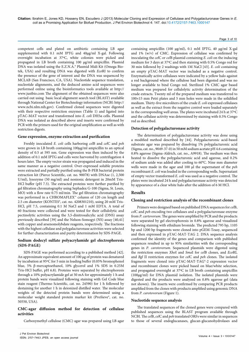
Primers were designed based on published DNA sequences for celB, celC and peh encoding two cellulases and a polygalacturonase enzyme from P. cartovorum. The genes were amplified by PCR and the products were separated by gel electrophoresis in 0.8% agarose and visualized following staining with ethidium bromide. The predicted 795 bp, 1105 bp and 1200 bp fragments were cloned into pGEM-Teasy, sequenced and then expressed in pTAC-MAT-TAG 2. DNA sequence analysis confirmed the identity of the genes and comparison with published sequences resulted in up to 95% similarities with the corresponding genes in P. carotovorum. Sequenced plasmids were digested using the restriction enzymes XhoI and SmaI for celB clones, and EcoRI, and Bgl II restriction enzymes for celC and peh clones. The isolated fragments were cloned into pTAC-MAT-TAG®-2 expression vector and recombinant clones were picked based on blue/white selection, and propagated overnight at 37ºC in LB broth containing ampicillin (100µg/ml) for DNA plasmid isolation. The isolated plasmids were digested and the products were analyzed on 0.8% agarose gel (data not shown). The inserts were confirmed by comparing PCR products amplified from the clones with products amplified using genomic DNA from P. carotovorum (Figure 1).
Nucleotide sequence analysis
The translated sequences of the cloned genes were compared with published sequences using the BLAST program available through NCBI. The celB, celC and peh translated ORFs were similar in sequences to those of several endoglucanases, glucan-glucanohydrolases and
Citation: Ibrahim E, Jones KD, Hosseny EN, Escudero J (2013) Molecular Cloning and Expression of Cellulase and Polygalacturonase Genes in E. coli as a Promising Application for Biofuel Production. J Pet Environ Biotechnol 4: 147. doi:10.4172/2157-7463.1000147
Page 4 of 10
Volume 4 • Issue 3 • 1000147J Pet Environ BiotechnolISSN: 2157-7463 JPEB, an open access journal
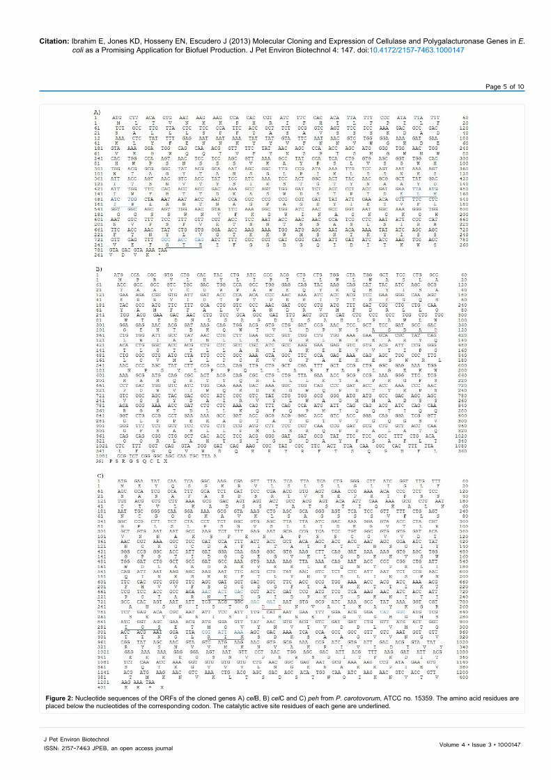
polygalacturonases, respectively. The nucleotide sequences along with their corresponding amino acids were found to exhibit a high degree of similarities with P. carotovorum and other organisms possessing cellulase and polygalacturonase activities (Table 2). Based on the available conserved domain sequences, the deduced amino acid sequences of the 3 tested ORFs were found to belong to glycosyl hydrolase families 12, 8 and 28 for celB, celC and peh, respectively [45,46]. The nucleotide length of the tested celB, celC and peh ORFs were determined to be 795 bp, 1105 bp and 1209 bp, with putatively encoding proteins of 266, 369 and 404 amino acids, and with estimated molecular weights of 29.5 kDa, 41.3 kDa and 42.5 kDa, respectively. The nucleotide sequences of the ORFs of the 3 cloned genes and their corresponding amino acids are shown in Figure 2.
CMC-agar diffusion method for detection of cellulase activity
Three transformants each of celC and celB were inoculated on CMC-agar including the negative control, E. coli containing an empty pTAC-MAT vector. The agar plates were incubated 3 days at 37°C and then stained with 0.5% Congo red for 30 min followed by 2 washings with 1M NaCl [43]. Enzymatically active cellulases were indicated by a yellow halo against a red background where the cellulose had been digested and was no longer available to bind Congo red. All of the tested
strains were positive for cellulases by visual inspection (data not shown) so one representative transformant of each of the cloned cellulases was used for gene expression and characterization. The transformed E. coli strains, including the empty-vector negative control strain, were grown and induced for gene expression for 4 hours. The cells were harvested and 35 µl of the supernatants were loaded separately in the corresponding wells in the CMC-agar. The plates were incubated 24 h at 37°C and the cellulase activity was determined by staining with 0.5% Congo red as described before. As shown in Figure 3, the crude supernatants of the celB and celC transformants contained cellulase activity as indicated by the appearance of the yellow halo against the red background. However, the CMC-cellulase activity of the celC protein is much higher than was detected by celB encoded protein as indicated by the larger diameter of the yellow halo. Furthermore, no activity was detected in the well containing the negative control supernatant. These results confirm the expression and cellulase activity of the cloned celB and celC products.
Detection of polygalacturonase activity
The peh-pTAC-MAT transformed E. coli was grown in LB broth containing ampicillin (100µg/ml), 0.1 mM IPTG, 40µg/ml X-gal to induce the expression of the cloned peh product, polygalacturonase. After
A BA B A B
Figure 1: Gel electrophoresis showing a comparison of the PCR products of the amplified ORFs of celB, celC and peh of P. carotovorum (lanes B in each figure) and the amplified fragments of the celB, celC and peh clones (lanes A), respectively. Lane 1 of each figure contains Hi-lo™ DNA ladder, (Minnesota Molecular, Inc) with relevant size markers indicated. Bands with a molecular size of approximately 795bp, 1105bp, and 1200bp were detected in the lanes loaded with the amplified products from celB, celC and peh clones, which were the same size as the amplified products from genomic DNA of P. carotovorum.
Tested clone Aligned organism Enzyme coded % Identity % positivity Accession no.
celB
P. carotovorum subsp. carotovorum beta(1,4)-glucan glucanohydrolase precursor 97% 98% AAC02965.2P.carotovorum subsp.carotovorum PC1 Cellulase 96% 97% YP_003017082.1
P. wasabiae WPP163z Cellulase 84% 90% YP_003260319.1Bacillus licheniformis WX-02 glycoside hydrolase family protein 66% 81% ZP_17658456.1
Paenibacillus mucilaginosus 3016 glycoside hydrolase 53% 74% YP_005314236.1
celC
P. carotovorum subsp. carotovorum WPP14 endo-1,4-D-glucanase 95% 95% ZP_03832232.1P. carotovorum subsp. carotovorum PC1 Cellulase 92% 94% refYP_003015672.1
P. wasabiae WPP163 endo-1,4-D-glucanase 87% 91% YP_003257546.1Serratia marcescens FGI94 endoglucanase Y 70% 79% YP_007342699.1
Yersinia intermedia ATCC 29909 Endoglucanase 63% 74% ZP_04636877.1
peh
P. carotovorum polygalacturonase 99% 99% AAA03624.1P. carotovorum polygalacturonase 99% 99% BAA74431.1
P. atrosepticum SCRI1043 endo-polygalacturonase 95% 97% YP_049201.1Erwinia pyrifoliae DSM 12163 polygalacturonase 60% 73% YP_005802329.1
Erwinia amylovora ATCC BAA-2158 polygalacturonase 59% 73% CBX81050.1
Table 2: Comparisons of celB, celC and peh translated amino acid sequences with various species using NCBI´s BLAST search.
Citation: Ibrahim E, Jones KD, Hosseny EN, Escudero J (2013) Molecular Cloning and Expression of Cellulase and Polygalacturonase Genes in E. coli as a Promising Application for Biofuel Production. J Pet Environ Biotechnol 4: 147. doi:10.4172/2157-7463.1000147
Page 5 of 10
Volume 4 • Issue 3 • 1000147J Pet Environ BiotechnolISSN: 2157-7463 JPEB, an open access journal
Figure 2: Nucleotide sequences of the ORFs of the cloned genes A) celB, B) celC and C) peh from P. carotovorum, ATCC no. 15359. The amino acid residues are placed below the nucleotides of the corresponding codon. The catalytic active site residues of each gene are underlined.

Citation: Ibrahim E, Jones KD, Hosseny EN, Escudero J (2013) Molecular Cloning and Expression of Cellulase and Polygalacturonase Genes in E. coli as a Promising Application for Biofuel Production. J Pet Environ Biotechnol 4: 147. doi:10.4172/2157-7463.1000147
Page 6 of 10
Volume 4 • Issue 3 • 1000147J Pet Environ BiotechnolISSN: 2157-7463 JPEB, an open access journal
4 hours of induction, the cells were harvested and the supernatant was collected and tested for enzymatic activity. The negative control strain of E. coli transformed with the empty pTAC-MAT vector was treated in the same manner. One hundred µl of the respective supernatants were incubated in the wells incorporated in the polygalacturonic acid agar plates and incubated for 24 h. Polygalacturonase activity was indicated by the appearance of a white halo around the wells after the addition of 6 M HCl. As shown in Figure 4, polygalacturonase activity was detected in the supernatant of the E. coli transformed with peh-pTACMAT but it was not detected in the well of the negative control (panels A and B, respectively). These results clearly indicate expression and activity of polygalacturonase from the cloned peh gene.
SDS-PAGE for molecular weight determination
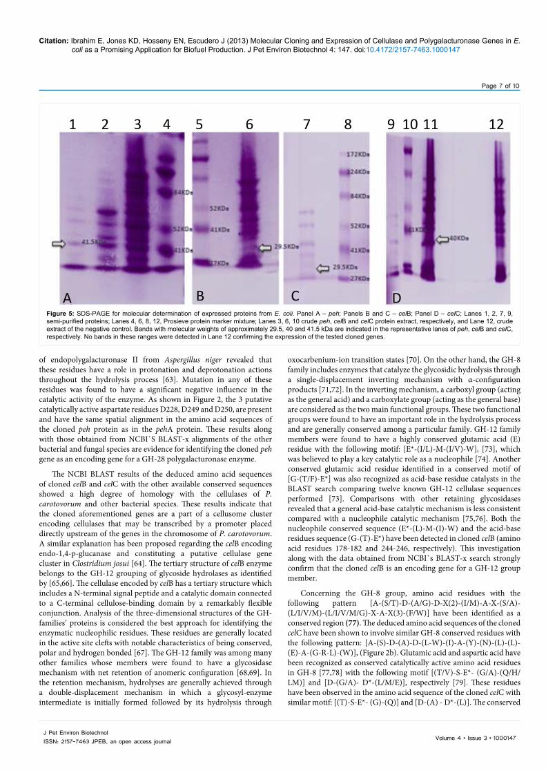
The molecular weights of the expressed celB, celC and peh products were determined using the SDS-PAGE method. The crude extracts of the expressed E. coli transformed with the cloned celB, celC and peh genes, as well as the corresponding semi-purified fractions with maximum cellulolytic and pectinolytic activities were used for molecular weight determination. The molecular weights of the protein products found in the supernatant of the negative control E. coli were also analyzed. As shown in Figure 5, protein bands of approximate molecular weights of 29.5, 40, 41.5 kDa were detected in the lanes loaded with the crude supernatants of the expressed celB, celC and peh along with their representative semi-purified fractions, respectively. Similar molecular weights were previously detected in the protein products of the cloned genes of P. carotovorum [34,47,48]. In contrast, the lane loaded with the crude supernatant of the negative control of E. coli had no bands in the aforementioned molecular weight ranges. These results indicate the putative expression of the 3 cloned genes.
DiscussionGlycoside hydrolases (GHs) are comprised of a wide range of
enzymes that are capable of hydrolyzing the glycosidic bonds in glycosides, glycans and glycoconjugates [49]. GHs are playing a critical role in biofuel production through their wide application in production of reducing sugars from pre-treated biomass materials. The reducing sugars formed are useful substrates for ethanol and butanol production which, indeed, can be used as renewable sources for gasoline [50].
Based on the available conserved domain sequences in the NCBI website, the deduced amino acid sequences of the 3 cloned ORFs were found to belong to GHs-12, 8, 28 for the cloned celB, celC and peh, respectively [45,46]. The enzymes belonging to GH-28 family are classified into several categories in accordance with their catalytic hydrolysis mechanism and are given specific E.C. numbers based
on the Nomenclature Committee of the International Union of Biochemistry and Molecular Biology (http://www.brenda-enzymes.org/). Comparison of the deduced amino acid sequences encoded by peh with those GH amino acid sequences of various species using NCBI´s BLAST search revealed that the cloned gene belongs to the polygalacturonase category of the GH-28 enzymes. Polygalacturonases (PG) are a group of enzymes that function in the hydrolysis of the α-linkage of the galacturonic acid (GalA) monomer residues in pectin (E.C.’s 3.2.1.15 [endo-PG] and 3.2.1.67 [exo-PG]) [51]. Genes encoding GH-28 enzymes have been identified in a number of plant pathogens and fungal species [52]. These degradative enzymes found in pathogenic microorganisms may act as virulence factors and play a critical role in plant cell wall maceration [53,54]. P. carotovorum is a known plant pathogen and the presence of the GH-28 polygalacturonase gene in the genome supports its pathogenic role in the hydrolysis of plant cell wall pectin. The role of the peh product in plant tissue maceration along with the other expressed enzymes has been investigated by many researchers [55,56]. The GH-28 polygalacturonases are characterized by the presence of 4 conserved amino acid groups (NTD, DD, HG, and RIK) which were thought to be implicated in the catalytic mechanism [57]. As indicated in Figure 2, the aforementioned conserved amino acid regions were found to be present in the deduced amino acid sequence of the peh encoded protein. Comparison of these results with those of others [58] reveals replacement of a histidine residue with an arginine residue in their identified pehN gene of E. chrysanthemi 3937. The possible replacement of the pehN gene was inferred to suggest a mechanism adapted to different substrate specificities [58]. Histidine (H) was also recognized as a conserved active site residue among fungal and bacterial GH-28 polygalacturonases with an identified motif of (G/S/D/E/N/K/R/H*)-x(2) (V/M/F/C)-x(2)-(G/S)-H*-G-(L/I/V/M/A/G)- x(1,2)-(L/I/V/M)-G-S [59]. The deduced amino acid sequence of the cloned peh was found to include the histidine active site residue in the following motif [(H*)- N-E- (F) -G-T- (G)- H*-G-(M)- S- (I)- G -S]. Rye et al. noted that the cleavage of glycosidic bonds by several glycoside hydrolases can be achieved by either a single- or double-displacement mechanism, which gives rise to inversion or retention of anomeric configuration, respectively [60]. Hydrolysis of the homogalacturonan and the rhamnogalacturonan components of the pectin chain by GH-28 polygalacturonases has been revealed to be a single-displacement inverting mechanism [59,61]. Pickersgill et al have mapped the active site of the pehA encoded GH-28 polygalacturonase of P. carotovorum. Three aspartate residues: D202, D223 and D224 have been found to be involved in the catalytic mechanism of this polygalacturonase [62]. The 3 catalytic aspartate residues have also been recognized to be functionally conserved between exo-and endo-acting polygalacturonases [59]. Site-directed mutagenesis studies
A B C
Figure 3: CMC agar for the detection of cellulase activity of the cloned celB and celC. A: negative control supernatant of E. coli with an empty vector. B: the supernatant of E. coli with celB gene. C: the supernatant of E. coli with celC gene. The detected yellow halo against the red background in B and C indicate the cellulase activity of the cloned gene products.
Figure 4: Cup plate method for determination of polygalacturonase activity. A: peh transformed E. coli supernatant. B: empty vector transformed E. coli supernatant. The white halo appearance after the addition of 6.0 M HCl indicates the presence of polygalacturonase activity from the expressed peh encoded protein.
Citation: Ibrahim E, Jones KD, Hosseny EN, Escudero J (2013) Molecular Cloning and Expression of Cellulase and Polygalacturonase Genes in E. coli as a Promising Application for Biofuel Production. J Pet Environ Biotechnol 4: 147. doi:10.4172/2157-7463.1000147
Page 7 of 10
Volume 4 • Issue 3 • 1000147J Pet Environ BiotechnolISSN: 2157-7463 JPEB, an open access journal
of endopolygalacturonase II from Aspergillus niger revealed that these residues have a role in protonation and deprotonation actions throughout the hydrolysis process [63]. Mutation in any of these residues was found to have a significant negative influence in the catalytic activity of the enzyme. As shown in Figure 2, the 3 putative catalytically active aspartate residues D228, D249 and D250, are present and have the same spatial alignment in the amino acid sequences of the cloned peh protein as in the pehA protein. These results along with those obtained from NCBI`S BLAST-x alignments of the other bacterial and fungal species are evidence for identifying the cloned peh gene as an encoding gene for a GH-28 polygalacturonase enzyme.
The NCBI BLAST results of the deduced amino acid sequences of cloned celB and celC with the other available conserved sequences showed a high degree of homology with the cellulases of P. carotovorum and other bacterial species. These results indicate that the cloned aforementioned genes are a part of a cellusome cluster encoding cellulases that may be transcribed by a promoter placed directly upstream of the genes in the chromosome of P. carotovorum. A similar explanation has been proposed regarding the celB encoding endo-1,4-p-glucanase and constituting a putative cellulase gene cluster in Clostridium josui [64]. The tertiary structure of celB enzyme belongs to the GH-12 grouping of glycoside hydrolases as identified by [65,66]. The cellulase encoded by celB has a tertiary structure which includes a N-terminal signal peptide and a catalytic domain connected to a C-terminal cellulose-binding domain by a remarkably flexible conjunction. Analysis of the three-dimensional structures of the GH-families’ proteins is considered the best approach for identifying the enzymatic nucleophilic residues. These residues are generally located in the active site clefts with notable characteristics of being conserved, polar and hydrogen bonded [67]. The GH-12 family was among many other families whose members were found to have a glycosidase mechanism with net retention of anomeric configuration [68,69]. In the retention mechanism, hydrolyses are generally achieved through a double-displacement mechanism in which a glycosyl-enzyme intermediate is initially formed followed by its hydrolysis through
oxocarbenium-ion transition states [70]. On the other hand, the GH-8 family includes enzymes that catalyze the glycosidic hydrolysis through a single-displacement inverting mechanism with α-configuration products [71,72]. In the inverting mechanism, a carboxyl group (acting as the general acid) and a carboxylate group (acting as the general base) are considered as the two main functional groups. These two functional groups were found to have an important role in the hydrolysis process and are generally conserved among a particular family. GH-12 family members were found to have a highly conserved glutamic acid (E) residue with the following motif: [E*-(I/L)-M-(I/V)-W], [73], which was believed to play a key catalytic role as a nucleophile [74]. Another conserved glutamic acid residue identified in a conserved motif of [G-(T/F)-E*] was also recognized as acid-base residue catalysts in the BLAST search comparing twelve known GH-12 cellulase sequences performed [73]. Comparisons with other retaining glycosidases revealed that a general acid-base catalytic mechanism is less consistent compared with a nucleophile catalytic mechanism [75,76]. Both the nucleophile conserved sequence (E*-(L)-M-(I)-W) and the acid-base residues sequence (G-(T)-E*) have been detected in cloned celB (amino acid residues 178-182 and 244-246, respectively). This investigation along with the data obtained from NCBI`s BLAST-x search strongly confirm that the cloned celB is an encoding gene for a GH-12 group member.
Concerning the GH-8 group, amino acid residues with the following pattern [A-(S/T)-D-(A/G)-D-X(2)-(I/M)-A-X-(S/A)-(L/I/V/M)-(L/I/V/M/G)-X-A-X(3)-(F/W)] have been identified as a conserved region (77). The deduced amino acid sequences of the cloned celC have been shown to involve similar GH-8 conserved residues with the following pattern: [A-(S)-D-(A)-D-(L-W)-(I)-A-(Y)-(N)-(L)-(L)-(E)-A-(G-R-L)-(W)], (Figure 2b). Glutamic acid and aspartic acid have been recognized as conserved catalytically active amino acid residues in GH-8 [77,78] with the following motif [(T/V)-S-E*- (G/A)-(Q/H/LM)] and [D-(G/A)- D*-(L/M/E)], respectively [79]. These residues have been observed in the amino acid sequence of the cloned celC with similar motif: [(T)-S-E*- (G)-(Q)] and [D-(A) - D*-(L)]. The conserved
1 2 3 4 5 6 7 8 9 10 11 12
A B C DFigure 5: SDS-PAGE for molecular determination of expressed proteins from E. coli. Panel A – peh; Panels B and C – celB; Panel D – celC; Lanes 1, 2, 7, 9, semi-purified proteins; Lanes 4, 6, 8, 12, Prosieve protein marker mixture; Lanes 3, 6, 10 crude peh, celB and celC protein extract, respectively, and Lane 12, crude extract of the negative control. Bands with molecular weights of approximately 29.5, 40 and 41.5 kDa are indicated in the representative lanes of peh, celB and celC, respectively. No bands in these ranges were detected in Lane 12 confirming the expression of the tested cloned genes.

Citation: Ibrahim E, Jones KD, Hosseny EN, Escudero J (2013) Molecular Cloning and Expression of Cellulase and Polygalacturonase Genes in E. coli as a Promising Application for Biofuel Production. J Pet Environ Biotechnol 4: 147. doi:10.4172/2157-7463.1000147
Page 8 of 10
Volume 4 • Issue 3 • 1000147J Pet Environ BiotechnolISSN: 2157-7463 JPEB, an open access journal
glutamic acid active site residue was suggested to act as a proton donor in the catalytic reaction, while the aspartic acid residue was inferred to have a catalytic role as a nucleophile [79]. The significant ablation that has been observed in the enzymatic activity upon the replacement of either active-site residue by site-directed mutagenesis emphasizes their main catalytic role [77].
The basal expression of the 3 tested clones has been examined using agar diffusion methods. Polygalacturonase activity was detected in the supernatant of the cloned peh as indicated by the appearance of a white halo surrounding the well in the polygalacturonic acid based medium (Figure 4). This observation was confirmed by the absence of the white precipitate around the negative control well. Similar findings have been described of the appearance of the white halo surrounding the well loaded with polygalacturonase of Fusarium moniliforme and no halo was detected with the same sample mixed with polygalacturonase-inhibitor protein of Phaseolus vulgaris (common bean) [44]. The cellulase activities of the cloned celB and celC products were also confirmed by the appearance of a yellow halo against a red background in CMC-based medium. As indicated in Figure 3, cellulase activity of the celC product is relatively high in comparison with that of the celB product. It has also been reported a marked reduction in the cellulase activity of celB of P. carotovorum - LY34 relative to that of celA product [80]. The relative dissimilarities in cellulase activity may be attributed to the heterogeneous mode of their catalytic activities. As shown in Table 2, the deduced amino acid sequences of celB and celC encoded proteins showed a high degree of similarities with that of β-(1,4)-glucan glucanohydrolase precursor and endo-1,4-D-glucanase of P. carotovorum, respectively. The endogluconases exhibit a general hydrolytic activity for internal glycosidic linkages with a consequential increase in the reducing sugar concentration and decrease in the polymer length [81,82]. Furthermore, the action of glucan glucanohydrolases was reported on cello-oligomers with glucose production as a primary product [83,84]. However, the action of the GH-12 cellulase of Thermotoga neapolitana on cellotriose and cellotretrose resulted in the production of small quantities of glucose after prolonged periods of incubation with cellobiose as the main product [85]. On the other hand, the activity of the celB enzyme on CMC substrate, reported by [86,87], suggests that this enzyme has both endo- and exo-cellulase activities [85]. This suggestion has been supported by other reports of cellulases with both endo- and exo-activities [88,89]. Analysis of β-glucan hydrolysis products of a GH-12 member β-(1,4)-glucanase from Aspergillus japonicas by gel-permeation chromatography revealed the formation of glucose in the initial part of the reaction [90]. This report also suggested the exo-acting activity of that enzyme. The suggestion of exo-acting activity of the celB enzyme is consistent with its original role inside the cell as a transmembrane channel for cellobiose [91,92]. CelC encodes the enzyme that is reported to be responsible for the hydrolysis of cellobiose-phosphate into hydrophilic glucose products [35].
The successful cloning of genes encoding cellulases and polygalacturonase enzymes will lead to the development of a low-cost effective biorefinery strategy to achieve significant reduction in the consumption of unsustainable resources for fuel. Understanding the kinetic action of the expressed enzymes needs further investigations. The role of such enzymes in lignocellulosic waste bioconversions will also be investigated in the ongoing research. Further studies are anticipated involving the optimization of the experimental conditions necessary for achieving maximum biomass conversions into fermentable sugars. An improved gas chromatography-mass spectrophotometry method that has been developed by researchers at TAMUK will be a helpful
tool in understanding the catalytic action of the expressed enzymes in the bioconversion process.
Acknowledgements
The authors are grateful to Prof. Keith E. Taylor, Department of Chemistry and Biochemistry, University of Windsor, for his helpful comments on the manuscript.
References1. Kerr RA (2007) Climate change. Global warming is changing the world. Science
316: 188-190.
2. Stephanopoulos G (2007) Challenges in engineering microbes for biofuels production. Science 315: 801-804.
3. Hill J, Nelson E, Tilman D, Polasky S, Tiffany D (2006) Environmental, economic, and energetic costs and benefits of biodiesel and ethanol biofuels. Proc Natl Acad Sci U S A 103: 11206-11210.
4. Schmidt LD, Dauenhauer PJ (2007) Chemical engineering: hybrid routes to biofuels. Nature 447: 914-915.
5. Huffer S, Roche CM, Blanch HW, Clark DS (2012) Escherichia coli for biofuel production: bridging the gap from promise to practice. Trends Biotechnol 30: 538-545.
6. Bokinsky G, Peralta-Yahya PP, George A, Holmes BM, Steen EJ, et al. (2011) Synthesis of three advanced biofuels from ionic liquid-pretreated switchgrass using engineered Escherichia coli. Proc Natl Acad Sci U S A 108: 19949-19954.
7. Steen EJ, Kang Y, Bokinsky G, Hu Z, Schirmer A, et al. (2010) Microbial production of fatty-acid-derived fuels and chemicals from plant biomass. Nature 463: 559-562.
8. Edwards MC, Henriksen ED, Yomano LP, Gardner BC, Sharma LN, et al. (2011) Addition of genes for cellobiase and pectinolytic activity in Escherichia coli for fuel ethanol production from pectin-rich lignocellulosic biomass. Appl Environ Microbiol 77: 5184-5191.
9. Hanai T, Atsumi S, Liao JC (2007) Engineered synthetic pathway for isopropanol production in Escherichia coli. Appl Environ Microbiol 73: 7814-7818.
10. Atsumi S, Cann AF, Connor MR, Shen CR, Smith KM, et al. (2008) Metabolic engineering of Escherichia coli for 1-butanol production. Metab Eng 10: 305-311.
11. Lee SK, Chou H, Ham TS, Lee TS, Keasling JD (2008) Metabolic engineering of microorganisms for biofuels production: from bugs to synthetic biology to fuels. Curr Opin Biotechnol 19: 556-563.
12. Rude MA, Schirmer A (2009) New microbial fuels: a biotech perspective. Curr Opin Microbiol 12: 274-281.
13. Edwards MC, Doran-Peterson J (2012) Pectin-rich biomass as feedstock for fuel ethanol production. Appl Microbiol Biotechnol 95: 565-575.
14. Dale BE, Leong CK, Pham TK, Esquivel VM, Rios I, et al. (1996) Hydrolysis of lignocellulosics at low enzyme levels: Application of the AFEX process. Bioresource Technol 56:111-116.
15. Sun Y, Cheng J (2002) Hydrolysis of lignocellulosic materials for ethanol production: a review. Bioresour Technol 83: 1-11.
16. Berlin A, Gilkes N, Kurabi A, Bura R, Tu M, et al. (2005) Weak lignin-binding enzymes: a novel approach to improve activity of cellulases for hydrolysis of lignocellulosics. Appl Biochem Biotechnol 121-124: 163-70.
17. Guo G-L, Hsu D-C, Chen W-H, Chen W-H, Hwang W-S (2009) Characterization of enzymatic saccharification for acid-pretreated lignocellulosic materials with different lignin composition. Enzyme Microb Tech 45: 80-87.
18. Rivas B, Torrado A, Torre P, Converti A, Domínguez JM (2008) Submerged citric acid fermentation on orange peel autohydrolysate. J Agric Food Chem 56: 2380-2387.
19. Boluda-Aguilar M, García-Vidal L, González-Castañeda Fdel P, López-Gómez A (2010) Mandarin peel wastes pretreatment with steam explosion for bioethanol production. Bioresour Technol 101: 3506-3513.
20. Wilkins MR, Widmer WW, Grohmann K, Cameron RG (2007) Hydrolysis of grapefruit peel waste with cellulase and pectinase enzymes. Bioresour Technol 98: 1596-1601.
21. Dashtban M, Schraft H, Qin W (2009) Fungal bioconversion of lignocellulosic residues; opportunities & perspectives. Int J Biol Sci 5: 578-595.
Citation: Ibrahim E, Jones KD, Hosseny EN, Escudero J (2013) Molecular Cloning and Expression of Cellulase and Polygalacturonase Genes in E. coli as a Promising Application for Biofuel Production. J Pet Environ Biotechnol 4: 147. doi:10.4172/2157-7463.1000147
Page 9 of 10
Volume 4 • Issue 3 • 1000147J Pet Environ BiotechnolISSN: 2157-7463 JPEB, an open access journal
22. Enari TM, Niku-Paavola ML (1987) Enzymatic hydrolysis of cellulose: is the current theory of the mechanisms of hydrolysis valid? Crit Rev Biotechnol 5: 67-87.
23. Maki M, Leung KT, Qin W (2009) The prospects of cellulase-producing bacteria for the bioconversion of lignocellulosic biomass. Int J Biol Sci 5: 500-516.
24. Gupta S, Kapoor M, Sharma KK, Nair LM, Kuhad RC (2008) Production and recovery of an alkaline exo-polygalacturonase from Bacillus subtilis RCK under solid-state fermentation using statistical approach. Bioresour Technol 99: 937-945.
25. Voragen AGJ, Coenen GJ, Verhoef RP, Schols HA (2009) Pectin, a versatile polysaccharide present in plant cell walls. Struct Chem 20: 263-275.
26. Martin N. DSRS, De Silva R., Gomes E (2004) Pectinases production by fungal strain in solid state fermentation agroindustrial by-product. Braz Arch Biol Technol 47: 813-819.
27. Sharma N, Rathore M, Sharma M (2013) Microbial pectinase: sources, characterization and applications. Reviews in Environmental Science and Bio/Technology 12: 45-60.
28. Singh SA, Appu Rao AG (2002) A simple fractionation protocol for, and a comprehensive study of the molecular properties of, two major endopolygalacturonases from Aspergillus niger. Biotechnol Appl Biochem 35: 115-123.
29. Takao M, Nakaniwa T, Yoshikawa K, Terashita T, Sakai T (2001) Molecular cloning, DNA sequence, and expression of the gene encoding for thermostable pectate lyase of thermophilic Bacillus sp. TS 47. Biosci Biotechnol Biochem 65: 322-329.
30. Alonso J, Canet W, Howell N, Alique R (2003) Purification and characterisation of carrot (Daucus carota L) pectinesterase. J Sci Food Agr 83: 1600-1606.
31. Pathak N, Sanwal GG (1998) Multiple forms of polygalacturonase from banana fruits. Phytochemistry 48: 249-255.
32. Collmer A, Keen NT (1986) The Role of Pectic Enzymes in Plant Pathogenesis. Annu Rev Phytopathol 24: 383-409.
33. Béguin P (1990) Molecular biology of cellulose degradation. Annu Rev Microbiol 44: 219-248.
34. Woo Park Y, Tech Lim S, Dae Yun H (1998) Cloning and characterization of a CMCase gene, celB, of Erwinia carotovora subsp. carotovora LY34 and its comparison to celA. Mol Cells 8: 280-285.
35. Lai X, Ingram LO (1993) Cloning and sequencing of a cellobiose phosphotransferase system operon from Bacillus stearothermophilus XL-65-6 and functional expression in Escherichia coli. J Bacteriol 175: 6441-6450.
36. Cote CK, Honeyman AL (2003) The LicT protein acts as both a positive and a negative regulator of loci within the bgl regulon of Streptococcus mutans. Microbiology 149: 1333-1340.
37. Warner JB, Lolkema JS (2003) A Crh-specific function in carbon catabolite repression in Bacillus subtilis. FEMS Microbiol Lett 220: 277-280.
38. Nasuno S, Starr MP (1966) Polygalacturonase of Erwinia carotovora. J Biol Chem 241: 5298-5306.
39. Miller GL (1959) Use of Dinitrosalicylic Acid Reagent for Determination of Reducing Sugar. Anal Chem 31: 426-428.
40. Nelson N (1944) A Photometric Adaptation of the Somogyi Method for the Determination of Glucose. J Biol Chem 153: 375-380.
41. SMOGYI M (1952) Notes on sugar determination. J Biol Chem 195: 19-23.
42. Laemmli UK (1970) Cleavage of structural proteins during the assembly of the head of bacteriophage T4. Nature 227: 680-685.
43. Teather RM, Wood PJ (1982) Use of Congo red-polysaccharide interactions in enumeration and characterization of cellulolytic bacteria from the bovine rumen. Appl Environ Microbiol 43: 777-780.
44. Sella L, Castiglioni C, Roberti S, D’Ovidio R, Favaron F (2004) An endo-polygalacturonase (PG) of Fusarium moniliforme escaping inhibition by plant polygalacturonase-inhibiting proteins (PGIPs) provides new insights into the PG-PGIP interaction. FEMS Microbiol Lett 240: 117-124.
45. Marchler-Bauer A, Bryant SH (2004) CD-Search: protein domain annotations on the fly. Nucleic Acids Res 32: W327-331.
46. Marchler-Bauer A, Anderson JB, Chitsaz F, Derbyshire MK, DeWeese-Scott C,
et al. (2009) CDD: specific functional annotation with the Conserved Domain Database. Nucleic Acids Res 37: D205-210.
47. Hinton JC, Gill DR, Lalo D, Plastow GS, Salmond GP (1990) Sequence of the peh gene of Erwinia carotovora: homology between Erwinia and plant enzymes. Mol Microbiol 4: 1029-1036.
48. Lei SP, Lin HC, Wang SS, Higaki P, Wilcox G (1992) Characterization of the Erwinia carotovora peh gene and its product polygalacturonase. Gene 117: 119-124.
49. Vuong TV, Wilson DB (2010) Glycoside hydrolases: catalytic base/nucleophile diversity. Biotechnol Bioeng 107: 195-205.
50. Wilson DB (2009) Cellulases and biofuels. Curr Opin Biotechnol 20: 295-299.
51. Markovic O, Janecek S (2001) Pectin degrading glycoside hydrolases of family 28: sequence-structural features, specificities and evolution. Protein Eng 14: 615-631.
52. Sprockett DD, Piontkivska H, Blackwood CB (2011) Evolutionary analysis of glycosyl hydrolase family 28 (GH28) suggests lineage-specific expansions in necrotrophic fungal pathogens. Gene 479: 29-36.
53. Scott-Craig JS, Panaccione DG, Cervone F, Walton JD (1990) Endopolygalacturonase is not required for pathogenicity of Cochliobolus carbonum on maize. Plant Cell 2: 1191-1200.
54. Gao S, Choi GH, Shain L, Nuss DL (1996) Cloning and targeted disruption of enpg-1, encoding the major in vitro extracellular endopolygalacturonase of the chestnut blight fungus, Cryphonectria parasitica. Appl Environ Microbiol 62: 1984-1990.
55. Saarilahti HT, Heino P, Pakkanen R, Kalkkinen N, Palva I, et al. (1990) Structural analysis of the pehA gene and characterization of its protein product, endopolygalacturonase, of Erwinia carotovora subspecies carotovora. Mol Microbiol 4: 1037-1044.
56. Pirhonen M, Saarilahti HT, Karlsson MB, Palva ET (1991) Identification of pathogenicity determinants of Erwinia carotovora subsp. carotovora by transposon mutagenesis. Plant-Microbe Interact 4: 276-283.
57. Bussink HJ, Buxton FP, Visser J (1991) Expression and sequence comparison of the Aspergillus niger and Aspergillus tubigensis genes encoding polygalacturonase II. Curr Genet 19: 467-474.
58. Hugouvieux-Cotte-Pattat N, Shevchik VE, Nasser W (2002) PehN, a polygalacturonase homologue with a low hydrolase activity, is coregulated with the other Erwinia chrysanthemi polygalacturonases. J Bacteriol 184: 2664-2673.
59. Abbott DW, Boraston AB (2007) The structural basis for exopolygalacturonase activity in a family 28 glycoside hydrolase. J Mol Biol 368: 1215-1222.
60. Rye CS, Withers SG (2000) Glycosidase mechanisms. Curr Opin Chem Biol 4: 573-580.
61. Pitson SM, Mutter M, van den Broek LA, Voragen AG, Beldman G (1998) Stereochemical course of hydrolysis catalysed by alpha-L-rhamnosyl and alpha-D-galacturonosyl hydrolases from Aspergillus aculeatus. Biochem Biophys Res Commun 242: 552-559.
62. Pickersgill R, Smith D, Worboys K, Jenkins J (1998) Crystal structure of polygalacturonase from Erwinia carotovora ssp. carotovora. J Biol Chem 273: 24660-24664.
63. van Santen Y, Benen JA, Schröter KH, Kalk KH, Armand S, et al. (1999) 1.68-A crystal structure of endopolygalacturonase II from Aspergillus niger and identification of active site residues by site-directed mutagenesis. J Biol Chem 274: 30474-30480.
64. Fujino T, Karita S, Ohmiya K (1993) Nucleotide sequences of the celB gene encoding endo-1,4-β-β-glucanase-2, ORF1 and ORF2 forming a putative cellulase gene cluster of Clostridium josui. J Ferment Bioeng 76: 243-250.
65. Wittmann S, Shareck F, Kluepfel D, Morosoli R (1994) Purification and characterization of the CelB endoglucanase from Streptomyces lividans 66 and DNA sequence of the encoding gene. Appl Environ Microbiol 60: 1701-1703.
66. Davies G, Henrissat B (1995) Structures and mechanisms of glycosyl hydrolases. Structure 3: 853-859.
67. Bartlett GJ, Porter CT, Borkakoti N, Thornton JM (2002) Analysis of catalytic residues in enzyme active sites. J Mol Biol 324: 105-121.
68. Schou C, Rasmussen G, Kaltoft MB, Henrissat B, Schülein M (1993)
Citation: Ibrahim E, Jones KD, Hosseny EN, Escudero J (2013) Molecular Cloning and Expression of Cellulase and Polygalacturonase Genes in E. coli as a Promising Application for Biofuel Production. J Pet Environ Biotechnol 4: 147. doi:10.4172/2157-7463.1000147
Page 10 of 10
Volume 4 • Issue 3 • 1000147J Pet Environ BiotechnolISSN: 2157-7463 JPEB, an open access journal
Stereochemistry, specificity and kinetics of the hydrolysis of reduced cellodextrins by nine cellulases. Eur J Biochem 217: 947-953.
69. Schülein M (1997) Enzymatic properties of cellulases from Humicola insolens. J Biotechnol 57: 71-81.
70. Davies G, Sinnott, ML, and Withers, SG (1997) Comprehensive Biological Catalysis. Academic Press, London, UK.
71. Fierobe HP, Bagnara-Tardif C, Gaudin C, Guerlesquin F, Sauve P, et al. (1993) Purification and characterization of endoglucanase C from Clostridium cellulolyticum. Catalytic comparison with endoglucanase A. Eur J Biochem 217: 557-565.
72. Collins T, Meuwis MA, Stals I, Claeyssens M, Feller G, et al. (2002) A novel family 8 xylanase, functional and physicochemical characterization. J Biol Chem 277: 35133-35139.
73. Altschul SF, Gish W, Miller W, Myers EW, Lipman DJ (1990) Basic local alignment search tool. J Mol Biol 215: 403-410.
74. Zechel DL, He S, Dupont C, Withers SG (1998) Identification of Glu-120 as the catalytic nucleophile in Streptomyces lividans endoglucanase celB. Biochem J 336 : 139-145.
75. Wang Q, Graham RW, Trimbur D, Warren RAJ, Withers SG (1994) Changing Enzymic Reaction Mechanisms by Mutagenesis: Conversion of a Retaining Glucosidase to an Inverting Enzyme. J Am Chem Soc 116: 11594-11595.
76. Lawson SL, Wakarchuk WW, Withers SG (1997) Positioning the acid/base catalyst in a glycosidase: studies with Bacillus circulans xylanase. Biochemistry 36: 2257-2265.
77. Kimoto H, Kusaoke H, Yamamoto I, Fujii Y, Onodera T, et al. (2002) Biochemical and genetic properties of Paenibacillus glycosyl hydrolase having chitosanase activity and discoidin domain. J Biol Chem 277: 14695-14702.
78. Alzari PM, Souchon H, Dominguez R (1996) The crystal structure of endoglucanase CelA, a family 8 glycosyl hydrolase from Clostridium thermocellum. Structure 4: 265-275.
79. Choi YJ, Kim EJ, Piao Z, Yun YC, Shin YC (2004) Purification and characterization of chitosanase from Bacillus sp. strain KCTC 0377BP and its application for the production of chitosan oligosaccharides. Appl Environ Microbiol 70: 4522-4531.
80. Park YW, Lim ST, Cho SJ, Yun HD (1997) Characterization of Erwinia carotovora subsp. carotovora LY34 endo-1,4-beta-glucanase genes and rapid identification of their gene products. Biochem Biophys Res Commun 241: 636-641.
81. Wood TM, Bhat KM (1988) Methods for measuring cellulase activities. Methods in Enzymology. (Vol. 160) Academic Press, London, UK.
82. Béguin P, Aubert JP (1994) The biological degradation of cellulose. FEMS Microbiol Rev 13: 25-58.
83. Rixon JE, Ferreira LM, Durrant AJ, Laurie JI, Hazlewood GP, et al. (1992) Characterization of the gene celD and its encoded product 1,4-beta-D-glucan glucohydrolase D from Pseudomonas fluorescens subsp. cellulosa. Biochem J 285: 947-955.
84. Goyal AK, Eveleigh DE (1996) Cloning, sequencing and analysis of the ggh-A gene encoding a 1,4-beta-D-glucan glucohydrolase from Microbispora bispora. Gene 172: 93-98.
85. Bok JD, Yernool DA, Eveleigh DE (1998) Purification, characterization, and molecular analysis of thermostable cellulases CelA and CelB from Thermotoga neapolitana. Appl Environ Microbiol 64: 4774-4781.
86. Bronnenmeier K, Kern A, Liebl W, Staudenbauer WL (1995) Purification of Thermotoga maritima enzymes for the degradation of cellulosic materials. Appl Environ Microbiol 61: 1399-1407.
87. Ruttersmith LD, Daniel RM (1991) Thermostable cellobiohydrolase from the thermophilic eubacterium Thermotoga sp. strain FjSS3-B.1. Purification and properties. Biochem J 277 : 887-890.
88. Barr BK, Hsieh YL, Ganem B, Wilson DB (1996) Identification of two functionally different classes of exocellulases. Biochemistry 35: 586-592.
89. Tomme P, Kwan E, Gilkes NR, Kilburn DG, Warren RA (1996) Characterization of CenC, an enzyme from Cellulomonas fimi with both endo- and exoglucanase activities. J Bacteriol 178: 4216-4223.
90. Grishutin SG, Gusakov AV, Dzedzyulya EI, Sinitsyn AP (2006) A lichenase-like family 12 endo-(1-->4)-beta-glucanase from Aspergillus japonicus: study of the substrate specificity and mode of action on beta-glucans in comparison with other glycoside hydrolases. Carbohydr Res 341: 218-229.
91. Hu KY, Saier MH Jr (2002) Phylogeny of phosphoryl transfer proteins of the phosphoenolpyruvate-dependent sugar-transporting phosphotransferase system. Res Microbiol 153: 405-415.
92. Kotrba P, Inui M, Yukawa H (2003) A single V317A or V317M substitution in Enzyme II of a newly identified beta-glucoside phosphotransferase and utilization system of Corynebacterium glutamicum R extends its specificity towards cellobiose. Microbiology 149: 1569-1580.
Submit your next manuscript and get advantages of OMICS Group submissionsUnique features:
• Userfriendly/feasiblewebsite-translationofyourpaperto50world’sleadinglanguages• AudioVersionofpublishedpaper• Digitalarticlestoshareandexplore
Special features:
• 250OpenAccessJournals• 20,000editorialteam• 21daysrapidreviewprocess• Qualityandquickeditorial,reviewandpublicationprocessing• IndexingatPubMed(partial),Scopus,EBSCO,IndexCopernicusandGoogleScholaretc• SharingOption:SocialNetworkingEnabled• Authors,ReviewersandEditorsrewardedwithonlineScientificCredits• Betterdiscountforyoursubsequentarticles
Submityourmanuscriptat:http://www.editorialmanager.com/environsci
Citation: Ibrahim E, Jones KD, Hosseny EN, Escudero J (2013) Molecular Cloning and Expression of Cellulase and Polygalacturonase Genes in E. coli as a Promising Application for Biofuel Production. J Pet Environ Biotechnol 4: 147. doi:10.4172/2157-7463.1000147


![Industrial Biocatalysts Nature[1]](https://img.pdfslide.net/doc/110x75/577cbd8e1a28aba7118de783/industrial-biocatalysts-nature1.jpg)