Embed Size (px)
Citation preview
iCell® CardiomyocytesBasic cardiac research, drug discovery, and toxicity
testing require an in vitro system that recapitulates native
biology. Cellular Dynamics International (CDI) meets this
need with iCell® Cardiomyocytes, human cardiomyocytes
with proven, predictive, and published advantages over
other cellular cardiac models for faithfully and robustly
reflecting cardiovascular disease endpoints and for
enabling early and accurate prediction of drug-induced
cardiotoxicity. The human biology, ease of use, and far-
reaching applicability of iCell Cardiomyocytes has had a
profound impact on academic research and the search for
new medicines and cures.
Genomic and Protein Characterization
iCell Cardiomyoctyes are differentiated from human
induced pluripotent stem cells. Whole-genome transcript
profiling and immunocytochemical characterization
demonstrate that iCell Cardiomyocytes exhibit a stable
human cardiac gene expression profile with proper
protein expression and localization necessary for cardiac
function.
Human cells: iCell Cardiomyocytes are terminally differentiated from human iPS cells and exhibit functional characteristics similar to native human cardiomyocytes.
Homogenous and reproducible: iCell Cardiomyocytes are highly pure, providing biologically relevant and reproducible results.
Acute and long-term testing: iCell Cardiomyocytes remain viable and pure in culture for weeks, enabling assessment of both acute and sub-chronic responses.
Easy to implement: iCell Cardiomyocytes are shipped cryopreserved with cell culture media specifically formulated for optimal cell performance. Simply thaw and use.
Advantages
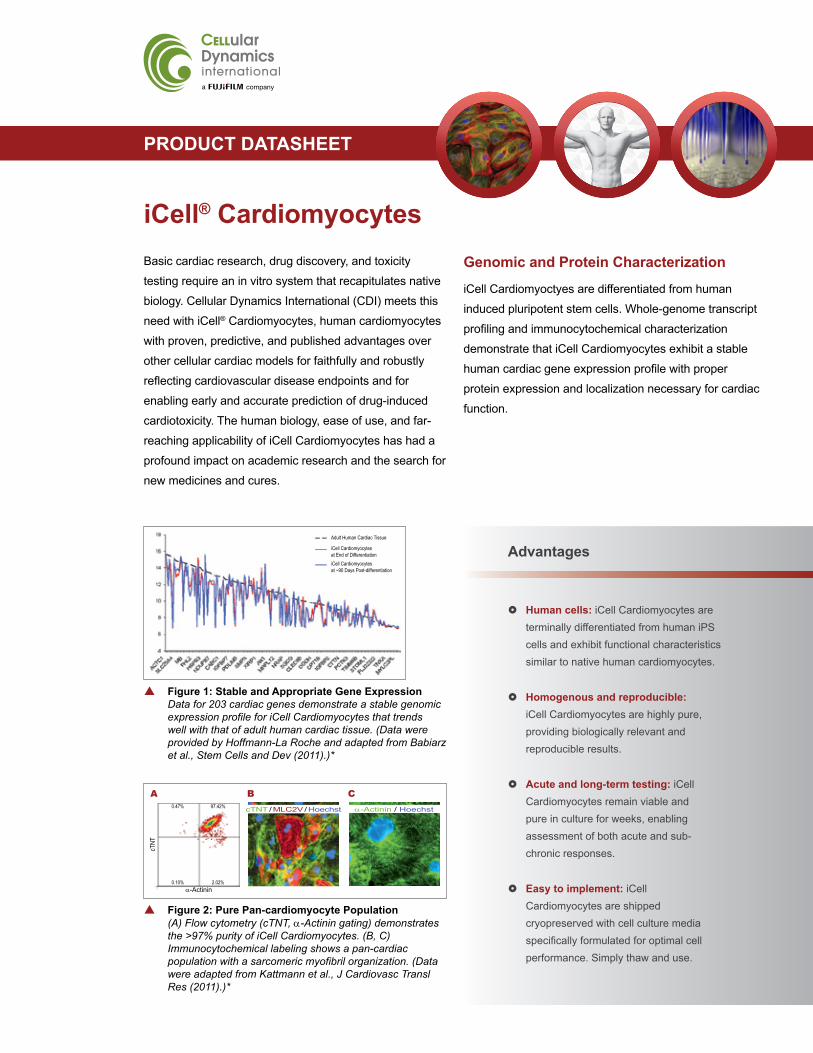
p Figure 1: Stable and Appropriate Gene Expression Data for 203 cardiac genes demonstrate a stable genomic expression profile for iCell Cardiomyocytes that trends well with that of adult human cardiac tissue. (Data were provided by Hoffmann-La Roche and adapted from Babiarz et al., Stem Cells and Dev (2011).)*
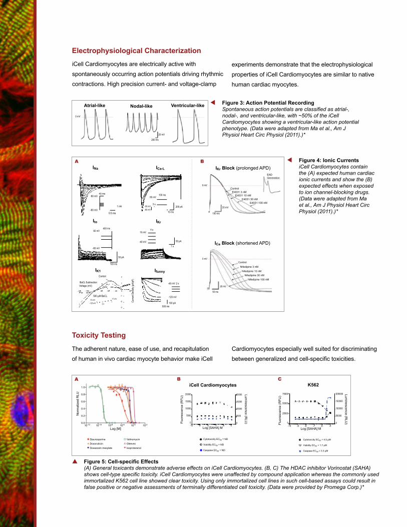
p Figure 2: Pure Pan-cardiomyocyte Population (A) Flow cytometry (cTNT, a-Actinin gating) demonstrates the >97% purity of iCell Cardiomyocytes. (B, C) Immunocytochemical labeling shows a pan-cardiac population with a sarcomeric myofibril organization. (Data were adapted from Kattmann et al., J Cardiovasc Transl Res (2011).)*
Ca-Actinin / Hoechst
BcTNT / MLC2V / Hoechst
A
cTNT
a-Actinin
0.47% 97.42%
0.10% 2.02%
Adult Human Cardiac Tissue
iCell Cardiomyocytes at End of DifferentiationiCell Cardiomyocytesat ~90 Days Post-differentiation
PRODUCT DATASHEET
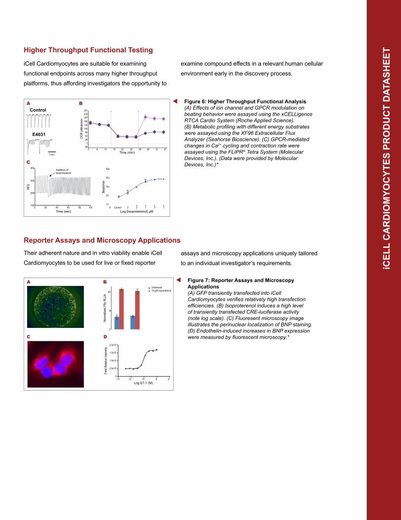
Electrophysiological Characterization
experiments demonstrate that the electrophysiological
properties of iCell Cardiomyocytes are similar to native
human cardiac myocytes.
t Figure 3: Action Potential RecordingSpontaneous action potentials are classified as atrial-, nodal-, and ventricular-like, with ~50% of the iCell Cardiomyocytes showing a ventricular-like action potential phenotype. (Data were adapted from Ma et al., Am J Physiol Heart Circ Physiol (2011).)*
Atrial-like Nodal-like Ventricular-like
0 mV
20 mV
200 ms
iCell Cardiomyocytes are electrically active with
spontaneously occurring action potentials driving rhythmic
contractions. High precision current- and voltage-clamp
Toxicity Testing
Cardiomyocytes especially well suited for discriminating
between generalized and cell-specific toxicities.
p Figure 5: Cell-specific Effects(A) General toxicants demonstrate adverse effects on iCell Cardiomyocytes. (B, C) The HDAC inhibitor Vorinostat (SAHA) shows cell-type specific toxicity. iCell Cardiomyocytes were unaffected by compound application whereas the commonly used immortalized K562 cell line showed clear toxicity. Using only immortalized cell lines in such cell-based assays could result in false positive or negative assessments of terminally differentiated cell toxicity. (Data were provided by Promega Corp.)*
20000
15000
10000
5000
0Fluo
resc
ence
(RFU
)
-10 -9 -8 -7 -6 -5Log [SAHA] M
20000
15000
10000
5000
0
Luminescence (R
LU)
Cytotoxicity EC50 = ND
Viability EC50 = ND
Caspase EC50 = ND
Cytotoxicity EC50 = 4.5 µM
Viability EC50 = 1.7 µM
Caspase EC50 = 3.3 µM
1.0
0.8
0.6
0.4
0.2
0.0
Nor
mal
ized
RLU
10-12 10-10 10-8 10-6 10-4 10-2
Log [M]
Staurosporine
Doxorubicin
Doxazosin mesylate
Valinomycin
Gleevec
Isoproterenol
iCell CardiomyocytesA B
K562C
Fluo
resc
ence
(RFU
)
-10 -9 -8 -7 -6 -5
200000
150000
100000
50000
0
Luminescence (R
LU)
75000
25000
0
50000
Log [SAHA] M
The adherent nature, ease of use, and recapitulation
of human in vivo cardiac myocyte behavior make iCell
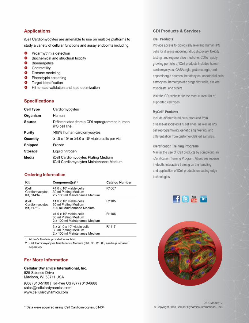
t Figure 4: Ionic CurrentsiCell Cardiomyocytes contain the (A) expected human cardiac ionic currents and show the (B) expected effects when exposed to ion channel-blocking drugs. (Data were adapted from Ma et al., Am J Physiol Heart Circ Physiol (2011).)*
ICa-L
200 pA
10 ms
60 mV100 ms
-50 mV-80 mV
3 s
IKr
50 pA
1 s
4 s
-40 mV
10 mV
50 pA
100 ms
-60 mV
60 mV400 ms
Ito
1 nA
0.5 ms-80 mV
40 ms60 mV
INa
A
ICa Block (shortened APD)
Control
Nifedipine 3 nM
Nifedipine 10 nMNifedipine 30 nM
Nifedipine 100 nM
0 mV
20 mV
50 ms
ControlE4031 3 nM
E4031 10 nME4031 30 nM
E4031 100 nM
20 mV
100 ms
0 mV
EADGeneration
IKr Block (prolonged APD)B
100 pA500 ms
-120 mV
-40 mV 2 s
IfunnyIK1Control
BaCl2 Subtraction
500 µM BaCl2
Voltage (mV)
-73 mV-123 mV 2 s
Curre
nt De
nsity
(pA/
pF)
0-100 -80 -60 -40 -20 20
2
1
-1
-2
-3
0-120
17 mV
iCEL
L C
AR
DIO
MYO
CYT
ES P
RO
DU
CT
DAT
ASH
EETHigher Throughput Functional Testing
Reporter Assays and Microscopy Applications
t Figure 6: Higher Throughput Functional Analysis (A) Effects of ion channel and GPCR modulation on beating behavior were assayed using the xCELLigence RTCA Cardio System (Roche Applied Science). (B) Metabolic profiling with different energy substrates were assayed using the XF96 Extracellular Flux Analyzer (Seahorse Bioscience). (C) GPCR-mediated changes in Ca2+ cycling and contraction rate were assayed using the FLIPR® Tetra System (Molecular Devices, Inc.). (Data were provided by Molecular Devices, Inc.)*
50
40
30
20
10 -5 Control 3 2 1 0 1
Log [Isoproterenol] µM
Beat
s/min
0 6 13 19 25 32 38 44 51 57Time (min)
241214187160133106805326-1
-28
OCR
pM
ole/
min
B
C400
300
200
100 0 20 40 60 80 100
Time (sec)
RFU
Addition of Isoproterenol
AControl
E4031
examine compound effects in a relevant human cellular
environment early in the discovery process.
Their adherent nature and in vitro viability enable iCell
Cardiomyocytes to be used for live or fixed reporter assays and microscopy applications uniquely tailored
to an individual investigator’s requirements.
iCell Cardiomyocytes are suitable for examining
functional endpoints across many higher throughput
platforms, thus affording investigators the opportunity to
t Figure 7: Reporter Assays and Microscopy Applications (A) GFP transiently transfected into iCell Cardiomyocytes verifies relatively high transfection efficiencies. (B) Isoproterenol induces a high level of transiently transfected CRE-luciferase activity (note log scale). (C) Fluoresent microscopy image illustrates the perinuclear localization of BNP staining. (D) Endothelin-induced increases in BNP expression were measured by fluorescent microscopy.*
40
2
0
Uninduced10 µM Isoproterenol
B
C
A
D2.0x106
1.5x10⁶
1.0x10⁶
0.5x10⁶
0 -14 -12 -10 -8 -6
Log ET-1 (M)
Field
Rela
tve In
tens
ity
CDI Products & Services
iCell Products Provide access to biologically relevant, human iPS cells for disease modeling, drug discovery, toxicity testing, and regenerative medicine. CDI’s rapidly growing portfolio of iCell products includes human cardiomyocytes, GABAergic, glutamatergic, and dopaminergic neurons, hepatocytes, endothelial cells, astrocytes, hematopoietic progenitor cells, skeletal myoblasts, and others.
Visit the CDI website for the most current list of supported cell types.
MyCell® Products Include differentiated cells produced from disease-associated iPS cell lines, as well as iPS cell reprogramming, genetic engineering, and differentiation from customer-defined samples.
iCertification Training Programs Master the use of iCell products by completing an iCertification Training Program. Attendees receive in-depth, interactive training on the handling and application of iCell products on cutting-edge technologies.
DS-CM180312 © Copyright 2018 Cellular Dynamics International, Inc.
Applications
iCell Cardiomyocytes are amenable to use on multiple platforms to
study a variety of cellular functions and assay endpoints including:
Proarrhythmia detection Biochemical and structural toxicity Bioenergetics Contractility Disease modeling Phenotypic screening Target identification Hit-to-lead validation and lead optimization
For More Information
Cellular Dynamics International, Inc. 525 Science Drive Madison, WI 53711 USA
(608) 310-5100 | Toll-free US (877) 310-6688 [email protected] www.cellulardynamics.com
Specifications
Cell Type Cardiomyocytes
Organism Human
Source Differentiated from a CDI reprogrammed human iPS cell line
Purity >95% human cardiomyocytes
Quantity ≥1.0 x 10⁶ or ≥4.0 x 10⁶ viable cells per vial
Shipped Frozen
Storage Liquid nitrogen
Media iCell Cardiomyocytes Plating Medium iCell Cardiomyocytes Maintenance Medium
Ordering Information
Kit Component(s)1, 2 Catalog Number
iCell Cardiomyocytes Kit, 01434
≥4.0 x 10⁶ viable cells 30 ml Plating Medium 2 x 100 ml Maintenance Medium
R1007
iCell Cardiomyocytes Kit, 11713
≥1.0 x 10⁶ viable cells 30 ml Plating Medium 100 ml Maintenance Medium
R1105
≥4.0 x 10⁶ viable cells 30 ml Plating Medium 2 x 100 ml Maintenance Medium
R1106
3 x ≥1.0 x 10⁶ viable cells 30 ml Plating Medium 2 x 100 ml Maintenance Medium
R1117
1 A User's Guide is provided in each kit.2 iCell Cardiomyocytes Maintenance Medium (Cat. No. M1003) can be purchased
separately.
* Data were acquired using iCell Cardiomyocytes, 01434.