Embed Size (px)
Citation preview
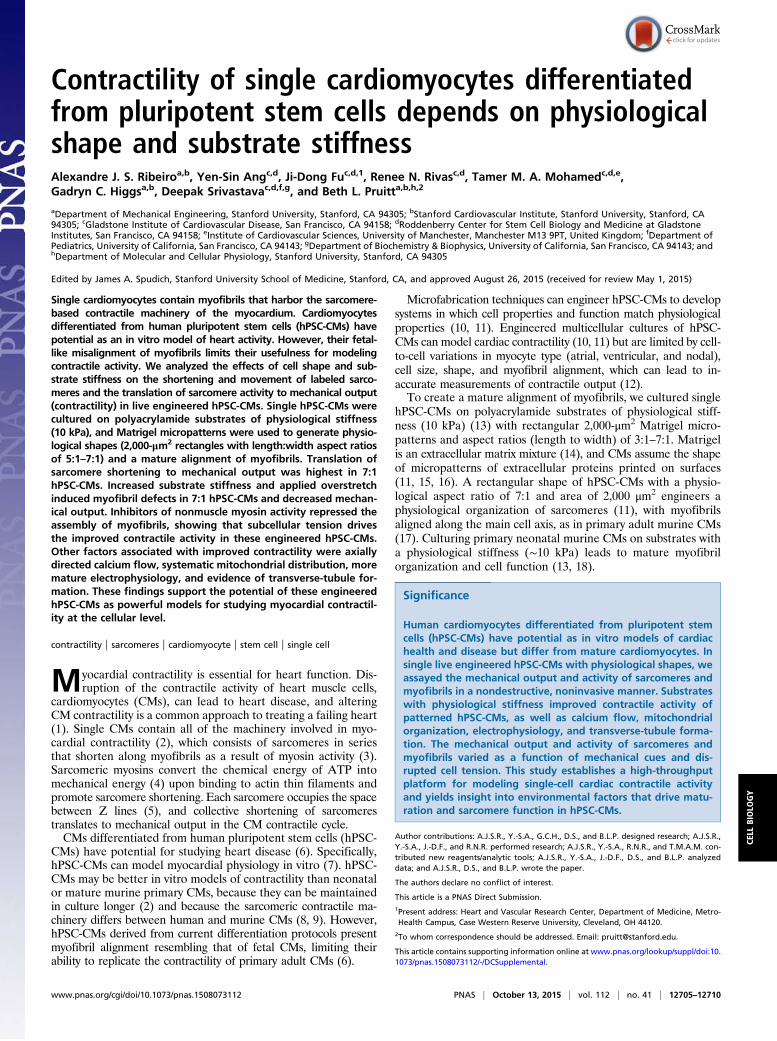
Contractility of single cardiomyocytes differentiatedfrom pluripotent stem cells depends on physiologicalshape and substrate stiffnessAlexandre J. S. Ribeiroa,b, Yen-Sin Angc,d, Ji-Dong Fuc,d,1, Renee N. Rivasc,d, Tamer M. A. Mohamedc,d,e,Gadryn C. Higgsa,b, Deepak Srivastavac,d,f,g, and Beth L. Pruitta,b,h,2
aDepartment of Mechanical Engineering, Stanford University, Stanford, CA 94305; bStanford Cardiovascular Institute, Stanford University, Stanford, CA94305; cGladstone Institute of Cardiovascular Disease, San Francisco, CA 94158; dRoddenberry Center for Stem Cell Biology and Medicine at GladstoneInstitutes, San Francisco, CA 94158; eInstitute of Cardiovascular Sciences, University of Manchester, Manchester M13 9PT, United Kingdom; fDepartment ofPediatrics, University of California, San Francisco, CA 94143; gDepartment of Biochemistry & Biophysics, University of California, San Francisco, CA 94143; andhDepartment of Molecular and Cellular Physiology, Stanford University, Stanford, CA 94305
Edited by James A. Spudich, Stanford University School of Medicine, Stanford, CA, and approved August 26, 2015 (received for review May 1, 2015)
Single cardiomyocytes contain myofibrils that harbor the sarcomere-based contractile machinery of the myocardium. Cardiomyocytesdifferentiated from human pluripotent stem cells (hPSC-CMs) havepotential as an in vitro model of heart activity. However, their fetal-like misalignment of myofibrils limits their usefulness for modelingcontractile activity. We analyzed the effects of cell shape and sub-strate stiffness on the shortening and movement of labeled sarco-meres and the translation of sarcomere activity to mechanical output(contractility) in live engineered hPSC-CMs. Single hPSC-CMs werecultured on polyacrylamide substrates of physiological stiffness(10 kPa), and Matrigel micropatterns were used to generate physio-logical shapes (2,000-μm2 rectangles with length:width aspect ratiosof 5:1–7:1) and a mature alignment of myofibrils. Translation ofsarcomere shortening to mechanical output was highest in 7:1hPSC-CMs. Increased substrate stiffness and applied overstretchinduced myofibril defects in 7:1 hPSC-CMs and decreased mechan-ical output. Inhibitors of nonmuscle myosin activity repressed theassembly of myofibrils, showing that subcellular tension drivesthe improved contractile activity in these engineered hPSC-CMs.Other factors associated with improved contractility were axiallydirected calcium flow, systematic mitochondrial distribution, moremature electrophysiology, and evidence of transverse-tubule for-mation. These findings support the potential of these engineeredhPSC-CMs as powerful models for studying myocardial contractil-ity at the cellular level.
contractility | sarcomeres | cardiomyocyte | stem cell | single cell
Myocardial contractility is essential for heart function. Dis-ruption of the contractile activity of heart muscle cells,
cardiomyocytes (CMs), can lead to heart disease, and alteringCM contractility is a common approach to treating a failing heart(1). Single CMs contain all of the machinery involved in myo-cardial contractility (2), which consists of sarcomeres in seriesthat shorten along myofibrils as a result of myosin activity (3).Sarcomeric myosins convert the chemical energy of ATP intomechanical energy (4) upon binding to actin thin filaments andpromote sarcomere shortening. Each sarcomere occupies the spacebetween Z lines (5), and collective shortening of sarcomerestranslates to mechanical output in the CM contractile cycle.CMs differentiated from human pluripotent stem cells (hPSC-
CMs) have potential for studying heart disease (6). Specifically,hPSC-CMs can model myocardial physiology in vitro (7). hPSC-CMs may be better in vitro models of contractility than neonatalor mature murine primary CMs, because they can be maintainedin culture longer (2) and because the sarcomeric contractile ma-chinery differs between human and murine CMs (8, 9). However,hPSC-CMs derived from current differentiation protocols presentmyofibril alignment resembling that of fetal CMs, limiting theirability to replicate the contractility of primary adult CMs (6).
Microfabrication techniques can engineer hPSC-CMs to developsystems in which cell properties and function match physiologicalproperties (10, 11). Engineered multicellular cultures of hPSC-CMs can model cardiac contractility (10, 11) but are limited by cell-to-cell variations in myocyte type (atrial, ventricular, and nodal),cell size, shape, and myofibril alignment, which can lead to in-accurate measurements of contractile output (12).To create a mature alignment of myofibrils, we cultured single
hPSC-CMs on polyacrylamide substrates of physiological stiff-ness (10 kPa) (13) with rectangular 2,000-μm2 Matrigel micro-patterns and aspect ratios (length to width) of 3:1–7:1. Matrigelis an extracellular matrix mixture (14), and CMs assume the shapeof micropatterns of extracellular proteins printed on surfaces(11, 15, 16). A rectangular shape of hPSC-CMs with a physio-logical aspect ratio of 7:1 and area of 2,000 μm2 engineers aphysiological organization of sarcomeres (11), with myofibrilsaligned along the main cell axis, as in primary adult murine CMs(17). Culturing primary neonatal murine CMs on substrates witha physiological stiffness (∼10 kPa) leads to mature myofibrilorganization and cell function (13, 18).
Significance
Human cardiomyocytes differentiated from pluripotent stemcells (hPSC-CMs) have potential as in vitro models of cardiachealth and disease but differ from mature cardiomyocytes. Insingle live engineered hPSC-CMs with physiological shapes, weassayed the mechanical output and activity of sarcomeres andmyofibrils in a nondestructive, noninvasive manner. Substrateswith physiological stiffness improved contractile activity ofpatterned hPSC-CMs, as well as calcium flow, mitochondrialorganization, electrophysiology, and transverse-tubule forma-tion. The mechanical output and activity of sarcomeres andmyofibrils varied as a function of mechanical cues and dis-rupted cell tension. This study establishes a high-throughputplatform for modeling single-cell cardiac contractile activityand yields insight into environmental factors that drive matu-ration and sarcomere function in hPSC-CMs.
Author contributions: A.J.S.R., Y.-S.A., G.C.H., D.S., and B.L.P. designed research; A.J.S.R.,Y.-S.A., J.-D.F., and R.N.R. performed research; A.J.S.R., Y.-S.A., R.N.R., and T.M.A.M. con-tributed new reagents/analytic tools; A.J.S.R., Y.-S.A., J.-D.F., D.S., and B.L.P. analyzeddata; and A.J.S.R., D.S., and B.L.P. wrote the paper.
The authors declare no conflict of interest.
This article is a PNAS Direct Submission.1Present address: Heart and Vascular Research Center, Department of Medicine, Metro-Health Campus, Case Western Reserve University, Cleveland, OH 44120.
2To whom correspondence should be addressed. Email: [email protected].
This article contains supporting information online at www.pnas.org/lookup/suppl/doi:10.1073/pnas.1508073112/-/DCSupplemental.
www.pnas.org/cgi/doi/10.1073/pnas.1508073112 PNAS | October 13, 2015 | vol. 112 | no. 41 | 12705–12710
CELL
BIOLO
GY
In this study, we analyzed the contractile mechanical output ofsingle engineered rectangular hPSC-CMs (11) on 10-kPa hydrogelsas a function of sarcomere shortening and myofibril organization.Our goals were (i) to determine whether sarcomere activity affectscontractility as a function of cell shape and substrate stiffness, and(ii) to understand how these cues regulate contractility. We calcu-lated the mechanical output of beating hPSC-CMs and simulta-neously imaged fluorescently labeled actin to quantify myofibrilorganization and sarcomere dynamics. We then tested the effects ofsubstrate stiffness and subcellular tension on the mechanical outputand myofibril organization of engineered hPSC-CMs. We alsoassayed calcium flow, mitochondrial organization, electrophysiol-ogy, and the presence of transverse-tubule (t-tubule) structures.
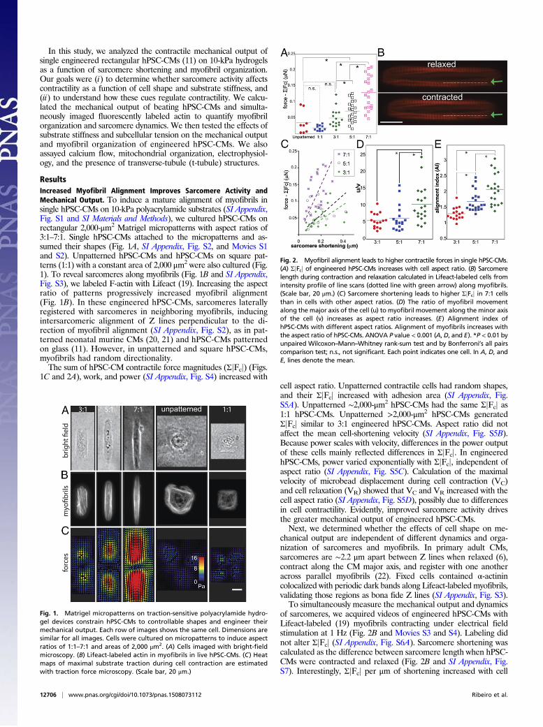
ResultsIncreased Myofibril Alignment Improves Sarcomere Activity andMechanical Output. To induce a mature alignment of myofibrils insingle hPSC-CMs on 10-kPa polyacrylamide substrates (SI Appendix,Fig. S1 and SI Materials and Methods), we cultured hPSC-CMs onrectangular 2,000-μm2 Matrigel micropatterns with aspect ratios of3:1–7:1. Single hPSC-CMs attached to the micropatterns and as-sumed their shapes (Fig. 1A, SI Appendix, Fig. S2, and Movies S1and S2). Unpatterned hPSC-CMs and hPSC-CMs on square pat-terns (1:1) with a constant area of 2,000 μm2 were also cultured (Fig.1). To reveal sarcomeres along myofibrils (Fig. 1B and SI Appendix,Fig. S3), we labeled F-actin with Lifeact (19). Increasing the aspectratio of patterns progressively increased myofibril alignment(Fig. 1B). In these engineered hPSC-CMs, sarcomeres laterallyregistered with sarcomeres in neighboring myofibrils, inducingintersarcomeric alignment of Z lines perpendicular to the di-rection of myofibril alignment (SI Appendix, Fig. S2), as in pat-terned neonatal murine CMs (20, 21) and hPSC-CMs patternedon glass (11). However, in unpatterned and square hPSC-CMs,myofibrils had random directionality.The sum of hPSC-CM contractile force magnitudes (ΣjFcj) (Figs.
1C and 2A), work, and power (SI Appendix, Fig. S4) increased withcell aspect ratio. Unpatterned contractile cells had random shapes,and their ΣjFcj increased with adhesion area (SI Appendix, Fig.S5A). Unpatterned ∼2,000-μm2 hPSC-CMs had the same ΣjFcj as1:1 hPSC-CMs. Unpatterned >2,000-μm2 hPSC-CMs generatedΣjFcj similar to 3:1 engineered hPSC-CMs. Aspect ratio did notaffect the mean cell-shortening velocity (SI Appendix, Fig. S5B).Because power scales with velocity, differences in the power outputof these cells mainly reflected differences in ΣjFcj. In engineeredhPSC-CMs, power varied exponentially with ΣjFcj, independent ofaspect ratio (SI Appendix, Fig. S5C). Calculation of the maximalvelocity of microbead displacement during cell contraction (VC)and cell relaxation (VR) showed that VC and VR increased with thecell aspect ratio (SI Appendix, Fig. S5D), possibly due to differencesin cell contractility. Evidently, improved sarcomere activity drivesthe greater mechanical output of engineered hPSC-CMs.Next, we determined whether the effects of cell shape on me-
chanical output are independent of different dynamics and orga-nization of sarcomeres and myofibrils. In primary adult CMs,sarcomeres are ∼2.2 μm apart between Z lines when relaxed (6),contract along the CM major axis, and register with one anotheracross parallel myofibrils (22). Fixed cells contained α-actinincolocalized with periodic dark bands along Lifeact-labeled myofibrils,validating those regions as bona fide Z lines (SI Appendix, Fig. S3).To simultaneously measure the mechanical output and dynamics
of sarcomeres, we acquired videos of engineered hPSC-CMs withLifeact-labeled (19) myofibrils contracting under electrical fieldstimulation at 1 Hz (Fig. 2B and Movies S3 and S4). Labeling didnot alter ΣjFcj (SI Appendix, Fig. S6A). Sarcomere shortening wascalculated as the difference between sarcomere length when hPSC-CMs were contracted and relaxed (Fig. 2B and SI Appendix, Fig.S7). Interestingly, ΣjFcj per μm of shortening increased with cell
Fig. 1. Matrigel micropatterns on traction-sensitive polyacrylamide hydro-gel devices constrain hPSC-CMs to controllable shapes and engineer theirmechanical output. Each row of images shows the same cell. Dimensions aresimilar for all images. Cells were cultured on micropatterns to induce aspectratios of 1:1–7:1 and areas of 2,000 μm2. (A) Cells imaged with bright-fieldmicroscopy. (B) Lifeact-labeled actin in myofibrils in live hPSC-CMs. (C) Heatmaps of maximal substrate traction during cell contraction are estimatedwith traction force microscopy. (Scale bar, 20 μm.)
Fig. 2. Myofibril alignment leads to higher contractile forces in single hPSC-CMs.(A) ΣjFcj of engineered hPSC-CMs increases with cell aspect ratio. (B) Sarcomerelength during contraction and relaxation calculated in Lifeact-labeled cells fromintensity profile of line scans (dotted line with green arrow) along myofibrils.(Scale bar, 20 μm.) (C) Sarcomere shortening leads to higher ΣjFcj in 7:1 cellsthan in cells with other aspect ratios. (D) The ratio of myofibril movementalong the major axis of the cell (u) to myofibril movement along the minor axisof the cell (v) increases as aspect ratio increases. (E) Alignment index ofhPSC-CMs with different aspect ratios. Alignment of myofibrils increases withthe aspect ratio of hPSC-CMs. ANOVA P value < 0.001 (A, D, and E). *P < 0.01 byunpaired Wilcoxon–Mann–Whitney rank-sum test and by Bonferroni’s all pairscomparison test; n.s., not significant. Each point indicates one cell. In A, D, andE, lines denote the mean.
12706 | www.pnas.org/cgi/doi/10.1073/pnas.1508073112 Ribeiro et al.
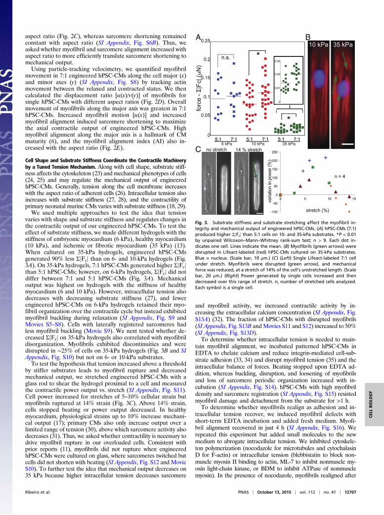
aspect ratio (Fig. 2C), whereas sarcomere shortening remainedconstant with aspect ratio (SI Appendix, Fig. S6B). Thus, weasked whether myofibril and sarcomere alignment increased withaspect ratio to more efficiently translate sarcomere shortening tomechanical output.Using particle-tracking velocimetry, we quantified myofibril
movement in 7:1 engineered hPSC-CMs along the cell major (x)and minor axes (y) (SI Appendix, Fig. S8) by tracking actinmovement between the relaxed and contracted states. We thencalculated the displacement ratio [u(x)/v(y)] of myofibrils forsingle hPSC-CMs with different aspect ratios (Fig. 2D). Overallmovement of myofibrils along the major axis was greatest in 7:1hPSC-CMs. Increased myofibril motion [u(x)] and increasedmyofibril alignment induced sarcomere shortening to maximizethe axial contractile output of engineered hPSC-CMs. Highmyofibril alignment along the major axis is a hallmark of CMmaturity (6), and the myofibril alignment index (AI) also in-creased with the aspect ratio (Fig. 2E).
Cell Shape and Substrate Stiffness Coordinate the Contractile Machineryby a Tuned Tension Mechanism. Along with cell shape, substrate stiff-ness affects the cytoskeleton (23) and mechanical phenotypes of cells(24, 25) and may regulate the mechanical output of engineeredhPSC-CMs. Generally, tension along the cell membrane increaseswith the aspect ratio of adherent cells (26). Intracellular tension alsoincreases with substrate stiffness (27, 28), and the contractility ofprimary neonatal murine CMs varies with substrate stiffness (18, 29).We used multiple approaches to test the idea that tension
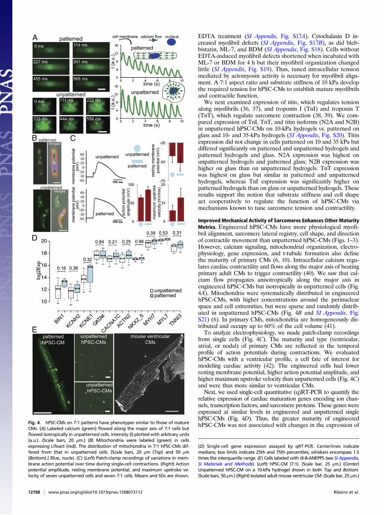
varies with shape and substrate stiffness and regulates changes inthe contractile output of our engineered hPSC-CMs. To test theeffect of substrate stiffness, we made different hydrogels with thestiffness of embryonic myocardium (6 kPa), healthy myocardium(10 kPa), and ischemic or fibrotic myocardium (35 kPa) (13).When cultured on 35-kPa hydrogels, engineered hPSC-CMsgenerated 90% less ΣjFcj than on 6- and 10-kPa hydrogels (Fig.3A). On 35-kPa hydrogels, 7:1 hPSC-CMs generated higher ΣjFcjthan 5:1 hPSC-CMs; however, on 6-kPa hydrogels, ΣjFcj did notdiffer between 7:1 and 5:1 hPSC-CMs (Fig. 3A). Mechanicaloutput was highest on hydrogels with the stiffness of healthymyocardium (6 and 10 kPa). However, intracellular tension alsodecreases with decreasing substrate stiffness (27), and fewerengineered hPSC-CMs on 6-kPa hydrogels retained their myo-fibril organization over the contractile cycle but instead exhibitedmyofibril buckling during relaxation (SI Appendix, Fig. S9 andMovies S5–S8). Cells with laterally registered sarcomeres hadless myofibril buckling (Movie S9). We next tested whether de-creased ΣjFcj on 35-kPa hydrogels also correlated with myofibrildisorganization. Myofibrils exhibited discontinuities and weredisrupted in ∼25% of cells on 35-kPa hydrogels (Fig. 3B and SIAppendix, Fig. S10) but not on 6- or 10-kPa substrates.To test the hypothesis that tension increased above a threshold
by stiffer substrates leads to myofibril rupture and decreasedmechanical output, we stretched engineered hPSC-CMs with aglass rod to shear the hydrogel proximal to a cell and measuredthe contractile power output vs. stretch (SI Appendix, Fig. S11).Cell power increased for stretches of 5–10% cellular strain butmyofibrils ruptured at 14% strain (Fig. 3C). Above 14% strain,cells stopped beating or power output decreased. In healthymyocardium, physiological strains up to 10% increase mechani-cal output (17); primary CMs also only increase output over alimited range of tension (30), above which sarcomere activity alsodecreases (31). Thus, we asked whether contractility is necessary todrive myofibril rupture in our overloaded cells. Consistent withprior reports (11), myofibrils did not rupture when engineeredhPSC-CMs were cultured on glass, where sarcomeres twitched butcells did not shorten with beating (SI Appendix, Fig. S12 and MovieS10). To further test the idea that mechanical output decreases on35 kPa because higher intracellular tension decreases sarcomere
and myofibril activity, we increased contractile activity by in-creasing the extracellular calcium concentration (SI Appendix, Fig.S13A) (32). The fraction of hPSC-CMs with disrupted myofibrils(SI Appendix, Fig. S13B andMovies S11 and S12) increased to 50%(SI Appendix, Fig. S13D).To determine whether intracellular tension is needed to main-
tain myofibril alignment, we incubated patterned hPSC-CMs inEDTA to chelate calcium and reduce integrin-mediated cell-sub-strate adhesion (33, 34) and disrupt myofibril tension (35) and theintracellular balance of forces. Beating stopped upon EDTA ad-dition, whereas buckling, disruption, and loosening of myofibrilsand loss of sarcomere periodic organization increased with in-cubation (SI Appendix, Fig. S14). hPSC-CMs with high myofibrildensity and sarcomere registration (SI Appendix, Fig. S15) resistedmyofibril damage and detachment from the substrate for >1 h.To determine whether myofibrils realign as adhesion and in-
tracellular tension recover, we induced myofibril defects withshort-term EDTA incubation and added fresh medium. Myofi-bril alignment recovered in just 4 h (SI Appendix, Fig. S16). Werepeated this experiment but added small molecules to the newmedium to abrogate intracellular tension. We inhibited cytoskele-ton polymerization (nocodazole for microtubules and cytochalasinD for F-actin) or intracellular tension (blebbistatin to block non-muscle myosin II binding to actin, ML-7 to inhibit nonmuscle my-osin light-chain kinase, or BDM to inhibit ATPase of nonmusclemyosin). In the presence of nocodazole, myofibrils realigned after
Fig. 3. Substrate stiffness and substrate stretching affect the myofibril in-tegrity and mechanical output of engineered hPSC-CMs. (A) hPSC-CMs (7:1)produced higher ΣjFcj than 5:1 cells on 10- and 35-kPa substrates. *P < 0.01by unpaired Wilcoxon–Mann–Whitney rank-sum test; n > 9. Each dot in-dicates one cell. Lines indicate the mean. (B) Myofibrils (green arrows) weredisrupted in Lifeact-labeled (red) hPSC-CMs cultured on 35-kPa substrates.Blue = nucleus. (Scale bar, 10 μm.) (C) (Left) Single Lifeact-labeled 7:1 cellunder stretch. Myofibrils were disrupted (green arrow), and mechanicalforce was reduced, at a stretch of 14% of the cell’s unstretched length. (Scalebar, 20 μm.) (Right) Power generated by single cells increased and thendecreased over this range of stretch. n, number of stretched cells analyzed.Each symbol is a single cell.
Ribeiro et al. PNAS | October 13, 2015 | vol. 112 | no. 41 | 12707
CELL
BIOLO
GY
EDTA treatment (SI Appendix, Fig. S17A). Cytochalasin D in-creased myofibril defects (SI Appendix, Fig. S17B), as did bleb-bistatin, ML-7, and BDM (SI Appendix, Fig. S18). Cells withoutEDTA-induced myofibril defects shortened when incubated withML-7 or BDM for 4 h but their myofibril organization changedlittle (SI Appendix, Fig. S19). Thus, tuned intracellular tensionmediated by actomyosin activity is necessary for myofibril align-ment. A 7:1 aspect ratio and substrate stiffness of 10 kPa developthe required tension for hPSC-CMs to establish mature myofibrilsand contractile function.We next examined expression of titin, which regulates tension
along myofibrils (36, 37), and troponin I (TnI) and troponin T(TnT), which regulate sarcomere contraction (38, 39). We com-pared expression of TnI, TnT, and titin isoforms (N2A and N2B)in unpatterned hPSC-CMs on 10-kPa hydrogels vs. patterned onglass and 10- and 35-kPa hydrogels (SI Appendix, Fig. S20). Titinexpression did not change in cells patterned on 10 and 35 kPa butdiffered significantly on patterned and unpatterned hydrogels andpatterned hydrogels and glass. N2A expression was highest onunpatterned hydrogels and patterned glass; N2B expression washigher on glass than on unpatterned hydrogels. TnT expressionwas highest on glass but similar in patterned and unpatternedhydrogels, whereas TnI expression was significantly higher onpatterned hydrogels than on glass or unpatterned hydrogels. Theseresults support the notion that substrate stiffness and cell shapeact cooperatively to regulate the function of hPSC-CMs viamechanisms known to tune sarcomere tension and contractility.
Improved Mechanical Activity of Sarcomeres Enhances Other MaturityMetrics. Engineered hPSC-CMs have more physiological myofi-bril alignment, sarcomere lateral registry, cell shape, and directionof contractile movement than unpatterned hPSC-CMs (Figs. 1–3).However, calcium signaling, mitochondrial organization, electro-physiology, gene expression, and t-tubule formation also definethe maturity of primary CMs (6, 10). Intracellular calcium regu-lates cardiac contractility and flows along the major axis of beatingprimary adult CMs to trigger contractility (40). We saw that cal-cium flow propagates anisotropically along the major axis inengineered hPSC-CMs but isotropically in unpatterned cells (Fig.4A). Mitochondria were systematically distributed in engineeredhPSC-CMs, with higher concentrations around the perinuclearspace and cell extremities, but were sparse and randomly distrib-uted in unpatterned hPSC-CMs (Fig. 4B and SI Appendix, Fig.S21) (6). In primary CMs, mitochondria are homogeneously dis-tributed and occupy up to 60% of the cell volume (41).To analyze electrophysiology, we made patch-clamp recordings
from single cells (Fig. 4C). The maturity and type (ventricular,atrial, or nodal) of primary CMs are reflected in the temporalprofile of action potentials during contractions. We evaluatedhPSC-CMs with a ventricular profile, a cell fate of interest formodeling cardiac activity (42). The engineered cells had lowerresting membrane potential, higher action potential amplitude, andhigher maximum upstroke velocity than unpatterned cells (Fig. 4C)and were thus more similar to ventricular CMs.Next, we used single-cell quantitative (q)RT-PCR to quantify the
relative expression of cardiac maturation genes encoding ion chan-nels, transcription factors, and sarcomere proteins. These genes wereexpressed at similar levels in engineered and unpatterned singlehPSC-CMs (Fig. 4D). Thus, the greater maturity of engineeredhPSC-CMs was not associated with changes in the expression ofFig. 4. hPSC-CMs on 7:1 patterns have phenotypes similar to those of mature
CMs. (A) Labeled calcium (green) flowed along the major axis of 7:1 cells butflowed isotropically in unpatterned cells. Intensity (I) plotted with arbitrary units(a.u.). (Scale bars, 20 μm.) (B) Mitochondria were labeled (green) in cellsexpressing Lifeact (red). The distribution of mitochondria in 7:1 hPSC-CMs dif-fered from that in unpatterned cells. [Scale bars, 20 μm (Top) and 50 μm(Bottom).] Blue, nuclei. (C) (Left) Patch-clamp recordings of variations in mem-brane action potential over time during single-cell contractions. (Right) Actionpotential amplitude, resting membrane potential, and maximum upstroke ve-locity of seven unpatterned cells and seven 7:1 cells. Means and SDs are shown.
(D) Single-cell gene expression assayed by qRT-PCR. Centerlines indicatemedians; box limits indicate 25th and 75th percentiles; whiskers encompass 1.5times the interquartile range. (E) Cells labeled with di-8-ANEPPS (see SI Appendix,SI Materials and Methods). (Left) hPSC-CM (7:1). (Scale bar, 25 μm.) (Center)Unpatterned hPSC-CM on a 10-kPa hydrogel shown in both Top and Bottom.(Scale bars, 50 μm.) (Right) Isolated adult mouse ventricular CM. (Scale bar, 25 μm.)
12708 | www.pnas.org/cgi/doi/10.1073/pnas.1508073112 Ribeiro et al.
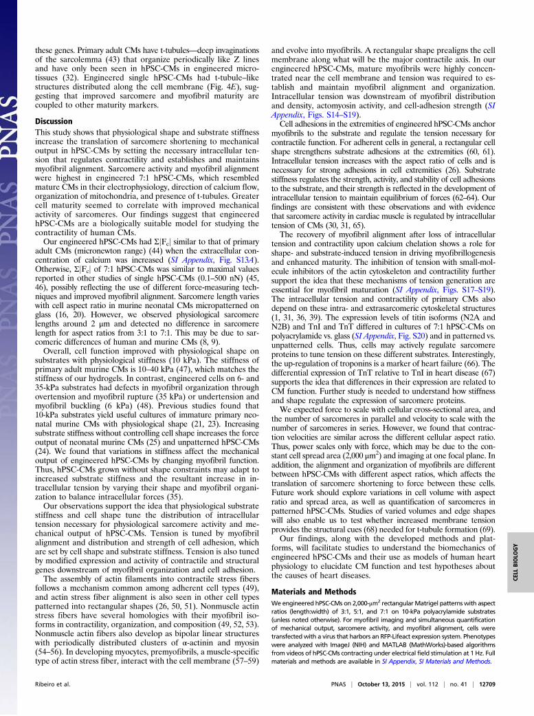
these genes. Primary adult CMs have t-tubules—deep invaginationsof the sarcolemma (43) that organize periodically like Z linesand have only been seen in hPSC-CMs in engineered micro-tissues (32). Engineered single hPSC-CMs had t-tubule–likestructures distributed along the cell membrane (Fig. 4E), sug-gesting that improved sarcomere and myofibril maturity arecoupled to other maturity markers.
DiscussionThis study shows that physiological shape and substrate stiffnessincrease the translation of sarcomere shortening to mechanicaloutput in hPSC-CMs by setting the necessary intracellular ten-sion that regulates contractility and establishes and maintainsmyofibril alignment. Sarcomere activity and myofibril alignmentwere highest in engineered 7:1 hPSC-CMs, which resembledmature CMs in their electrophysiology, direction of calcium flow,organization of mitochondria, and presence of t-tubules. Greatercell maturity seemed to correlate with improved mechanicalactivity of sarcomeres. Our findings suggest that engineeredhPSC-CMs are a biologically suitable model for studying thecontractility of human CMs.Our engineered hPSC-CMs had ΣjFcj similar to that of primary
adult CMs (micronewton range) (44) when the extracellular con-centration of calcium was increased (SI Appendix, Fig. S13A).Otherwise, ΣjFcj of 7:1 hPSC-CMs was similar to maximal valuesreported in other studies of single hPSC-CMs (0.1–500 nN) (45,46), possibly reflecting the use of different force-measuring tech-niques and improved myofibril alignment. Sarcomere length varieswith cell aspect ratio in murine neonatal CMs micropatterned onglass (16, 20). However, we observed physiological sarcomerelengths around 2 μm and detected no difference in sarcomerelength for aspect ratios from 3:1 to 7:1. This may be due to sar-comeric differences of human and murine CMs (8, 9).Overall, cell function improved with physiological shape on
substrates with physiological stiffness (10 kPa). The stiffness ofprimary adult murine CMs is 10–40 kPa (47), which matches thestiffness of our hydrogels. In contrast, engineered cells on 6- and35-kPa substrates had defects in myofibril organization throughovertension and myofibril rupture (35 kPa) or undertension andmyofibril buckling (6 kPa) (48). Previous studies found that10-kPa substrates yield useful cultures of immature primary neo-natal murine CMs with physiological shape (21, 23). Increasingsubstrate stiffness without controlling cell shape increases the forceoutput of neonatal murine CMs (25) and unpatterned hPSC-CMs(24). We found that variations in stiffness affect the mechanicaloutput of engineered hPSC-CMs by changing myofibril function.Thus, hPSC-CMs grown without shape constraints may adapt toincreased substrate stiffness and the resultant increase in in-tracellular tension by varying their shape and myofibril organi-zation to balance intracellular forces (35).Our observations support the idea that physiological substrate
stiffness and cell shape tune the distribution of intracellulartension necessary for physiological sarcomere activity and me-chanical output of hPSC-CMs. Tension is tuned by myofibrilalignment and distribution and strength of cell adhesion, whichare set by cell shape and substrate stiffness. Tension is also tunedby modified expression and activity of contractile and structuralgenes downstream of myofibril organization and cell adhesion.The assembly of actin filaments into contractile stress fibers
follows a mechanism common among adherent cell types (49),and actin stress fiber alignment is also seen in other cell typespatterned into rectangular shapes (26, 50, 51). Nonmuscle actinstress fibers have several homologies with their myofibril iso-forms in contractility, organization, and composition (49, 52, 53).Nonmuscle actin fibers also develop as bipolar linear structureswith periodically distributed clusters of α-actinin and myosin(54–56). In developing myocytes, premyofibrils, a muscle-specifictype of actin stress fiber, interact with the cell membrane (57–59)
and evolve into myofibrils. A rectangular shape prealigns the cellmembrane along what will be the major contractile axis. In ourengineered hPSC-CMs, mature myofibrils were highly concen-trated near the cell membrane and tension was required to es-tablish and maintain myofibril alignment and organization.Intracellular tension was downstream of myofibril distributionand density, actomyosin activity, and cell-adhesion strength (SIAppendix, Figs. S14–S19).Cell adhesions in the extremities of engineered hPSC-CMs anchor
myofibrils to the substrate and regulate the tension necessary forcontractile function. For adherent cells in general, a rectangular cellshape strengthens substrate adhesions at the extremities (60, 61).Intracellular tension increases with the aspect ratio of cells and isnecessary for strong adhesions in cell extremities (26). Substratestiffness regulates the strength, activity, and stability of cell adhesionsto the substrate, and their strength is reflected in the development ofintracellular tension to maintain equilibrium of forces (62–64). Ourfindings are consistent with these observations and with evidencethat sarcomere activity in cardiac muscle is regulated by intracellulartension of CMs (30, 31, 65).The recovery of myofibril alignment after loss of intracellular
tension and contractility upon calcium chelation shows a role forshape- and substrate-induced tension in driving myofibrillogenesisand enhanced maturity. The inhibition of tension with small-mol-ecule inhibitors of the actin cytoskeleton and contractility furthersupport the idea that these mechanisms of tension generation areessential for myofibril maturation (SI Appendix, Figs. S17–S19).The intracellular tension and contractility of primary CMs alsodepend on these intra- and extrasarcomeric cytoskeletal structures(1, 31, 36, 39). The expression levels of titin isoforms (N2A andN2B) and TnI and TnT differed in cultures of 7:1 hPSC-CMs onpolyacrylamide vs. glass (SI Appendix, Fig. S20) and in patterned vs.unpatterned cells. Thus, cells may actively regulate sarcomereproteins to tune tension on these different substrates. Interestingly,the up-regulation of troponins is a marker of heart failure (66). Thedifferential expression of TnT relative to TnI in heart disease (67)supports the idea that differences in their expression are related toCM function. Further study is needed to understand how stiffnessand shape regulate the expression of sarcomere proteins.We expected force to scale with cellular cross-sectional area, and
the number of sarcomeres in parallel and velocity to scale with thenumber of sarcomeres in series. However, we found that contrac-tion velocities are similar across the different cellular aspect ratio.Thus, power scales only with force, which may be due to the con-stant cell spread area (2,000 μm2) and imaging at one focal plane. Inaddition, the alignment and organization of myofibrils are differentbetween hPSC-CMs with different aspect ratios, which affects thetranslation of sarcomere shortening to force between these cells.Future work should explore variations in cell volume with aspectratio and spread area, as well as quantification of sarcomeres inpatterned hPSC-CMs. Studies of varied volumes and edge shapeswill also enable us to test whether increased membrane tensionprovides the structural cues (68) needed for t-tubule formation (69).Our findings, along with the developed methods and plat-
forms, will facilitate studies to understand the biomechanics ofengineered hPSC-CMs and their use as models of human heartphysiology to elucidate CM function and test hypotheses aboutthe causes of heart diseases.
Materials and MethodsWeengineered hPSC-CMs on 2,000-μm2 rectangularMatrigel patterns with aspectratios (length:width) of 3:1, 5:1, and 7:1 on 10-kPa polyacrylamide substrates(unless noted otherwise). For myofibril imaging and simultaneous quantificationof mechanical output, sarcomere activity, and myofibril alignment, cells weretransfectedwith a virus that harbors an RFP-Lifeact expression system. Phenotypeswere analyzed with ImageJ (NIH) and MATLAB (MathWorks)-based algorithmsfrom videos of hPSC-CMs contracting under electrical field stimulation at 1 Hz. Fullmaterials and methods are available in SI Appendix, SI Materials and Methods.
Ribeiro et al. PNAS | October 13, 2015 | vol. 112 | no. 41 | 12709
CELL
BIOLO
GY
ACKNOWLEDGMENTS. We thank C. Liu, A. Heidersbach, and E. Booth fordiscussions. This study was supported by American Heart Association Fellow-ships 14POST18360018 (to A.J.S.R.) and 13POST17390040 (to Y.-S.A.) and Award
13SDG14580035 (to J.-D.F.), the National Science Foundation under MIKS-1136790 (to B.L.P.), the National Institutes of Health under R01-EB006745 (toB.L.P.), and seed grants from the Stanford Cardiovascular Institute and Bio-X.
1. Dorn GW, II, Molkentin JD (2004) Manipulating cardiac contractility in heart failure:Data from mice and men. Circulation 109(2):150–158.
2. Louch WE, Sheehan KA, Wolska BM (2011) Methods in cardiomyocyte isolation, cul-ture, and gene transfer. J Mol Cell Cardiol 51(3):288–298.
3. de Tombe PP, ter Keurs HE (2012) The velocity of cardiac sarcomere shortening:Mechanisms and implications. J Muscle Res Cell Motil 33(6):431–437.
4. Lodish HF, et al. (2012) Myosin-powered movements. Molecular Cell Biology (W. H.Freeman, New York), 7th Ed, pp 801–805.
5. Knöll R, Buyandelger B, Lab M (2011) The sarcomeric Z-disc and Z-discopathies.J Biomed Biotechnol 2011:569628.
6. Yang X, Pabon L, Murry CE (2014) Engineering adolescence: Maturation of humanpluripotent stem cell-derived cardiomyocytes. Circ Res 114(3):511–523.
7. Parameswaran S, Kumar S, Verma RS, Sharma RK (2013) Cardiomyocyte culture—Anupdate on the in vitro cardiovascular model and future challenges. Can J PhysiolPharmacol 91(12):985–998.
8. Reiser PJ, Portman MA, Ning XH, Schomisch Moravec C (2001) Human cardiac myosinheavy chain isoforms in fetal and failing adult atria and ventricles. Am J Physiol HeartCirc Physiol 280(4):H1814–H1820.
9. Milani-Nejad N, Janssen PM (2014) Small and large animal models in cardiac con-traction research: Advantages and disadvantages. Pharmacol Ther 141(3):235–249.
10. Sheehy SP, et al. (2014) Quality metrics for stem cell-derived cardiac myocytes. StemCell Rep 2(3):282–294.
11. Wang G, et al. (2014) Modeling the mitochondrial cardiomyopathy of Barth syndromewith induced pluripotent stem cell and heart-on-chip technologies. Nat Med 20(6):616–623.
12. French A, et al. (2015) Enabling consistency in pluripotent stem cell-derived productsfor research and development and clinical applications through material standards.Stem Cells Transl Med 4(3):217–223.
13. Engler AJ, et al. (2008) Embryonic cardiomyocytes beat best on a matrix with heart-like elasticity: Scar-like rigidity inhibits beating. J Cell Sci 121(Pt 22):3794–3802.
14. Hughes CS, Postovit LM, Lajoie GA (2010) Matrigel: A complex protein mixture re-quired for optimal growth of cell culture. Proteomics 10(9):1886–1890.
15. Boudou T, et al. (2012) A microfabricated platform to measure and manipulate themechanics of engineered cardiac microtissues. Tissue Eng Part A 18(9-10):910–919.
16. Bray MA, Sheehy SP, Parker KK (2008) Sarcomere alignment is regulated by myocyteshape. Cell Motil Cytoskeleton 65(8):641–651.
17. Trayanova NA, Rice JJ (2011) Cardiac electromechanical models: From cell to organ.Front Physiol 2:43.
18. Chopra A, Tabdanov E, Patel H, Janmey PA, Kresh JY (2011) Cardiac myocyte re-modeling mediated by N-cadherin-dependent mechanosensing. Am J Physiol HeartCirc Physiol 300(4):H1252–H1266.
19. Riedl J, et al. (2008) Lifeact: A versatile marker to visualize F-actin. Nat Methods 5(7):605–607.
20. Kuo PL, et al. (2012) Myocyte shape regulates lateral registry of sarcomeres andcontractility. Am J Pathol 181(6):2030–2037.
21. McCain ML, Yuan H, Pasqualini FS, Campbell PH, Parker KK (2014) Matrix elasticityregulates the optimal cardiac myocyte shape for contractility. Am J Physiol Heart CircPhysiol 306(11):H1525–H1539.
22. Friedrich BM, Buxboim A, Discher DE, Safran SA (2011) Striated acto-myosin fibers canreorganize and register in response to elastic interactions with the matrix. Biophys J100(11):2706–2715.
23. Chopra A, et al. (2012) Reprogramming cardiomyocyte mechanosensing by crosstalkbetween integrins and hyaluronic acid receptors. J Biomech 45(5):824–831.
24. Hazeltine LB, et al. (2012) Effects of substrate mechanics on contractility of car-diomyocytes generated from human pluripotent stem cells. Int J Cell Biol 2012:508294.
25. Hersch N, et al. (2013) The constant beat: Cardiomyocytes adapt their forces by equalcontraction upon environmental stiffening. Biol Open 2(3):351–361.
26. Oakes PW, Banerjee S, Marchetti MC, Gardel ML (2014) Geometry regulates tractionstresses in adherent cells. Biophys J 107(4):825–833.
27. Mih JD, Marinkovic A, Liu F, Sharif AS, Tschumperlin DJ (2012) Matrix stiffness re-verses the effect of actomyosin tension on cell proliferation. J Cell Sci 125(Pt 24):5974–5983.
28. Banerjee S, Sknepnek R, Marchetti MC (2014) Optimal shapes and stresses of adherentcells on patterned substrates. Soft Matter 10(14):2424–2430.
29. Young JL, Kretchmer K, Ondeck MG, Zambon AC, Engler AJ (2014) Mechanosensitivekinases regulate stiffness-induced cardiomyocyte maturation. Sci Rep 4:6425.
30. ter Keurs HE, Rijnsburger WH, van Heuningen R, Nagelsmit MJ (1980) Tension de-velopment and sarcomere length in rat cardiac trabeculae. Evidence of length-dependent activation. Circ Res 46(5):703–714.
31. Granzier HL, Irving TC (1995) Passive tension in cardiac muscle: Contribution of col-lagen, titin, microtubules, and intermediate filaments. Biophys J 68(3):1027–1044.
32. Godier-Furnémont AF, et al. (2015) Physiologic force-frequency response in en-gineered heart muscle by electromechanical stimulation. Biomaterials 60:82–91.
33. Sjaastad MD, Nelson WJ (1997) Integrin-mediated calcium signaling and regulation ofcell adhesion by intracellular calcium. BioEssays 19(1):47–55.
34. Zhang K, Chen J (2012) The regulation of integrin function by divalent cations. CellAdhes Migr 6(1):20–29.
35. Mullen CA, et al. (2014) Cell morphology and focal adhesion location alters internalcell stress. J R Soc Interface 11(101):20140885.
36. Cazorla O, et al. (2000) Differential expression of cardiac titin isoforms and modu-lation of cellular stiffness. Circ Res 86(1):59–67.
37. Kass DA, Bronzwaer JG, Paulus WJ (2004) What mechanisms underlie diastolic dys-function in heart failure? Circ Res 94(12):1533–1542.
38. Solaro RJ (2003) The special structure and function of troponin I in regulation of cardiaccontraction and relaxation. Adv Exp Med Biol 538:389–401, discussion 401–402.
39. McCall SJ, et al. (2006) Development and cardiac contractility: Cardiac troponin Tisoforms and cytosolic calcium in rabbit. Pediatr Res 60(3):276–281.
40. Bers DM (2000) Calcium fluxes involved in control of cardiac myocyte contraction. CircRes 87(4):275–281.
41. Piquereau J, et al. (2013) Mitochondrial dynamics in the adult cardiomyocytes: Whichroles for a highly specialized cell? Front Physiol 4:102.
42. Winslow RL, et al. (2011) Integrative modeling of the cardiac ventricular myocyte.Wiley Interdiscip Rev Syst Biol Med 3(4):392–413.
43. Ibrahim M, Gorelik J, Yacoub MH, Terracciano CM (2011) The structure and functionof cardiac t-tubules in health and disease. Proc Biol Sci 278(1719):2714–2723.
44. Korte FS, Herron TJ, Rovetto MJ, McDonald KS (2005) Power output is linearly relatedto MyHC content in rat skinned myocytes and isolated working hearts. Am J PhysiolHeart Circ Physiol 289(2):H801–H812.
45. Taylor RE, et al. (2013) Sacrificial layer technique for axial force post assay of imma-ture cardiomyocytes. Biomed Microdevices 15(1):171–181.
46. Rodriguez ML, et al. (2014) Measuring the contractile forces of human induced plu-ripotent stem cell-derived cardiomyocytes with arrays of microposts. J Biomech Eng136(5):051005.
47. Chang WT, Yu D, Lai YC, Lin KY, Liau I (2013) Characterization of the mechanody-namic response of cardiomyocytes with atomic force microscopy. Anal Chem 85(3):1395–1400.
48. Bathe M, Heussinger C, Claessens MM, Bausch AR, Frey E (2008) Cytoskeletal bundlemechanics. Biophys J 94(8):2955–2964.
49. Tojkander S, Gateva G, Lappalainen P (2012) Actin stress fibers—Assembly, dynamicsand biological roles. J Cell Sci 125(Pt 8):1855–1864.
50. Roca-Cusachs P, et al. (2008) Micropatterning of single endothelial cell shape reveals atight coupling between nuclear volume in G1 and proliferation. Biophys J 94(12):4984–4995.
51. Wang D, et al. (2014) Tissue-specific mechanical and geometrical control of cell via-bility and actin cytoskeleton alignment. Sci Rep 4:6160.
52. Peterson LJ, et al. (2004) Simultaneous stretching and contraction of stress fibersin vivo. Mol Biol Cell 15(7):3497–3508.
53. Ono S (2010) Dynamic regulation of sarcomeric actin filaments in striated muscle.Cytoskeleton (Hoboken) 67(11):677–692.
54. Langanger G, et al. (1986) The molecular organization of myosin in stress fibers ofcultured cells. J Cell Biol 102(1):200–209.
55. Cramer LP, Siebert M, Mitchison TJ (1997) Identification of novel graded polarity actinfilament bundles in locomoting heart fibroblasts: Implications for the generation ofmotile force. J Cell Biol 136(6):1287–1305.
56. Butt T, et al. (2010) Myosin motors drive long range alignment of actin filaments.J Biol Chem 285(7):4964–4974.
57. Rhee D, Sanger JM, Sanger JW (1994) The premyofibril: Evidence for its role in my-ofibrillogenesis. Cell Motil Cytoskeleton 28(1):1–24.
58. LoRusso SM, Rhee D, Sanger JM, Sanger JW (1997) Premyofibrils in spreading adultcardiomyocytes in tissue culture: Evidence for reexpression of the embryonic programfor myofibrillogenesis in adult cells. Cell Motil Cytoskeleton 37(3):183–198.
59. Dabiri GA, Turnacioglu KK, Sanger JM, Sanger JW (1997) Myofibrillogenesis visualizedin living embryonic cardiomyocytes. Proc Natl Acad Sci USA 94(17):9493–9498.
60. Lemmon CA, Romer LH (2010) A predictive model of cell traction forces based on cellgeometry. Biophys J 99(9):L78–L80.
61. Banerjee S, Marchetti MC (2013) Controlling cell–matrix traction forces by extracel-lular geometry. New J Phys 15:035015.
62. Pelham RJ, Jr, Wang Yl (1997) Cell locomotion and focal adhesions are regulated bysubstrate flexibility. Proc Natl Acad Sci USA 94(25):13661–13665.
63. Huang J, Peng X, Xiong C, Fang J (2011) Influence of substrate stiffness on cell-sub-strate interfacial adhesion and spreading: A mechano-chemical coupling model.J Colloid Interface Sci 355(2):503–508.
64. Yeung T, et al. (2005) Effects of substrate stiffness on cell morphology, cytoskeletalstructure, and adhesion. Cell Motil Cytoskeleton 60(1):24–34.
65. Solaro RJ (2011) Left ventricular diastolic and systolic pressure, ejection, and relaxa-tion reflect sarcomeric mechanical properties. Regulation of Cardiac Contractility, edsGranger DN, Granger J (Morgan & Claypool Life Sciences, San Rafael, CA), pp 12–14.
66. Maynard SJ, Menown IB, Adgey AA (2000) Troponin T or troponin I as cardiac markersin ischaemic heart disease. Heart 83(4):371–373.
67. Rittoo D, Jones A, Lecky B, Neithercut D (2014) Elevation of cardiac troponin T, butnot cardiac troponin I, in patients with neuromuscular diseases: Implications for thediagnosis of myocardial infarction. J Am Coll Cardiol 63(22):2411–2420.
68. Parton RG, del Pozo MA (2013) Caveolae as plasma membrane sensors, protectors andorganizers. Nat Rev Mol Cell Biol 14(2):98–112.
69. Sens P, Turner MS (2004) Theoretical model for the formation of caveolae and similarmembrane invaginations. Biophys J 86(4):2049–2057.
12710 | www.pnas.org/cgi/doi/10.1073/pnas.1508073112 Ribeiro et al.