Embed Size (px)
Citation preview
The
Jour
nal o
f G
ener
al P
hysi
olo
gy
A RT I C L E
The Rockefeller University Press $30.00J. Gen. Physiol. Vol. 133 No. 1 29–42www.jgp.org/cgi/doi/10.1085/jgp.200810080 29
I N T R O D U C T I O N
ClC genes are expressed in all phyla from bacteria to
mammals. Members of the ClC superfamily of transport
proteins function as anion channels or Cl � /H + exchang-
ers in plasma and intracellular organelle membranes
and play key roles in diverse and fundamental physio-
logical processes, including transepithelial Cl � transport,
organelle acidifi cation, regulation of cytoplasmic Cl �
levels and skeletal muscle membrane excitability, and
regulation of nitrate content in plants and cation homeo-
stasis in yeast. Nine ClC genes are expressed in humans
and mutations in at least four of these are associated
with muscle, bone, kidney, and neurological diseases
( Jentsch et al., 2002, 2005 ; Jentsch, 2008 ; Miller, 2006 ).
Despite intensive study and their functional importance,
little is known about how ClC channels are regulated
and regulatory signaling pathways have not been defi ned.
The nematode Caenorhabditis elegans provides numerous
experimental advantages for defining the molecular
bases of fundamental physiological processes, including
cellular signaling pathways ( Barr, 2003 ; Strange, 2003 ).
Correspondence to Kevin Strange: k e v i n . s t r a n g e @ v a n d e r b i l t . e d u
Abbreviations used in this paper: GCK, germinal center kinase; KD,
kinase-dead; LC-MS/MS, liquid chromatography-tandem mass spectrome-
try; MS, mass spectrometry.
We recently demonstrated that the C. elegans ClC gene
clh-3 encodes two anion channel splice variants. CLH-3b
is expressed in the worm oocyte and is activated during
oocyte meiotic cell cycle progression, a process termed
meiotic maturation, and in response to oocyte swelling
( Rutledge et al., 2001 ; Denton et al., 2004 ). Meiotic
maturation is the physiologically relevant stimulus for
channel activation ( Rutledge et al., 2001 ), which func-
tions to synchronize oocyte cell cycle events with ovu-
lation and fertilization ( Rutledge et al., 2001 ; Strange,
2002 ; Yin et al., 2004 ).
Activation of CLH-3b in response to meiotic matura-
tion or cell swelling occurs via serine/threonine dephos-
phorylation events that are mediated by the type I
protein phosphatases GLC-7 � and GLC-7 � ( Rutledge
et al., 2002 ). Inhibition of CLH-3b is mediated by the
recently identifi ed Ste20 kinase, germinal center kinase
(GCK)-3 ( Denton et al., 2005 ). Ste20-type kinases com-
prise a large superfamily that is divided into p21-acti-
vated kinase and GCK subfamilies ( Dan et al., 2001 ).
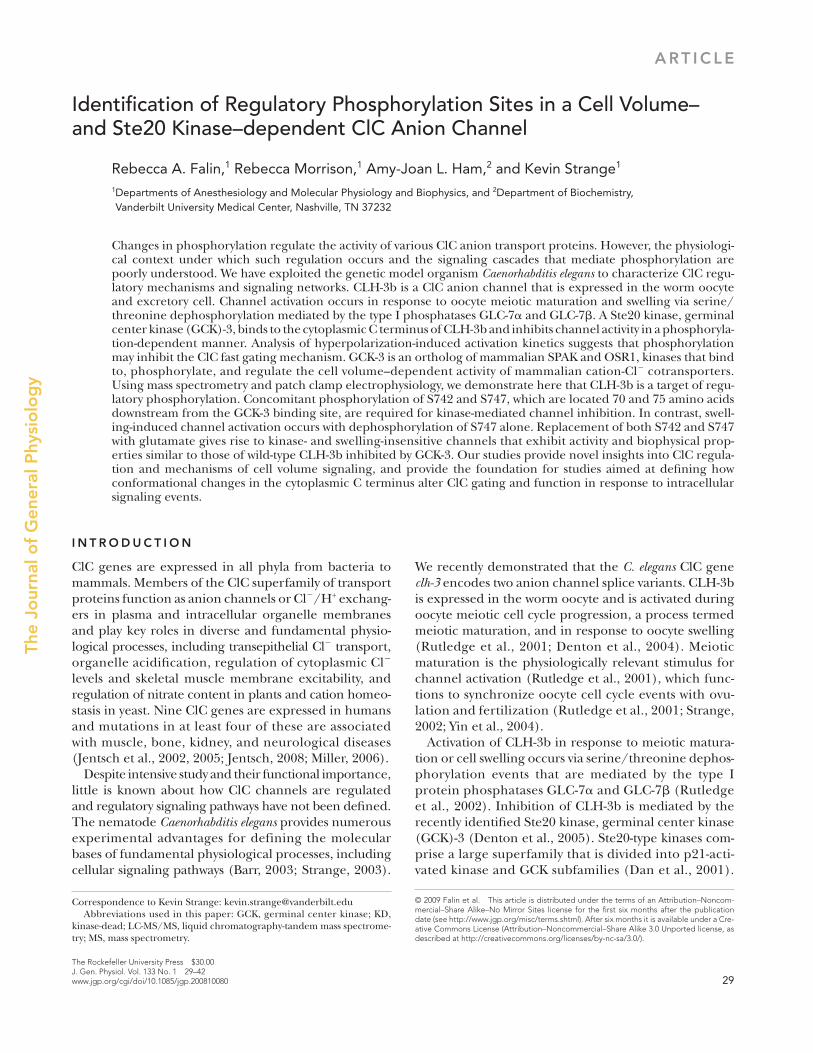
Identifi cation of Regulatory Phosphorylation Sites in a Cell Volume – and Ste20 Kinase – dependent ClC Anion Channel
Rebecca A. Falin , 1 Rebecca Morrison , 1 Amy-Joan L. Ham , 2 and Kevin Strange 1
1 Departments of Anesthesiology and Molecular Physiology and Biophysics, and 2 Department of Biochemistry, Vanderbilt University Medical Center, Nashville, TN 37232
Changes in phosphorylation regulate the activity of various ClC anion transport proteins. However, the physiologi-cal context under which such regulation occurs and the signaling cascades that mediate phosphorylation are poorly understood. We have exploited the genetic model organism Caenorhabditis elegans to characterize ClC regu-latory mechanisms and signaling networks. CLH-3b is a ClC anion channel that is expressed in the worm oocyte and excretory cell. Channel activation occurs in response to oocyte meiotic maturation and swelling via serine/threonine dephosphorylation mediated by the type I phosphatases GLC-7 � and GLC-7 � . A Ste20 kinase, germinal center kinase (GCK)-3, binds to the cytoplasmic C terminus of CLH-3b and inhibits channel activity in a phosphoryla-tion-dependent manner. Analysis of hyperpolarization-induced activation kinetics suggests that phosphorylation may inhibit the ClC fast gating mechanism. GCK-3 is an ortholog of mammalian SPAK and OSR1, kinases that bind to, phosphorylate, and regulate the cell volume – dependent activity of mammalian cation-Cl � cotransporters. Using mass spectrometry and patch clamp electrophysiology, we demonstrate here that CLH-3b is a target of regu-latory phosphorylation. Concomitant phosphorylation of S742 and S747, which are located 70 and 75 amino acids downstream from the GCK-3 binding site, are required for kinase-mediated channel inhibition. In contrast, swell-ing-induced channel activation occurs with dephosphorylation of S747 alone. Replacement of both S742 and S747 with glutamate gives rise to kinase- and swelling-insensitive channels that exhibit activity and biophysical prop-erties similar to those of wild-type CLH-3b inhibited by GCK-3. Our studies provide novel insights into ClC regula-tion and mechanisms of cell volume signaling, and provide the foundation for studies aimed at defi ning how conformational changes in the cytoplasmic C terminus alter ClC gating and function in response to intracellular signaling events.
© 2009 Falin et al. This article is distributed under the terms of an Attribution–Noncom-mercial–Share Alike–No Mirror Sites license for the fi rst six months after the publication date (see http://www.jgp.org/misc/terms.shtml). After six months it is available under a Cre-ative Commons License (Attribution–Noncommercial–Share Alike 3.0 Unported license, as described at http://creativecommons.org/licenses/by-nc-sa/3.0/).

30 Regulatory Phosphorylation Sites in a Ste20 Kinase – dependent ClC Channel
Denton et al., 2005 ). Swelling-induced current amplitude was measured at � 100 mV. Characterization of all mutant channels was performed on at least two independently transfected groups of cells.
Patch electrodes were pulled from 1.5-mm outer diameter – silanized borosilicate microhematocrit tubes. Electrode resistances ranged from 4 to 8 M Ω . Currents were measured with an Axo-patch 200B (MDS Analytical Technologies) patch clamp amplifi er. Electrical connections to the patch clamp amplifi er were made using Ag/AgCl wires and 3 M KCl/agar bridges. Data acquisition and analysis were performed using pClamp 10 software (MDS Analytical Technologies).
Data Analysis Whole cell currents were evoked by stepping membrane voltage for 1 s between � 140 and +60 mV in 20-mV increments from a holding potential of 0 mV. Test pulses were followed by a 1-s inter-val at 0 mV. Current-to-voltage relationships were constructed from mean CLH-3b current values recorded over the last 20 msec of each test pulse.
Coexpression of CLH-3b with GCK-3 causes striking changes in channel voltage sensitivity and the kinetics of hyperpolarization-induced activation ( Denton et al., 2005 ). Channel activation volt-ages were estimated from current-to-voltage relationships. A line was drawn by linear regression analysis of currents measured be-tween 0 and 60 mV. A second line was drawn by linear regression analysis of currents measured between the fi rst voltage at which inward current was detected and a second voltage 20 mV more negative. The intercept of these two lines is defi ned as the activa-tion voltage.
The kinetics of hyperpolarization-induced channel activation are presented as 50% rise time, which is the time required for the current to reach half-maximal activation during a test pulse to � 100 mV. Time constants for hyperpolarization-induced activa-tion were also determined by fi tting current traces with mono- or bi-exponential functions over the fi rst 500 msec of test pulses af-ter decay of the capacitance transient.
Liquid Chromatography-Tandem MS (LC-MS/MS) Analysis and Protein Identifi cation Chinese hamster ovary cells were cultured at 37 ° C in Ham ’ s F12 medium (Invitrogen) containing 10% fetal bovine serum (Hyclone Laboratories, Inc.), 50 U/ml penicillin, and 50 μ g/ml streptomycin. Cells grown in 100-mm diameter tissue culture plates to � 50% confl uency were transfected using FuGENE 6 (Roche) with 8 μ g of V5-tagged CLH-3b and 3 μ g of either GCK-3 or kinase-dead (KD) GCK-3 cDNAs ligated into pcDNA3.1. Approximately 28 h after transfection, 10 culture plates were rinsed twice with PBS (2.7 mM KCl, 144 mM NaCl, 1.5 mM KH 2 PO 4 , and 8.1 mM Na 2 HPO 4 , pH 7.4) and placed on ice. Cells were lysed by adding 0.4 ml of ice-cold modifi ed RIPA lysis buffer (PBS containing 0.05% SDS and 1% Triton X-100) containing complete protease inhibitor cocktail (Roche) and 1 mM of sodium orthovandate. After 10 – 15 min, lysed cells were scraped, collected, passed through a 23-gauge needle, and incubated on ice for an additional 10 – 15 min. Lysates were centrifuged for 20 min at 10,000 g and 4 ° C, and pooled supernatants were pre-cleared with protein A/G PLUS agarose (Santa Cruz Biotechnology, Inc.). Cleared supernatants were incubated overnight with protein A/G PLUS agarose and anti-V5 antibody (Invitrogen). Immunoprecipitated proteins were washed fi ve times with modifi ed RIPA buffer and heated to 60 ° C for 40 min in NuPage LDS sample buffer (Invitrogen) containing 2 M of freshly prepared urea.
Immunoprecipitated CLH-3b protein was resolved by SDS-PAGE and digested in gel with trypsin or chymotrypsin by adaptations of standard methods ( Hellman et al., 1995 ; Jones et al., 2003 ). LC-MS/MS analysis of the resulting peptides was performed using
Members of Ste20 superfamily regulate numerous fun-
damental physiological processes, including the cell cy-
cle, apoptosis, cellular stress responses, morphogenesis,
and oocyte meiotic maturation ( Dan et al., 2001 ; Strange
et al., 2006 ; Ling et al., 2008 ). GCK-3 is a homologue of
mammalian SPAK and OSR1, which bind to, phosphor-
ylate, and regulate the cell volume – sensitive activity of
cation-Cl � cotransporters ( Strange et al., 2006 ; Delpire
and Gagnon, 2008 ).
To determine whether CLH-3b itself is a target of regu-
latory phosphorylation, we performed mass spectromet-
ric phosphopeptide analysis. GCK-3 binds to a 101 – amino
acid splice insert on the cytoplasmic C terminus of the
channel, and binding is required for channel inhibition
( Denton et al., 2005 ). Mass spectrometry (MS) and mu-
tagenesis studies identifi ed two phosphorylated serine
residues downstream of the GCK-3 binding. These resi-
dues conform to the recently identifi ed Ste20 phosphor-
ylation motif ( Zhou et al., 2004 ). Phosphorylation of
both residues is required for channel inhibition. Our re-
sults provide novel insights into ClC regulatory signaling
pathways and, along with previous studies ( Denton et al.,
2005 ; He et al., 2006 ), suggest structural mechanisms by
which phosphorylation regulates channel activity.
M AT E R I A L S A N D M E T H O D S
Transfection and Whole Cell Patch Clamp Recording Human embryonic kidney (HEK293) cells were cultured in 35-mm diameter tissue culture plates in Eagle ’ s minimal essential me-dium (MEM; Invitrogen) containing 10% fetal bovine serum (Hyclone Laboratories, Inc.), nonessential amino acids, sodium pyruvate, 50 U/ml penicillin, and 50 μ g/ml streptomycin. After reaching 40 – 50% confl uency, cells were transfected using Fu-GENE 6 (Roche) with 1 μ g of green fl uorescent protein (GFP), 1 μ g CLH-3b, and 1 μ g GCK-3 cDNAs ligated into pcDNA3.1. Point mutations in CLH-3b and GCK-3 were generated using a Quik-Change Site-Directed Mutagenesis kit (Agilent Technologies). All mutants were confi rmed by DNA sequencing.
After transfection, cells were incubated at 37 ° C for 24 – 30 h. Approximately 2 h before patch clamp experiments, cells were detached from growth plates by exposure to 0.25% trypsin con-taining 1 mM EDTA (Invitrogen) for 45 s. Detached cells were sus-pended in MEM, pelleted by centrifugation, resuspended in fresh MEM, and then plated onto poly- l -lysine – coated coverslips. Plated coverslips were placed in a bath chamber mounted onto the stage of an inverted microscope. Cells were visualized by fl uorescence and differential interference contrast microscopy.
Transfected cells were identifi ed by GFP fl uorescence and patch clamped using a bath solution containing 90 mM NMDG-Cl, 5 mM MgSO 4 , 1 mM CaCl 2 , and 12 mM HEPES free acid titrated to pH 7.0 with CsOH, 8 mM Tris, 5 mM glucose, 80 mM sucrose, and 2 mM glutamine (pH 7.4, 295 mOsm), and a pipette solution containing 116 mM NMDG-Cl, 2 mM MgSO 4 , 20 mM HEPES, 6 mM CsOH, 1 mM EGTA, 2 mM ATP, 0.5 mM GTP, and 10 mM sucrose (pH 7.2, 275 mOsm). Cells were swollen by exposure to a hypo-tonic (225 mOsm) bath solution that contained no added sucrose. Exposure to hypotonic solution was limited to 1 min to avoid con-tamination of CLH-3b current traces by activation of the ubiquitous outwardly rectifying Cl � current I Cl,swell ( Rutledge et al., 2002 ;
Falin et al. 31
MS/MS/MS scans) and maximum injection time of 100 s for each scan on the LTQ and 500 s on the LTQ-Orbitrap. The LTQ-Orbi-trap was run such that the full MS scans were collected at 60,000 resolution and AGC target value of 100,000.
Data were searched as described previously ( Lapierre et al., 2007 ) against a FASTA database containing C. elegans sequences from ver-sion WS151 of the WormBase database (http://www.wormbase.org) along with the Escherichia coli sequences from the Uniref100 da-tabase (http://www.uniprot.org) downloaded in December 2005. The database was reversed so that false discovery rates could be determined, and the reversed version of each protein sequence was appended to the forward database for a total of 95,488 se-quences. Additional analysis of MS/MS and MS/MS/MS data was performed using a P-Mod algorithm and software developed by Hansen et al. (2005) . All modifi ed spectra were manually inspected and verifi ed.
Statistical Analyses Data are presented as means ± SE or SD. Statistical signifi cance was determined using Student ’ s two-tailed t test for paired or un-paired means. P-values of ≤ 0.05 were taken to indicate statistical signifi cance. Graphs are plotted on common scales to facilitate comparison between experimental groups.
a Thermo LTQ ion trap mass spectrometer or a Thermo LTQ-Orbitrap as described previously ( Popescu et al., 2006 ; Lapierre et al., 2007 ). Dynamic exclusion settings were set for a list size of 50 and an exclusion time of 60 s with a repeat count of 1.
Samples were also analyzed in a targeted fashion in which one full MS scan from 400 – 2,000 m/z was acquired, followed by the acquisition of MS/MS or MS/MS/MS scans collecting MS/MS spectra for the doubly charged version of the SITHLSFGR peptide (509.27 m/z) and the corresponding mono-phosphorylated pep-tide (549.26 m/z) and doubly phosphorylated peptide (589.24 m/z). In addition, MS/MS/MS spectra of the neutral loss of phosphoric acid ion (that results from the phosphorylation) were targeted for the peptide with one or two phosphorylations (549.26 to 500.27 m/z; 589.24 to 540.56 m/z; 589.26 to 491.27 m/z). MS/MS and MS/MS/MS spectra were collected using an isolation width of 2 m/z for the data-dependent methods and 4 m/z for the targeted method. In these experiments, three unmodifi ed tryptic peptides from the CLH-3b protein (KILTVEEK at 480.3 m/z, LVHGSSG-GIFENESR at 794.885 m/z, and YVDSQIGTK at 505.76 m/z) were also targeted to look for relative changes in phosphorylation as described previously ( Erickson et al., 2008 ). All methods used an activation time of 30 ms and activation Q of 0.250 and 30% normalized collision energy using 1 microscan (2 microscans for
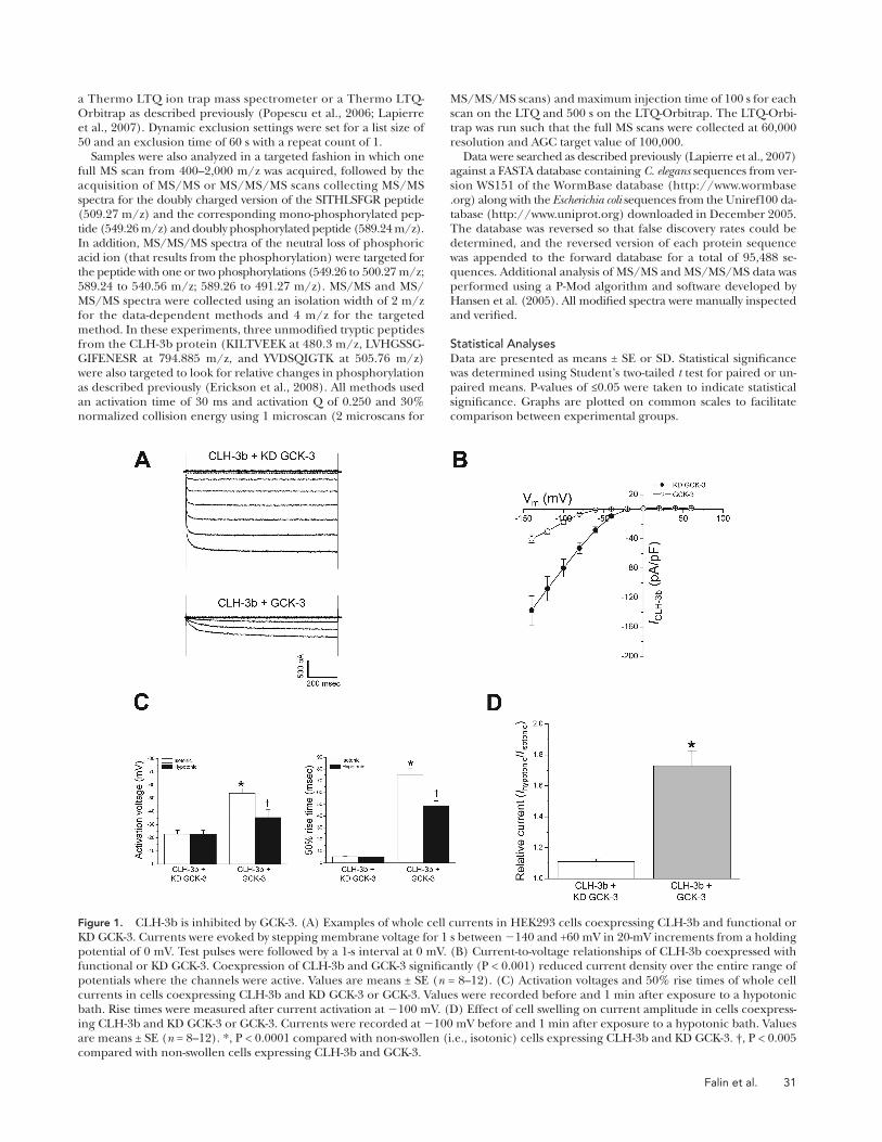
Figure 1. CLH-3b is inhibited by GCK-3. (A) Examples of whole cell currents in HEK293 cells coexpressing CLH-3b and functional or KD GCK-3. Currents were evoked by stepping membrane voltage for 1 s between � 140 and +60 mV in 20-mV increments from a holding potential of 0 mV. Test pulses were followed by a 1-s interval at 0 mV. (B) Current-to-voltage relationships of CLH-3b coexpressed with functional or KD GCK-3. Coexpression of CLH-3b and GCK-3 signifi cantly (P < 0.001) reduced current density over the entire range of potentials where the channels were active. Values are means ± SE ( n = 8 – 12). (C) Activation voltages and 50% rise times of whole cell currents in cells coexpressing CLH-3b and KD GCK-3 or GCK-3. Values were recorded before and 1 min after exposure to a hypotonic bath. Rise times were measured after current activation at � 100 mV. (D) Effect of cell swelling on current amplitude in cells coexpress-ing CLH-3b and KD GCK-3 or GCK-3. Currents were recorded at � 100 mV before and 1 min after exposure to a hypotonic bath. Values are means ± SE ( n = 8 – 12). *, P < 0.0001 compared with non-swollen (i.e., isotonic) cells expressing CLH-3b and KD GCK-3. † , P < 0.005 compared with non-swollen cells expressing CLH-3b and GCK-3.

32 Regulatory Phosphorylation Sites in a Ste20 Kinase – dependent ClC Channel
whole cell current observed 1 min after induction of cell
swelling was � 11%. In contrast, the activity of CLH-3b
coexpressed with GCK-3 was increased � 73% by 1 min
of cell swelling ( Fig. 1 D ). Exposure of cells to hypo-
tonic bath for 1 min decreased the rise time signifi cantly
(P < 0.0001) and shifted the activation voltage to a more
depolarized value (P < 0.005) in cells expressing GCK-3,
but had no signifi cant (P > 0.7) effect on these parame-
ters in control cells ( Fig. 1 C ).
Data shown in Fig. 1 are consistent with our previous
results ( Denton et al., 2005 ). These fi ndings demonstrate
that (1) CLH-3b expressed in the absence of functional
GCK-3 is constitutively active, (2) GCK-3 inhibits channel
activity, and (3) cell swelling reverses the inhibitory effect
of GCK-3. Swelling-induced reversal of inhibition is due
to net dephosphorylation brought about by a decrease in
GCK-3 activity and/or an increase in the activity of phos-
phatases ( Rutledge et al., 2002 ).
S742 and S747 Phosphorylation Increases in the Presence of GCK-3 Fig. 2 shows a block diagram of CLH-3b intramembrane
and intracellular domains. GCK-3 binds to a SPAK bind-
ing motif ( Piechotta et al., 2002 ) located at the beginning
of a 101 – amino acid splice insert unique to CLH-3b, and
binding is required for channel inhibition ( Denton et al.,
2005 ). This splice insert is termed a “ regulatory domain. ”
To determine if CLH-3b is a target of regulatory phos-
phorylation, we performed data-dependent LC-MS/MS
analysis of tryptic digests of CLH-3b immunoprecipi-
tated from cells coexpressing GCK-3 or KD GCK-3. Ini-
tial analyses revealed that the SITHLSFGR tryptic peptide
was phosphorylated at S747 as well as a site on the N ter-
minus at S742 or T744 (Fig. S1, A – D). However, we were
unable to differentiate between phosphorylation of S742
or T744 due to the lack of suffi cient fragment ions to
distinguish these two sites.
To further resolve phosphorylation changes at these
sites, we performed an additional set of immunoprecipita-
tion studies and targeted the singly and doubly phos-
phorylated SITHLSFGR peptide and three unmodifi ed
reference peptides (see Table I ) for both LC-MS/MS and
LC-MS/MS/MS analysis. Tryptic digests were analyzed in
triplicate, and MS data were manually inspected to deter-
mine the peak areas for SITHLSFGR peptides, which were
Online Supplemental Material Fig. S1 provides chromatograms and spectra from the targeted MS analysis of the phosphorylated SITHLSFGR peptide consis-tent with phosphorylation at S747 and an N-terminal residue, ei-ther S742 or T744. Fig. S2 details the data-dependent LC-MS analysis of the SITHLSFGR peptide phosphorylated at both S742 and S747. The T744A mutant exhibits wild-type channel proper-ties, and this is illustrated in Fig. S3. To provide additional evi-dence that phosphorylation of both S742 and S747 is required for inhibition by GCK-3, properties of the S742A, S747E and S742E, S747A mutants are given in Fig. S4. Figs. S1 – S4 are available at http://www.jgp.org/cgi/content/full/jgp.200810080/DC1.
RESULTS
GCK-3 Inhibits CLH-3b Current Amplitude and Alters Channel Gating Kinetics and Voltage Sensitivity Fig. 1 A shows examples of whole cell current traces re-
corded from cells coexpressing CLH-3b and GCK-3 or
control cells coexpressing the channel and a GCK-3 KD
mutant. Mean current-to-voltage relationships are shown
in Fig. 1 B . At all voltages where channel activity was
detected, current amplitude was signifi cantly (P < 0.005)
greater in control cells ( Fig. 1 B ). Thus, as shown previ-
ously ( Denton et al., 2005 ), GCK-3 inhibits whole cell
current amplitude.
Reduced current amplitude ( Fig. 1, A and B ) could re-
fl ect inhibition of CLH-3b by GCK-3 or reduced mem-
brane expression of functional channels. It must be
stressed that GCK-3 induces three distinct changes in
CLH-3b biophysical properties. Specifi cally, GCK-3 (1)
induces an � 2.4-fold hyperpolarizing shift in activation
voltage from � � 23 to � � 54 mV ( Fig. 1 C ), (2) induces
an � 15-fold slowing of the 50% rise time from � 5 to � 75
msec ( Fig. 1 C ), and (3) eliminates the fast activation time
constant (see Fig. 9 ) ( Denton et al., 2005 ). These changes
in the biophysical characteristics of the current rule out
the possibility that coexpression of CLH-3b with GCK-3
simply reduces the number of functional channels. Im-
portantly, the biophysical properties of GCK-3 – inhibited
CLH-3b are similar to those observed in C. elegans oocytes
before activation of the channel by swelling or meiotic
maturation ( Rutledge et al., 2001, 2002 ).
Because CLH-3b coexpressed with KD GCK-3 is con-
stitutively active, cell swelling has little effect on current
amplitude. As shown in Fig. 1 D , the mean increase in
Figure 2. CLH-3b functional domains. Open and shaded bars are predicted intramembrane and intracellular domains, respectively. Relative size of domains is approximately to scale. The regulatory domain is a 101 – amino acid splice insert unique to CLH-3b ( Denton et al., 2004 ). Sequence of regulatory domain is shown. Green highlighting indicates location of GCK-3 binding site ( Denton et al., 2005 ). Serines 742 and 747 are highlighted in red.

Falin et al. 33
are phosphorylated in the same peptide. Table I shows the
relative changes in the abundance of the SITHLSFGR
peptide phosphorylated at S747 or at both S742 and S747
in cells expressing GCK-3 as compared with KD GCK.
Phosphorylation of the doubly phosphorylated peptide
increased � 16-fold in cells expressing functional GCK-3.
Phosphorylation of S747 Is Required for GCK-3 – mediated Channel Inhibition To determine whether S747 plays a role in channel reg-
ulation, we mutated it to alanine and coexpressed the
normalized to the peak areas of the three reference pep-
tides. As shown in Table I , an approximately eightfold
increase in phosphorylation at S747 was evident in cells
coexpressing CLH-3b and functional GCK-3. We were also
able to resolve in these studies an SITHLSFGR peptide
where phosphorylation of both S742 and S747 but not
T744 was evident (Fig. S2, A and B). Fig. 3 is a representa-
tive chromatogram demonstrating the increase in the nor-
malized mass spectral signal for the doubly phosphorylated
peptide in the presence of GCK-3 as compared with KD
GSK-3, further demonstrating that both S742 and S747
TA B L E I
Relative Change in Phosphorylation of the SITHLSFGR Peptide in Cells Coexpressing CLH-3b and Functional GCK-3 or KD GCK-3
Peptide modifi cation a Description Relative phosphorylation
SITHLS(-18)FGR Single phosphorylation at S747 7.8 ± 1.8
S(80)ITHLS(80)FGR Double phosphorylation at S742 and S747 16 ± 3
Samples of CLH-3b protein isolated from cells expressing CLH-3b in the presence of either active GCK-3 or KD GCK-3 were evaluated by targeted MS/MS
analysis (see Materials and methods). Peak areas were determined for each of the modifi ed SITHLSFGR peptides indicated and the peak areas for the
unmodifi ed reference peptides KILTVEEK, LVHGSSGGIFENESR, and YVDSQIGTK. The peak areas for the phosphorylated SITHLSFGR peptide were
then normalized to the peak areas for each of the unmodifi ed reference peptides. Relative phosphorylation was calculated as the ratio of peak areas of the
SITHLSFGR peptide from cells expressing functional GCK-3 or KD GCK-3. Values are means ± SD for three separate MS runs.
a Phosphorylation can be identifi ed by an increase in peptide mass of +80, which corresponds to the addition of a phosphate group (HPO 3 ). It can also
be identifi ed by the neutral loss of H 3 PO 4 from the phosphorylated peptide. When compared to a peptide that is not phosphorylated, a net decrease in
peptide mass of � 18 is detected. ( � 18) and (80) refer to the peptide mass changes that were observed. The MS/MS/MS spectra of the neutral loss of
phosphoric acid from the singly phosphorylated peptide allowed us to distinguish the phosphorylation at S747 versus S742 (see Figs. S1, B – D).
Figure 3. Representative extracted ion chro-matograms from targeted MS/MS analysis of the SITHLSFGR peptide phosphorylated at both S742 and S747 in cells expressing KD GCK-3 (A) or functional GCK-3 (B). Shading shows the peak areas for the doubly phosphorylated SITHLSFGR peptide. These areas were normalized to the CLH-3b tryptic peptide KILTVEEK and are reported on each plot. In cells expressing functional GCK-3, phosphorylation of SITHLSFGR increases 15-fold. Similar fold changes were obtained in three replicate analyses and with normalization of the peak areas to two other CHL-3b tryptic peptides, LVHGSSGGIFENESR and YVDSQIGTK.

34 Regulatory Phosphorylation Sites in a Ste20 Kinase – dependent ClC Channel
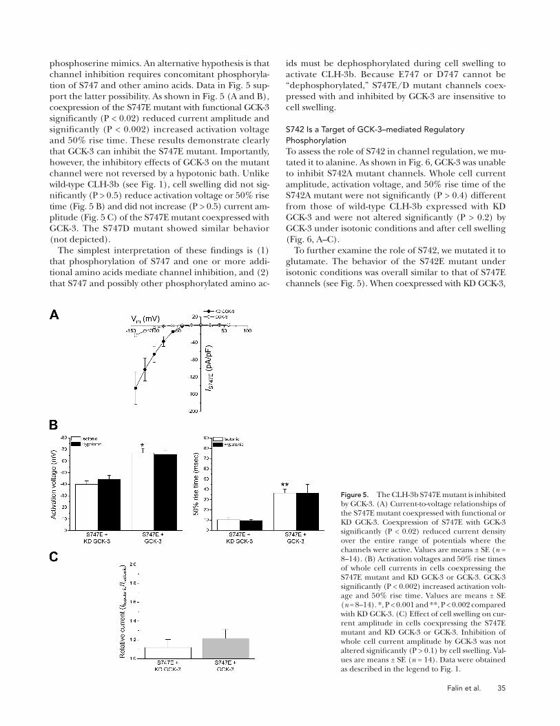
To further characterize the functional role of S747, we
replaced it with glutamate. When coexpressed with KD
GCK-3, current amplitude of the S747E mutant was
similar to that of wild-type CLH-3b ( Fig. 5 A ). Activation
voltages and 50% rise times were � � 40 mV and � 10
msec. These values were not signifi cantly (P > 0.9) altered
by cell swelling ( Fig. 5 B ).
Interestingly, when compared with wild-type CLH-3b,
the S747E mutant exhibited a signifi cant (P < 0.05) left-
ward shift in activation voltage from � � 23 to � � 40 mV
in the absence of GCK-3 activity and from � � 54 to
� � 72 mV when coexpressed with the functional kinase
(compare Figs. 1 D and 5 B ). In addition, this mutation
induced a small ( � 50%) but signifi cant (P < 0.001) in-
crease in rise time (compare Figs. 1 D and 5 B ). Results
qualitatively similar to those shown in Fig. 5 were ob-
tained using a S747D mutant where S747 was replaced
with an aspartate residue (not depicted).
Collectively, the results indicate that the S747E and
S747D mutants are constitutively active and suggest that
aspartate or glutamate substitutions may not be effective
S747A mutant with active or KD GCK-3. As shown in Fig.
4 A , GCK-3 had no signifi cant (P > 0.5) inhibitory effect
on whole cell current amplitude. Mean activation voltages
and 50% rise times ( Fig. 4 B ) were unaffected (P > 0.2)
by GCK-3 or cell swelling and were not signifi cantly
(P > 0.2) different from those of wild-type CLH-3b coex-
pressed with KD GCK-3 ( Fig. 4 B ). Cell swelling caused
a small but signifi cantly (P < 0.04) greater increase in
whole cell current amplitude of the S747A mutant ex-
pressed with GCK-3 versus KD GCK-3 ( Fig. 4 C ). How-
ever, swelling-induced activation of the S747A channel
was dramatically suppressed compared with wild-type
CLH-3b ( Fig. 4 C ), and swelling had no signifi cant (P >
0.2) effect on activation voltage or rise time ( Fig. 4 B ).
Collectively, these results indicate that the S747A mu-
tant is constitutively active and that its activity is largely
unaffected by GCK-3. Phosphorylation of S747 thus
plays an important role in CLH-3b regulation.
The effects of phosphorylation on protein function
can often be mimicked by replacement of phosphory-
lated amino acids with glutamate or aspartate residues.
Figure 4. Mutation of S747 to alanine prevents GCK-3 – mediated inhibition of CLH-3b. (A) Cur-rent-to-voltage relationships of the S747A mutant coexpressed with functional or KD GCK-3. Values are means ± SE ( n = 6 – 9). (B) Activation voltages and 50% rise times of whole cell currents in cells coexpressing the S747A mutant and KD GCK-3 or GCK-3. Values are means ± SE ( n = 6 – 9). (C) Ef-fect of cell swelling on current amplitude in cells coexpressing the S747A mutant and KD GCK-3 or GCK-3. Values are means ± SE ( n = 6 – 9). *, P < 0.04. Data were obtained as described in the legend to Fig. 1 .
Falin et al. 35
ids must be dephosphorylated during cell swelling to
activate CLH-3b. Because E747 or D747 cannot be
“ dephosphorylated, ” S747E/D mutant channels coex-
pressed with and inhibited by GCK-3 are insensitive to
cell swelling.
S742 Is a Target of GCK-3 – mediated Regulatory Phosphorylation To assess the role of S742 in channel regulation, we mu-
tated it to alanine. As shown in Fig. 6 , GCK-3 was unable
to inhibit S742A mutant channels. Whole cell current
amplitude, activation voltage, and 50% rise time of the
S742A mutant were not signifi cantly (P > 0.4) different
from those of wild-type CLH-3b expressed with KD
GCK-3 and were not altered signifi cantly (P > 0.2) by
GCK-3 under isotonic conditions and after cell swelling
( Fig. 6, A – C ).
To further examine the role of S742, we mutated it to
glutamate. The behavior of the S742E mutant under
isotonic conditions was overall similar to that of S747E
channels (see Fig. 5 ). When coexpressed with KD GCK-3,
phosphoserine mimics. An alternative hypothesis is that
channel inhibition requires concomitant phosphoryla-
tion of S747 and other amino acids. Data in Fig. 5 sup-
port the latter possibility. As shown in Fig. 5 (A and B) ,
coexpression of the S747E mutant with functional GCK-3
signifi cantly (P < 0.02) reduced current amplitude and
signifi cantly (P < 0.002) increased activation voltage
and 50% rise time. These results demonstrate clearly
that GCK-3 can inhibit the S747E mutant. Importantly,
however, the inhibitory effects of GCK-3 on the mutant
channel were not reversed by a hypotonic bath. Unlike
wild-type CLH-3b (see Fig. 1 ), cell swelling did not sig-
nifi cantly (P > 0.5) reduce activation voltage or 50% rise
time ( Fig. 5 B ) and did not increase (P > 0.5) current am-
plitude ( Fig. 5 C ) of the S747E mutant coexpressed with
GCK-3. The S747D mutant showed similar behavior
(not depicted).
The simplest interpretation of these fi ndings is (1)
that phosphorylation of S747 and one or more addi-
tional amino acids mediate channel inhibition, and (2)
that S747 and possibly other phosphorylated amino ac-
Figure 5. The CLH-3b S747E mutant is inhibited by GCK-3. (A) Current-to-voltage relationships of the S747E mutant coexpressed with functional or KD GCK-3. Coexpression of S747E with GCK-3 signifi cantly (P < 0.02) reduced current density over the entire range of potentials where the channels were active. Values are means ± SE ( n = 8 – 14). (B) Activation voltages and 50% rise times of whole cell currents in cells coexpressing the S747E mutant and KD GCK-3 or GCK-3. GCK-3 signifi cantly (P < 0.002) increased activation volt-age and 50% rise time. Values are means ± SE ( n = 8 – 14). *, P < 0.001 and **, P < 0.002 compared with KD GCK-3. (C) Effect of cell swelling on cur-rent amplitude in cells coexpressing the S747E mutant and KD GCK-3 or GCK-3. Inhibition of whole cell current amplitude by GCK-3 was not altered signifi cantly (P > 0.1) by cell swelling. Val-ues are means ± SE ( n = 14). Data were obtained as described in the legend to Fig. 1 .

36 Regulatory Phosphorylation Sites in a Ste20 Kinase – dependent ClC Channel
strate that T744 plays no role in GCK-3 – dependent
channel regulation.
GCK-3 – mediated Inhibition of CLH-3b Requires Concomitant Phosphorylation of S742 and S747 Data shown in Figs. 3 – 7 indicate that both S742 and
S747 must be phosphorylated in order for CLH-3b to be
inhibited by GCK-3. Consistent with this idea, we found
that GCK-3 was unable to inhibit the double-mutant
channels S742A, S747E and S742E, S747A (Fig. S4). To
further assess the combined role of these two amino
acid residues in channel regulation, we mutated them
both to glutamate. As shown in Fig. 8 A , whole cell
current amplitude of the S742E, S747E mutant was dra-
matically reduced and was not signifi cantly (P > 0.2) dif-
ferent from that of cells coexpressing wild-type CLH-3b
and GCK-3 (see Fig. 1 B ).
As noted earlier, reduction of current amplitude
alone may reflect reduced membrane expression of
functional CLH-3b. However, mutation of both S742
and S747 induced changes in current properties resem-
bling those of wild-type channels coexpressed with and
current amplitude, activation voltages, and 50% rise
times were not signifi cantly (P > 0.7) different from
those of wild-type CLH-3b. Coexpression of the S742E
mutant with GCK-3 signifi cantly (P < 0.01) inhibited
whole cell current amplitude, increased activation voltage,
and slowed activation kinetics ( Fig. 7, A and B ). How-
ever, unlike the S747E mutant, cell swelling increased
the activity of S742E channels coexpressed with GCK-3.
Whole cell current amplitude increased � 70% in cells
coexpressing S742E and GCK-3 compared with � 10%
in cells coexpressing S742E and KD GCK-3 (P < 0.02)
( Fig. 7 C ). Cell swelling also caused significant (P <
0.002) decreases in activation voltage and 50% rise times
of the S742E mutant coexpressed with GCK-3 ( Fig. 7 B ).
Similar results were observed using a S742D mutant
where S742 was replaced with an aspartate residue
(not depicted).
Because of initial uncertainty about whether T744
was a target of regulatory phosphorylation, we also
mutated this residue to alanine. As shown in Fig. S3,
the T744A mutation had no effect on channel prop-
erties or inhibition by GCK-3. These results demon-
Figure 6. Mutation of S742 to alanine prevents GCK-3 – mediated inhibition of CLH-3b. (A) Cur-rent-to-voltage relationships of the S742A mu-tant coexpressed with functional or KD GCK-3. Values are means ± SE ( n = 7). (B) Activation voltages and 50% rise times of whole cell cur-rents in cells coexpressing the S742A mutant and KD GCK-3 or GCK-3. Values are means ± SE ( n = 7). (C) Effect of cell swelling on cur-rent amplitude in cells coexpressing the S742A mutant and KD GCK-3 or GCK-3. Values are means ± SE ( n = 7). Data were obtained as described in the legend to Fig. 1 .

Falin et al. 37
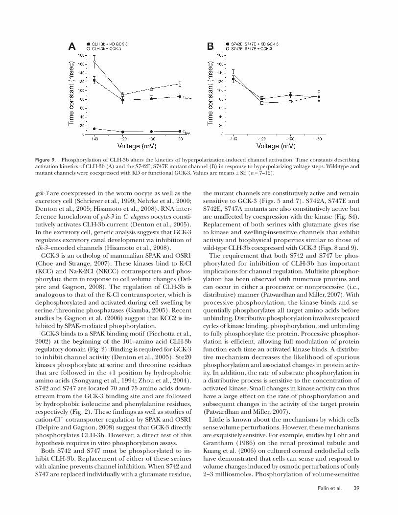
Phosphorylation Inhibits the Fast Component of Channel Gating The kinetics of hyperpolarization-induced activation of
wild-type CLH-3b expressed alone or with KD GCK-3 are
well described by the sum of two exponentials that de-
fi ne fast and slow time constants ( � fast and � slow ) ( Fig. 9 A )
( Denton et al., 2005 ). In contrast, the activation kinetics
of channels coexpressed with GCK-3 are described by a
single time constant similar to that of � slow ( Fig. 9 A ). As
discussed previously, these results suggest that GCK-3 may
inhibit a fast gating process ( Denton et al., 2005 ).
We derived time constants for voltage-dependent acti-
vation of all mutant channels. As expected, the activa-
tion kinetics of mutants that were inhibited by GCK-3
were defi ned by a single slow time constant when they
were coexpressed with the kinase. Similarly, the activa-
tion kinetics of mutant channels that were unaffected
by GCK-3 were described by fast and slow time constants
in the presence and absence of functional kinase (not
depicted). Importantly, the activation kinetics of S742E,
S747E mutant channels were described by a single slow
time constant when they were expressed with either KD
inhibited by GCK-3. The activation voltage of the S742E,
S747E mutant was hyperpolarized and 50% rise time was
increased signifi cantly (P < 0.02) compared with consti-
tutively active wild-type CLH-3b coexpressed with KD
GCK-3 ( Fig. 8 B ). Coexpression of S742E, S747E with
GCK-3 had no signifi cant (P > 0.2) effect on current am-
plitude and activation voltage, but it did cause a small
( � 20%; P < 0.01) increase in rise time ( Fig. 8, A and B ).
None of the functional parameters of S742E, S747E chan-
nels coexpressed with either GCK-3 or KD GCK-3 were
altered signifi cantly (P > 0.8) by cell swelling ( Fig. 8,
B and C ). Similar results were obtained using a S742D,
S747D mutant (not depicted). Collectively, these results
demonstrate that combined replacement of S742 and
S747 with phosphomimetic amino acids gives rise to chan-
nels that behave as if they have been inhibited by GCK-3;
S742E, S747E mutant channels show greatly reduced
whole cell current amplitude, a strong hyperpolariz-
ing shift in activation voltage, and slowed activation
kinetics. S742E, S747E mutant channels cannot be de-
phosphorylated and therefore cannot be activated by
cell swelling.
Figure 7. The CLH-3b S742E mutant is inhibited by GCK-3. (A) Current-to-voltage relationships of the S742E mutant coexpressed with functional or KD GCK-3. Coexpression of S742E with GCK-3 signifi cantly (P < 0.01) reduced current density over the entire range of potentials where the channels were active. Values are means ± SE ( n = 7 – 16). (B) Activation voltages and 50% rise times of whole cell currents in cells coexpressing the S742E mutant and KD GCK-3 or GCK-3. Values are means ± SE ( n = 7 – 16). *, P < 0.001 compared with KD GCK-3 isotonic. **, P < 0.002 compared with GCK-3 isotonic. (C) Effect of cell swelling on current amplitude in cells coexpressing the S742E mutant and KD GCK-3 or GCK-3. Values are means ± SE ( n = 16). *, P < 0.02. Data were obtained as described in the legend to Fig. 1 .

38 Regulatory Phosphorylation Sites in a Ste20 Kinase – dependent ClC Channel
ClC-3 belongs to a subfamily of ClC transport proteins
that function in intracellular membranes as electrogenic
Cl � /H + exchangers ( Chen and Hwang, 2008 ). However,
several groups have shown that heterologous expres-
sion of ClC-3 induces outwardly rectifying anion currents
that are modulated by phosphorylation. As with ClC-2,
phosphorylation has different effects under different
experimental conditions. For example, Cl � currents in-
duced by expression of human ClC-3 can be activated
by intracellular dialysis with autonomously active cal-
cium/calmodulin-dependent protein kinase II ( Huang
et al., 2001 ; Robinson et al., 2004 ). In contrast, Cl � cur-
rents observed in cells transfected with guinea pig ClC-3
are inhibited by phosphorylation mediated by PKA or
PKC ( Duan et al., 1999 ; Nagasaki et al., 2000 ). It is un-
clear if the varying effects of phosphorylation on ClC
activity refl ect species differences and/or differences in
experimental conditions.
We have exploited the genetically tractable model or-
ganism C. elegans to characterize the physiological roles
and regulatory mechanisms of ClC channels. clh-3 and
GCK-3 or functional GCK-3 ( Fig. 9 B ). These results fur-
ther support our conclusion that the S742E, S747E
mutant behaves like wild-type CLH-3b that has been in-
hibited by GCK-3.
D I S C U S S I O N
Changes in phosphorylation have been shown to regu-
late the activity of various ClC anion transport proteins.
However, the physiological context under which such
regulation occurs and the signaling cascades that medi-
ate phosphorylation are poorly understood. In addition,
phosphorylation has been shown to have opposing ef-
fects on ClCs under different experimental conditions.
For example, swelling-induced activation of rat ClC-2 is
inhibited by the type I protein phosphatase inhibitor ca-
lyculin A, suggesting that dephosphorylation activates
the channel ( Rutledge et al., 2002 ). In contrast, PKA-
mediated phosphorylation activates a rabbit ClC-2 splice
variant ( Malinowska et al., 1995 ), but has no effect on rat
ClC-2 ( Park et al., 2001 ).
Figure 8. CLH-3b S74E, S747E mutant chan-nels are constitutively inhibited and unaffected by GCK-3. (A) Current-to-voltage relationships of the S742E, S747E double mutant coexpressed with functional or KD GCK-3. Values are means ± SE ( n = 6 – 7). (B) Activation voltages and 50% rise times of whole cell currents in cells coexpressing S742E, S747E and KD GCK-3 or GCK-3. Values are means ± SE ( n = 6 – 7). *, P < 0.01 compared with KD GCK-3 isotonic. (C) Effect of cell swell-ing on current amplitude in cells coexpressing S742E, S747E and KD GCK-3 or GCK-3. Values are means ± SE ( n = 6 – 7). Data were obtained as described in the legend to Fig. 1 .
Falin et al. 39
the mutant channels are constitutively active and remain
sensitive to GCK-3 ( Figs. 5 and 7 ). S742A, S747E and
S742E, S747A mutants are also constitutively active but
are unaffected by coexpression with the kinase (Fig. S4).
Replacement of both serines with glutamate gives rise
to kinase and swelling-insensitive channels that exhibit
activity and biophysical properties similar to those of
wild-type CLH-3b coexpressed with GCK-3 ( Figs. 8 and 9 ).
The requirement that both S742 and S747 be phos-
phorylated for inhibition of CLH-3b has important
implications for channel regulation. Multisite phosphor-
ylation has been observed with numerous proteins and
can occur in either a processive or nonprocessive (i.e.,
distributive) manner ( Patwardhan and Miller, 2007 ). With
processive phosphorylation, the kinase binds and se-
quentially phosphorylates all target amino acids before
unbinding. Distributive phosphorylation involves repeated
cycles of kinase binding, phosphorylation, and unbinding
to fully phosphorylate the protein. Processive phosphor-
ylation is effi cient, allowing full modulation of protein
function each time an activated kinase binds. A distribu-
tive mechanism decreases the likelihood of spurious
phosphorylation and associated changes in protein activ-
ity. In addition, the rate of substrate phosphorylation in
a distributive process is sensitive to the concentration of
activated kinase. Small changes in kinase activity can thus
have a large effect on the rate of phosphorylation and
subsequent changes in the activity of the target protein
( Patwardhan and Miller, 2007 ).
Little is known about the mechanisms by which cells
sense volume perturbations. However, these mechanisms
are exquisitely sensitive. For example, studies by Lohr and
Grantham (1986) on the renal proximal tubule and
Kuang et al. (2006) on cultured corneal endothelial cells
have demonstrated that cells can sense and respond to
volume changes induced by osmotic perturbations of only
2 – 3 milliosmoles. Phosphorylation of volume-sensitive
gck-3 are coexpressed in the worm oocyte as well as the
excretory cell ( Schriever et al., 1999 ; Nehrke et al., 2000 ;
Denton et al., 2005 ; Hisamoto et al., 2008 ). RNA inter-
ference knockdown of gck-3 in C. elegans oocytes consti-
tutively activates CLH-3b current ( Denton et al., 2005 ).
In the excretory cell, genetic analysis suggests that GCK-3
regulates excretory canal development via inhibition of
clh-3 – encoded channels ( Hisamoto et al., 2008 ).
GCK-3 is an ortholog of mammalian SPAK and OSR1
( Choe and Strange, 2007 ). These kinases bind to K-Cl
(KCC) and Na-K-2Cl (NKCC) cotransporters and phos-
phorylate them in response to cell volume changes ( Del-
pire and Gagnon, 2008 ). The regulation of CLH-3b is
analogous to that of the K-Cl contransporter, which is
dephosphorylated and activated during cell swelling by
serine/threonine phosphatases ( Gamba, 2005 ). Recent
studies by Gagnon et al. (2006) suggest that KCC2 is in-
hibited by SPAK-mediated phosphorylation.
GCK-3 binds to a SPAK binding motif ( Piechotta et al.,
2002 ) at the beginning of the 101 – amino acid CLH-3b
regulatory domain ( Fig. 2 ). Binding is required for GCK-3
to inhibit channel activity ( Denton et al., 2005 ). Ste20
kinases phosphorylate at serine and threonine residues
that are followed in the +1 position by hydrophobic
amino acids ( Songyang et al., 1994 ; Zhou et al., 2004 ).
S742 and S747 are located 70 and 75 amino acids down-
stream from the GCK-3 binding site and are followed
by hydrophobic isoleucine and phenylalanine residues,
respectively ( Fig. 2 ). These fi ndings as well as studies of
cation-Cl � cotransporter regulation by SPAK and OSR1
( Delpire and Gagnon, 2008 ) suggest that GCK-3 directly
phosphorylates CLH-3b. However, a direct test of this
hypothesis requires in vitro phosphorylation assays.
Both S742 and S747 must be phosphorylated to in-
hibit CLH-3b. Replacement of either of these serines
with alanine prevents channel inhibition. When S742 and
S747 are replaced individually with a glutamate residue,
Figure 9. Phosphorylation of CLH-3b alters the kinetics of hyperpolarization-induced channel activation. Time constants describing activation kinetics of CLH-3b (A) and the S742E, S747E mutant channel (B) in response to hyperpolarizing voltage steps. Wild-type and mutant channels were coexpressed with KD or functional GCK-3. Values are means ± SE ( n = 7 – 12).
40 Regulatory Phosphorylation Sites in a Ste20 Kinase – dependent ClC Channel
( Denton et al., 2005 ). As we have proposed previously
( Denton et al., 2005 ), this suggests indirectly that phos-
phorylation may inhibit the fast gating mechanism.
Deletion of the CLH-3b regulatory domain gives rise
to channels with voltage sensitivity and kinetics of hyper-
polarization-induced activation that resemble those of
wild-type channels coexpressed with GCK-3 ( He et al.,
2006 ). It is interesting to speculate that the CLH-3b regu-
latory domain may interact with other parts of the chan-
nel. Phosphorylation of S742 and S747 could disrupt this
interaction leading to channel inhibition. Deletion
of the entire regulatory domain would also disrupt pro-
tein – protein interactions and mimic phosphorylation.
How could changes in the conformation of the cytoplas-
mic C terminus lead to changes in channel gating? ClC
proteins are comprised of 18 � -helical domains (desig-
nated “ A-R ” ), 17 of which are membrane embedded
( Dutzler et al., 2002, 2003 ). The R-helix forms part of the
channel pore and selectivity fi lter ( Dutzler et al., 2002,
2003 ). In eukaryotic ClC proteins, the R-helix connects via
a short linker to a large cytoplasmic C terminus. Dutzler
et al. (2002) proposed that this linker may provide a path-
way by which conformational changes in the cytoplasmic
C terminus could be transmitted to the intramembrane
domain and thereby alter channel gating and pore prop-
erties. Recent studies on ClC-1 ( Hebeisen and Fahlke,
2005 ), CLH-3b ( He et al., 2006 ), and ClC-Kb ( Martinez
and Maduke, 2008 ) have demonstrated that C terminus
mutations give rise to extracellular structure/function
changes. Clearly though, extensive additional studies will
be required to defi ne how phosphorylation events alter
cytoplasmic C terminus conformation and how those con-
formational changes in turn regulate CLH-3b gating. This work was supported by National Institutes of Health
(NIH) R01 grants DK51610 and DK61168 to K. Strange and NIH training grants 5T32 NS007491-07 and 5F32 DK080576-02 to R.A. Falin. Experiments described in this paper were proposed and designed by R.A. Falin, R. Morrison, A.-J.L. Ham, and K. Strange. Experimental procedures were carried out by R.A. Falin, R. Morrison, and A.-J.L. Ham. All authors participated in the analysis and interpretation of data, in the writing of the manu-script, and in the approval of the fi nal version of the manuscript for publication.
Edward N. Pugh Jr. served as editor.
Submitted: 17 July 2008 Accepted: 17 November 2008
R E F E R E N C E S Barr , M.M. 2003 . Super models. Physiol. Genomics . 13 : 15 – 24 .
Chen , T.Y. , and T.C. Hwang . 2008 . CLC-0 and CFTR: chloride chan-
nels evolved from transporters. Physiol. Rev. 88 : 351 – 387 .
Choe , K.P. , and K. Strange . 2007 . Evolutionarily conserved WNK
and Ste20 kinases are essential for acute volume recovery and sur-
vival following hypertonic shrinkage in Caenorhabditis elegans. Am. J. Physiol. 293 : C915 – C927 .
Dan , I. , N.M. Watanabe , and A. Kusumi . 2001 . The Ste20 group
kinases as regulators of MAP kinase cascades. Trends Cell Biol. 11 : 220 – 230 .
channels and cation-Cl � cotransporters via a distributive
mechanism may contribute to this sensitivity.
It is presently unclear whether changes in swelling-in-
duced dephosphorylation and activation of CLH-3b are
brought about by reductions in GCK-3 activity and/or in-
creases in the activity of phosphatases. Indirect evidence
from studies of cation-Cl � cotransporters suggests that
they are regulated by a volume-sensitive kinase that oper-
ates in the presence of constant and unregulated phos-
phatase activity ( Jennings and al-Rohil, 1990 ; Lytle, 1998 ;
Jennings, 1999 ). Thus, increases or decreases in kinase
activity brought about by cell volume perturbations in-
duce concomitant increases or decreases in cotransporter
phosphorylation. It remains to be determined whether
GCK-3 and/or other kinases are the volume-sensitive
components of the CLH-3b regulatory pathway.
Interestingly, activation of CLH-3b in response to cell
swelling occurs with dephosphorylation of S747 alone.
S747E mutant channels coexpressed with GCK-3 are not
activated by cell volume increase ( Fig. 5 ). In contrast,
S742E channels inhibited by GCK-3 exhibit swelling-in-
duced increases in current amplitude and decreases in
activation voltage and rise time comparable to that of
wild-type CLH-3b (compare Figs. 1, C and D, and 7, B
and C ). These results suggest two possibilities. First, swell-
ing-induced dephosphorylation of the channel may be
an ordered process requiring S747 to be dephosphory-
lated before S742. Partial dephosphorylation may in turn
allow partial activation of CLH-3b in response to differ-
ent levels or types of stimuli. Alternatively, the phosphor-
ylation of S742 may simply be a permissive event required
for protein conformational changes to take place in
response to phosphorylation and dephosphorylation
of S747 that in turn inhibit or activate CLH-3b. Unfortu-
nately, these possibilities cannot be tested using mam-
malian expression systems. Mammalian cells express an
outwardly rectifying anion current, I Cl, swell , that is acti-
vated dramatically by cell swelling ( Hartzell et al., 2008 ).
The presence of this current limits the amount of time
cells can be swelled and therefore prevents assessment of
the full degree to which wild-type and mutant CLH-3b
channels can be activated. Alternative expression systems
may be useful for assessing how dephosphorylation regu-
lates channel function.
ClC channels are homodimers, and each monomer
forms a pore that is independently gated by a so-called
“ fast ” gate that opens and closes on a millisecond time-
scale. A “ common ” gate with much slower kinetics closes
both pores simultaneously ( Miller, 2006 ; Jentsch, 2008 ).
The kinetics of hyperpolarization-induced activation of
wild-type CLH-3b expressed in the absence of functional
GCK-3 are described by the fast and slow time constants
( Fig. 9 A ) ( Denton et al., 2005 ). Voltage-dependent ac-
tivation of CLH-3b is described by a single slow time
constant when the channel is inhibited by GCK-3 or by
mutation of both S742 and S747 to glutamate ( Fig. 9 )

Falin et al. 41
Jentsch , T.J. , V. Stein , F. Weinreich , and A.A. Zdebik . 2002 . Molecular
structure and physiological function of chloride channels. Physiol. Rev. 82 : 503 – 568 .
Jentsch , T.J. , M. Poet , J.C. Fuhrmann , and A.A. Zdebik . 2005 .
Physiological functions of CLC Cl - channels gleaned from human
genetic disease and mouse models. Annu. Rev. Physiol. 67 : 779 – 807 .
Jones , J.A. , L. Kaphalia , M. Treinen-Moslen , and D.C. Liebler . 2003 .
Proteomic characterization of metabolites, protein adducts, and
biliary proteins in rats exposed to 1,1-dichloroethylene or diclo-
fenac. Chem. Res. Toxicol. 16 : 1306 – 1317 .
Kuang , K. , M. Yiming , Z. Zhu , P. Iserovich , F.P. Diecke , and J.
Fischbarg . 2006 . Lack of threshold for anisotonic cell volume regu-
lation. J. Membr. Biol. 211 : 27 – 33 .
Lapierre , L.A. , K.M. Avant , C.M. Caldwell , A.J. Ham , S. Hill , J.A. Williams ,
A.J. Smolka , and J.R. Goldenring . 2007 . Characterization of immu-
noisolated human gastric parietal cells tubulovesicles: identifi cation
of regulators of apical recycling. Am. J. Physiol. 292 : G1249 – G1262 .
Ling , P. , T.J. Lu , C.J. Yuan , and M.D. Lai . 2008 . Biosignaling of mam-
malian Ste20-related kinases. Cell. Signal. 20 : 1237 – 1247 .
Lohr , J.W. , and J.J. Grantham . 1986 . Isovolumetric regulation of
isolated S 2 proximal tubules in anisotonic media. J. Clin. Invest. 78 : 1165 – 1172 .
Lytle , C. 1998 . A volume-sensitive protein kinase regulates the
Na-K-2Cl cotransporter in duck red blood cells. Am. J. Physiol. 274 : C1002 – C1010 .
Malinowska , D.H. , E.Y. Kupert , A. Bahinski , A.M. Sherry , and J.
Cuppoletti . 1995 . Cloning, functional expression, and charac-
terization of a PKA-activated gastric Cl - channel. Am. J. Physiol. 268 : C191 – C200 .
Martinez , G.Q. , and M. Maduke . 2008 . A cytoplasmic domain muta-
tion in ClC-Kb affects long-distance communication across the
membrane. PLoS ONE. 3 : e2746 .
Miller , C. 2006 . ClC chloride channels viewed through a transporter
lens. Nature . 440 : 484 – 489 .
Nagasaki , M. , L. Ye , D. Duan , B. Horowitz , and J.R. Hume . 2000 .
Intracellular cyclic AMP inhibits native and recombinant volume-
regulated chloride channels from mammalian heart. J. Physiol. 523 : 705 – 717 .
Nehrke , K. , T. Begenisich , J. Pilato , and J.E. Melvin . 2000 . C. elegans ClC-type chloride channels: novel variants and functional expres-
sion. Am. J. Physiol. 279 : C2052 – C2066 .
Park , K. , T. Begenisich , and J.E. Melvin . 2001 . Protein kinase A ac-
tivation phosphorylates the rat ClC-2 Cl- channel but does not
change activity. J. Membr. Biol. 182 : 31 – 37 .
Patwardhan , P. , and W.T. Miller . 2007 . Processive phosphorylation:
mechanism and biological importance. Cell. Signal. 19 : 2218 – 2226 .
Piechotta , K. , J. Lu , and E. Delpire . 2002 . Cation chloride cotrans-
porters interact with the stress-related kinases Ste20-related pro-
line-alanine-rich kinase (SPAK) and oxidative stress response 1
(OSR1). J. Biol. Chem. 277 : 50812 – 50819 .
Popescu , D.C. , A.J. Ham , and B.H. Shieh . 2006 . Scaffolding protein
INAD regulates deactivation of vision by promoting phosphoryla-
tion of transient receptor potential by eye protein kinase C in
Drosophila. J. Neurosci. 26 : 8570 – 8577 .
Robinson , N.C. , P. Huang , M.A. Kaetzel , F.S. Lamb , and D.J. Nelson .
2004 . Identifi cation of an N-terminal amino acid of the CLC-3
chloride channel critical in phosphorylation-dependent activation
of a CaMKII-activated chloride current. J. Physiol. 556 : 353 – 368 .
Rutledge , E. , L. Bianchi , M. Christensen , C. Boehmer , R. Morrison ,
A. Broslat , A.M. Beld , A. George , D. Greenstein , and K. Strange .
2001 . CLH-3, a ClC-2 anion channel ortholog activated during
meiotic maturation in C. elegans oocytes. Curr. Biol. 11 : 161 – 170 .
Rutledge , E. , J. Denton , and K. Strange . 2002 . Cell cycle- and swell-
ing-induced activation of a C. elegans ClC channel is mediated by
CeGLC-7 � / � phosphatases. J. Cell Biol. 158 : 435 – 444 .
Delpire , E. , and K.B. Gagnon . 2008 . SPAK and OSR1: STE20 kinases
involved in the regulation of ion homoeostasis and volume con-
trol in mammalian cells. Biochem. J. 409 : 321 – 331 .
Denton , J. , K. Nehrke , E. Rutledge , R. Morrison , and K. Strange .
2004 . Alternative splicing of N- and C-termini of a C. elegans ClC
channel alters gating and sensitivity to external Cl - and H + . J. Physiol. 555 : 97 – 114 .
Denton , J. , K. Nehrke , X. Yin , R. Morrison , and K. Strange . 2005 .
GCK-3, a newly identifi ed Ste20 kinase, binds to and regulates
the activity of a cell cycle – dependent ClC anion channel. J. Gen. Physiol. 125 : 113 – 125 .
Duan , D. , S. Cowley , B. Horowitz , and J.R. Hume . 1999 . A serine res-
idue in ClC-3 links phosphorylation-dephosphorylation to chlo-
ride channel regulation by cell volume. J. Gen. Physiol. 113 : 57 – 70 .
Dutzler , R. , E.B. Campbell , M. Cadene , B.T. Chait , and R.
MacKinnon . 2002 . X-ray structure of a ClC chloride channel
at 3.0 A reveals the molecular basis of anion selectivity. Nature . 415 : 287 – 294 .
Dutzler , R. , E.B. Campbell , and R. MacKinnon . 2003 . Gating the
selectivity fi lter in ClC chloride channels. Science . 300 : 108 – 112 .
Erickson , J.R. , M.L. Joiner , X. Guan , W. Kutschke , J. Yang , C.V.
Oddis , R.K. Bartlett , J.S. Lowe , S.E. O ’ Donnell , N. Aykin-Burns , et
al . 2008 . A dynamic pathway for calcium-independent activation
of CaMKII by methionine oxidation. Cell . 133 : 462 – 474 .
Gagnon , K.B. , R. England , and E. Delpire . 2006 . Volume sensitiv-
ity of cation-Cl- cotransporters is modulated by the interaction of
two kinases: Ste20-related proline-alanine-rich kinase and WNK4.
Am. J. Physiol. 290 : C134 – C142 .
Gamba , G. 2005 . Molecular physiology and pathophysiology of electro-
neutral cation-chloride cotransporters. Physiol. Rev. 85 : 423 – 493 .
Hansen , B.T. , S.W. Davey , A.J.L. Ham , and D.C. Liebler . 2005 . P-
Mod: an algorithm and software to map modifi cations to peptide
sequences from tandem MS data. J. Proteome Res. 4 : 358 – 368 .
Hartzell , H.C. , Z. Qu , K. Yu , Q. Xiao , and L.T. Chien . 2008 .
Molecular physiology of bestrophins: multifunctional mem-
brane proteins linked to best disease and other retinopathies.
Physiol. Rev. 88 : 639 – 672 .
He , L. , J. Denton , K. Nehrke , and K. Strange . 2006 . Carboxy terminus
splice variation alters ClC channel gating and extracellular cysteine
reactivity. Biophys. J. 90 : 3570 – 3581 .
Hebeisen , S. , and C. Fahlke . 2005 . Carboxy-terminal truncations
modify the outer pore vestibule of muscle chloride channels.
Biophys. J. 89 : 1710 – 1720 .
Hellman , U. , C. Wernstedt , J. Gonez , and C.H. Heldin . 1995 .
Improvement of an “ In-Gel ” digestion procedure for the micro-
preparation of internal protein fragments for amino acid sequenc-
ing. Anal. Biochem. 224 : 451 – 455 .
Hisamoto , N. , T. Moriguchi , S. Urushiyama , S. Mitani , H. Shibuya ,
and K. Matsumoto . 2008 . Caenorhabditis elegans WNK-STE20 path-
way regulates tube formation by modulating ClC channel activity.
EMBO Rep. 9 : 70 – 75 .
Huang , P. , J. Liu , A. Di , N.C. Robinson , M.W. Musch , M.A. Kaetzel ,
and D.J. Nelson . 2001 . Regulation of human CLC-3 channels by
multifunctional Ca 2+ /calmodulin-dependent protein kinase. J. Biol. Chem. 276 : 20093 – 20100 .
Jennings , M.L. 1999 . Volume-sensitive K + /Cl � cotransport in rabbit
erythrocytes. Analysis of the rate-limiting activation and inactiva-
tion events. J. Gen. Physiol. 114 : 743 – 758 .
Jennings , M.L. , and N. al-Rohil . 1990 . Kinetics of activation and inac-
tivation of swelling-stimulated K + /Cl � transport. The volume-sensi-
tive parameter is the rate constant for inactivation. J. Gen. Physiol. 95 : 1021 – 1040 .
Jentsch , T.J. 2008 . CLC chloride channels and transporters: from
genes to protein structure, pathology and physiology. Crit. Rev. Biochem. Mol. Biol. 43 : 3 – 36 .

42 Regulatory Phosphorylation Sites in a Ste20 Kinase – dependent ClC Channel
Strange , K. , J. Denton , and K. Nehrke . 2006 . Ste20-type kinases: evo-
lutionarily conserved regulators of ion transport and cell volume.
Physiology (Bethesda) . 21 : 61 – 68 .
Yin , X. , N.J. Gower , H.A. Baylis , and K. Strange . 2004 . Inositol 1,4,5-
trisphosphate signaling regulates rhythmic contractile activity of
smooth muscle-like sheath cells in the nematode Caenorhabditis elegans. Mol. Biol. Cell . 15 : 3938 – 3949 .
Zhou , T. , M. Raman , Y. Gao , S. Earnest , Z. Chen , M. Machius , M.H.
Cobb , and E.J. Goldsmith . 2004 . Crystal structure of the TAO2 ki-
nase domain: activation and specifi city of a Ste20p MAP3K. Structure . 12 : 1891 – 1900 .
Schriever , A.M. , T. Friedrich , M. Pusch , and T.J. Jentsch . 1999 .
CLC chloride channels in Caenorhabditis elegans. J. Biol. Chem. 274 : 34238 – 34244 .
Songyang , Z. , S. Blechner , N. Hoagland , M.F. Hoekstra , H.
Piwnica-Worms , and L.C. Cantley . 1994 . Use of an oriented pep-
tide library to determine the optimal substrates of protein kinases.
Curr. Biol. 4 : 973 – 982 .
Strange , K. 2002 . Of mice and worms: novel insights into ClC-2 an-
ion channel physiology. News Physiol. Sci. 17 : 11 – 16 .
Strange , K. 2003 . From genes to integrative physiology: ion channel and
transporter biology in Caenorhabditis elegans. Physiol. Rev. 83 : 377 – 415 .