Embed Size (px)
Citation preview

Hindawi Publishing CorporationMediators of InflammationVolume 2013, Article ID 967987, 7 pageshttp://dx.doi.org/10.1155/2013/967987
Clinical StudyIL-17 Expression in Dermatitis Herpetiformis andBullous Pemphigoid
Agnieszka Zebrowska,1 Malgorzata Wagrowska-Danilewicz,2
Marian Danilewicz,2 Olga Stasikowska-Kanicka,2 Anna Cynkier,1
Anna Sysa-Jedrzejowska,1 and Elzbieta Waszczykowska1
1 Department of Dermatology and Venereology, Medical University of Lodz, 5 Krzemieniecka Street, 94-017 Lodz, Poland2 Laboratory of Nephropathology of Medical University of Lodz, 251 Pomorska Street, 92-213 Lodz, Poland
Correspondence should be addressed to Agnieszka Zebrowska; [email protected]
Received 4 March 2013; Accepted 25 June 2013
Academic Editor: Alex Kleinjan
Copyright © 2013 Agnieszka Zebrowska et al. This is an open access article distributed under the Creative Commons AttributionLicense, which permits unrestricted use, distribution, and reproduction in any medium, provided the original work is properlycited.
Dermatitis herpetiformis (DH) and bullous pemphigoid (BP) are skin diseases associated with eosinophilic and neutrophilicinfiltrations. Although cytokines are critical for the inflammatory process, there are single findings concerning concentration ofIL-17 in bullous diseases.The goal of this study was to assess IL-17 expression in DH and BP patients. Skin biopsies were taken from10 DH, 14 BP patients and from 10 healthy subjects.The localization and expression of IL-17 was studied by immunohistochemistryand the serum concentration was measured by immunoassays. Expression of IL-17 in the epidermis and in influxed cells in dermiswas detected in skin biopsies. Expression of IL-17 was statistically higher in epidermis and infiltration cells in specimens from BPthan from DH patients. Examined interleukin expression was detected in perilesional skin of all patients but it was much lowerthan in lesional skin.The expression of IL-17 was not observed in biopsies from healthy people. Serum level of IL-17 was statisticallyhigher in BP and DH groups as compared to control group. Our results provide the evidence that IL-17 may play an essential rolein activating and recruiting eosinophils and neutrophils, which ultimately contribute to the tissue damage in DH and BP.
1. Introduction
Dermatitis herpetiformis (DH) is one of the subepidermalautoimmune bullous diseases characterized by skin andintestinal lesions. Skin lesions include polymorphic erup-tion accompanied by severe pruritus. Intestinal lesions arecharacterized by atrophy of intestinal villi resulting fromimmunological process [1]. Diagnosis of DH is established onthe results of direct immunofluorescence test (DIF) revealinggranular deposits of IgA in the papillae and the presenceof circulating IgA antibodies directed against endomysiumand/or tissue and epidermal transglutaminase (tTG, eTG)[2, 3]. Skin lesions in DH are histologically characterizedby neutrophilic infiltrate leading to destruction of basementmembrane zone (BMZ) proteins, anchoring fibers, and blisterformation [4–6].
Bullous pemphigoid (BP) is a blistering disease, charac-terized by inflammatory infiltrate in the dermis, presence of
IgG and C3 deposits along the basement membrane zone,and circulating IgG autoantibodies. Autoantibodies bindingto autoantigens (glycoproteins: 230 kD (BPAG1) and 180 kD(BPAG2)) localized in the basement membrane of the epi-dermis activate a series of immunological and enzymaticphenomena leading to destruction of basement membranecomponents and anchoring fibers and blister formation as inDH [7, 8].
Inflammatory infiltrates in the dermis, formed by eosin-ophils and neutrophils and bound in vivo deposits along thebasementmembrane in BP or in the top of papillae inDH, areobserved. Ultrastructural studies also confirmed the presenceof intensive inflammatory infiltrate at dermo-epidermal junc-tion, as well as destruction of hemidesmosomes and com-ponents of extracellular matrix [9].
Formation of the infiltrates is preceded by early accu-mulation of leukocytes, depending on activity of adhesionmolecules. The binding of autoantibodies leads to activation
2 Mediators of Inflammation
of keratinocytes, release IL-6 and IL-8, of activation of C5component of the complement and metalloproteinases—enzymes produced by eosinophils and neutrophils attractedto the basement membrane by selectins and integrins andchemokines [10, 11]. Chemokines are important chemoattrac-tants for both eosinophils and neutrophils [12, 13]. Chemok-ines and cytokines play their role through receptors. Someof them are highly specific whereas others may interact withmore than one ligand [12, 13].
Few studies available suggested cytokines’ role in gener-ation of inflammatory influx in autoimmune blistering dis-eases [14–17].
There is increasing evidence that Th17 cells and thecytokines they release such as interleukin-17 (IL-17) are im-portant regulators of innate and adaptive immune responsesin many Th1- and/or Th2-mediated autoimmune diseasessuch as rheumatoid arthritis, systemic lupus erythematosus,and allergic asthma [18].There is also evidence thatTh17 cellsmay have a role in pathogenesis of blistering skin diseases.Interleukin-17 is important in initiation and maintenance ofmany autoimmune reactions and it is involved in productionof proinflammatory cytokines, matrix metalloproteinases,neutrophils, and eosinophils, all of which are importantpathogenic factors in bullous pemphigoid and dermatitisherpetiformis [19]. The hypothesis is that interleukin-17 mayhave an important pathogenic role in DH and BP.
It was previously reported that Th17 cells are recruited tothe lesional skin in pemphigus vulgaris (PV) and pemphigusfoliaceus (PF). Immunohistochemical studies showed thatboth IL-17+ and Foxp3+ cells were present in higher numbersin cells in BP lesions, compared with control skin. IL-17/CD4 ratio in BP was significantly higher than that in PF.Foxp3/CD4 ratio in BP was significantly lower than that ineither PV or PF. There were no obvious correlations betweenthese cells and the disease severity of BP. The previousstudy suggests that, compared with pemphigus, BP showsmore Th17 cell-related inflammation and less Treg-relatedregulation [20].There was no data about IL-17 serum levels inblistering disease comparing this level with tissue expression.There was no literature data with reference to the role of IL-17in pathogenesis of dermatitis herpetiformis.
Il-17 can be assessed in the skin lesions and sera of patientsand can be used as a marker of disease activity and responseto therapy. The information obtained could also lead to thedevelopment of novel therapeutic strategies for this and otherautoimmune blistering diseases.
The goal of this study was to assess interleukin-17 expres-sion in skin lesions and perilesional area and serum levels inpatients with DH and BP.
2. Materials and Methods
2.1. Patients. The study included 34 persons: 14 untreatedpatients with BP (range: 58 to 84 years, average: 68,5) and10 with DH (range: 18 to 70 years, average: 49,8) in an activestage of the disease. A control group consisted of 10 healthyindividuals (range: 19 to 80 years, average: 52,6 years).
All the patients signed informed consent before enteringthe study and the study protocol (RNN/132/07/KB) was
approved by The Local Ethical Committee of Medical Uni-versity of Lodz.
Eight out of 10DH patients had skin lesions character-ized by vesicles and itching papules; the others had ery-thematous papules. In all the cases histological picturesshowed perivascular neutrophilic infiltrates, the presence ofPierrard’s abscesses, and in all patients small subepidermalblisters. In 7/10 samples large unilocular blisters displayingmultiple neutrophilic papillary microabscesses were found.The histopathologic findings according to Ackerman in allcases were fully developed [21]. Direct immunofluorescencetests revealed the presence of granular deposits of IgA inskin papillae and indirect immunofluorescence tests werepositive for IgAEmA (Oesophagus Monkey IgAEmA, Medi-zinische Labordiagnostica) in all the patients (titer 1 : 40–1 : 640, median 1 : 80). Antitissue transglutaminase antibod-ies measured using an immunoassay (Celikey, Pharmacia& Upjohn) were present in 7/10 cases (median 5.1 IU/mL(range: 0,0–186,3; IU/mL). Diagnosis of DH was establishedbased on clinical presentation and results of histological andimmunological examination.
Pemphigoid was diagnosed based on clinical picture andhistological and immunological findings [7]. The patientswere at an active stage of the disease. In 12 out of 14patients skin blisters, vesicles, and itching papules werefound, whereas others had only small vesicles and urticarialpapules.The histopathologic findings according toAckermanin all cases were fully developed [21]. In all the patients directimmunofluorescence test revealed IgG/C3 linear depositsalong BMZ. In salt split test deposits were observed inepidermal part of the blister. Using indirect immunoflu-orescence test circulating IgG antibodies were found in14/14 patients, whereas ELISA test showed the anti-NC16autoantibodies (MBL, Nagoya, Japan) present in serum of 11out of 14 patients. Typical histological features of BP includingneutrophilic infiltrates, eosinophils, lymphocytes, and, in 12cases subepidermal blisters supported the clinical diagnosis.
2.1.1. Tissue Specimens. The biopsies were taken from thebuttock or trunk skin before administration of any (topical orsystemic) treatment. Skin lesions lasted between 2 weeks and3 months. Biopsy specimens were taken from buttock skin ofhealthy volunteers.
2.1.2. Immunohistochemistry. Paraffin-embedded sections (3-4 𝜇m thick) were used for routine H&E staining and forimmunohistochemistry with DAKO EnVision detection sys-tem using immunoperoxidase method. The following pri-mary monoclonal antibodies were used: antiinterleukin 17obtained from R&D, UK.
For immunohistochemistry the paraffin-embedded sec-tions were placed on adhesive plates, dried at 56∘C for 24hours, and later deparaffinized in a series of xylenes and alco-hols with decreasing concentrations. Activity of endogenousperoxidasewas inhibitedwith 3%hydrogen peroxide solutionin methanol for 5 minutes.
In order to retrieve the antigenicity of tissues and allowthem to react with antibodies, specific procedures were usedfor each of the tested antibody, according to manufacturers’
Mediators of Inflammation 3
instruction. After incubation with diluted antibodies for 60minutes at room temperature, they were washed with Trisbuffer twice. DAKO EnVision double-step visualization sys-tem was then applied in order to visualize the antigen-antibody reaction. In cases of positive immunohistochemicalreaction cellular nuclei were stained with the Meyer haema-toxylin for 2 minutes. After dehydration and processingthrough series of acetones and xylenes the sections were fixedin Canadian balm.
2.1.3. Morphometry. Histological morphometry was per-formed by means of image analysis system consisting of aPC equipped with a Pentagram graphical tablet, Indeo Fastcard (frame grabber, true color, real time), produced by Indeo(Taiwan), and color TV camera Panasonic (Japan) coupledto a Carl Zeiss microscope (Germany). This system was pro-grammed (MultiScan 8.08 software, produced by ComputerScanning Systems, Poland) to calculate the number of objects(semiautomatic function). The coloured microscopic imageswere saved serially in the memory of a computer, and thenquantitative examinations had been carried out. The IL-17-positive cells were counted (semiautomatic function) in asequence of 7–10 consecutive computer images of 400x highpower fields—0.0047mm2 each. The results were expressedas percentages of IL-17-positive cells of all lymphocytes deter-mined by their morphology. In each case 500 lymphocyteswere counted.
2.2. Statistical Methods. All values were expressed as themean ± SD (standard deviation). Differences between groupswere tested using unpaired Student’s t-test preceded byevaluation of normality and Levene’s test.TheMann-WhitneyU test was used where appropriate. Results were consideredstatistically significant if 𝑃 < 0.05.
2.3. Serum IL-17 Levels. IL-17 levels were measured in serumin all patients and healthy controls undergoing skin biopsy.Five cc of venous blood were drawn from the ulnar veinand after centrifugation serum was stored at −20∘C for animmunoassay.
The enzyme-linked immunoassays were used to measureIL-17. Immunoassays were obtained from R&D, UK.
IL-17 levels are shown in pg/mL as mean +/− SD whereast-TN are presented in IU/mL. The Mann-Whitney test wasapplied in statistical analysis and results with 𝑃 < 0.05 wereconsidered statistically significant.
3. Results
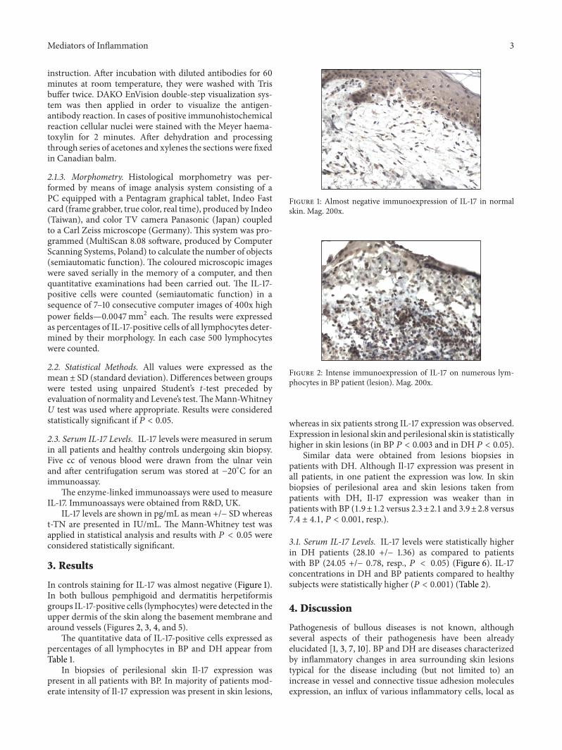
In controls staining for IL-17 was almost negative (Figure 1).In both bullous pemphigoid and dermatitis herpetiformisgroups IL-17-positive cells (lymphocytes) were detected in theupper dermis of the skin along the basement membrane andaround vessels (Figures 2, 3, 4, and 5).
The quantitative data of IL-17-positive cells expressed aspercentages of all lymphocytes in BP and DH appear fromTable 1.
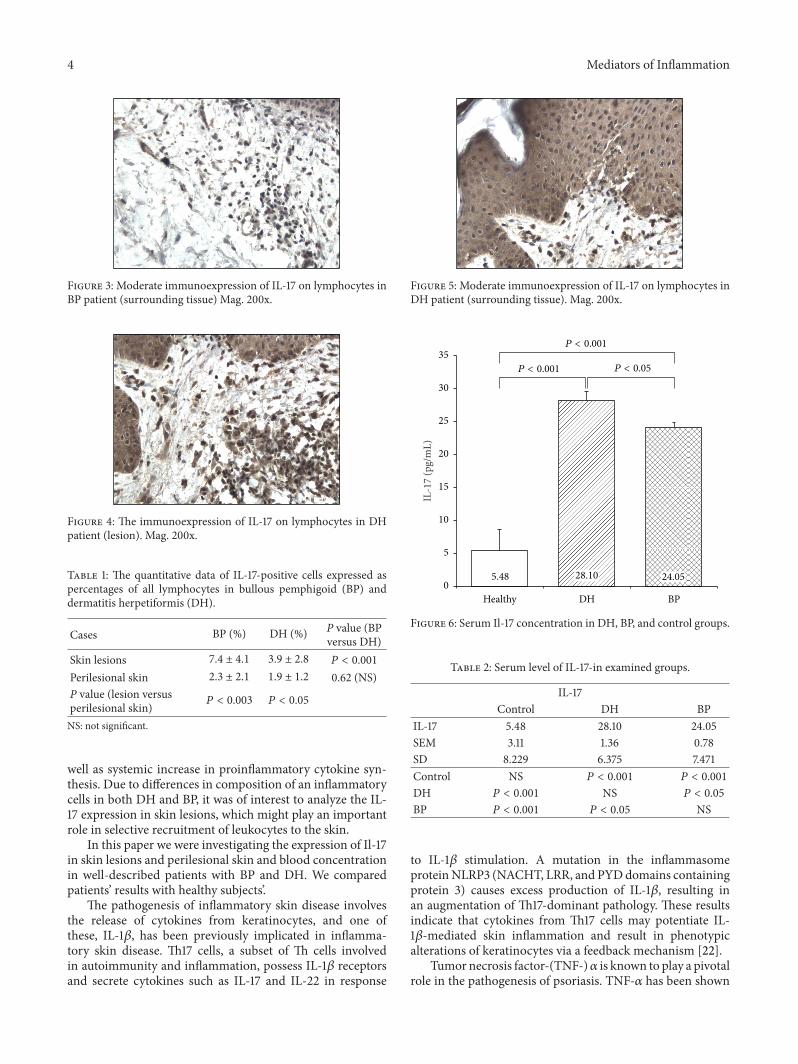
In biopsies of perilesional skin Il-17 expression waspresent in all patients with BP. In majority of patients mod-erate intensity of Il-17 expression was present in skin lesions,
Figure 1: Almost negative immunoexpression of IL-17 in normalskin. Mag. 200x.
Figure 2: Intense immunoexpression of IL-17 on numerous lym-phocytes in BP patient (lesion). Mag. 200x.
whereas in six patients strong IL-17 expression was observed.Expression in lesional skin and perilesional skin is statisticallyhigher in skin lesions (in BP 𝑃 < 0.003 and in DH 𝑃 < 0.05).
Similar data were obtained from lesions biopsies inpatients with DH. Although Il-17 expression was present inall patients, in one patient the expression was low. In skinbiopsies of perilesional area and skin lesions taken frompatients with DH, Il-17 expression was weaker than inpatients with BP (1.9±1.2 versus 2.3±2.1 and 3.9±2.8 versus7.4 ± 4.1, 𝑃 < 0.001, resp.).
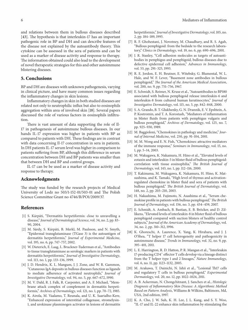
3.1. Serum IL-17 Levels. IL-17 levels were statistically higherin DH patients (28.10 +/− 1.36) as compared to patientswith BP (24.05 +/− 0.78, resp., 𝑃 < 0.05) (Figure 6). IL-17concentrations in DH and BP patients compared to healthysubjects were statistically higher (𝑃 < 0.001) (Table 2).
4. Discussion
Pathogenesis of bullous diseases is not known, althoughseveral aspects of their pathogenesis have been alreadyelucidated [1, 3, 7, 10]. BP and DH are diseases characterizedby inflammatory changes in area surrounding skin lesionstypical for the disease including (but not limited to) anincrease in vessel and connective tissue adhesion moleculesexpression, an influx of various inflammatory cells, local as
4 Mediators of Inflammation
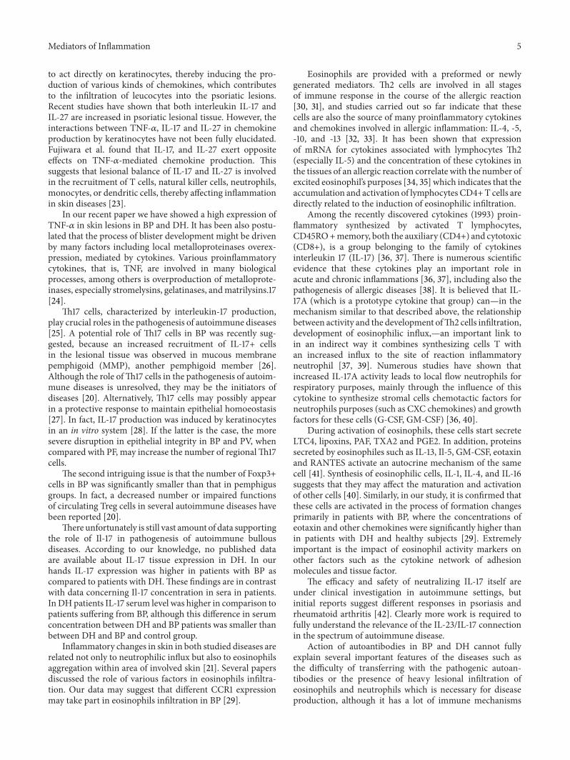
Figure 3: Moderate immunoexpression of IL-17 on lymphocytes inBP patient (surrounding tissue) Mag. 200x.
Figure 4: The immunoexpression of IL-17 on lymphocytes in DHpatient (lesion). Mag. 200x.
Table 1: The quantitative data of IL-17-positive cells expressed aspercentages of all lymphocytes in bullous pemphigoid (BP) anddermatitis herpetiformis (DH).
Cases BP (%) DH (%) P value (BPversus DH)
Skin lesions 7.4 ± 4.1 3.9 ± 2.8 𝑃 < 0.001
Perilesional skin 2.3 ± 2.1 1.9 ± 1.2 0.62 (NS)P value (lesion versusperilesional skin) 𝑃 < 0.003 𝑃 < 0.05
NS: not significant.
well as systemic increase in proinflammatory cytokine syn-thesis. Due to differences in composition of an inflammatorycells in both DH and BP, it was of interest to analyze the IL-17 expression in skin lesions, which might play an importantrole in selective recruitment of leukocytes to the skin.
In this paper we were investigating the expression of Il-17in skin lesions and perilesional skin and blood concentrationin well-described patients with BP and DH. We comparedpatients’ results with healthy subjects’.
The pathogenesis of inflammatory skin disease involvesthe release of cytokines from keratinocytes, and one ofthese, IL-1𝛽, has been previously implicated in inflamma-tory skin disease. Th17 cells, a subset of Th cells involvedin autoimmunity and inflammation, possess IL-1𝛽 receptorsand secrete cytokines such as IL-17 and IL-22 in response
Figure 5: Moderate immunoexpression of IL-17 on lymphocytes inDH patient (surrounding tissue). Mag. 200x.
5.48 28.10 24.050
5
10
15
20
25
30
35
Healthy DH BP
IL-1
7 (p
g/m
L)
P < 0.001
P < 0.05P < 0.001
Figure 6: Serum Il-17 concentration in DH, BP, and control groups.
Table 2: Serum level of IL-17-in examined groups.
IL-17Control DH BP
IL-17 5.48 28.10 24.05SEM 3.11 1.36 0.78SD 8.229 6.375 7.471Control NS 𝑃 < 0.001 𝑃 < 0.001
DH 𝑃 < 0.001 NS 𝑃 < 0.05
BP 𝑃 < 0.001 𝑃 < 0.05 NS
to IL-1𝛽 stimulation. A mutation in the inflammasomeproteinNLRP3 (NACHT, LRR, and PYDdomains containingprotein 3) causes excess production of IL-1𝛽, resulting inan augmentation of Th17-dominant pathology. These resultsindicate that cytokines from Th17 cells may potentiate IL-1𝛽-mediated skin inflammation and result in phenotypicalterations of keratinocytes via a feedback mechanism [22].
Tumor necrosis factor-(TNF-)𝛼 is known to play a pivotalrole in the pathogenesis of psoriasis. TNF-𝛼 has been shown
Mediators of Inflammation 5
to act directly on keratinocytes, thereby inducing the pro-duction of various kinds of chemokines, which contributesto the infiltration of leucocytes into the psoriatic lesions.Recent studies have shown that both interleukin IL-17 andIL-27 are increased in psoriatic lesional tissue. However, theinteractions between TNF-𝛼, IL-17 and IL-27 in chemokineproduction by keratinocytes have not been fully elucidated.Fujiwara et al. found that IL-17, and IL-27 exert oppositeeffects on TNF-𝛼-mediated chemokine production. Thissuggests that lesional balance of IL-17 and IL-27 is involvedin the recruitment of T cells, natural killer cells, neutrophils,monocytes, or dendritic cells, thereby affecting inflammationin skin diseases [23].
In our recent paper we have showed a high expression ofTNF-𝛼 in skin lesions in BP and DH. It has been also postu-lated that the process of blister development might be drivenby many factors including local metalloproteinases overex-pression, mediated by cytokines. Various proinflammatorycytokines, that is, TNF, are involved in many biologicalprocesses, among others is overproduction of metalloprote-inases, especially stromelysins, gelatinases, andmatrilysins.17[24].
Th17 cells, characterized by interleukin-17 production,play crucial roles in the pathogenesis of autoimmune diseases[25]. A potential role of Th17 cells in BP was recently sug-gested, because an increased recruitment of IL-17+ cellsin the lesional tissue was observed in mucous membranepemphigoid (MMP), another pemphigoid member [26].Although the role ofTh17 cells in the pathogenesis of autoim-mune diseases is unresolved, they may be the initiators ofdiseases [20]. Alternatively, Th17 cells may possibly appearin a protective response to maintain epithelial homoeostasis[27]. In fact, IL-17 production was induced by keratinocytesin an in vitro system [28]. If the latter is the case, the moresevere disruption in epithelial integrity in BP and PV, whencompared with PF, may increase the number of regionalTh17cells.
The second intriguing issue is that the number of Foxp3+cells in BP was significantly smaller than that in pemphigusgroups. In fact, a decreased number or impaired functionsof circulating Treg cells in several autoimmune diseases havebeen reported [20].
There unfortunately is still vast amount of data supportingthe role of Il-17 in pathogenesis of autoimmune bullousdiseases. According to our knowledge, no published dataare available about IL-17 tissue expression in DH. In ourhands IL-17 expression was higher in patients with BP ascompared to patients with DH.These findings are in contrastwith data concerning Il-17 concentration in sera in patients.In DHpatients IL-17 serum level was higher in comparison topatients suffering from BP, although this difference in serumconcentration between DH and BP patients was smaller thanbetween DH and BP and control group.
Inflammatory changes in skin in both studied diseases arerelated not only to neutrophilic influx but also to eosinophilsaggregation within area of involved skin [21]. Several papersdiscussed the role of various factors in eosinophils infiltra-tion. Our data may suggest that different CCR1 expressionmay take part in eosinophils infiltration in BP [29].
Eosinophils are provided with a preformed or newlygenerated mediators. Th2 cells are involved in all stagesof immune response in the course of the allergic reaction[30, 31], and studies carried out so far indicate that thesecells are also the source of many proinflammatory cytokinesand chemokines involved in allergic inflammation: IL-4, -5,-10, and -13 [32, 33]. It has been shown that expressionof mRNA for cytokines associated with lymphocytes Th2(especially IL-5) and the concentration of these cytokines inthe tissues of an allergic reaction correlate with the number ofexcited eosinophil’s purposes [34, 35] which indicates that theaccumulation and activation of lymphocytes CD4+T cells aredirectly related to the induction of eosinophilic infiltration.
Among the recently discovered cytokines (1993) proin-flammatory synthesized by activated T lymphocytes,CD45RO+memory, both the auxiliary (CD4+) and cytotoxic(CD8+), is a group belonging to the family of cytokinesinterleukin 17 (IL-17) [36, 37]. There is numerous scientificevidence that these cytokines play an important role inacute and chronic inflammations [36, 37], including also thepathogenesis of allergic diseases [38]. It is believed that IL-17A (which is a prototype cytokine that group) can—in themechanism similar to that described above, the relationshipbetween activity and the development ofTh2 cells infiltration,development of eosinophilic influx,—an important link toin an indirect way it combines synthesizing cells T withan increased influx to the site of reaction inflammatoryneutrophil [37, 39]. Numerous studies have shown thatincreased IL-17A activity leads to local flow neutrophils forrespiratory purposes, mainly through the influence of thiscytokine to synthesize stromal cells chemotactic factors forneutrophils purposes (such as CXC chemokines) and growthfactors for these cells (G-CSF, GM-CSF) [36, 40].
During activation of eosinophils, these cells start secreteLTC4, lipoxins, PAF, TXA2 and PGE2. In addition, proteinssecreted by eosinophiles such as IL-13, Il-5, GM-CSF, eotaxinand RANTES activate an autocrine mechanism of the samecell [41]. Synthesis of eosinophilic cells, IL-1, IL-4, and IL-16suggests that they may affect the maturation and activationof other cells [40]. Similarly, in our study, it is confirmed thatthese cells are activated in the process of formation changesprimarily in patients with BP, where the concentrations ofeotaxin and other chemokines were significantly higher thanin patients with DH and healthy subjects [29]. Extremelyimportant is the impact of eosinophil activity markers onother factors such as the cytokine network of adhesionmolecules and tissue factor.
The efficacy and safety of neutralizing IL-17 itself areunder clinical investigation in autoimmune settings, butinitial reports suggest different responses in psoriasis andrheumatoid arthritis [42]. Clearly more work is required tofully understand the relevance of the IL-23/IL-17 connectionin the spectrum of autoimmune disease.
Action of autoantibodies in BP and DH cannot fullyexplain several important features of the diseases such asthe difficulty of transferring with the pathogenic autoan-tibodies or the presence of heavy lesional infiltration ofeosinophils and neutrophils which is necessary for diseaseproduction, although it has a lot of immune mechanisms
6 Mediators of Inflammation
and relations between them in bullous diseases described[43]. The hypothesis is that interleukin-17 has an importantpathogenic role in BP and DH and can describe features ofthe disease not explained by the autoantibody theory. Thiscytokine can be assessed in the sera of patients and can beused as a marker of disease activity and response to therapy.The information obtained could also lead to the developmentof novel therapeutic strategies for this and other autoimmuneblistering diseases.
5. Conclusions
BP andDH are diseases with unknown pathogenesis, varyingin clinical picture, and have many common issues regardingthe inflammatory changes in skin lesions.
Inflammatory changes in skin in both studied diseases arerelated not only to neutrophilic influx but also to eosinophilsaggregation within area of involved skin [18]. Several papersdiscussed the role of various factors in eosinophils infiltra-tion.
There is vast amount of data supporting the role of Il-17 in pathogenesis of autoimmune bullous diseases. In ourhands IL-17 expression was higher in patients with BP ascompared to patients with DH.These findings are in contrastwith data concerning Il-17 concentration in sera in patients.In DHpatients IL-17 serum level was higher in comparison topatients suffering from BP, although this difference in serumconcentration between DH and BP patients was smaller thanthat between DH and BP and control groups.
IL-17 can be be used as a marker of disease activity andresponse to therapy.
Acknowledgments
The study was funded by the research projects of MedicalUniversity of Lodz no 503/1-152-01/503-01 and The PolishScience Committee Grant no 4746/B/PO1/2009/37.
References
[1] S. Karpati, “Dermatitis herpetiformis: close to unravelling adisease,” Journal of Dermatological Science, vol. 34, no. 2, pp. 83–90, 2004.
[2] M. Sardy, S. Karpati, B. Merkl, M. Paulsson, and N. Smyth,“Epidermal transglutaminase (TGase 3) is the autoantigen ofdermatitis herpetiformis,” Journal of Experimental Medicine,vol. 195, no. 6, pp. 747–757, 2002.
[3] W. Dieterich, E. Laag, L. Bruckner-Tuderman et al., “Antibodiesto tissue transglutaminase as serologic markers in patients withdermatitis herpetiformis,” Journal of Investigative Dermatology,vol. 113, no. 1, pp. 133–136, 1999.
[4] J. D. Hendrix, K. L. Mangum, J. J. Zone, and W. R. Gammon,“Cutaneous IgA deposits in bullous diseases function as ligandsto mediate adherence of activated neutrophils,” Journal ofInvestigative Dermatology, vol. 94, no. 5, pp. 667–672, 1990.
[5] M. V. Dahl, R. J. Falk, R. Carpenter, and A. F. Michael, “Mem-brane attack complex of complement in dermatitis herpeti-formis,” Archives of Dermatology, vol. 121, no. 1, pp. 70–72, 1985.
[6] K. Airola, M. Vaalamo, T. Reunala, and U. K. Saarialho-Kere,“Enhanced expression of interstitial collagenase, stromelysin-1, and urokinase plasminogen activator in lesions of dermatitis
herpetiformis,” Journal of InvestigativeDermatology, vol. 105, no.2, pp. 184–189, 1995.
[7] R. F. Ghohestani, J. Novotney, M. Chaudhary, and R. S. Agah,“Bullous pemphigoid: from the bedside to the research labora-tory,” Clinics in Dermatology, vol. 19, no. 6, pp. 690–696, 2001.
[8] J. R. Stanley, “Cell adhesion molecules as targets of autoanti-bodies in pemphigus and pemphigoid, bullous diseases due todefective epidermal cell adhesion,” Advances in Immunology,vol. 53, pp. 291–325, 1993.
[9] R. E. Jordon, E. H. Beutner, E. Witebsky, G. Blumental, W. L.Hale, and W. F. Lever, “Basement zone antibodies in bullouspemphigoid,” The Journal of the American Medical Association,vol. 200, no. 9, pp. 751–756, 1967.
[10] E. Schmidt, S. Reimer, N. Kruse et al., “Autoantibodies to BP180associated with bullous pemphigoid release interleukin-6 andinterleukin-8 from cultured human keratinocytes,” Journal ofInvestigative Dermatology, vol. 115, no. 5, pp. 842–848, 2000.
[11] S. A. Grando, B. T. Glukhenky, G. N. Drannik, E. V. Epshtein, A.P. Kostromin, and T. A. Korostash, “Mediators of inflammationin blister fluids from patients with pemphigus vulgaris andbullous pemphigoid,” Archives of Dermatology, vol. 125, no. 7,pp. 925–930, 1989.
[12] M. Baggioloni, “Chemokines in pathology and medicine,” Jour-nal of Internal Medicine, vol. 250, pp. 91–104, 2001.
[13] M. M.Wong and E. N. Fish, “Chemokines: attractive mediatorsof the immune response,” Seminars in Immunology, vol. 15, no.1, pp. 5–14, 2003.
[14] M. Wakugawa, K. Nakamura, H. Hino et al., “Elevated levels ofeotaxin and interleukin-5 in blister fluid of bullous pemphigoid:correlation with tissue eosinophilia,” The British Journal ofDermatology, vol. 143, no. 1, pp. 112–116, 2000.
[15] T. Kakinuma, M. Wakugawa, K. Nakamura, H. Hino, K. Mat-sushima, and K. Tamaki, “High level of thymus and activation-regulated chemokine in blister fluid and sera of patients withbullous pemphigoid,” The British Journal of Dermatology, vol.148, no. 2, pp. 203–210, 2003.
[16] H. Nakashima, M. Fujimoto, N. Asashima et al., “Serum che-mokine profile in patients with bullous pemphigoid,”TheBritishJournal of Dermatology, vol. 156, no. 3, pp. 454–459, 2007.
[17] E. Schmidt, A. Ambach, B. Bastian, E. B. Brocker, and D. Zil-likens, “Elevated levels of interleukin-8 in blister fluid of bullouspemphigoid compared with suction blisters of healthy controlsubjects,” Journal of the American Academy of Dermatology, vol.34, no. 2, pp. 310–312, 1996.
[18] K. Ghoreschi, A. Laurence, X. Yang, K. Hirahara, and J. J.O’Shea, “T helper 17 cell heterogeneity and pathogenicity inautoimmune disease,” Trends in Immunology, vol. 32, no. 9, pp.395–401, 2011.
[19] L. E. Harrington, R. D. Hatton, P. R. Mangan et al., “Interleukin17-producing CD4+ effector T cells develop via a lineage distinctfrom the T helper type 1 and 2 lineages,” Nature Immunology,vol. 6, no. 11, pp. 1123–1132, 2005.
[20] M. Arakawa, T. Dainichi, N. Ishii et al., “Lesional Th17 cellsand regulatory T cells in bullous pemphigoid,” ExperimentalDermatology, vol. 20, no. 12, pp. 1022–1024, 2011.
[21] A. B. Ackerman, N. Chongchitnant, J. Sanchex et al., HistologicDiagnosis of Inflammatory Skin Disease: A Algorithmic MethodBased on Pattern Analysis, Williams &Wilkins, Baltimore, Md,USA, 2nd edition, 1997.
[22] K. A. Cho, J. W. Suh, K. H. Lee, J. L. Kang, and S. Y. Woo,“IL-17 and IL-22 enhance skin inflammation by stimulating the
Mediators of Inflammation 7
secretion of il-1𝛽 by keratinocytes via the ROS-NLRP3-caspase-1 pathway,” International Immunology, vol. 24, no. 3, pp. 147–158,2012.
[23] S. Fujiwara, H. Nagai, S. Oniki, T. Yoshimoto, and C. Nishigori,“Interleukin (IL)-17 versus IL-27: opposite effects on tumornecrosis factor-𝛼-mediated chemokine production in humankeratinocytes,” Experimental Dermatology, vol. 21, no. 1, pp. 70–72, 2012.
[24] A. I. Oikarinen, T. Reunala, and J. J. Zone, “Proteolytic enzymesin blister fluids from patients with dermatitis herpetiformis,”The British Journal of Dermatology, vol. 114, no. 3, pp. 295–302,1986.
[25] E. Bettelli, M. Oukka, and V. K. Kuchroo, “TH-17 cells in thecircle of immunity and autoimmunity,”Nature Immunology, vol.8, no. 4, pp. 345–350, 2007.
[26] A.M. Suelves, T. Z. Zhao, S. S. Siddique, andC. S. Foster, “Profileof local interleukin expression in a cohort of ocular cicatricialpemphigoid patients,” Investigative Ophthalmology and VisualScience, vol. 53, no. 13, pp. 8112–8117, 2012.
[27] L. Hammerich, F. Heymann, and F. Tacke, “Role of IL-17and Th17 cells in liver diseases,” Clinical and DevelopmentalImmunology, vol. 2011, Article ID 345803, 12 pages, 2011.
[28] F. X. Bernard, F. Morel, M. Camus et al., “Keratinocytes underfire of proinflammatory cytokines: bona fide innate immunecells involved in the physiopathology of chronic atopic dermati-tis and psoriasis,” Journal of Allergy, vol. 2012, Article ID 718725,10 pages, 2012.
[29] A. Zebrowska, A. Erkiert-Polguj, M. Wągrowska-Danilewicz etal., “Eotaxin (CCL11), TARC [thymus and activation-regulatedchemokine (CCL17)], MCP-1 (CCL2) and CCR-1, CXCR-1,CXCR-2 expression in dermatitis herpetiformis and bullouspemphigoid,” Archives of Medical Science, vol. 5, no. 3, pp. 475–485, 2009.
[30] M. Akdis, K. Blaser, and C. A. Akdis, “T regulatory cells inallergy: novel concepts in the pathogenesis, prevention, andtreatment of allergic diseases,” Journal of Allergy and ClinicalImmunology, vol. 116, no. 5, pp. 961–968, 2005.
[31] M. Akdis, K. Blaser, and C. A. Akdis, “T regulatory cells inallergy,” Chemical Immunology and Allergy, vol. 91, pp. 159–173,2006.
[32] P. S. Foster, M. Martinez-Moczygemba, D. P. Huston, and D. B.Corry, “Interleukins-4, -5, and -13: emerging therapeutic targetsin allergic disease,” Pharmacology andTherapeutics, vol. 94, no.3, pp. 253–264, 2002.
[33] A. P. Kaplan, “Chemokines, chemokine receptors and allergy,”International Archives of Allergy and Immunology, vol. 124, no.4, pp. 423–431, 2001.
[34] M. Benson, I. Strannegard,G.Wennergren, and O. Strannegard,“Low levels of interferon-𝛾 in nasal fluid accompany raisedlevels of T-helper 2 cytokines in children with ongoing allergicrhinitis,” Pediatric Allergy and Immunology, vol. 11, no. 1, pp. 20–28, 2000.
[35] A. KleinJan, M. D. Dijkstra, S. S. Boks, L. A. Severijnen, P. G.H. Mulder, and W. J. Fokkens, “Increase in IL-8, IL-10, IL-13,and RANTES mRNA levels (in situ hybridization) in the nasalmucosa after nasal allergen provocation,” Journal of Allergy andClinical Immunology, vol. 103, no. 3 I, pp. 441–450, 1999.
[36] M. Kawaguchi, M. Adachi, N. Oda, F. Kokubu, and S. K.Huang, “IL-17 cytokine family,” Journal of Allergy and ClinicalImmunology, vol. 114, no. 6, pp. 1265–1273, 2004.
[37] J. Witowski, K. Ksiązek, and A. Jorres, “Interleukin-17: amediator of inflammatory responses,” Cellular and MolecularLife Sciences, vol. 61, no. 5, pp. 567–579, 2004.
[38] M. R. Kim, R. Manoukian, R. Yeh et al., “Transgenic overex-pression of human IL-17E results in eosinophilia, B-lymphocytehyperplasia, and altered antibody production,” Blood, vol. 100,no. 7, pp. 2330–2340, 2002.
[39] A. Linden, “Interleukin 17 and airway remodeling,” PulmonaryPharmacology andTherapeutics, vol. 19, no. 1, pp. 47–50, 2006.
[40] M. Laan, Z. H. Cui, H. Hoshino et al., “Neutrophil recruitmentby human IL-17 via C-X-C chemokine release in the airways,”Journal of Immunology, vol. 162, no. 4, pp. 2347–2352, 1999.
[41] A. KleinJan, A. F. Holm, M. D. Dijkstra et al., “Preventivetreatment of intranasal fluticasone propionate reduces cytokinemRNA expressing cells before and during a single nasal allergenprovocation,” Clinical and Experimental Allergy, vol. 30, no. 10,pp. 1476–1485, 2000.
[42] W. Hueber, D. D. Patel, T. Dryja et al., “Effects of AIN457, a fullyhuman antibody to interleukin-17A, on psoriasis, rheumatoidarthritis, and uveitis,” Science Translational Medicine, vol. 2, no.52, Article ID 52ra72, 2010.
[43] M. Sticherling and C. Erfurt-Berge, “Autoimmune blisteringdiseases of the skin,” Autoimmunity Reviews, vol. 11, no. 3, pp.226–230, 2012.

Submit your manuscripts athttp://www.hindawi.com
Stem CellsInternational
Hindawi Publishing Corporationhttp://www.hindawi.com Volume 2014
Hindawi Publishing Corporationhttp://www.hindawi.com Volume 2014
MEDIATORSINFLAMMATION
of
Hindawi Publishing Corporationhttp://www.hindawi.com Volume 2014
Behavioural Neurology
EndocrinologyInternational Journal of
Hindawi Publishing Corporationhttp://www.hindawi.com Volume 2014
Hindawi Publishing Corporationhttp://www.hindawi.com Volume 2014
Disease Markers
Hindawi Publishing Corporationhttp://www.hindawi.com Volume 2014
BioMed Research International
OncologyJournal of
Hindawi Publishing Corporationhttp://www.hindawi.com Volume 2014
Hindawi Publishing Corporationhttp://www.hindawi.com Volume 2014
Oxidative Medicine and Cellular Longevity
Hindawi Publishing Corporationhttp://www.hindawi.com Volume 2014
PPAR Research
The Scientific World JournalHindawi Publishing Corporation http://www.hindawi.com Volume 2014
Immunology ResearchHindawi Publishing Corporationhttp://www.hindawi.com Volume 2014
Journal of
ObesityJournal of
Hindawi Publishing Corporationhttp://www.hindawi.com Volume 2014
Hindawi Publishing Corporationhttp://www.hindawi.com Volume 2014
Computational and Mathematical Methods in Medicine
OphthalmologyJournal of
Hindawi Publishing Corporationhttp://www.hindawi.com Volume 2014
Diabetes ResearchJournal of
Hindawi Publishing Corporationhttp://www.hindawi.com Volume 2014
Hindawi Publishing Corporationhttp://www.hindawi.com Volume 2014
Research and TreatmentAIDS
Hindawi Publishing Corporationhttp://www.hindawi.com Volume 2014
Gastroenterology Research and Practice
Hindawi Publishing Corporationhttp://www.hindawi.com Volume 2014
Parkinson’s Disease
Evidence-Based Complementary and Alternative Medicine
Volume 2014Hindawi Publishing Corporationhttp://www.hindawi.com