Embed Size (px)
Citation preview
of December 19, 2018.This information is current as
Pathogenic AntigenPemphigoid Targeting Humanized A Novel Active Mouse Model for Bullous
Akiyama and Hiroshi ShimizuMoriuchi, Hongjiang Qiao, Hideki Nakamura, MasashiSawamura, Gang Wang, Yasuki Tateishi, Qiang Li, Reine Hideyuki Ujiie, Akihiko Shibaki, Wataru Nishie, Daisuke
http://www.jimmunol.org/content/184/4/2166doi: 10.4049/jimmunol.0903101January 2010;
2010; 184:2166-2174; Prepublished online 20J Immunol
Referenceshttp://www.jimmunol.org/content/184/4/2166.full#ref-list-1
, 12 of which you can access for free at: cites 48 articlesThis article
average*
4 weeks from acceptance to publicationFast Publication! •
Every submission reviewed by practicing scientistsNo Triage! •
from submission to initial decisionRapid Reviews! 30 days* •
Submit online. ?The JIWhy
Subscriptionhttp://jimmunol.org/subscription
is online at: The Journal of ImmunologyInformation about subscribing to
Permissionshttp://www.aai.org/About/Publications/JI/copyright.htmlSubmit copyright permission requests at:
Email Alertshttp://jimmunol.org/alertsReceive free email-alerts when new articles cite this article. Sign up at:
Print ISSN: 0022-1767 Online ISSN: 1550-6606. Immunologists, Inc. All rights reserved.Copyright © 2010 by The American Association of1451 Rockville Pike, Suite 650, Rockville, MD 20852The American Association of Immunologists, Inc.,
is published twice each month byThe Journal of Immunology
by guest on Decem
ber 19, 2018http://w
ww
.jimm
unol.org/D
ownloaded from
by guest on D
ecember 19, 2018
http://ww
w.jim
munol.org/
Dow
nloaded from
The Journal of Immunology
A Novel Active Mouse Model for Bullous PemphigoidTargeting Humanized Pathogenic Antigen
Hideyuki Ujiie, Akihiko Shibaki, Wataru Nishie, Daisuke Sawamura, Gang Wang,
Yasuki Tateishi, Qiang Li, Reine Moriuchi, Hongjiang Qiao, Hideki Nakamura,
Masashi Akiyama, and Hiroshi Shimizu
Bullous pemphigoid (BP), the most common autoimmune blistering disease, is caused by autoantibodies against type XVII collagen
(COL17). To establish an active stable BP animal model that demonstrates the persistent inflammatory skin lesions initiated by the
anti-human COL17 Abs, we used COL17-humanized (COL17m2/2,h+) mice that we recently produced. First, we generated immu-
nodeficient Rag-22/2/COL17–humanized mice by crossing Rag-22/2 mice with COL17-humanized mice. Then, splenocytes from
wild-type mice that had been immunized by grafting of human COL17-transgenic mouse skin were transferred into Rag-22/2
/COL17–humanized mice. The recipient mice continuously produced anti-human COL17 IgG Abs in vivo and developed blisters
and erosions corresponding to clinical, histological, and immunopathological features of BP, although eosinophil infiltration, one of
the characteristic histological findings observed in BP patients, was not detected in the recipients. Although the depletion of CD8+
T cells from the immunized splenocytes was found to produce no effects in the recipients, the depletion of CD4+ T cells as well as
CD45R+ B cells was found to inhibit the production of anti-humanCOL17 IgGAbs in the recipients, resulting in no apparent clinical
phenotype. Furthermore, we demonstrated that cyclosporin A significantly suppressed the production of anti-human COL17 IgG
Abs and prevented the development of the BP phenotype in the treated recipients. Although this model in an immunodeficient mouse
does not exactly reproduce the inductionmechanism of BP in human patients, this unique experimental system targeting humanized
pathogenic Ag allows us to investigate ongoing autoimmune responses to human molecules in experimental animal models. The
Journal of Immunology, 2010, 184: 2166–2174.
To investigate the pathogenic mechanisms of autoimmunediseases, thedevelopmentofanimalmodels isessential (1,2).However, interspeciesmolecular differences in autoantigens
make it difficult to develop autoimmune animal disease models insome cases. We recently overcame this issue by using the uniquetechnique of humanization of autoantigens to generate an animalmodel for bullous pemphigoid (BP) (1).BP is the most common autoimmune blistering disorder that is
induced by autoantibodies against type XVII collagen (COL17,also called BP180 or BPAG2), a hemidesmosomal type II trans-membrane protein that spans the lamina lucida and projects into thelamina densa of the epidermal basement membrane zone (3–7).The noncollagenous 16A domain located at the membrane-prox-imal region of COL17 is known as the major pathogenic epitopefor BP (8, 9). Our group recently generated COL17-humanized
mice (COL17m2/2,h+) that lack mouse COL17 but express humanCOL17 (hCOL17) (1). Autoantibodies from BP patients fail torecognize mouse COL17 due to differences in the amino acidsequence between human and mouse. In contrast, BP autoanti-bodies react to hCOL17 molecules expressed in COL17-human-ized mice and induce BP-like skin lesions in the neonates. Thus,this passive-transfer BP mouse model directly demonstrated thepathogenicity of human BP autoantibodies (1). Our system makesit possible to investigate immune reactions mediated by Absspecific to human molecules even in animal models.However, passive-transfer animal models demonstrate only
transient disease activity. In this study, we have developed an active,stable BP model to further advance our knowledge of the BP path-ogenic mechanisms for the dynamic process of developing chronicinflammatory skin lesions observed in BP patients. To develop sucha model, we adoptively transferred the splenocytes immunized withhCOL17 into immunodeficient COL17-humanized recipients (10).This active, stable autoimmune disease model targeting humanizedpathogenic Ag enables us to investigate ongoing autoimmune re-sponses to human molecules in experimental animals.
Materials and MethodsMice
C57BL/6J mice were purchased from Japan Clea (Hamamatsu, Japan).C57BL/6-background Rag-22/2 mice were received as a gift from theCentral Institute for Experimental Animals (Kawasaki, Japan). We crossedCOL17m2/2,h+ (COL17-humanized)mice that we had recently generated (1)with Rag-22/2 mice. Mice that carried the heterozygous null mutations ofboth the Rag-2 and mouse Col17 genes and the transgene of human COL17(Rag-2+/2/COL17m+/2,h+) were bred to produce Rag-22/2/COL17m2/2,h+
(Rag-22/2/COL17–humanized) mice. All of the animal procedures wereconducted according to guidelines provided by the Hokkaido UniversityInstitutional Animal Care and Use Committee under an approved protocol.
Department of Dermatology, Hokkaido University Graduate School of Medicine,Sapporo, Japan
Received for publication September 21, 2009. Accepted for publication December 9,2009.
This work was supported in part by Grants-in-Aid for Scientific Research (A) (No.21249063 to H.S.) and (C) (No. 20591312 to A.S.) from the Ministry of Education,Culture, Sports, Science and Technology of Japan and by the Program for Promotion ofFundamental Studies in Health Sciences of the National Institute of Biomedical Innova-tion (No. 06-42 to H.S.).
Address correspondence and reprint requests to Dr. Hiroshi Shimizu and Dr. AkihikoShibaki, Department of Dermatology, Hokkaido University Graduate School of Medicine,N.15 W.7, Kita-ku, Sapporo 060-8638, Japan. E-mail addresses: [email protected] or [email protected]
Abbreviations used in this paper: BP, bullous pemphigoid; COL17, type XVII col-lagen; CsA, cyclosporin A; DEJ, dermal–epidermal junction; hCOL17, humanCOL17; hNC16A, human COL17 noncollagenous 16A domain; IF, immunofluores-cence; LD, lamina densa; Tg, transgenic; Treg, regulatory T cell; WT, wild-type.
Copyright� 2010 by TheAmericanAssociation of Immunologists, Inc. 0022-1767/10/$16.00
www.jimmunol.org/cgi/doi/10.4049/jimmunol.0903101
by guest on Decem
ber 19, 2018http://w
ww
.jimm
unol.org/D
ownloaded from
Immunization of mice by hCOL17-transgenic skin grafting
Immunization of the mice by hCOL17-transgenic (Tg) skin graft was per-formed according to the method reported by Olasz et al. (11). Briefly, full-thickness 1-cm2 pieces of dorsal skin were removed from sacrificedhCOL17-Tg mice (COL17m+/+,h+) and grafted onto the backs of gender-matched 6-wk-old C57BL/6 wild-type (WT) mice. After topical applicationof antibiotic ointment, the grafted site was covered with gauze and an elasticbandage for 14 d. In selected experiments, WTmouse skin was grafted ontothe backs of WT mice. Ab production was confirmed at 5 wk after skingrafting by indirect immunofluorescence (IF) analysis, as described below.
IF analysis
Indirect IF using mice sera was performed on the skin samples from human,COL17-humanizedmice, orWTmice using standard protocols (1). In selectedexperiments, indirect IFwas performedon 1MNaCl-split normal human skin.We used FITC-conjugatedAbs against mouse IgG (Jackson ImmunoResearchLaboratories, West Grove, PA), mouse IgG1, IgG2a, IgG2b, and IgG3 (BDPharmingen, San Diego, CA), and mouse IgG2c (Bethyl Laboratories,Montgomery, TX) as the secondary Abs.
ELISA
To determine the Ab titer against hCOL17 noncollagenous 16A domain(hNC16A) in the serum samples from the experimental mice, 96-wellmicrotiter plates coated with recombinant hNC16A protein purchased fromMedical & Biological Laboratories (Nagoya, Japan) were incubated withdiluted mouse sera for 1 h at room temperature. After being washed, boundAbs were developed with a 40,000-fold–diluted, HRP-labeled Ab specific tomouse IgG (Jackson ImmunoResearch Laboratories), and the OD was readat 450 nm using an ELISA plate reader (Mithras; Berthold Technologies,Bad Wildbad, Germany). The ELISA index value was defined by the fol-lowing formula: index = (OD450 of tested serum – OD450 of negative con-trol)/(OD450 of positive control – OD450 of negative control) 3 100 (10).
Immunoblotting
Immunoblotting was performed as described previously (1). Recombinantproteins were subjected to SDS-PAGE and electrotransferred onto nitro-cellulose membrane (Trans-Blot; Bio-Rad, Hercules, CA). The membraneswere blocked and incubated at room temperature for 1 h with diluted seraobtained from experimental mice. After being washed, the membraneswere incubated with alkaline phosphatase-conjugated anti-mouse IgG(Zymed Laboratories, South San Francisco, CA). The bound Abs weredetected by the Western Blue Stabilized Substrate for Alkaline Phospha-tase (Promega, Madison, WI).
Complement fixation study
ComplementactivationinducedbyAbsobtainedfromtheimmunizedWTmiceagainst the COL17 in human skin samples was investigated by IFmicroscopyaspreviouslydescribed,withminormodifications (12).Cryosectionsof humanskin were incubated with IgG (10mg/ml) from immunizedWTmice for 1 h at37˚C. Freshly preparedmouse serumwas then added as a complement source.One hour after incubation, in situ deposition of mouse complement C3 wasdetected with FITC-conjugated Abs specific to mouse C3 (Cappel; ValeantPharmaceuticals, Costa Mesa, CA).
Adoptive transfer of splenocytes
Splenocytes were isolated and pooled from several immunized WT mice at35dafter the skingraftingandadministered toRag-22/2/COL17–humanizedor Rag-22/2 mice by i.v. injection into the tail vein with 2.0 3 108 spleno-cytes in 500 ml PBS per mouse (10).
ELISPOT assay
ELISPOT assay was performed as previously described (10, 13) with someminor modifications. Polyvinylidene difluoride-bottomed 96-well multi-screen plates (Millipore, Bedford, MA) were coated with 30 mg/ml re-combinant hNC16A protein. In some experiments, 30 mg/ml recombinantmouse noncollagenous 14A domain protein was coated as negative con-trols. Mononuclear cells isolated from the spleen, bone marrow, and lymphnodes of the Rag-22/2/COL17–humanized recipients were incubated onthe plate at 37˚C in a 5% CO2 incubator for 4 h. IgG bound to themembrane was visualized as spots with alkaline phosphatase-conjugatedanti-mouse IgG Abs. The number of spots was counted under a dissectingmicroscope (SMZ1500; Nikon, Tokyo, Japan), and the frequency of anti-hNC16A IgG-producing B cells was defined as the number of spots in 105
mononuclear cells.
Evaluation of recipient mice
Weekly, the recipient mice were examined for their general condition andcutaneous lesions (i.e., erythema, hair loss, blisters, erosions, and crusts).Extent of skin disease was scored as follows: 0, no lesions; 1, lesions on,10% of the skin surface; 2, lesions on 10–20% of the skin surface; 3,lesions on 20–40% of the skin surface; 4, lesions on 40–60% of the skinsurface; 5, lesions on .60% of the skin surface, as previously described(14) with minor modifications. Serum samples were also obtained fromrecipient mice weekly and assayed by indirect IF microscopy and hNC16AELISA. Biopsies of lesional and perilesional skin were obtained between 2and 5 wk after adoptive transfer for light microscopy (H&E), for toluidineblue staining to evaluate mast cell infiltration and degranulation, and fordirect IF using FITC-conjugated Abs against mouse IgG, IgG1, IgG2a,IgG2b, IgG2c, IgG3, and C3.
Immunoelectron microscopy
Postembedding immunoelectron microscopy of cryofixed and cryosub-stituted skin samples taken from the Rag-22/2/COL17–humanized mice at5 wk after the adoptive transfer of immunized splenocytes was performedas previously described (15, 16) with minor modifications. Small pieces offresh skin were cryofixed by plunging them into liquid propane at 2190˚Cusing a freeze–plunge apparatus (Leica Microsystems, Cambridge, U.K.).Skin samples were then cryosubstituted with methanol at 280˚C using anautomated freeze substitution system (Leica Microsystems) and embeddedin Lowicryl K11M (Ladd Research Industries, Williston, VT) at 260˚C.Ultrathin sections were incubated with 5-nm gold-labeled goat anti-mouseIgG (Biocell Laboratories, Rancho Dominguez, CA) and observed witha transmission electron microscope (H-7100; Hitachi High-Technologies,Tokyo, Japan).
Preparation of IgG fractions from mice and passive-transferstudies
Sera were obtained from Rag-22/2/COL17–humanized mice at 8 d afterthe adoptive transfer of immunized splenocytes. Total IgG was preparedfrom the sera by affinity chromatography using a HiTrap Protein G HP (GEHealthcare Biosciences, Uppsala, Sweden). We performed passive transferof IgG into mice as previously described (1). A 60-ml dose of sterile IgG inPBS was administered to neonatal COL17-humanized mice by i.p. in-jection (0.1, 0.5, or 1.0 mg/g body weight). As a control, we prepared thetotal IgG fractions from WT mice and i.p. injected them into neonatalCOL17-humanized mice (1.0 mg/g body weight). We judged skin phe-notype at 48 h after the injection. The animals were then sacrificed, andskin sections were taken for histological examination.
Depletion of CD4+ or CD8+ T cells or CD45R+ B cells fromimmunized splenocytes
For adoptive transfer of immunized splenocytes without CD4+ or CD8+ Tcells or CD45R+ B cells, we depleted each fraction from splenocytes of theimmunized WT mice by using microbeads conjugated to monoclonal anti-mouse CD4 (L3T4), anti-mouse CD8a (Ly-2), or anti-mouse CD45R (B220)Abs (Miltenyi Biotec, Auburn, CA). The depletions of CD4+ or CD8+
T cells or CD45R+ B cells were confirmed by flow cytometric analysis ona FACSAria (BD Pharmingen) as described below. Approximately 1.0 to 2.03 108 splenocytes depleted with CD4+ or CD8+ T cells or CD45R+ B cellswere adoptively transferred to Rag-22/2/COL17–humanized mice.
Administration of cyclosporin A to the BP model mice
Cyclosporin A (CsA) (Novartis Pharma, Basel, Switzerland) dissolved inolive oil was given i.p. at the dose of 35 mg/kg (100 ml) from 2 d after theadoptive transfer and continued daily for 14 d. The dosewas chosen based ona previous study (17) and our preliminary data. As a control, the samevolume of olive oil was injected into the BP model mice. The treated BPmodelmicewere observed for 5 wk to evaluate the efficacy of the treatments.
Flow cytometry
The following mAbs were purchased from BD Pharmingen: 145-2C11-FITC (anti-CD3ε), H129.19-FITC (anti-CD4), 53-6.7-PE (anti-CD8), andRA3-6B2-PE (anti-CD45R/B220). One million cells were stained andsubjected to analysis using a FACSAria.
Statistical analysis
TocompareELISA indexvaluesofAbs, theweightsofmice, and thenumbersof splenocytes, Student t tests were applied. We determined the statisticaldifferences between groups of indirect IF titer and disease severity using the
The Journal of Immunology 2167
by guest on Decem
ber 19, 2018http://w
ww
.jimm
unol.org/D
ownloaded from
Mann-Whitney U test or ANOVA with the Scheffe F test. Data were ex-pressed as mean6 SE. We considered p values of,0.05 as significant.
ResultsHigh titer of anti-hCOL17 IgG is induced in WT mice byhCOL17-Tg skin graft immunization
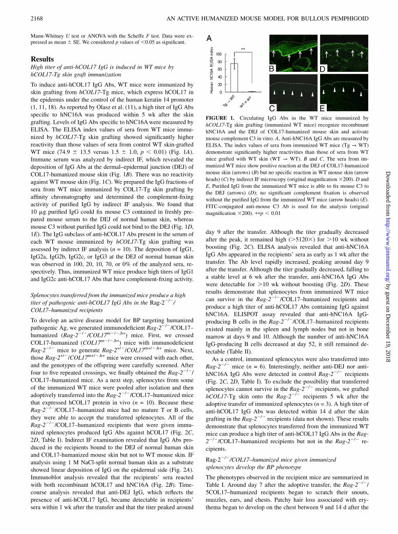
To induce anti-hCOL17 IgG Abs, WT mice were immunized byskin grafting from hCOL17-Tg mice, which express hCOL17 inthe epidermis under the control of the human keratin 14 promoter(1, 11, 18). As reported by Olasz et al. (11), a high titer of IgG Absspecific to hNC16A was produced within 5 wk after the skingrafting. Levels of IgG Abs specific to hNC16Awere measured byELISA. The ELISA index values of sera from WT mice immu-nized by hCOL17-Tg skin grafting showed significantly higherreactivity than those values of sera from control WT skin-graftedWT mice (74.9 6 13.5 versus 1.5 6 1.0, p , 0.01) (Fig. 1A).Immune serum was analyzed by indirect IF, which revealed thedeposition of IgG Abs at the dermal–epidermal junction (DEJ) ofCOL17-humanized mouse skin (Fig. 1B). There was no reactivityagainst WT mouse skin (Fig. 1C). We prepared the IgG fractions ofsera from WT mice immunized by COL17-Tg skin grafting byaffinity chromatography and determined the complement-fixingactivity of purified IgG by indirect IF analysis. We found that10 mg purified IgG could fix mouse C3 contained in freshly pre-pared mouse serum to the DEJ of normal human skin, whereasmouse C3 without purified IgG could not bind to the DEJ (Fig. 1D,1E). The IgG subclass of anti-hCOL17 Abs present in the serum ofeach WT mouse immunized by hCOL17-Tg skin grafting wasassessed by indirect IF analysis (n = 10). The deposition of IgG1,IgG2a, IgG2b, IgG2c, or IgG3 at the DEJ of normal human skinwas observed in 100, 20, 10, 70, or 0% of the analyzed sera, re-spectively. Thus, immunized WT mice produce high titers of IgG1and IgG2c anti-hCOL17 Abs that have complement-fixing activity.
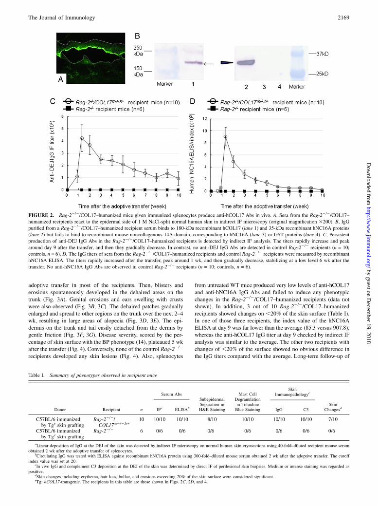
Splenocytes transferred from the immunized mice produce a hightiter of pathogenic anti-hCOL17 IgG Abs in the Rag-22/2/COL17–humanized recipients
To develop an active disease model for BP targeting humanizedpathogenic Ag, we generated immunodeficient Rag-22/2/COL17–humanized (Rag-22/2/COL17m2/2,h+) mice. First, we crossedCOL17-humanized (COL17m2/2,h+) mice with immunodeficientRag-22/2 mice to generate Rag-2+/2/COL17m+/2,h+ mice. Next,those Rag-2+/2/COL17m+/2,h+ mice were crossed with each other,and the genotypes of the offspring were carefully screened. Afterfour to five repeated crossings, we finally obtained the Rag-22/2/COL17–humanized mice. As a next step, splenocytes from someof the immunized WT mice were pooled after isolation and thenadoptively transferred into the Rag-22/2/COL17–humanized micethat expressed hCOL17 protein in vivo (n = 10). Because theseRag-22/2/COL17–humanized mice had no mature T or B cells,they were able to accept the transferred splenocytes. All of theRag-22/2/COL17–humanized recipients that were given immu-nized splenocytes produced IgG Abs against hCOL17 (Fig. 2C,2D, Table I). Indirect IF examination revealed that IgG Abs pro-duced in the recipients bound to the DEJ of normal human skinand COL17-humanized mouse skin but not to WT mouse skin. IFanalysis using 1 M NaCl-split normal human skin as a substrateshowed linear deposition of IgG on the epidermal side (Fig. 2A).Immunoblot analysis revealed that the recipients’ sera reactedwith both recombinant hCOL17 and hNC16A (Fig. 2B). Time-course analysis revealed that anti-DEJ IgG, which reflects thepresence of anti-hCOL17 IgG, became detectable in recipients’sera within 1 wk after the transfer and that the titer peaked around
day 9 after the transfer. Although the titer gradually decreasedafter the peak, it remained high (.51203) for .10 wk withoutboosting (Fig. 2C). ELISA analysis revealed that anti-hNC16AIgG Abs appeared in the recipients’ sera as early as 1 wk after thetransfer. The Ab level rapidly increased, peaking around day 9after the transfer. Although the titer gradually decreased, falling toa stable level at 6 wk after the transfer, anti-hNC16A IgG Abswere detectable for .10 wk without boosting (Fig. 2D). Theseresults demonstrate that splenocytes from immunized WT micecan survive in the Rag-22/2/COL17–humanized recipients andproduce a high titer of anti-hCOL17 Abs containing IgG againsthNC16A. ELISPOT assay revealed that anti-hNC16A IgG-producing B cells in the Rag-22/2/COL17–humanized recipientsexisted mainly in the spleen and lymph nodes but not in bonemarrow at days 9 and 10. Although the number of anti-hNC16AIgG-producing B cells decreased at day 52, it still remained de-tectable (Table II).As a control, immunized splenocytes were also transferred into
Rag-22/2 mice (n = 6). Interestingly, neither anti-DEJ nor anti-hNC16A IgG Abs were detected in control Rag-22/2 recipients(Fig. 2C, 2D, Table I). To exclude the possibility that transferredsplenocytes cannot survive in the Rag-22/2 recipients, we graftedhCOL17-Tg skin onto the Rag-22/2 recipients 5 wk after theadoptive transfer of immunized splenocytes (n = 3). A high titer ofanti-hCOL17 IgG Abs was detected within 14 d after the skingrafting in the Rag-22/2 recipients (data not shown). These resultsdemonstrate that splenocytes transferred from the immunized WTmice can produce a high titer of anti-hCOL17 IgG Abs in the Rag-22/2/COL17–humanized recipients but not in the Rag-22/2 re-cipients.
Rag-22/2/COL17–humanized mice given immunizedsplenocytes develop the BP phenotype
The phenotypes observed in the recipient mice are summarized inTable I. Around day 7 after the adoptive transfer, the Rag-22/2/5COL17–humanized recipients began to scratch their snouts,muzzles, ears, and chests. Patchy hair loss associated with ery-thema began to develop on the chest between 9 and 14 d after the
FIGURE 1. Circulating IgG Abs in the WT mice immunized by
hCOL17-Tg skin grafting (immunized WT mice) recognize recombinant
hNC16A and the DEJ of COL17-humanized mouse skin and activate
mouse complement C3 in vitro. A, Anti-hNC16A IgG Abs are measured by
ELISA. The index values of sera from immunized WT mice (Tg → WT)
demonstrate significantly higher reactivities than those of sera from WT
mice grafted with WT skin (WT → WT). B and C, The sera from im-
munized WT mice show positive reaction at the DEJ of COL17-humanized
mouse skin (arrows) (B) but no specific reaction in WT mouse skin (arrow
heads) (C) by indirect IF microscopy (original magnification3200). D and
E, Purified IgG from the immunized WT mice is able to fix mouse C3 to
the DEJ (arrows) (D); no significant complement fixation is observed
without the purified IgG from the immunized WT mice (arrow heads) (E).
FITC-conjugated anti-mouse C3 Ab is used for the analysis (original
magnification 3200). ppp , 0.01
2168 AN ACTIVE HUMANIZED MOUSE MODEL FOR BULLOUS PEMPHIGOID
by guest on Decem
ber 19, 2018http://w
ww
.jimm
unol.org/D
ownloaded from
adoptive transfer in most of the recipients. Then, blisters anderosions spontaneously developed in the dehaired areas on thetrunk (Fig. 3A). Genital erosions and ears swelling with crustswere also observed (Fig. 3B, 3C). The dehaired patches graduallyenlarged and spread to other regions on the trunk over the next 2–4wk, resulting in large areas of alopecia (Fig. 3D, 3E). The epi-dermis on the trunk and tail easily detached from the dermis bygentle friction (Fig. 3F, 3G). Disease severity, scored by the per-centage of skin surface with the BP phenotype (14), plateaued 5 wkafter the transfer (Fig. 4). Conversely, none of the control Rag-22/2
recipients developed any skin lesions (Fig. 4). Also, splenocytes
from untreated WT mice produced very low levels of anti-hCOL17and anti-hNC16A IgG Abs and failed to induce any phenotypicchanges in the Rag-22/2/COL17–humanized recipients (data notshown). In addition, 3 out of 10 Rag-22/2/COL17–humanizedrecipients showed changes on ,20% of the skin surface (Table I).In one of those three recipients, the index value of the hNC16AELISA at day 9 was far lower than the average (85.3 versus 907.8),whereas the anti-hCOL17 IgG titer at day 9 checked by indirect IFanalysis was similar to the average. The other two recipients withchanges of ,20% of the surface showed no obvious difference inthe IgG titers compared with the average. Long-term follow-up of
Table I. Summary of phenotypes observed in recipient mice
Donor Recipient n
Serum AbsSubepidermalSeparation inH&E Staining
Mast CellDegranulationin ToluidineBlue Staining
SkinImmunopathologyc
IFa ELISAb IgG C3Skin
Changesd
C57BL/6 immunizedby Tge skin grafting
Rag-22/2/COL17m2/2,h+
10 10/10 10/10 8/10 10/10 10/10 10/10 7/10
C57BL/6 immunizedby Tge skin grafting
Rag-22/2 6 0/6 0/6 0/6 0/6 0/6 0/6 0/6
aLinear deposition of IgG at the DEJ of the skin was detected by indirect IF microscopy on normal human skin cryosections using 40-fold–diluted recipient mouse serumobtained 2 wk after the adoptive transfer of splenocytes.
bCirculating IgG was tested with ELISA against recombinant hNC16A protein using 300-fold–diluted mouse serum obtained 2 wk after the adoptive transfer. The cutoffindex value was set at 20.
cIn vivo IgG and complement C3 deposition at the DEJ of the skin was determined by direct IF of perilesional skin biopsies. Medium or intense staining was regarded aspositive.
dSkin changes including erythema, hair loss, bullae, and erosions exceeding 20% of the skin surface were considered significant.eTg: hCOL17-transgenic. The recipients in this table are those shown in Figs. 2C, 2D, and 4.
FIGURE 2. Rag-22/2/COL17–humanized mice given immunized splenocytes produce anti-hCOL17 Abs in vivo. A, Sera from the Rag-22/2/COL17–
humanized recipients react to the epidermal side of 1 M NaCl-split normal human skin in indirect IF microscopy (original magnification 3200). B, IgG
purified from a Rag-22/2/COL17–humanized recipient serum binds to 180-kDa recombinant hCOL17 (lane 1) and 35-kDa recombinant hNC16A proteins
(lane 2) but fails to bind to recombinant mouse noncollagenous 14A domain, corresponding to hNC16A (lane 3) or GST proteins (lane 4). C, Persistent
production of anti-DEJ IgG Abs in the Rag-22/2/COL17–humanized recipients is detected by indirect IF analysis. The titers rapidly increase and peak
around day 9 after the transfer, and then they gradually decrease. In contrast, no anti-DEJ IgG Abs are detected in control Rag-22/2 recipients (n = 10;
controls, n = 6). D, The IgG titers of sera from the Rag-22/2/COL17–humanized recipients and control Rag-22/2 recipients were measured by recombinant
hNC16A ELISA. The titers rapidly increased after the transfer, peak around 1 wk, and then gradually decrease, stabilizing at a low level 6 wk after the
transfer. No anti-hNC16A IgG Abs are observed in control Rag-22/2 recipients (n = 10; controls, n = 6).
The Journal of Immunology 2169
by guest on Decem
ber 19, 2018http://w
ww
.jimm
unol.org/D
ownloaded from
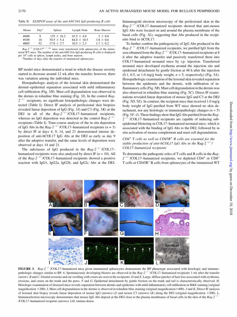
BP model mice demonstrated a trend in which the disease severitystarted to decrease around 12 wk after the transfer; however, therewas variation among the individual mice.Histopathologic analysis of the lesional skin demonstrated the
dermal–epidermal separation associated with mild inflammatorycell infiltration (Fig. 3H). Mast cell degranulation was observed inthe dermis in toluidine blue staining (Fig. 3I). In the control Rag-22/2 recipients, no significant histopathologic changes were de-tected (Table I). Direct IF analysis of perilesional skin biopsiesrevealed linear deposition of IgG (Fig. 3J) and C3 (Fig. 3K) at theDEJ in all of the Rag-22/2/COL17–humanized recipients,whereas no IgG deposition was detected in the control Rag-22/2
recipients (Table I). Time-course analysis of the in situ depositionof IgG Abs in the Rag-22/2/COL17–humanized recipients (n = 3)by direct IF at days 4, 9, 14, and 21 demonstrated intense de-position of anti-hCOL17 IgG Abs at the DEJ as early as day 9after the adoptive transfer, and the same levels of deposition wereobserved at days 14 and 21.The subclasses of IgG produced in the Rag-22/2/COL17–
humanized recipients were also analyzed by direct IF (n = 10). Allof the Rag-22/2/COL17–humanized recipients showed a positivereaction with IgG1, IgG2a, IgG2b, and IgG2c Abs at the DEJ.
Immunogold electron microscopy of the perilesional skin in theRag-22/2/COL17–humanized recipients showed that anti-mouseIgG Abs were located on and around the plasma membrane of thebasal cells (Fig. 3L), suggesting that Abs produced in the recipi-ents bind to hCOL17.To further confirm the pathogenicity of IgG Abs produced in the
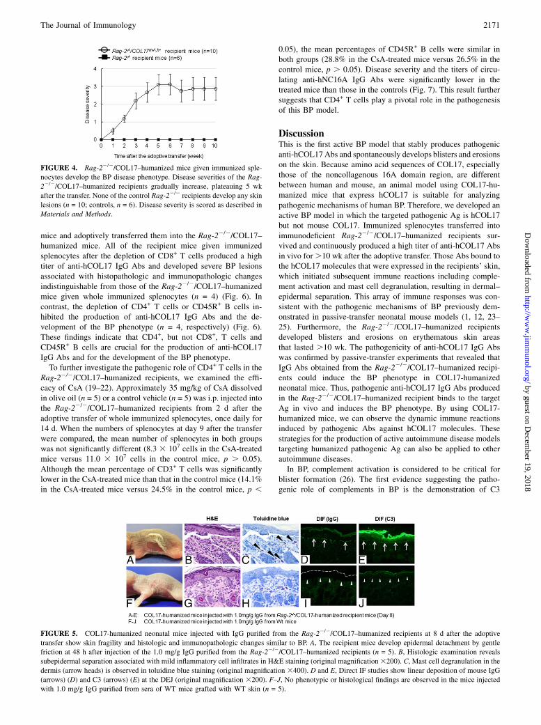
Rag-22/2/COL17–humanized recipients, we purified IgG from thesera obtained from the Rag-22/2/COL17–humanized recipients at 8d after the adoptive transfer and passively transferred them intoCOL17-humanized neonatal mice by i.p. injection. Transferredneonatal mice developed erythema around the injection site andepidermal detachment by gentle friction at 48 h after the injection(0.1, 0.5, or 1.0 mg/g body weight, n = 5, respectively) (Fig. 5A).Histopathologic examination of the lesional skin revealed separationbetween the epidermis and the dermis, with infiltration of in-flammatory cells (Fig. 5B).Mast cell degranulation in the dermiswasalso observed in toluidine blue staining (Fig. 5C). Direct IF exami-nations revealed linear deposition of mouse IgG and C3 at the DEJ(Fig. 5D, 5E). In contrast, the recipient mice that received 1.0 mg/gbody weight of IgG purified from WT mice showed no skin de-tachment, nor any histologic or immunopathologic changes (n = 5)(Fig. 5F–J). These findings show that IgGAbs purified from theRag-22/2/COL17–humanized recipients are capable of inducing sub-epidermal blistering in COL17–humanized neonatal mice, which isassociated with the binding of IgG Abs to the DEJ, followed by insitu activation of mouse complement and mast cell degranulation.
CD4+ T cells as well as CD45R+ B cells are essential for thestable production of anti-hCOL17 IgG Abs in the Rag-22/2/COL17–humanized recipients
To determine the pathogenic roles of T cells and B cells in the Rag-22/2/COL17–humanized recipients, we depleted CD4+ or CD8+
T cells or CD45R+ B cells from splenocytes of the immunized WT
Table II. ELISPOT assay of the anti-hNC16A IgG-producing B cells
Mouse Daya Spleen Lymph Node Bone Marrow
#488 9 135 6 16.2 43.5 6 4.0 2 6 0.8#109 10 70.5 6 5.4 84.0 6 10.5 3.8 6 0.6#805 52 17.0 6 2.7 10.5 6 2.3 1.7 6 0.2
Rag-22/2/COL17m2/2,h+ mice were transferred with splenocytes of the immu-nized WT mice. The number of the anti-hNC16A IgG-producing B cells is displayedper 105 cells in spleen, lymph nodes, and bone marrow.
aNumber of days after the transfer of immunized splenocytes.
FIGURE 3. Rag-22/2/COL17–humanized mice given immunized splenocytes demonstrate the BP phenotype associated with histologic and immuno-
pathologic changes similar to BP. A, Spontaneously developing blisters are observed in the Rag-22/2/COL17–humanized recipients 3 wk after the transfer
(arrow). B andC, Genital erosions and ear swelling with crusts are seen in the recipients.D and E, Large, diffuse patches of hair loss associated with erythema,
erosions, and crusts on the trunk and the paws. F and G, Epidermal detachment by gentle friction on the trunk and tail is characteristically observed. H,
Histologic examination of diseased mice reveals separation between dermis and epidermis with mild inflammatory cell infiltration in H&E staining (original
magnification3200). I, Mast cell degranulation in the dermis is observed in toluidine blue staining (original magnification3400). J and K, Direct IF analysis
of lesional skin biopsy reveals linear deposition of mouse IgG (arrows) (J) and mouse C3 (arrows) (K) along the DEJ (original magnification 3200). L,
Immunoelectron microscopy demonstrates that mouse IgG Abs deposit at the DEJ close to the plasma membranes of basal cells in the skin of the Rag-22/2
/COL17–humanized recipient (arrows). LD, lamina densa.
2170 AN ACTIVE HUMANIZED MOUSE MODEL FOR BULLOUS PEMPHIGOID
by guest on Decem
ber 19, 2018http://w
ww
.jimm
unol.org/D
ownloaded from
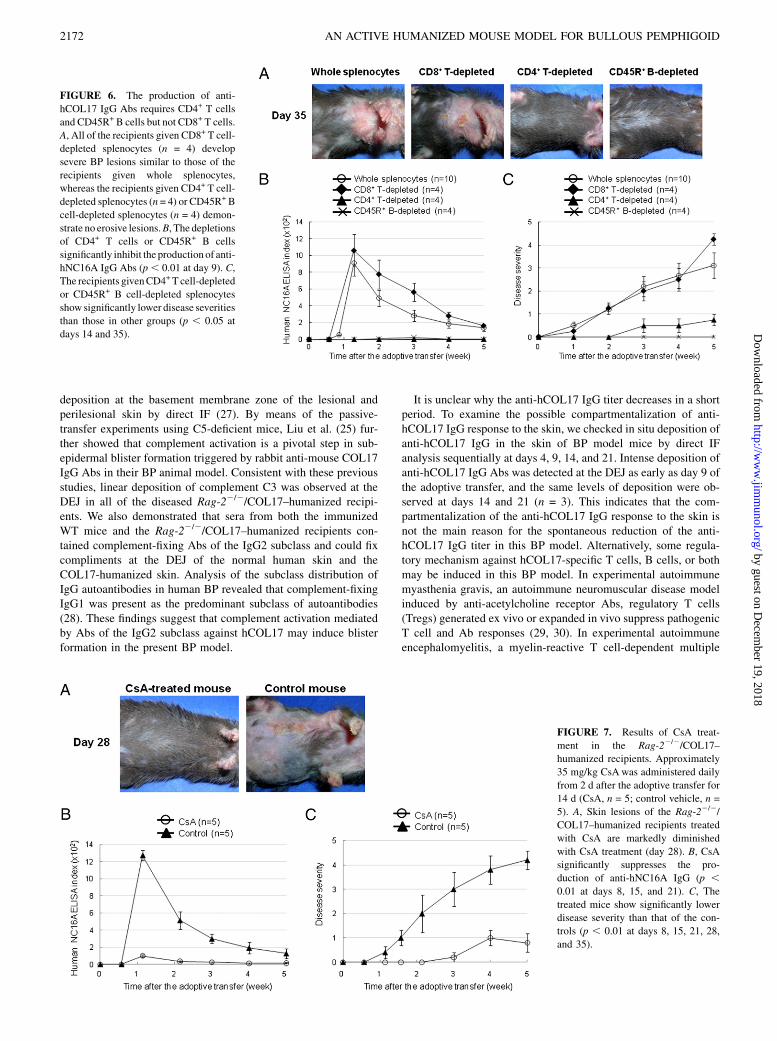
mice and adoptively transferred them into the Rag-22/2/COL17–humanized mice. All of the recipient mice given immunizedsplenocytes after the depletion of CD8+ T cells produced a hightiter of anti-hCOL17 IgG Abs and developed severe BP lesionsassociated with histopathologic and immunopathologic changesindistinguishable from those of the Rag-22/2/COL17–humanizedmice given whole immunized splenocytes (n = 4) (Fig. 6). Incontrast, the depletion of CD4+ T cells or CD45R+ B cells in-hibited the production of anti-hCOL17 IgG Abs and the de-velopment of the BP phenotype (n = 4, respectively) (Fig. 6).These findings indicate that CD4+, but not CD8+, T cells andCD45R+ B cells are crucial for the production of anti-hCOL17IgG Abs and for the development of the BP phenotype.To further investigate the pathogenic role of CD4+ T cells in the
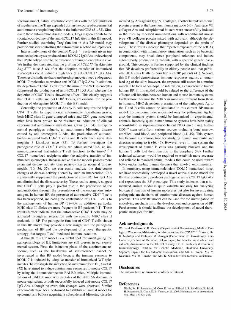
Rag-22/2/COL17–humanized recipients, we examined the effi-cacy of CsA (19–22). Approximately 35 mg/kg of CsA dissolvedin olive oil (n = 5) or a control vehicle (n = 5) was i.p. injected intothe Rag-22/2/COL17–humanized recipients from 2 d after theadoptive transfer of whole immunized splenocytes, once daily for14 d. When the numbers of splenocytes at day 9 after the transferwere compared, the mean number of splenocytes in both groupswas not significantly different (8.3 3 107 cells in the CsA-treatedmice versus 11.0 3 107 cells in the control mice, p . 0.05).Although the mean percentage of CD3+ T cells was significantlylower in the CsA-treated mice than that in the control mice (14.1%in the CsA-treated mice versus 24.5% in the control mice, p ,
0.05), the mean percentages of CD45R+ B cells were similar inboth groups (28.8% in the CsA-treated mice versus 26.5% in thecontrol mice, p . 0.05). Disease severity and the titers of circu-lating anti-hNC16A IgG Abs were significantly lower in thetreated mice than those in the controls (Fig. 7). This result furthersuggests that CD4+ T cells play a pivotal role in the pathogenesisof this BP model.
DiscussionThis is the first active BP model that stably produces pathogenicanti-hCOL17 Abs and spontaneously develops blisters and erosionson the skin. Because amino acid sequences of COL17, especiallythose of the noncollagenous 16A domain region, are differentbetween human and mouse, an animal model using COL17-hu-manized mice that express hCOL17 is suitable for analyzingpathogenic mechanisms of human BP. Therefore, we developed anactive BP model in which the targeted pathogenic Ag is hCOL17but not mouse COL17. Immunized splenocytes transferred intoimmunodeficient Rag-22/2/COL17–humanized recipients sur-vived and continuously produced a high titer of anti-hCOL17 Absin vivo for.10 wk after the adoptive transfer. Those Abs bound tothe hCOL17 molecules that were expressed in the recipients’ skin,which initiated subsequent immune reactions including comple-ment activation and mast cell degranulation, resulting in dermal–epidermal separation. This array of immune responses was con-sistent with the pathogenic mechanisms of BP previously dem-onstrated in passive-transfer neonatal mouse models (1, 12, 23–25). Furthermore, the Rag-22/2/COL17–humanized recipientsdeveloped blisters and erosions on erythematous skin areasthat lasted .10 wk. The pathogenicity of anti-hCOL17 IgG Abswas confirmed by passive-transfer experiments that revealed thatIgG Abs obtained from the Rag-22/2/COL17–humanized recipi-ents could induce the BP phenotype in COL17-humanizedneonatal mice. Thus, pathogenic anti-hCOL17 IgG Abs producedin the Rag-22/2/COL17–humanized recipient binds to the targetAg in vivo and induces the BP phenotype. By using COL17-humanized mice, we can observe the dynamic immune reactionsinduced by pathogenic Abs against hCOL17 molecules. Thesestrategies for the production of active autoimmune disease modelstargeting humanized pathogenic Ag can also be applied to otherautoimmune diseases.In BP, complement activation is considered to be critical for
blister formation (26). The first evidence suggesting the patho-genic role of complements in BP is the demonstration of C3
FIGURE 4. Rag-22/2/COL17–humanized mice given immunized sple-
nocytes develop the BP disease phenotype. Disease severities of the Rag-
22/2/COL17–humanized recipients gradually increase, plateauing 5 wk
after the transfer. None of the control Rag-22/2 recipients develop any skin
lesions (n = 10; controls, n = 6). Disease severity is scored as described in
Materials and Methods.
FIGURE 5. COL17-humanized neonatal mice injected with IgG purified from the Rag-22/2/COL17–humanized recipients at 8 d after the adoptive
transfer show skin fragility and histologic and immunopathologic changes similar to BP. A, The recipient mice develop epidermal detachment by gentle
friction at 48 h after injection of the 1.0 mg/g IgG purified from the Rag-22/2/COL17–humanized recipients (n = 5). B, Histologic examination reveals
subepidermal separation associated with mild inflammatory cell infiltrates in H&E staining (original magnification3200). C, Mast cell degranulation in the
dermis (arrow heads) is observed in toluidine blue staining (original magnification 3400). D and E, Direct IF studies show linear deposition of mouse IgG
(arrows) (D) and C3 (arrows) (E) at the DEJ (original magnification 3200). F–J, No phenotypic or histological findings are observed in the mice injected
with 1.0 mg/g IgG purified from sera of WT mice grafted with WT skin (n = 5).
The Journal of Immunology 2171
by guest on Decem
ber 19, 2018http://w
ww
.jimm
unol.org/D
ownloaded from
deposition at the basement membrane zone of the lesional andperilesional skin by direct IF (27). By means of the passive-transfer experiments using C5-deficient mice, Liu et al. (25) fur-ther showed that complement activation is a pivotal step in sub-epidermal blister formation triggered by rabbit anti-mouse COL17IgG Abs in their BP animal model. Consistent with these previousstudies, linear deposition of complement C3 was observed at theDEJ in all of the diseased Rag-22/2/COL17–humanized recipi-ents. We also demonstrated that sera from both the immunizedWT mice and the Rag-22/2/COL17–humanized recipients con-tained complement-fixing Abs of the IgG2 subclass and could fixcompliments at the DEJ of the normal human skin and theCOL17-humanized skin. Analysis of the subclass distribution ofIgG autoantibodies in human BP revealed that complement-fixingIgG1 was present as the predominant subclass of autoantibodies(28). These findings suggest that complement activation mediatedby Abs of the IgG2 subclass against hCOL17 may induce blisterformation in the present BP model.
It is unclear why the anti-hCOL17 IgG titer decreases in a shortperiod. To examine the possible compartmentalization of anti-hCOL17 IgG response to the skin, we checked in situ deposition ofanti-hCOL17 IgG in the skin of BP model mice by direct IFanalysis sequentially at days 4, 9, 14, and 21. Intense deposition ofanti-hCOL17 IgG Abs was detected at the DEJ as early as day 9 ofthe adoptive transfer, and the same levels of deposition were ob-served at days 14 and 21 (n = 3). This indicates that the com-partmentalization of the anti-hCOL17 IgG response to the skin isnot the main reason for the spontaneous reduction of the anti-hCOL17 IgG titer in this BP model. Alternatively, some regula-tory mechanism against hCOL17-specific T cells, B cells, or bothmay be induced in this BP model. In experimental autoimmunemyasthenia gravis, an autoimmune neuromuscular disease modelinduced by anti-acetylcholine receptor Abs, regulatory T cells(Tregs) generated ex vivo or expanded in vivo suppress pathogenicT cell and Ab responses (29, 30). In experimental autoimmuneencephalomyelitis, a myelin-reactive T cell-dependent multiple
FIGURE 6. The production of anti-
hCOL17 IgG Abs requires CD4+ T cells
and CD45R+ B cells but not CD8+ T cells.
A, All of the recipients given CD8+ T cell-
depleted splenocytes (n = 4) develop
severe BP lesions similar to those of the
recipients given whole splenocytes,
whereas the recipients given CD4+ T cell-
depleted splenocytes (n = 4) or CD45R+ B
cell-depleted splenocytes (n = 4) demon-
strate no erosive lesions.B, The depletions
of CD4+ T cells or CD45R+ B cells
significantly inhibit the production of anti-
hNC16A IgG Abs (p, 0.01 at day 9). C,
The recipients givenCD4+T cell-depleted
or CD45R+ B cell-depleted splenocytes
show significantly lower disease severities
than those in other groups (p , 0.05 at
days 14 and 35).
FIGURE 7. Results of CsA treat-
ment in the Rag-22/2/COL17–
humanized recipients. Approximately
35 mg/kg CsAwas administered daily
from 2 d after the adoptive transfer for
14 d (CsA, n = 5; control vehicle, n =
5). A, Skin lesions of the Rag-22/2/
COL17–humanized recipients treated
with CsA are markedly diminished
with CsA treatment (day 28). B, CsA
significantly suppresses the pro-
duction of anti-hNC16A IgG (p ,0.01 at days 8, 15, and 21). C, The
treated mice show significantly lower
disease severity than that of the con-
trols (p , 0.01 at days 8, 15, 21, 28,
and 35).
2172 AN ACTIVE HUMANIZED MOUSE MODEL FOR BULLOUS PEMPHIGOID
by guest on Decem
ber 19, 2018http://w
ww
.jimm
unol.org/D
ownloaded from
sclerosis model, natural resolution correlates with the accumulationofmyelin-reactiveTregs expandedduring the course of experimentalautoimmune encephalomyelitis in the inflamed CNS (31, 32). Sim-ilar to these autoimmunediseasemodels, Tregsmay contribute to thespontaneous decline of the anti-hCOL17 IgG titer in this BP model.Further studies examining Treg function in this BP model mayprovide clues for controlling the autoimmune reaction inBPpatients.Interestingly, none of the control Rag-22/2 recipients given im-
munized splenocytes produced anti-hCOL17 IgG Abs or developedthe BP phenotype despite the presence of living splenocytes in vivo.We further demonstrated that the grafting of hCOL17-Tg skin ontoRag-22/2 mice 5 wk after the adoptive transfer of immunizedsplenocytes could induce a high titer of anti-hCOL17 IgG Abs.These results indicate that transferred splenocytes need endogenoushCOL17 molecules to produce anti-hCOL17 IgG Abs. In addition,the depletion of CD4+ T cells from the immunized WT splenocytessuppressed the production of anti-hCOL17 IgG Abs, whereas thedepletion of CD8+ T cells showed no effects. This clearly suggeststhat CD4+ T cells, and not CD8+ T cells, are essential for the pro-duction of Abs against hCOL17 in this BP model.Generally, the production of Abs by B cells requires the help of
CD4+ T cells. In experimental autoimmune myasthenia gravis,both MHC class II gene-disrupted mice and CD4 gene knockoutmice have been proven to be resistant to induction of clinicalexperimental autoimmune myasthenia gravis (33, 34). In experi-mental pemphigus vulgaris, an autoimmune blistering diseasecaused by anti-desmoglein 3 Abs, the production of autoanti-bodies required both CD4+ T cells and B cells from naive des-moglein 3 knockout mice (35). To further investigate thepathogenic role of CD4+ T cells, we administered CsA, an im-munosuppressant that inhibits T cell function, to the Rag-22/2/COL17–humanized recipients after the adoptive transfer of im-munized splenocytes. Because active disease models possess morepersistent disease activity than passive-transfer neonatal diseasemodels (10, 36, 37), we can easily analyze the time-coursechanges of disease activity altered by such an intervention. CsAsignificantly suppressed the production of anti-hNC16A IgG Absand diminished the disease severity. These results strongly suggestthat CD4+ T cells play a pivotal role in the production of theautoantibodies through the presentation of the endogenous auto-antigen. In human BP, the presence of autoreactive CD4+ T cellshas been reported, indicating the contribution of CD4+ T cells tothe pathogenesis of human BP (38–40). In addition, particularMHC class II alleles are more frequent in BP patients (41). Theseresults further indicate that the autoreactive CD4+ T cells may beactivated through an interaction with the specific MHC class IImolecule in BP. The pathogenic function of CD4+ T cells shownin this BP model may provide a new insight into the pathogenicmechanism of BP and the development of a novel therapeuticstrategy that targets T cell-mediated immune reactions.Although this BP model is a useful tool for investigating the
pathophysiology of BP, limitations are still present in our experi-mental system. First, the induction phase of the autoimmune re-sponse, such as the breakdown of self-tolerance, cannot beinvestigated in this BP model because the immune response tohCOL17 is induced by adoptive transfer of immunized WT sple-nocytes. To investigate the induction of autoimmunity in BP, Xu et al.(42) have aimed to induce autoimmune responses to mouse COL17by using the immunocompetent BALB/c mice. Multiple immuni-zations of BALB/c mice with peptides of the hNC16A domain, itsmouse equivalent, or both successfully induced anti-mouse COL17IgG Abs, although no overt skin changes were observed. Similarexperiments have been performed to establish an animal model forepidermolysis bullosa acquisita, a subepidermal blistering disorder
induced by Abs against type VII collagen, another hemidesmosomalprotein present at the basement membrane zone (43). Anti-type VIIcollagen Abs and subepidermal blisters were successfully inducedin the mice by repeated immunizations with recombinant mousetype VII collagen protein mixed with adjuvant, although the de-velopment of the disease phenotype depended on the strain ofmice. These results indicate that repeated exposure of the self Agin conjunction with inflammatory stimulation, such as by bacterialcomponents, may break down peripheral tolerance and induceautoantibody production in patients with a specific genetic back-ground. This concept is further supported by the clinical findingsthat BP develops preferentially in elderly people and that partic-ular HLA class II alleles correlate with BP patients (41). Second,this BP model demonstrates immune responses against a human-ized Ag of the skin; however, the response still occurs in a murinemilieu. The lack of eosinophilic infiltration, a characteristic trait ofhuman BP, in this model could be related to the difference of theeffector cell function between human and mouse immune systems.Furthermore, because the MHCs in mice are different from thosein humans, MHC-dependent presentation of the pathogenic Ag tothe T and B cells cannot be simulated in this current BP mousemodel. To overcome these issues, not only the pathogenic Ag butalso the immune system should be humanized in experimentalanimals. Recently, quasi-human immune systems have been stablyreconstituted in supra-immunodeficient NOG mice using humanCD34+ stem cells from various sources including bone marrow,umbilical cord blood, and peripheral blood (44, 45). This systemhas become a common tool for studying human immunity anddiseases relating to it (46, 47). However, even in that system thedevelopment of human B cells was partially blocked, and thehuman T cells lost their function in the periphery (48). Furthertechnical advances would be required to establish more accurateand reliable humanized animal models that could be used towardbetter understanding human diseases that involve autoimmunity.In summary, using immunodeficient COL17-humanized mice,
we have successfully developed a novel active disease model forBP that continuously produces pathogenic anti-hCOL17 IgG Absand reproduces the BP phenotype. This study indicates that a hu-manized animal model is quite valuable not only for analyzingbiological function of human molecules but also for investigatingpathogenic mechanisms of autoimmune diseases against humanproteins. This new BP model can be used for the investigation ofunderlying mechanisms in the development and progression of BP.Furthermore, it should facilitate the development of novel thera-peutic strategies for BP.
AcknowledgmentsWe thank Professor K. B. Yancey (Department of Dermatology, Medical Col-
lege ofWisconsin, Milwaukee, WI) for providing the COL17m+/+,h+ mice, Dr.
K. Nishifuji and Professor M. Amagai (Department of Dermatology, Keio
University School of Medicine, Tokyo, Japan) for their technical advice and
valuable discussions on the ELISPOT assay, Dr. K. Iwabuchi (Division of
Immunobiology, Institute for Genetic Medicine, Hokkaido University,
Sapporo, Japan) for his valuable discussions, and Ms. N. Ikeda, Ms. Y.
Kashima, Ms. M. Tanabe, and Ms. K. Sakai for their technical assistance.
DisclosuresThe authors have no financial conflicts of interest.
References1. Nishie, W., D. Sawamura, M. Goto, K. Ito, A. Shibaki, J. R. McMillan, K. Sakai,
H. Nakamura, E. Olasz, K. B. Yancey, et al. 2007. Humanization of autoantigen.Nat. Med. 13: 378–383.
The Journal of Immunology 2173
by guest on Decem
ber 19, 2018http://w
ww
.jimm
unol.org/D
ownloaded from
2. Taneja, V., and C. S. David. 2001. Lessons from animal models for human au-toimmune diseases. Nat. Immunol. 2: 781–784.
3. Diaz, L. A., H. Ratrie, III, W. S. Saunders, S. Futamura, H. L. Squiquera,G. J. Anhalt, and G. J. Giudice. 1990. Isolation of a human epidermal cDNAcorresponding to the 180-kD autoantigen recognized by bullous pemphigoid andherpes gestationis sera. Immunolocalization of this protein to the hemi-desmosome. J. Clin. Invest. 86: 1088–1094.
4. Giudice, G. J., D. J. Emery, and L. A. Diaz. 1992. Cloning and primary structuralanalysis of the bullous pemphigoid autoantigen BP180. J. Invest. Dermatol. 99:243–250.
5. Hopkinson, S. B., K. S. Riddelle, and J. C. Jones. 1992. Cytoplasmic domain ofthe 180-kD bullous pemphigoid antigen, a hemidesmosomal component: mo-lecular and cell biologic characterization. J. Invest. Dermatol. 99: 264–270.
6. Bedane, C., J. R. McMillan, S. D. Balding, P. Bernard, C. Prost, J. M. Bonnetblanc,L. A. Diaz, R. A. Eady, and G. J. Giudice. 1997. Bullous pemphigoid and cicatricialpemphigoid autoantibodies react with ultrastructurally separable epitopes on theBP180 ectodomain: evidence that BP180 spans the lamina lucida. J. Invest. Der-matol. 108: 901–907.
7. Ishiko, A., H. Shimizu, A. Kikuchi, T. Ebihara, T. Hashimoto, and T. Nishikawa.1993. Human autoantibodies against the 230-kD bullous pemphigoid antigen(BPAG1) bind only to the intracellular domain of the hemidesmosome, whereasthose against the 180-kD bullous pemphigoid antigen (BPAG2) bind along theplasma membrane of the hemidesmosome in normal human and swine skin. J.Clin. Invest. 91: 1608–1615.
8. Zillikens, D., P. A. Rose, S. D. Balding, Z. Liu, M. Olague-Marchan, L. A. Diaz,and G. J. Giudice. 1997. Tight clustering of extracellular BP180 epitopes recog-nized by bullous pemphigoid autoantibodies. J. Invest. Dermatol. 109: 573–579.
9. Giudice, G. J., D. J. Emery, B. D. Zelickson, G. J. Anhalt, Z. Liu, and L. A. Diaz.1993. Bullous pemphigoid and herpes gestationis autoantibodies recognizea common non-collagenous site on the BP180 ectodomain. J. Immunol. 151:5742–5750.
10. Amagai, M., K. Tsunoda, H. Suzuki, K. Nishifuji, S. Koyasu, and T. Nishikawa.2000. Use of autoantigen-knockout mice in developing an active autoimmunedisease model for pemphigus. J. Clin. Invest. 105: 625–631.
11. Olasz, E. B., J. Roh, C. L. Yee, K. Arita, M. Akiyama, H. Shimizu, J. C. Vogel,and K. B. Yancey. 2007. Human bullous pemphigoid antigen 2 transgenic skinelicits specific IgG in wild-type mice. J. Invest. Dermatol. 127: 2807–2817.
12. Nelson, K. C., M. Zhao, P. R. Schroeder, N. Li, R. A. Wetsel, L. A. Diaz, andZ. Liu. 2006. Role of different pathways of the complement cascade in exper-imental bullous pemphigoid. J. Clin. Invest. 116: 2892–2900.
13. Nishifuji, K., M. Amagai, M. Kuwana, T. Iwasaki, and T. Nishikawa. 2000.Detection of antigen-specific B cells in patients with pemphigus vulgaris byenzyme-linked immunospot assay: requirement of T cell collaboration for au-toantibody production. J. Invest. Dermatol. 114: 88–94.
14. Sitaru, C., S. Mihai, C. Otto, M. T. Chiriac, I. Hausser, B. Dotterweich, H. Saito,C. Rose, A. Ishiko, and D. Zillikens. 2005. Induction of dermal-epidermalseparation in mice by passive transfer of antibodies specific to type VII collagen.J. Clin. Invest. 115: 870–878.
15. Shimizu, H., J. N. McDonald, A. R. Kennedy, and R. A. Eady. 1989. Demon-stration of intra- and extracellular localization of bullous pemphigoid antigenusing cryofixation and freeze substitution for postembedding immunoelectronmicroscopy. Arch. Dermatol. Res. 281: 443–448.
16. Shimizu, H., A. Ishida-Yamamoto, and R. A. Eady. 1992. The use of silver-enhanced 1-nm gold probes for light and electron microscopic localization ofintra- and extracellular antigens in skin. J. Histochem. Cytochem. 40: 883–888.
17. Takae, Y., T. Nishikawa, and M. Amagai. 2009. Pemphigus mouse model as a toolto evaluate various immunosuppressive therapies. Exp. Dermatol. 18: 252–260.
18. Shibaki, A., A. Sato, J. C. Vogel, F. Miyagawa, and S. I. Katz. 2004. Induction ofGVHD-like skin disease by passively transferred CD8+ T-cell receptor transgenicTcells intokeratin14-ovalbumin transgenicmice. J. Invest.Dermatol.123:109–115.
19. Bianchi, L., S. Gatti, and G. Nini. 1992. Bullous pemphigoid and severe er-ythrodermic psoriasis: combined low-dose treatment with cyclosporine andsystemic steroids. J. Am. Acad. Dermatol. 27: 278.
20. Curley, R. K., and C. A. Holden. 1991. Steroid-resistant bullous pemphigoidtreated with cyclosporin A. Clin. Exp. Dermatol. 16: 68–69.
21. Thivolet, J., H. Barthelemy, G. Rigot-Muller, and A. Bendelac. 1985. Effects ofcyclosporin on bullous pemphigoid and pemphigus. Lancet 325: 334–335.
22. Barthelemy, H., J. Thivolet, F. Cambazard, A. Bendelac, G. Mauduit, F. Granier,and A. Frappaz. 1986. [Cyclosporin in the treatment of bullous pemphigoid:preliminary study]. Ann. Dermatol. Venereol. 113: 309–313.
23. Chen, R., G. Ning, M. L. Zhao, M. G. Fleming, L. A. Diaz, Z. Werb, and Z. Liu.2001. Mast cells play a key role in neutrophil recruitment in experimental bul-lous pemphigoid. J. Clin. Invest. 108: 1151–1158.
24. Liu, Z., L. A. Diaz, J. L. Troy, A. F. Taylor, D. J. Emery, J. A. Fairley, andG. J. Giudice. 1993. A passive transfer model of the organ-specific autoimmunedisease, bullous pemphigoid, using antibodies generated against the hemi-desmosomal antigen, BP180. J. Clin. Invest. 92: 2480–2488.
25. Liu, Z., G. J. Giudice, S. J. Swartz, J. A. Fairley, G. O. Till, J. L. Troy, andL. A. Diaz. 1995. The role of complement in experimental bullous pemphigoid.J. Clin. Invest. 95: 1539–1544.
26. Jordon, R. E., S. Kawana, and K. A. Fritz. 1985. Immunopathologic mechanismsin pemphigus and bullous pemphigoid. J. Invest. Dermatol. 85: 72s–78s.
27. Provost, T. T., and T. B. Tomasi, Jr. 1973. Evidence for complement activationvia the alternate pathway in skin diseases, I. Herpes gestationis, systemic lupuserythematosus, and bullous pemphigoid. J. Clin. Invest. 52: 1779–1787.
28. Sitaru, C., S. Mihai, and D. Zillikens. 2007. The relevance of the IgG subclass ofautoantibodies for blister induction in autoimmune bullous skin diseases. Arch.Dermatol. Res. 299: 1–8.
29. Aricha, R., T. Feferman, S. Fuchs, and M. C. Souroujon. 2008. Ex vivo generatedregulatory T cells modulate experimental autoimmune myasthenia gravis. J.Immunol. 180: 2132–2139.
30. Sheng, J. R., L. Li, B. B. Ganesh, C. Vasu, B. S. Prabhakar, andM. N. Meriggioli. 2006. Suppression of experimental autoimmune myastheniagravis by granulocyte-macrophage colony-stimulating factor is associated withan expansion of FoxP3+ regulatory T cells. J. Immunol. 177: 5296–5306.
31. McGeachy, M. J., L. A. Stephens, and S. M. Anderton. 2005. Natural recovery andprotection from autoimmune encephalomyelitis: contribution of CD4+CD25+ reg-ulatory cells within the central nervous system. J. Immunol. 175: 3025–3032.
32. O’Connor, R. A., K. H. Malpass, and S. M. Anderton. 2007. The inflamed centralnervous system drives the activation and rapid proliferation of Foxp3+ regulatoryT cells. J. Immunol. 179: 958–966.
33. Kaul, R., M. Shenoy, E. Goluszko, and P. Christadoss. 1994. Major histocom-patibility complex class II gene disruption prevents experimental autoimmunemyasthenia gravis. J. Immunol. 152: 3152–3157.
34. Zhang, G. X., B. G. Xiao, M. Bakhiet, P. van der Meide, H. Wigzell, H. Link,and T. Olsson. 1996. Both CD4+ and CD8+ T cells are essential to induce ex-perimental autoimmune myasthenia gravis. J. Exp. Med. 184: 349–356.
35. Aoki-Ota, M., K. Tsunoda, T. Ota, T. Iwasaki, S. Koyasu, M. Amagai, andT. Nishikawa. 2004. A mouse model of pemphigus vulgaris by adoptive transferof naive splenocytes from desmoglein 3 knockout mice. Br. J. Dermatol. 151:346–354.
36. Christadoss, P., M. Poussin, and C. Deng. 2000. Animal models of myastheniagravis. Clin. Immunol. 94: 75–87.
37. Mendel, I., N. Kerlero de Rosbo, and A. Ben-Nun. 1995. A myelin oligoden-drocyte glycoprotein peptide induces typical chronic experimental autoimmuneencephalomyelitis in H-2b mice: fine specificity and T cell receptor Vb ex-pression of encephalitogenic T cells. Eur. J. Immunol. 25: 1951–1959.
38. Budinger, L., L. Borradori, C. Yee, R. Eming, S. Ferencik, H. Grosse-Wilde,H. F. Merk, K. Yancey, and M. Hertl. 1998. Identification and characterization ofautoreactive T cell responses to bullous pemphigoid antigen 2 in patients andhealthy controls. J. Clin. Invest. 102: 2082–2089.
39. Lin, M. S., C. L. Fu, G. J. Giudice, M. Olague-Marchan, A. M. Lazaro,P. Stastny, and L. A. Diaz. 2000. Epitopes targeted by bullous pemphigoid Tlymphocytes and autoantibodies map to the same sites on the bullous pemphi-goid 180 ectodomain. J. Invest. Dermatol. 115: 955–961.
40. Thoma-Uszynski, S., W. Uter, S. Schwietzke, G. Schuler, L. Borradori, andM. Hertl. 2006. Autoreactive T and B cells from bullous pemphigoid (BP) pa-tients recognize epitopes clustered in distinct regions of BP180 and BP230. J.Immunol. 176: 2015–2023.
41. Delgado, J. C., D. Turbay, E. J. Yunis, J. J. Yunis, E. D. Morton, K. Bhol,R. Norman, C. A. Alper, R. A. Good, and R. Ahmed. 1996. A common majorhistocompatibility complex class II allele HLA-DQB1p 0301 is present inclinical variants of pemphigoid. Proc. Natl. Acad. Sci. USA 93: 8569–8571.
42. Xu, L., N. Robinson, S. D. Miller, and L. S. Chan. 2001. Characterization ofBALB/c mice B lymphocyte autoimmune responses to skin basement membranecomponent type XVII collagen, the target antigen of autoimmune skin diseasebullous pemphigoid. Immunol. Lett. 77: 105–111.
43. Sitaru, C., M. T. Chiriac, S.Mihai, J. Buning, A. Gebert, A. Ishiko, and D. Zillikens.2006. Induction of complement-fixing autoantibodies against type VII collagen re-sults in subepidermal blistering in mice. J. Immunol. 177: 3461–3468.
44. Ishikawa, F., M. Yasukawa, B. Lyons, S. Yoshida, T. Miyamoto, G. Yoshimoto,T. Watanabe, K. Akashi, L. D. Shultz, and M. Harada. 2005. Development offunctional human blood and immune systems in NOD/SCID/IL2 receptor gchainnull mice. Blood 106: 1565–1573.
45. Ito, M., H. Hiramatsu, K. Kobayashi, K. Suzue, M. Kawahata, K. Hioki,Y. Ueyama, Y. Koyanagi, K. Sugamura, K. Tsuji, et al. 2002. NOD/SCID/gc
null
mouse: an excellent recipient mouse model for engraftment of human cells.Blood 100: 3175–3182.
46. Kumar, P., H. S. Ban, S. S. Kim, H. Wu, T. Pearson, D. L. Greiner, A. Laouar,J. Yao, V. Haridas, K. Habiro, et al. 2008. T cell-specific siRNA delivery sup-presses HIV-1 infection in humanized mice. Cell 134: 577–586.
47. Yajima, M., K. Imadome, A. Nakagawa, S. Watanabe, K. Terashima,H. Nakamura, M. Ito, N. Shimizu, M. Honda, N. Yamamoto, and S. Fujiwara.2008. A new humanized mouse model of Epstein-Barr virus infection that re-produces persistent infection, lymphoproliferative disorder, and cell-mediatedand humoral immune responses. J. Infect. Dis. 198: 673–682.
48. Watanabe, Y., T. Takahashi, A. Okajima, M. Shiokawa, N. Ishii, I. Katano, R. Ito,M. Ito, M. Minegishi, N. Minegishi, et al. 2009. The analysis of the functions ofhuman B and T cells in humanized NOD/shi-scid/gcnull (NOG) mice (hu-HSCNOG mice). Int. Immunol. 21: 843–858.
2174 AN ACTIVE HUMANIZED MOUSE MODEL FOR BULLOUS PEMPHIGOID
by guest on Decem
ber 19, 2018http://w
ww
.jimm
unol.org/D
ownloaded from