Embed Size (px)
Citation preview
of February 3, 2019.This information is current as
Inflammation Independent of IL-4and Promotes Allergic-Induced Airway
T Cells+IL-33 Induces Antigen-Specific IL-5
McKenzie, Mauro M. Teixeira, Foo Y. Liew and Damo XuMousa Komai-Koma, Nick Pitman, Yubin Li, Andrew N. J.Remo C. Russo, Bartosz Stolarski, Cristiana Couto Garcia, Mariola Kurowska-Stolarska, Pete Kewin, Grace Murphy,
http://www.jimmunol.org/content/181/7/4780doi: 10.4049/jimmunol.181.7.4780
2008; 181:4780-4790; ;J Immunol
Referenceshttp://www.jimmunol.org/content/181/7/4780.full#ref-list-1
, 28 of which you can access for free at: cites 59 articlesThis article
average*
4 weeks from acceptance to publicationFast Publication! •
Every submission reviewed by practicing scientistsNo Triage! •
from submission to initial decisionRapid Reviews! 30 days* •
Submit online. ?The JIWhy
Subscriptionhttp://jimmunol.org/subscription
is online at: The Journal of ImmunologyInformation about subscribing to
Permissionshttp://www.aai.org/About/Publications/JI/copyright.htmlSubmit copyright permission requests at:
Email Alertshttp://jimmunol.org/alertsReceive free email-alerts when new articles cite this article. Sign up at:
Errata
/content/181/11/8170.1.full.pdfor:
next pageAn erratum has been published regarding this article. Please see
Print ISSN: 0022-1767 Online ISSN: 1550-6606. Immunologists All rights reserved.Copyright © 2008 by The American Association of1451 Rockville Pike, Suite 650, Rockville, MD 20852The American Association of Immunologists, Inc.,
is published twice each month byThe Journal of Immunology
by guest on February 3, 2019http://w
ww
.jimm
unol.org/D
ownloaded from
by guest on February 3, 2019
http://ww
w.jim
munol.org/
Dow
nloaded from
by guest on February 3, 2019http://w
ww
.jimm
unol.org/D
ownloaded from
by guest on February 3, 2019
http://ww
w.jim
munol.org/
Dow
nloaded from
by guest on February 3, 2019http://w
ww
.jimm
unol.org/D
ownloaded from
by guest on February 3, 2019
http://ww
w.jim
munol.org/
Dow
nloaded from
by guest on February 3, 2019http://w
ww
.jimm
unol.org/D
ownloaded from
by guest on February 3, 2019
http://ww
w.jim
munol.org/
Dow
nloaded from
by guest on February 3, 2019http://w
ww
.jimm
unol.org/D
ownloaded from
by guest on February 3, 2019
http://ww
w.jim
munol.org/
Dow
nloaded from
by guest on February 3, 2019http://w
ww
.jimm
unol.org/D
ownloaded from
by guest on February 3, 2019
http://ww
w.jim
munol.org/
Dow
nloaded from
by guest on February 3, 2019http://w
ww
.jimm
unol.org/D
ownloaded from
by guest on February 3, 2019
http://ww
w.jim
munol.org/
Dow
nloaded from
IL-33 Induces Antigen-Specific IL-5� T Cells and PromotesAllergic-Induced Airway Inflammation Independent of IL-41
Mariola Kurowska-Stolarska,* Pete Kewin,* Grace Murphy,* Remo C. Russo,†
Bartosz Stolarski,* Cristiana Couto Garcia,† Mousa Komai-Koma,* Nick Pitman,* Yubin Li,*Andrew N. J. McKenzie,‡ Mauro M. Teixeira,† Foo Y. Liew,2* and Damo Xu2*
Type 2 cytokines (IL-4, IL-5, and IL-13) play a pivotal role in helminthic infection and allergic disorders. CD4� T cells whichproduce type 2 cytokines can be generated via IL-4-dependent and -independent pathways. Although the IL-4-dependent pathwayis well documented, factors that drive IL-4-independent Th2 cell differentiation remain obscure. We report here that the newcytokine IL-33, in the presence of Ag, polarizes murine and human naive CD4� T cells into a population of T cells which producemainly IL-5 but not IL-4. This polarization requires IL-1R-related molecule and MyD88 but not IL-4 or STAT6. The IL-33-induced T cell differentiation is also dependent on the phosphorylation of MAPKs and NF-�B but not the induction of GATA3 orT-bet. In vivo, ST2�/� mice developed attenuated airway inflammation and IL-5 production in a murine model of asthma.Conversely, IL-33 administration induced the IL-5-producing T cells and exacerbated allergen-induced airway inflammation inwild-type as well as IL-4�/� mice. Finally, adoptive transfer of IL-33-polarized IL-5�IL-4�T cells triggered airway inflammationin naive IL-4�/� mice. Thus, we demonstrate here that, in the presence of Ag, IL-33 induces IL-5-producing T cells and promotesairway inflammation independent of IL-4. The Journal of Immunology, 2008, 181: 4780–4790.
I t is generally accepted that CD4� T cells that express type 2cytokines can develop from at least two pathways: the IL-4-dependent and -independent pathways (1–9). Although the
IL-4-dependent classical Th2 cell development pathway has beenextensively studied, the mechanism of IL-4-independent Th2 differ-entiation remains obscure. These cells, originally identified in IL-4null mice, express IL-5 and/or IL-13 and play an important role inhelminthic infection (2–6), vaccination (7), and asthma (8, 9).
We have previously reported that IL-1R-related molecule(ST2),3 a member of the IL-1 receptor superfamily, is preferen-tially expressed on Th2 but not Th1 cells (10). Subsequent reportsextended this finding and showed that soluble ST2 (sST2), a decoyreceptor of ST2 ligand, could suppress type 2 responses and airwayinflammation (11, 12). Interestingly, although IL-4 is capable ofinducing ST2, it is not required for ST2 expression on CD4� cells(12). In line with the above, ST2�/� mice developed impaired type
2 functions but had normal Th2 cells, suggesting potential redun-dancy and heterogeneity of T cell populations involved in type 2immunity (13–15). Recently, IL-33 has been identified as the li-gand of ST2 (16). IL-33, a member of the IL-1 family, signals viaa heterodimeric receptor complex consisting of ST2 and IL-1Raccessory protein (16–18) and triggers the activation of NF-�Band all three MAPKs: p38, ERK1/2, and JNK1/2 in mast cells (16,19, 20). IL-33 is expressed by a variety of cell types (16). Admin-istration of IL-33 into naive mice induced innate type 2 immuneresponse associated with airway hypersensitivity (16, 21). Moreover,it was found that both human and murine mast cells when stimulatedin vitro with IL-33 produced a wide spectrum of cytokines and che-mokines (19, 20, 22). In addition, IL-33 enhanced IL-4-driven Th2cell responses (16, 21, 23, 24) and acted as a selective chemoattractantfor Th2 cell recruitment (25). However, the potential role of IL-33 inthe differentiation of uncommitted Ag-specific CD4� T cells and con-tribution of these cells to allergen-induced airway inflammation in anIL-4-free environment is unknown.
We report here that IL-33-polarized murine and human naiveCD4� T cells into CD4� T cells that produce IL-5 and IL-13independent of IL-4. In vivo, ST2�/� mice produce less IL-5 anddisplay less inflammation in the lungs compared with wild-type(WT) mice in a model of OVA-induced allergic airway inflamma-tion. Conversely, administration of IL-33 during Ag priming mark-edly increased the percentage of CD4�IL-5� and exacerbatedOVA-specific airway inflammation in WT and IL-4�/� mice. Fi-nally, adoptive transfer of IL-33-differentiated IL-5�IL-4�T cellstriggered airway inflammation in naive IL-4�/� mice. Thus, wedemonstrate here that IL-33 induces IL-5-producing T cells andpromotes eosinophilic airway inflammation independent of IL-4.
Materials and MethodsMice
BALB/c, C57BL/6 mice (Harlan Olac), and STAT6�/� mice (The JacksonLaboratory) were used. ST2�/� mice on the BALB/c background(26) were backcrossed with D0.11.10 for 10 generations to generate
*Division of Immunology, Infection and Inflammation, Glasgow Biomedical Re-search Centre, University of Glasgow, Glasgow United Kingdom; †Departamento deBioquimica e Imunologia, Instituto de Ciencias Biologicas, Universidade Federal deMinas Gerais, Pampulha, Brazil; and ‡Medical Research Council Laboratory of Mo-lecular Biology, Cambridge, United Kingdom
Received for publication May 20, 2008. Accepted for publication July 27, 2008.
The costs of publication of this article were defrayed in part by the payment of pagecharges. This article must therefore be hereby marked advertisement in accordancewith 18 U.S.C. Section 1734 solely to indicate this fact.1 This study received financial support from the Medical Research Council U.K., theWellcome Trust, and the Conselho Nacional de Desenvolvimento Cientifico e Tec-nologico, Brazil.2 Address correspondence and reprint requests to Dr. Foo Y. Liew and Dr. Damo Xu,Division of Immunology, Infection and Inflammation, 120 University Place, GlasgowBiomedical Research Centre, University of Glasgow, Glasgow G12 8TA, UnitedKingdom. E-mail addresses: [email protected] and [email protected] Abbreviations used in this paper: ST2, IL-1R-related molecule; IL-1RAcP, IL-1receptor accessory protein; BAL, bronchoalveolar lavage; DLN, draining lymphnode; EPO, eosinophil peroxidase; WT, wild type; i.n., intranasal(ly); PAS, periodicacid-Schiff; iNKT, invariant NKT; TRIF, Toll/IL-1R domain-containing adaptor in-ducing IFN-�.
Copyright © 2008 by The American Association of Immunologists, Inc. 0022-1767/08/$2.00
The Journal of Immunology
www.jimmunol.org
by guest on February 3, 2019http://w
ww
.jimm
unol.org/D
ownloaded from
ST2�/�/D0.11.10 mice. IL-4R�/� mice on the BALB/c background andIL-4�/� on the D0.11.10 background were provided by Dr. J. Alexander(University of Strathclyde, Glasgow, U.K.). Mice were kept at the Biolog-ical Services facilities of the University of Glasgow in accordance with theU.K. Home Office guidelines. IL-4�/� mice were also bred and housed atthe animal facility of the Instituto de Ciencias Biologicas, UniversidadeFederal de Minas Gerais (Belo Horizonte, Brazil).
Recombinant IL-33
IL-33 proteins were produced and purified as previously described (25).Endotoxin was removed by purification with polymyxin B columns. Thepurity of IL-33 was �97%. Endotoxin levels were � 0.1 endotoxinunits/�g protein according to the Limulus amebocyte lysate QCL-1000pyrogen test (Cambrex).
00.20.40.60.81
1.21.4
0
0.5
1
1.5
00.5
11.5
22.5
00.5
11.5
22.5
02468
1012
01020304050
00.10.20.30.40.5
00.050.1
0.150.2
0.25
2
0
1
0.6
0.3
0
0.25
0.15
0IL
-13
IL-4
IL-5
ng/m
l
A B C D
ND
CD3IL-33
+ + +- 2 10
CD3IL-33
+ ++0.5 50.05
+-
OVAIL-33
+ + +- 2 10
101 104103102
10 1
10 4
103
10 2
101 104103102
10 1
104
103
10 2
IFN-γ
101 104103102
101
10 4
10 3
10 2
101 104103102
10 1
104
103
10 2
101 104103102
101
10 4
10 3
10 2
101 104103102
10 1
10 4
103
10 2
IL-4
IL-5
isotypes OVA OVA+IL-33
WTST2
WT human CD4WTST2-/- -/-
+
IFN
-γ
*
* ** *
** *
*
**
*
02468
10
CD3IL-33
+ + +- 2 10
OVAIL-33
+ +- 10%
ST2
L C
D4
cells * *
+
+
Exp
ress
ion
of S
T2L
mR
NA
E
F
101 104103102
101
104
103
102
101 104103102
101
104
103
102
10 1 10 410 310 2
101
104
103
102
ST2
CD
4
OVA+IL-33OVAisotypes
IgG
IgG
G
2.37 8.2
00.51
1.52
2.5 *
nor
mal
ized
to c
ontro
l
*
*
CD3 + +
H
anti CD3anti CD3 +IL-33
8
6
4
2
0
1.6
1.20.8
0.4
0
5040302010
0
21.61.20.8
6070
0.4
IL-13IL-5 101 104103102
10 1
104
103
10 2
101 104103102
10 1
104
103
10 2
101 104103102
10 1
104
103
10 2
I
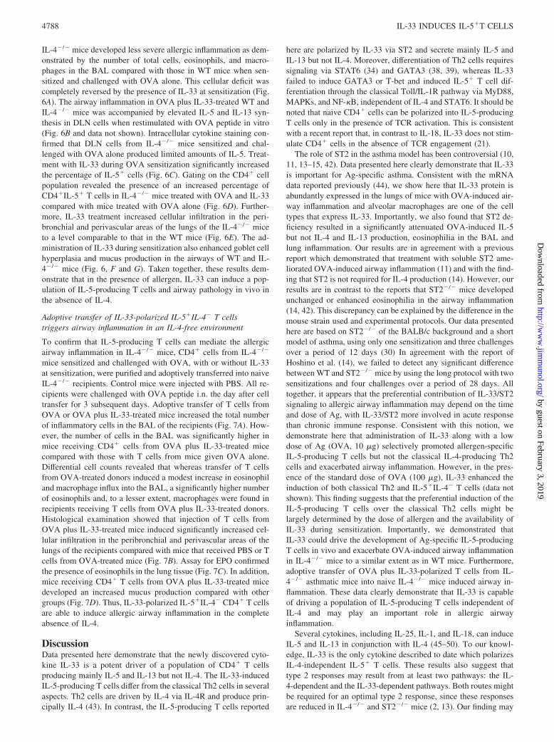
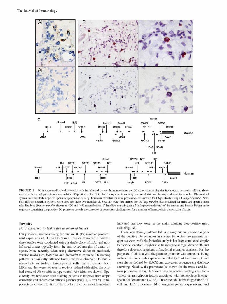
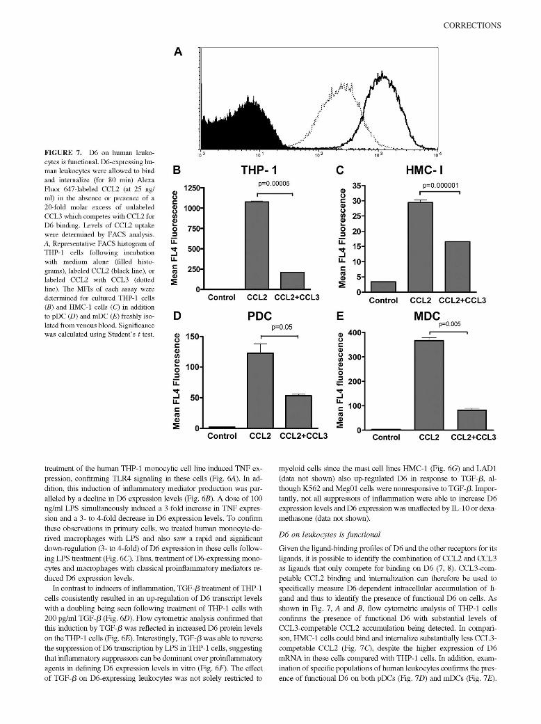
FIGURE 1. IL-33 polarizes CD4� T cells toproduce IL-5 and IL-13 via ST2. A–C, NaiveCD4� cells from BALB/c (A), D011.10 (B),ST2�/� (A), ST2�/� � D011.10 (B) mice, andhuman cord blood CD4� cells (C) were stimulatedwith anti-CD3 Abs (2 �g/ml) (A and C) or OVApeptide (10 nM; B) with or without IL-33 for 72 h.Supernatants were examined for cytokines byELISA. D, BALB/c CD4� cells stimulated withanti-CD3 Abs with or without IL-33 for 6 dayswere washed and restimulated with anti-CD3 Absalone for 72 h. E and F, CD4� cells from D011.10mice were stimulated with OVA (10 nM) and my-eloid dendritic cells with or without IL-33 (10 ng/ml) for 5 days followed by PMA/ionomycin re-stimulation and intracellular staining forcytokines. The representative dot plots of three in-dependent experiments with similar results areshown. G–I, CD4� cells from D011.10 mice werestimulated with OVA peptide (10 nM) in the pres-ence or absence of IL-33 for 72 h. Expression ofST2L was evaluated by FACS (G) and quantita-tive PCR (H). I, Representative results of ST2Lexpression on CD4� cells are shown. Data arepresented as mean � SEM and are representativeof three independent experiments. �, p � 0.05compared with control (without IL-33).
4781The Journal of Immunology
by guest on February 3, 2019http://w
ww
.jimm
unol.org/D
ownloaded from
CD4� T cell purification and culture
Cord blood was obtained from informed consenting mothers. Human andmurine CD4� T cells were purified by negative selection (AutoMACS;Miltenyi Biotec). T cells (purity �98%) were cultured in RPMI 1640 sup-plemented with 10% FCS, 2 mM L-glutamine, 100 U/ml penicillin, 100�g/ml streptomycin, and 0.05 M 2-ME. CD4� T cells (2 � 106 cells/ml)were activated with plate-bound anti-CD3 Abs (2–3 �g/ml; BD Bio-sciences), IL-4, IL-12 (all from PeproTech), and different doses of IL-33 ora combination of the cytokines. CD4� T cells from D0.11.10 mice werecultured with mitomycin C-treated APC (spleens from ST2KO mice),OVA peptide (10 nM), and cytokines as above. After 36 or 72 h, RNA andsupernatants were collected for PCR and ELISA, respectively. To test thestability of IL-33-induced IL-5-producing T cells, cells stimulated withanti-CD3 Abs with or without IL-33 for 6 days were washed and restim-ulated with anti-CD3 Abs alone. After 72 h, supernatants were collectedand ELISA was performed. To evaluate intracellular cytokine expressionby IL-33-stimulated naive T cells, purified naive CD4�CD62L� T cells(1.2 � 106) were cultured with bone marrow-derived myeloid dendriticcells (4 � 105) (27), OVA peptide (10 nM), and IL-33 (10 ng/ml) in24-well plates. After 3 days, fresh medium supplemented with IL-2 (10U/ml) was added to the culture for another 3 days. Next, cells were washedand stimulated again under the same conditions. Cells after the first andsecond round of stimulation were washed and activated with PMA (500ng/ml) and ionomycin (50 ng/ml; both from Sigma-Aldrich) in the absenceof IL-33 for 4 h followed by intracellular cytokine staining. To test the roleof NF-�B and MAPK, CD4� cells from BALB/c mice were stimulatedwith anti-CD3 Abs (4 �g/ml) and IL-33 (10 ng/ml). After 5 days, cellswere restimulated with immobilized anti-CD3 Abs for 24 h. Cells werethen washed and rested for 2 h and incubated with IL-33 alone or in thepresence of the following inhibitors or their controls: SN50M, SN50 (18�M) SB203560 (10 �M) PD098059 (60 �M), JNK II (50 �M), or DMSO.
ELISA
Murine and human cytokines IL-4, IL-5, IL-6, IL-10, IL-13, IFN-�, andIL-17 were analyzed by ELISA using paired Abs (BD Biosciences). Serumlevels of IgE and IgG1 were measured using an OptEIA ELISA kit (BDBiosciences).
Flow cytometry
Cultured or freshly isolated cells from draining lymph nodes (DLN) orbronchoalveolar lavage (BAL) were stimulated with PMA/ionomycin for4 h; GolgiStop was added during the final 3 h. The cells were incubatedwith anti-mouse CD16/32 Abs (BD Biosciences) followed by PerCP-con-jugated anti-CD4 (BD Biosciences) and FITC-conjugated anti-ST2L (MDBiosciences) or F4/80 Ab and CCR3 Ab or appropriate isotype controls.Cells were then fixed with Cytofix/Cytoperm buffer (BD Biosciences), per-meabilized with Perm/Wash buffer (BD Biosciences), and incubated withFITC-conjugated anti-IFN-�, PE-conjugated anti-IL-4, allophycocyanin-conjugated anti-IL-5 (all from BD Biosciences), or rat anti-IL-33 Ab orbiotinylated goat anti-IL-13 Ab (R&D Systems) or isotype controls fol-lowed by incubation with secondary Abs or streptavidin if necessary.
Western blot
CD4� T cells were stimulated with immobilized anti-CD3 Abs (4 �g/ml)in the presence of IL-33 (10 ng/ml). After 6 days, cells were restimulatedwith immobilized anti-CD3 Abs for 24 h and rested for 2 h. The cells werethen incubated with IL-33 at different time points followed by extraction ofcytoplasmic and nuclear proteins using Nuclear Extract kits (Active Motif).Cytoplasmic proteins were used in Western blotting with anti-phospho-p65, phospho-ERK1/2, phospho-p38, phospho-JNK, and p38 Abs (CellSignaling Technology). Nuclear proteins were used to evaluate NF-�Bactivity with a TransAM NF-�� p62 kit (Active Motif).
Quantitative PCR
This was conducted as described previously (28, 29).
OVA-induced airway inflammation
Protocol 1. All mice were sensitized i.p. with 100 �g of OVA in 2% alum(aluminum hydroxide gel adjuvant; Brenntag) on day 0, then challengedintranasally (i.n.) on days 8–10 with 10 �g of OVA or PBS (30).Protocol 2. To detect the effect of exogenous IL-33, the dose of OVA wasreduced. Mice were immunized i.p. with 10 �g of OVA in 2% alum on day0. On days 0–2, these mice were injected i.p. with IL-33 (2 �g/mouse) orPBS. Control mice were given PBS or IL-33 alone. Mice were challengedi.n. on days 8–10 with 2 �g of OVA, except the PBS group which received
00.51
1.52
2.5
00.51
1.52
2.5
50
0.
1.1
2.
52
3.
535
IL-5
IL-1
3ng
/ml
CD3IL-33
+ + +- 2 10
CD3IL-33
+ + +- 2 10
+
10-
---IL-4
A B CWTIL-4
WTSTAT6-/- -/-
** ***#
* **
#
#
#
#
**
* *
**
**#
##
00.51
1.52
2.5
WTJa18 -/-
* * ** * *
CD3IL-33
+- 2 50
+10
00.51
1.52
2.53
3.5
* ** * * *
02468
101214
+ +
EDGATA3 T-bet
CD3IL-33IL-12IL-4
+ ++ + ++
++ +- 2 10 100
+-- ----
- --- +
-
--
-+ +
+CD3IL-33IL-12IL-4
+ ++ + ++
++ +- 2 10 100
+-- ----
- --- +
-
--
-+ +
+
** * *
0123456
0
1
2
3
norm
aliz
ed to
con
trol
norm
aliz
ed to
con
trol
Exp
ress
ion
of T
-bet
Exp
ress
ion
of G
ATA
3
FIGURE 2. IL-33 polarizes IL-5� T cells indepen-dent of IL-4. A–C, CD4� cells from BALB/c (A and B),C57BL/6 (C), IL-4�/� (A), STAT6�/� (B), or J�18�/�
(C) mice were stimulated as described in Fig. 1, A–C, inthe presence or absence of IL-33 or IL-4. D and E,CD4� cells from BALB/c mice were stimulated withanti-CD3 Abs (2 �g/ml) in the presence or absence ofIL-33 (10 ng/ml), IL-4 (10 ng/ml), IL-12 (10 ng/ml), ora combination of these cytokines for 36 h followed byquantitative PCR. Data are presented as mean � SEMand are representative of at least two independent ex-periments; �, p � 0.05 compared with control (withoutIL-33) and #, p � 0.05 WT vs relevant deficient mice.
4782 IL-33 INDUCES IL-5�T CELLS
by guest on February 3, 2019http://w
ww
.jimm
unol.org/D
ownloaded from
i.n. PBS instead of OVA. All mice were sacrificed on day 11 and serum,BAL, lungs, and DLN were analyzed as described previously (31). Thelung sections were stained with H&E or periodic acid-Schiff (PAS) andexamined under light microscopy. Peribronchial and perivascular inflam-mation was scored using a semiquantitative scoring system assessing thedegree of eosinophilic inflammation: 0 � no eosinophils; 1 � eosinophilsmake up �10% of total infiltrate or total infiltrate is �20 cells; 2 � eo-sinophils make up 10–50% of total infiltrate; and 3 � eosinophils make up�50% of total infiltrate. Immunohistochemical staining was performed onfrozen lung sections using anti-mouse IL-33 Ab (Nessy-1; Axxora LifeScience) with a biotinylated pan-specific secondary Ab followed by detec-tion with the avidin-biotin complex/diaminobenzidine system (both VectorLaboratories). For the quantitative analysis of mucus area, images covering81.754 �m2 of lung sections were captured and mucus (red-stained areas)was measured with the software Image Pro-Plus (Media Cybernetics). Toestimate eosinophil levels in the lungs, the eosinophil peroxidase (EPO)colorimetric assay was performed as described previously (32).
Adoptive transfer
CD4� cells were isolated from IL-4�/� mice sensitized with OVA or OVAplus IL-33 and challenged with OVA as described above (protocol 2).These T cells (2 � 106) or PBS were injected i.v. into naive IL-4�/�
recipients on day 0. On days 1–3, mice were challenged with OVA (10�g/mouse). Mice were sacrificed 24 h after the last challenge.
Statistical analysis
ANOVA followed by Tukey’s test or Student’s t test was applied to in vitrostudies. Analysis between individual in vivo groups was examined byANOVA followed by the Student-Newman-Keuls test or Student’s t test.Experiments were performed at least twice. All data are expressed asmean � SEM. A value of p � 0.05 was considered to be significant.
ResultsIL-33 polarizes IL-5�IL-4� T cells
Although IL-33 induces IL-5 and IL-13 production by IL-4-dif-ferentiated Th2 cells (16, 21), its role in activation of uncommittednaive T cells is unknown. To address this issue, naive CD4� Tcells from WT or ST2�/� mice were cultured with anti-CD3 Abwith or without IL-33 for 72 h. WT but not ST2�/� cells producedmarkedly higher concentrations of IL-5 and IL-13 when stimulatedwith IL-33 compared with cells cultured with anti-CD3 Ab alone(Fig. 1A). Unexpectedly, IL-33 failed to induce any IL-4 synthesis,the signature cytokine for classical Th2 cells. IL-33 was also un-able to induce IFN-�, IL-17, or IL-10 production (Fig. 1A and datanot shown). We further confirmed this observation by usingCD4�CD62L� naive T cells from WT or ST2�/� OVATcR-trans-genic mice. In the presence of APC and OVA peptide, IL-33 en-hanced the synthesis of IL-5 and IL-13 (but not IL-4 or IFN-�) byWT but not ST2�/�OVATcR-transgenic T cells (Fig. 1B). Fur-thermore, human cord blood CD4� T cells, which are mainly com-prised of naive cells, also produced enhanced levels of IL-5 andIL-13 but not IFN-� or IL-4 when cultured with anti-CD3 andIL-33 compared with anti-CD3 alone (Fig. 1C). Importantly,CD4�T cells stimulated with anti-CD3 and IL-33 in the firstround of stimulation still produced an increased amount of IL-5and IL-13 but not IL-4 and IFN-� after restimulation with anti-CD3 alone (Fig. 1D). Consistent with the cytokine ELISA data,intracellular staining shows that IL-33-polarized CD4� T cellpopulation selectively expressed IL-5 and IL-13 but not IL-4and IFN-� after the first and second round of stimulation (Fig.1, E and F, and data not shown). IL-33 also enhanced ST2protein and mRNA expression in CD4� T cells (Fig. 1, G–I),suggesting a self-amplification circuit in the induction of IL-5-producing cells. Thus, IL-33, in the presence of Ag, preferen-tially polarizes a population of IL-5�IL-13�IL-4� T cells.
IL-33 induces IL-5-producing T cells independent of IL-4,IL-4R, and STAT6
IL-4, IL-4R, and STAT6 are essential for classical Th2 cell devel-opment (33–35). To further characterize the IL-33-polarized IL-5�T cells, we tested whether IL-33 is able to activate CD4� T cellsin the absence of IL-4, IL-4R�, or STAT6. As shown in Fig. 2A,
P-p65
P-p38
P-JNK1/2
p38
P-ERK1/2
p38
30 15 0 IL-33 (10ng/ml)
min
0 15IL-33 (10ng/ml)
43kDa
54kDa46kDa
43kDa
43kDa
44kDa42kDa
min
65kDa
00.20.40.60.81
1.21.4
00.51
1.52
2.5
00.51
1.52
2.53
05
10152025
0 50 100150200
CD3IL-33
+ + +- 2 10
IL-1
3IL
-4IF
N-γ
IL-5
ng/m
l
CD3IL-33
+ + +- 2 10
IL-4
+-
- -- 10
00.20.40.60.81
IL-5
IL-5
control
DMSO
SN50
SN50M
SB203560
PD098059
U0126
DMSO
SN50
SN50M
SB203560
PD098059
U0126
IL-3
3 10
ng/m
l
WTMyD88
WTTRIF
LCPSCD3IL-33
+++
pcnc
-15min30min
-/-
-/-ng
/ml
0 5 10 15 20 25
IL-13ng/ml
B E
D
CA
0 5000 10000 15000 20000**
**
*
* * *
***
*
##
####
0
2
4
6
8
* * * *
IL-1
3
0
5
10
15
20
IFN
-γ
FIGURE 3. IL-33 induces cytokine production via MyD88 and p38. Aand B, Naive CD4� cells from C57BL/6 (A and B), MyD88�/� (A), orTRIF�/� (B) mice were stimulated as described in Fig. 1, A–C. C–E, CD4�
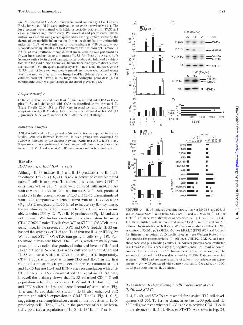
T cells stimulated with immobilized anti-CD3 Abs were rested for 2 hfollowed by incubation with IL-33 and/or various inhibitors: NF-�B (SN50or control SN50M), p38 (SB203560), or ERK1/2 (PD098059 and U0126)for different time points. C, Cytosolic proteins were Western blotted withAbs specific for phosphorylated (P) p65, p38, JNK1/2, ERK1/2, and non-phosphorylated p38 (loading control). D, Nuclear proteins were evaluatedin a TransAM NF-�B p65 assay (nc, negative control; pc, positive controlprovided by the assay kit; LCPS, luminescence count per second). E, Theamount of IL-5 and IL-13 was determined by ELISA. Data are presentedas mean � SEM and are representative of at least two independent exper-iments. �, p � 0.05 compared with control (without IL-33) and #, p � 0.05,IL-33 plus inhibitors vs IL-33 alone.
4783The Journal of Immunology
by guest on February 3, 2019http://w
ww
.jimm
unol.org/D
ownloaded from
IL-33 increased IL-5 and IL-13 production in both WT and IL-4�/� cells, although the level of IL-13 remained slightly lower inIL-4�/� compared with WT. Similar results were observed withCD4� T cells from IL-4R��/� mice (data not shown) andSTAT6�/� mice (Fig. 2B). Invariant NKT (iNKT) cells can alsoexpress CD4 and have been shown to be involved in type 2 im-munity (36). To exclude a role for iNKT cells in this context, wepolarized CD4� T cells from iNKT-deficient J�18�/� mice (37).CD4� T cells from WT and J�18�/� mice produced comparablelevels of IL-5 and IL-13 when activated with IL-33 and anti-CD3(Fig. 2C), indicating that NKT cells did not contribute to the IL-33-induced IL-5-producing T cells. IL-33 did not significantly af-fect the production of IFN-� or IL-4 in IL-4�/�, IL-4R�/�,STAT6�/�, or J�18�/� mice (data not shown). Taken together,these results demonstrate that IL-33 triggers IL-5 and IL-13 pro-duction from naive CD4� T cells independent of IL-4, IL-4R�,and STAT6 signaling.
We also investigated the involvement of GATA3 and T-bet inIL-33-induced T cell polarization. IL-4 and IL-12 induced the tran-scription of GATA3 and T-bet, respectively (38, 39). In contrast,IL-33 failed to induce GATA3 or T-bet even at high concentra-tions (Fig. 2, D and E). These results indicate that IL-33 polarizesthe IL-5-producing T cell via a distinct signaling pathway fromTh1 or classical Th2 cells.
IL-33 polarizes IL-5-producing T cells via MyD88
As a member of the IL-1 family, IL-33 may signal via the MyD88-dependent and -independent pathways (21, 40, 41). We thereforeinvestigated the role of MyD88 and Toll/IL-1R domain-containingadaptor-inducing IFN-� (TRIF) in IL-33-mediated T cell polariza-tion. As shown in Fig. 3A, IL-33 failed to induce IL-5 and IL-13secretion by MyD88�/� T cells. In contrast, IL-33 stimulated nor-mal levels of IL-5 and IL-13 production by CD4� cells from
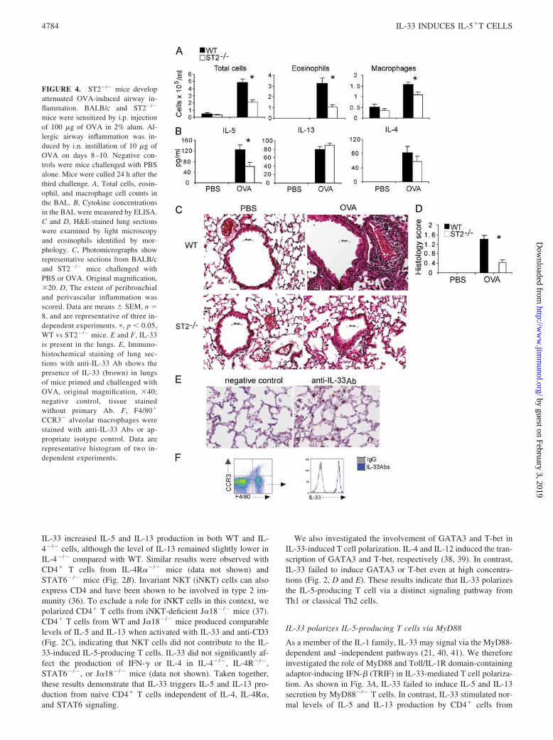
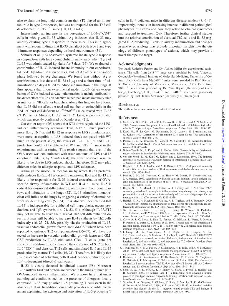
FIGURE 4. ST2�/� mice developattenuated OVA-induced airway in-flammation. BALB/c and ST2�/�
mice were sensitized by i.p. injectionof 100 �g of OVA in 2% alum. Al-lergic airway inflammation was in-duced by i.n. instillation of 10 �g ofOVA on days 8–10. Negative con-trols were mice challenged with PBSalone. Mice were culled 24 h after thethird challenge. A, Total cells, eosin-ophil, and macrophage cell counts inthe BAL. B, Cytokine concentrationsin the BAL were measured by ELISA.C and D, H&E-stained lung sectionswere examined by light microscopyand eosinophils identified by mor-phology. C, Photomicrographs showrepresentative sections from BALB/cand ST2�/� mice challenged withPBS or OVA. Original magnification,�20. D, The extent of peribronchialand perivascular inflammation wasscored. Data are means � SEM, n �8, and are representative of three in-dependent experiments. �, p � 0.05,WT vs ST2�/� mice. E and F, IL-33is present in the lungs. E, Immuno-histochemical staining of lung sec-tions with anti-IL-33 Ab shows thepresence of IL-33 (brown) in lungsof mice primed and challenged withOVA, original magnification, �40;negative control, tissue stainedwithout primary Ab. F, F4/80�
CCR3� alveolar macrophages werestained with anti-IL-33 Abs or ap-propriate isotype control. Data arerepresentative histogram of two in-dependent experiments.
4784 IL-33 INDUCES IL-5�T CELLS
by guest on February 3, 2019http://w
ww
.jimm
unol.org/D
ownloaded from
TRIF�/� mice (Fig. 3B). Thus, IL-33 polarization of IL-5- pro-ducing T cells was MyD88 dependent but TRIF independent. Wefurther explored the involvement of the key signaling componentsin IL-33 signaling. It has been reported that IL-33 triggers all threeMAPK pathways and the NF-�B pathway in mast cells (16, 20).Little, however, is known about the signaling pathways triggeredby IL-33 in T cells and their contribution to cytokine production.We found that IL-33 induced the phosphorylation of NF-�Bp65and the MAPK p38, JNK1/2, and ERK1/2 (Fig. 3C). The NF-�Bactivity induced by IL-33 was further confirmed by nuclear trans-location of NF-�Bp65 (Fig. 3D). IL-33-induced IL-5 synthesis wassuppressed by the inhibitors of p38 (SB203560), ERK1/2(PD098059 and U0126), and JNK1/2 (JNK II) but not by the in-hibitor of NF-�B (SN50) (Fig. 3E and data not shown). In contrast,the production of IL-13 was suppressed by p38, NF-�B, and, to a
lesser extent, by ERK1/2 and JNK1/2 inhibitors (Fig. 3E and datanot shown). Thus, IL-33 polarizes IL-5- producing T cells via theST2, MyD88, MAPK, and NF-�B pathways. Interestingly, IL-33appears to induce IL-5 and IL-13 production by different signalingmechanisms.
ST2�/� mice develop attenuated allergic airway inflammation
The role of ST2 in the asthma model is controversial (10, 11,13–15, 42). We sought to further determine the role of ST2/IL-33in type 2 responses by using WT and ST2�/� BALB/c mice in theOVA-specific model of airway inflammation (30). WT andST2�/� mice were sensitized once with OVA (100 �g), then chal-lenged i.n. with 10 �g of OVA for 3 subsequent days. ST2�/�
mice showed significantly less severe airway inflammation com-pared with WT mice. This was evident in a reduced number of
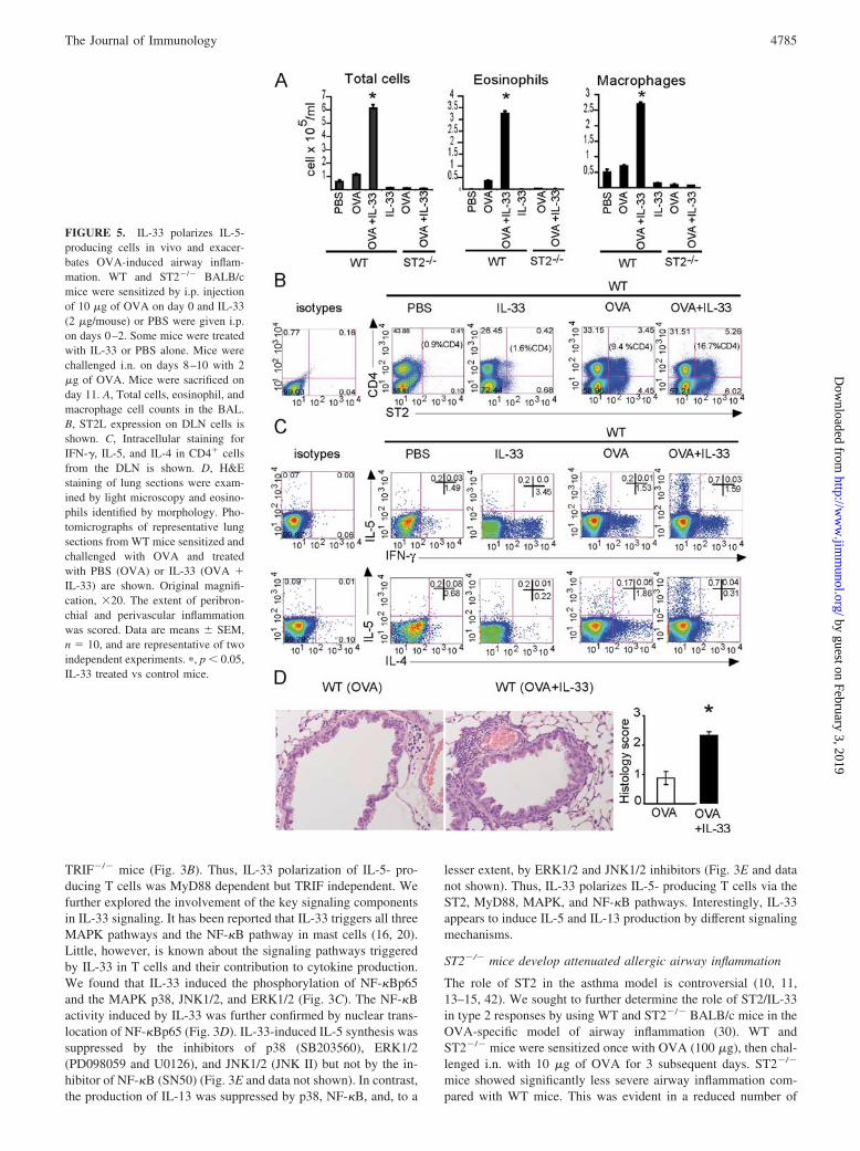
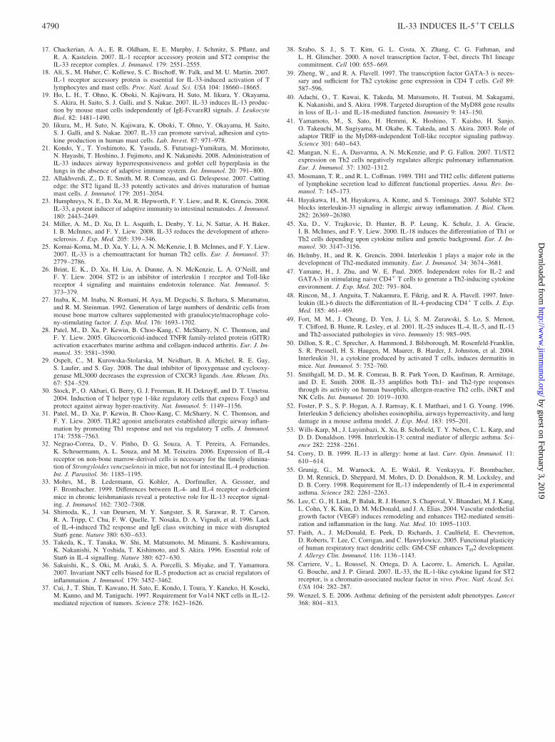
FIGURE 5. IL-33 polarizes IL-5-producing cells in vivo and exacer-bates OVA-induced airway inflam-mation. WT and ST2�/� BALB/cmice were sensitized by i.p. injectionof 10 �g of OVA on day 0 and IL-33(2 �g/mouse) or PBS were given i.p.on days 0–2. Some mice were treatedwith IL-33 or PBS alone. Mice werechallenged i.n. on days 8–10 with 2�g of OVA. Mice were sacrificed onday 11. A, Total cells, eosinophil, andmacrophage cell counts in the BAL.B, ST2L expression on DLN cells isshown. C, Intracellular staining forIFN-�, IL-5, and IL-4 in CD4� cellsfrom the DLN is shown. D, H&Estaining of lung sections were exam-ined by light microscopy and eosino-phils identified by morphology. Pho-tomicrographs of representative lungsections from WT mice sensitized andchallenged with OVA and treatedwith PBS (OVA) or IL-33 (OVA �IL-33) are shown. Original magnifi-cation, �20. The extent of peribron-chial and perivascular inflammationwas scored. Data are means � SEM,n � 10, and are representative of twoindependent experiments. �, p � 0.05,IL-33 treated vs control mice.
4785The Journal of Immunology
by guest on February 3, 2019http://w
ww
.jimm
unol.org/D
ownloaded from
total cells, eosinophils, and macrophages in the BAL of theST2�/� mice compared with WT mice (Fig. 4A). The BAL fromST2�/� mice also contained significantly less IL-5 than that of theWT mice, but IL-13 and IL-4 levels were not significantly different(Fig. 4B). The serum IgE and IgG levels were similar in theST2�/� and WT mice (data not shown). ST2�/� mice also exhib-ited reduced lung pathology after Ag challenge, showing signifi-cantly less inflammatory cell infiltration in the peribronchial andperivascular areas of the lungs than that of the WT mice (Fig. 4, Cand D). The contribution of endogenous IL-33 in OVA-inducedairway inflammation was further supported by our immunohisto-chemical analysis which clearly demonstrated the presence of en-dogenous IL-33 in the lungs of mice sensitized and challengedwith OVA (Fig. 4E). Since the morphological appearance of IL-33-positive cells suggested that these might be macrophages, weisolated alveolar macrophages and stained them with anti-IL-33-specific Ab. Indeed, as shown in Fig. 4F, F4/80�CCR3� alveolarmacrophages abundantly expressed IL-33. Altogether these resultstherefore demonstrate that IL-33/ST2 signals are required for anoptimal allergen induced-airway inflammation and IL-5 but notIL-4 production.
IL-33 induces IL-5�IL-4� T cells in vivo and exacerbatesAg-induced airway inflammation
Our result from ST2�/� mice, as well as previous data showing thebeneficial effect of soluble ST2 in airway inflammation (11), sug-gested that IL-33 participates in Ag-specific airway inflammation.We therefore further investigated whether IL-33 could induce IL-5-producing T cells in vivo and exacerbate OVA-triggered airwayinflammation. To be able to define the effect of exogenous IL-33,we reduced the dose of OVA in our model. As shown in Fig. 5A,mice sensitized (10 �g) and challenged (2 �g) with OVA aloneexhibited a low inflammatory response in the BAL. In contrast,mice sensitized with OVA plus IL-33 displayed markedly elevatedtotal cell numbers, eosinophils, and macrophages in the BAL (Fig.5A). The IL-33-enhanced inflammation was not evident in ST2�/�
mice. Importantly, OVA-nonsensitized WT mice injected withIL-33 and challenged with OVA showed no increase in the totalcell count and eosinophilia compared with the PBS group. We thenanalyzed the ST2 expression and cytokine profile on DLN T cells.IL-33 administration without Ag sensitization slightly increasedthe percentage of CD4� ST2� cells compared with the PBS group
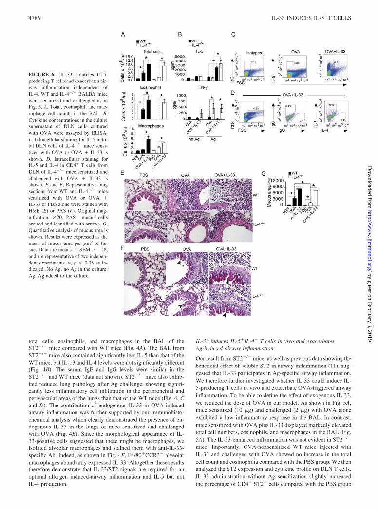
FIGURE 6. IL-33 polarizes IL-5-producing T cells and exacerbates air-way inflammation independent ofIL-4. WT and IL-4�/� BALB/c micewere sensitized and challenged as inFig. 5. A, Total, eosinophil, and mac-rophage cell counts in the BAL. B,Cytokine concentrations in the culturesupernatant of DLN cells culturedwith OVA were assayed by ELISA.C, Intracellular staining for IL-5 in to-tal DLN cells of IL-4�/� mice sensi-tized with OVA or OVA � IL-33 isshown. D, Intracellular staining forIL-5 and IL-4 in CD4� T cells fromDLN of IL-4�/� mice sensitized andchallenged with OVA � IL-33 isshown. E and F, Representative lungsections from WT and IL-4�/� micesensitized with OVA or OVA �IL-33 or PBS alone were stained withH&E (E) or PAS (F). Original mag-nification, �20. PAS� mucus cellsare red and identified with arrows. G,Quantitative analysis of mucus area isshown. Results were expressed as themean of mucus area per �m2 of tis-sue. Data are means � SEM, n � 8,and are representative of two indepen-dent experiments. �, p � 0.05 as in-dicated. No Ag, no Ag in the culture;Ag, Ag added to the culture.
4786 IL-33 INDUCES IL-5�T CELLS
by guest on February 3, 2019http://w
ww
.jimm
unol.org/D
ownloaded from
(1.6 and 0.9%, respectively; Fig. 5B). Cells from WT mice sensi-tized and challenged with OVA showed a substantial increase inthe percentage of ST2�CD4� cells (9.4%). This was further in-creased by the IL-33 treatment at sensitization (16.7%). Intracel-lular staining of DLN CD4� T cells revealed that IL-33 alone hadno effect on the type 2 cytokine profile, but increases the percent-age of IFN-�� cell population compared with the PBS group. Incontrast, CD4� cells from OVA-sensitized and challenged miceshowed an increase in percentage of IL-4� cells but not IL-5�
cells compared with either IL-33 alone or the PBS-treated groups.Importantly, the administration of IL-33 during the OVA sensiti-zation markedly enhanced the percentage of IL-5� T cells butdecreased the percentage of IL-4� T cells and did not change thepercentage of IFN-�� cells compared with the OVA group (Fig.5C). The injection of IL-33 in the OVA- sensitizing phase alsotriggered IL-13 production in WT mice (66.6 � 4.4 pg/ml), whichwas undetectable in other groups. IL-33 did not affect the cytokineproduction by CD4� T cells in ST2�/� mice (data not shown).
Thus, IL-33 markedly and specifically increased the number ofCD4�IL-5� T cells in vivo during Ag priming via ST2. Lunghistology analysis showed that IL-33 given during OVA sensiti-zation significantly increased inflammatory cell infiltration in theperibronchial and perivascular areas of the lungs compared withthat from mice injected with OVA alone (Fig. 5D). Mice givenPBS or IL-33 without Ag did not have any detectable histologicalchanges (data not shown). Together, these results suggest thatIL-33 is a pathogenic factor for Ag-specific airway inflammationaccompanied by the polarization of CD4�IL-5� T cells in vivo.
IL-33 exacerbates allergic airway inflammation independentof IL-4
To further investigate the relationship between IL-33 and IL-4 inthe polarization of IL-5-producing T cells and allergic response,we determined whether IL-33 can induce allergic airway inflam-mation in IL-4�/� mice. IL-4�/� and WT mice were sensitizedwith OVA or OVA plus IL-33 and challenged with OVA as before.
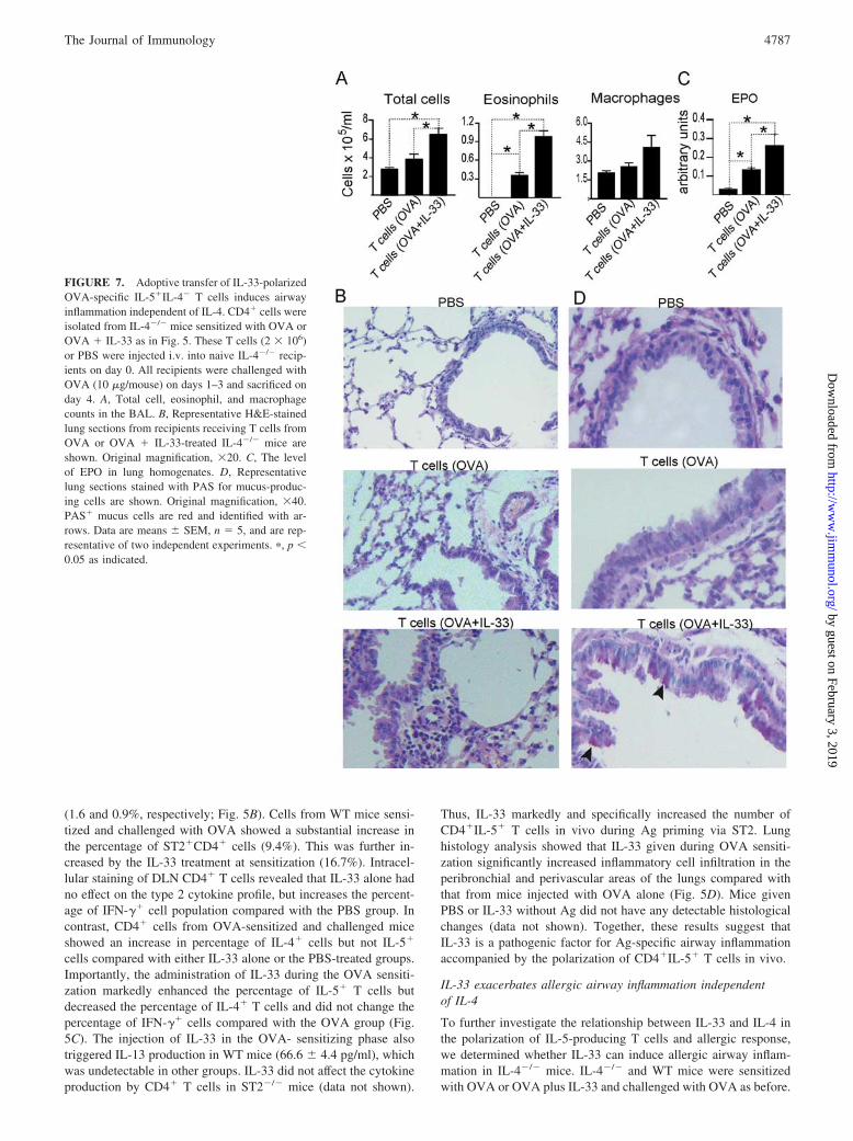
FIGURE 7. Adoptive transfer of IL-33-polarizedOVA-specific IL-5�IL-4� T cells induces airwayinflammation independent of IL-4. CD4� cells wereisolated from IL-4�/� mice sensitized with OVA orOVA � IL-33 as in Fig. 5. These T cells (2 � 106)or PBS were injected i.v. into naive IL-4�/� recip-ients on day 0. All recipients were challenged withOVA (10 �g/mouse) on days 1–3 and sacrificed onday 4. A, Total cell, eosinophil, and macrophagecounts in the BAL. B, Representative H&E-stainedlung sections from recipients receiving T cells fromOVA or OVA � IL-33-treated IL-4�/� mice areshown. Original magnification, �20. C, The levelof EPO in lung homogenates. D, Representativelung sections stained with PAS for mucus-produc-ing cells are shown. Original magnification, �40.PAS� mucus cells are red and identified with ar-rows. Data are means � SEM, n � 5, and are rep-resentative of two independent experiments. �, p �0.05 as indicated.
4787The Journal of Immunology
by guest on February 3, 2019http://w
ww
.jimm
unol.org/D
ownloaded from
IL-4�/� mice developed less severe allergic inflammation as dem-onstrated by the number of total cells, eosinophils, and macro-phages in the BAL compared with those in WT mice when sen-sitized and challenged with OVA alone. This cellular deficit wascompletely reversed by the presence of IL-33 at sensitization (Fig.6A). The airway inflammation in OVA plus IL-33-treated WT andIL-4�/� mice was accompanied by elevated IL-5 and IL-13 syn-thesis in DLN cells when restimulated with OVA peptide in vitro(Fig. 6B and data not shown). Intracellular cytokine staining con-firmed that DLN cells from IL-4�/� mice sensitized and chal-lenged with OVA alone produced limited amounts of IL-5. Treat-ment with IL-33 during OVA sensitization significantly increasedthe percentage of IL-5� cells (Fig. 6C). Gating on the CD4� cellpopulation revealed the presence of an increased percentage ofCD4�IL-5� T cells in IL-4�/� mice treated with OVA and IL-33compared with mice treated with OVA alone (Fig. 6D). Further-more, IL-33 treatment increased cellular infiltration in the peri-bronchial and perivascular areas of the lungs of the IL-4�/� miceto a level comparable to that in the WT mice (Fig. 6E). The ad-ministration of IL-33 during sensitization also enhanced goblet cellhyperplasia and mucus production in the airways of WT and IL-4�/� mice (Fig. 6, F and G). Taken together, these results dem-onstrate that in the presence of allergen, IL-33 can induce a pop-ulation of IL-5-producing T cells and airway pathology in vivo inthe absence of IL-4.
Adoptive transfer of IL-33-polarized IL-5�IL-4� T cellstriggers airway inflammation in an IL-4-free environment
To confirm that IL-5-producing T cells can mediate the allergicairway inflammation in IL-4�/� mice, CD4� cells from IL-4�/�
mice sensitized and challenged with OVA, with or without IL-33at sensitization, were purified and adoptively transferred into naiveIL-4�/� recipients. Control mice were injected with PBS. All re-cipients were challenged with OVA peptide i.n. the day after celltransfer for 3 subsequent days. Adoptive transfer of T cells fromOVA or OVA plus IL-33-treated mice increased the total numberof inflammatory cells in the BAL of the recipients (Fig. 7A). How-ever, the number of cells in the BAL was significantly higher inmice receiving CD4� cells from OVA plus IL-33-treated micecompared with those with T cells from mice given OVA alone.Differential cell counts revealed that whereas transfer of T cellsfrom OVA-treated donors induced a modest increase in eosinophiland macrophage influx into the BAL, a significantly higher numberof eosinophils and, to a lesser extent, macrophages were found inrecipients receiving T cells from OVA plus IL-33-treated donors.Histological examination showed that injection of T cells fromOVA plus IL-33-treated mice induced significantly increased cel-lular infiltration in the peribronchial and perivascular areas of thelungs of the recipients compared with mice that received PBS or Tcells from OVA-treated mice (Fig. 7B). Assay for EPO confirmedthe presence of eosinophils in the lung tissue (Fig. 7C). In addition,mice receiving CD4� T cells from OVA plus IL-33-treated micedeveloped an increased mucus production compared with othergroups (Fig. 7D). Thus, IL-33-polarized IL-5�IL-4� CD4� T cellsare able to induce allergic airway inflammation in the completeabsence of IL-4.
DiscussionData presented here demonstrate that the newly discovered cyto-kine IL-33 is a potent driver of a population of CD4� T cellsproducing mainly IL-5 and IL-13 but not IL-4. The IL-33-inducedIL-5-producing T cells differ from the classical Th2 cells in severalaspects. Th2 cells are driven by IL-4 via IL-4R and produce prin-cipally IL-4 (43). In contrast, the IL-5-producing T cells reported
here are polarized by IL-33 via ST2 and secrete mainly IL-5 andIL-13 but not IL-4. Moreover, differentiation of Th2 cells requiressignaling via STAT6 (34) and GATA3 (38, 39), whereas IL-33failed to induce GATA3 or T-bet and induced IL-5� T cell dif-ferentiation through the classical Toll/IL-1R pathway via MyD88,MAPKs, and NF-�B, independent of IL-4 and STAT6. It should benoted that naive CD4� cells can be polarized into IL-5-producingT cells only in the presence of TCR activation. This is consistentwith a recent report that, in contrast to IL-18, IL-33 does not stim-ulate CD4� cells in the absence of TCR engagement (21).
The role of ST2 in the asthma model has been controversial (10,11, 13–15, 42). Data presented here clearly demonstrate that IL-33is important for Ag-specific asthma. Consistent with the mRNAdata reported previously (44), we show here that IL-33 protein isabundantly expressed in the lungs of mice with OVA-induced air-way inflammation and alveolar macrophages are one of the celltypes that express IL-33. Importantly, we also found that ST2 de-ficiency resulted in a significantly attenuated OVA-induced IL-5but not IL-4 and IL-13 production, eosinophilia in the BAL andlung inflammation. Our results are in agreement with a previousreport which demonstrated that treatment with soluble ST2 ame-liorated OVA-induced airway inflammation (11) and with the find-ing that ST2 is not required for IL-4 production (14). However, ourresults are in contrast to the reports that ST2�/� mice developedunchanged or enhanced eosinophilia in the airway inflammation(14, 42). This discrepancy can be explained by the difference in themouse strain used and experimental protocols. Our data presentedhere are based on ST2�/� of the BALB/c background and a shortmodel of asthma, using only one sensitization and three challengesover a period of 12 days (30) In agreement with the report ofHoshino et al. (14), we failed to detect any significant differencebetween WT and ST2�/� mice by using the long protocol with twosensitizations and four challenges over a period of 28 days. Alltogether, it appears that the preferential contribution of IL-33/ST2signaling to allergic airway inflammation may depend on the timeand dose of Ag, with IL-33/ST2 more involved in acute responsethan chronic immune response. Consistent with this notion, wedemonstrate here that administration of IL-33 along with a lowdose of Ag (OVA, 10 �g) selectively promoted allergen-specificIL-5-producing T cells but not the classical IL-4-producing Th2cells and exacerbated airway inflammation. However, in the pres-ence of the standard dose of OVA (100 �g), IL-33 enhanced theinduction of both classical Th2 and IL-5�IL-4� T cells (data notshown). This finding suggests that the preferential induction of theIL-5-producing T cells over the classical Th2 cells might belargely determined by the dose of allergen and the availability ofIL-33 during sensitization. Importantly, we demonstrated thatIL-33 could drive the development of Ag-specific IL-5-producingT cells in vivo and exacerbate OVA-induced airway inflammationin IL-4�/� mice to a similar extent as in WT mice. Furthermore,adoptive transfer of OVA plus IL-33-polarized T cells from IL-4�/� asthmatic mice into naive IL-4�/� mice induced airway in-flammation. These data clearly demonstrate that IL-33 is capableof driving a population of IL-5-producing T cells independent ofIL-4 and may play an important role in allergic airwayinflammation.
Several cytokines, including IL-25, IL-1, and IL-18, can induceIL-5 and IL-13 in conjunction with IL-4 (45–50). To our knowl-edge, IL-33 is the only cytokine described to date which polarizesIL-4-independent IL-5� T cells. These results also suggest thattype 2 responses may result from at least two pathways: the IL-4-dependent and the IL-33-dependent pathways. Both routes mightbe required for an optimal type 2 response, since these responsesare reduced in IL-4�/� and ST2�/� mice (2, 13). Our finding may
4788 IL-33 INDUCES IL-5�T CELLS
by guest on February 3, 2019http://w
ww
.jimm
unol.org/D
ownloaded from
also explain the long-held conundrum that ST2 played an impor-tant role in type 2 responses, but was not required for the Th2 celldevelopment in ST2�/� mice (10, 11, 13–15).
Interestingly, an increase in the percentage of IFN-��CD4�
cells in mice given IL-33 without Ag indicates that IL-33 mayamplify existing type 1 responses in these mice. This is in agree-ment with recent findings that IL-33 can affect both type 2 and type1 immune responses depending on local environment (51).
Schmitz et al. (16) observed a systemic innate type 2 responsein conjunction with lung eosinophilia in naive mice when 2 �g ofIL-33 was administrated i.p. daily for 7 days (16). We evaluated acontribution of IL-33-induced innate immunity to our experimen-tal model by administration of IL-33 but not Ag at the sensitizationphase followed by Ag challenge. We found that without Ag atsensitization, a low dose of IL-33 (2 �g) and a short time of ad-ministration (3 days) failed to induce inflammation in the lungs. Itthus appears that in our experimental model, IL-33- driven exacer-bation of OVA-induced airway inflammation is mainly attributed tothe direct effect of IL-33 on adaptive rather than innate immunity suchas mast cells, NK cells, or basophils. Along this line, we have foundthat IL-33 did not affect the total cell number or eosinophilia in theBAL of mast cell-deficient (KitWsh/KitWsh) mice treated with IL-33(N. Pitman, G. Murphy, D. Xu, and F. Y. Liew, unpublished data),which was recently confirmed by Kondo et al. (21).
Our earlier report (26) shows that ST2 down-regulated the LPS-induced inflammatory response. Thus, ST2�/� mice producedmore IL-1, TNF-�, and IL-12 in response to LPS stimulation andwere more susceptible to LPS-induced shock compared with WTmice. In the present study, the levels of IL-1, TNF-�, and IL-12production could not be detected in WT and ST2�/� mice in theexperimental asthma setting. This result suggests that even if theOVA used was contaminated with trace amounts of LPS (�0.18endotoxin units/�g by Limulus test), the effect observed was un-likely to be due to LPS-induced shock. Therefore, ST2 may playdifferent roles in allergic response and LPS tolerance.
Although the molecular mechanism by which IL-33 preferen-tially induces IL-5/IL-13 is currently unknown, IL-5 and IL-13 arelikely to be responsible for IL-33-induced exacerbation of OVA-specific airway inflammation in WT and IL-4�/� mice. IL-5 iscritical for eosinophil differentiation, recruitment from bone mar-row, and migration to the lungs (52). IL-13 stimulates mononu-clear cell influx to the lungs by triggering a variety of chemokinesfrom resident lung cells (53, 54). It is also well documented thatIL-13 is indispensable for epithelial cell hyperplasia, mucus pro-duction, and IgE synthesis (16, 21, 53, 54). Although IL-33/ST2may not be able to drive the classical Th2 cell differentiation di-rectly, it may still be able to increase IL-4 synthesis by Th2 cellsindirectly (16, 23, 24, 55), possibly via the production of IL-13,vascular endothelial growth factor, and GM-CSF which have beenreported to enhance Th2 cell polarization (55–57). We have de-tected an increase in vascular endothelial growth factor and GM-CSF production by IL-33-stimulated CD4� T cells (data notshown). In addition, IL-33 enhanced the expression of ST2 on bothIL-5�CD4� and classical Th2 cells and stimulated IL-5 and IL-13production by mature Th2 cells (16, 21). Therefore, it is likely thatIL-33 is capable of activating both IL-4-dependent (indirectly) andIL-4-independent (directly) pathways.
IL-33 is clearly detected in clinical disease (58). MoreoverIL-33 mRNA (44) and protein are present in the lungs of mice withOVA-induced airway inflammation. We propose here that underpathological conditions such as the presence of allergens, locallyexpressed IL-33 may polarize IL-5-producing T cells even in theabsence of IL-4. In addition, our study provides a possible mech-anism explaining the existence and activation of IL-5-producing T
cells in IL-4-deficient mice in different disease models (3, 6–9).Importantly, there is an increasing interest in different pathologicalphenotypes of asthma and how they relate to clinical syndromesand respond to treatment (59). Therefore, further clinical studiesinto the relative contribution of classical Th2 cells and IL-33-trig-gered IL-5-producing T cells to airway inflammation and changesin airway physiology may provide important insights into the eti-ology of different phenotypes of asthma, which may provide anovel therapeutic target.
AcknowledgmentsWe thank Roderick Ferrier and Dr. Ashley Miller for experimental assis-tance. The cells from J�18�/� mice were provided by Prof. VincenzoCerundolo (Weatherall Institute of Molecular Medicine, University of Ox-ford, U.K.). Cells from MyD88�/� mice were provided by Prof. RichardK. Grencis (University of Manchester, Manchester, U.K.). Cells fromTRIF�/� mice were provided by Dr Clare Bryant (University of Cam-bridge, Cambridge, U.K.). IL-4�/� and IL-4R�/� mice were generouslyprovided by Prof. James Alexander (University of Strathclyde).
DisclosuresThe authors have no financial conflict of interest.
References1. McKenzie, G. J., P. G. Fallon, C. L. Emson, R. K. Grencis, and A. N. McKenzie.
1999. Simultaneous disruption of interleukin (IL)-4 and IL-13 defines individualroles in T helper cell type 2-mediated responses. J. Exp. Med. 189: 1565–1572.
2. Kopf, M., G. Le Gros, M. Bachmann, M. C. Lamers, H. Bluethmann, andG. Kohler. 1993. Disruption of the murine IL-4 gene blocks Th2 cytokine re-sponses. Nature 362: 245–248.
3. Pearce, E. J., A. Cheever, S. Leonard, M. Covalesky, R. Fernandez-Botran,G. Kohler, and M. Kopf. 1996. Schistosoma mansoni in IL-4-deficient mice. Int.Immunol. 8: 435–444.
4. Noben-Trauth, N., P. Kropf, and I. Muller. 1996. Susceptibility to Leishmaniamajor infection in interleukin-4-deficient mice. Science 271: 987–990.
5. von der Weid, T., M. Kopf, G. Kohler, and J. Langhorne. 1994. The immuneresponse to Plasmodium chabaudi malaria in interleukin-4-deficient mice. Eur.J. Immunol. 24: 2285–2293.
6. Hogarth, P. J., M. J. Taylor, and A. E. Bianco. 1998. IL-5-dependent immunityto microfilariae is independent of IL-4 in a mouse model of onchocerciasis. J. Im-munol. 160: 5436–5440.
7. Brewer, J. M., M. Conacher, C. A. Hunter, M. Mohrs, F. Brombacher, andJ. Alexander. 1999. Aluminum hydroxide adjuvant initiates strong antigen-spe-cific Th2 responses in the absence of IL-4- or IL-13-mediated signaling. J. Im-munol. 163: 6448–6454.
8. Hogan, S. P., A. Mould, H. Kikutani, A. J. Ramsay, and P. S. Foster. 1997.Aeroallergen-induced eosinophilic inflammation, lung damage, and airways hy-perreactivity in mice can occur independently of IL-4 and allergen-specific im-munoglobulins. J. Clin. Invest. 99: 1329–1339.
9. Herrick, C. A., H. MacLeod, E. Glusac, R. E. Tigelaar, and K. Bottomly. 2000.Th2 responses induced by epicutaneous or inhalational protein exposure are dif-ferentially dependent on IL-4. J. Clin. Invest. 105: 765–775.
10. Xu, D., W. L. Chan, B. P. Leung, F. Huang, R. Wheeler, D. Piedrafita,J. H. Robinson, and F. Y. Liew. 1998. Selective expression of a stable cell surfacemolecule on type 2 but not type 1 helper T cells. J. Exp. Med. 187: 787–794.
11. Coyle, A. J., C. Lloyd, J. Tian, T. Nguyen, C. Erikkson, L. Wang, P. Ottoson,P. Persson, T. Delaney, S. Lehar, et al. 1999. Crucial role of the interleukin 1receptor family member T1/ST2 in T helper cell type 2-mediated lung mucosalimmune responses. J. Exp. Med. 190: 895–902.
12. Lohning, M., A. Stroehmann, A. J. Coyle, J. L. Grogan, S. Lin,J. C. Gutierrez-Ramos, D. Levinson, A. Radbruch, and T. Kamradt. 1998. T1/ST2is preferentially expressed on murine Th2 cells, independent of interleukin 4,interleukin 5, and interleukin 10, and important for Th2 effector function. Proc.Natl. Acad. Sci. USA 95: 6930–6935.
13. Townsend, M. J., P. G. Fallon, D. J. Matthews, H. E. Jolin, and A. N. McKenzie.2000. T1/ST2-deficient mice demonstrate the importance of T1/ST2 in develop-ing primary T helper cell type 2 responses. J. Exp. Med. 191: 1069–1076.
14. Hoshino, K., S. Kashiwamura, K. Kuribayashi, T. Kodama, T. Tsujimura,K. Nakanishi, T. Matsuyama, K. Takeda, and S. Akira. 1999. The absence ofinterleukin 1 receptor-related T1/ST2 does not affect T helper cell type 2 devel-opment and its effector function. J. Exp. Med. 190: 1541–1548.
15. Senn, K. A., K. D. McCoy, K. J. Maloy, G. Stark, E. Frohli, T. Rulicke, andR. Klemenz. 2000. T1-deficient and T1-Fc-transgenic mice develop a normalprotective Th2-type immune response following infection with Nippostrongylusbrasiliensis. Eur. J. Immunol. 30: 1929–1938.
16. Schmitz, J., A. Owyang, E. Oldham, Y. Song, E. Murphy, T. K. McClanahan,G. Zurawski, M. Moshrefi, J. Qin, X. Li, et al. 2005. IL-33, an interleukin-1-likecytokine that signals via the IL-1 receptor-related protein ST2 and induces Thelper type 2-associated cytokines. Immunity 23: 479–490.
4789The Journal of Immunology
by guest on February 3, 2019http://w
ww
.jimm
unol.org/D
ownloaded from
17. Chackerian, A. A., E. R. Oldham, E. E. Murphy, J. Schmitz, S. Pflanz, andR. A. Kastelein. 2007. IL-1 receptor accessory protein and ST2 comprise theIL-33 receptor complex. J. Immunol. 179: 2551–2555.
18. Ali, S., M. Huber, C. Kollewe, S. C. Bischoff, W. Falk, and M. U. Martin. 2007.IL-1 receptor accessory protein is essential for IL-33-induced activation of Tlymphocytes and mast cells. Proc. Natl. Acad. Sci. USA 104: 18660–18665.
19. Ho, L. H., T. Ohno, K. Oboki, N. Kajiwara, H. Suto, M. Iikura, Y. Okayama,S. Akira, H. Saito, S. J. Galli, and S. Nakae. 2007. IL-33 induces IL-13 produc-tion by mouse mast cells independently of IgE-Fcvar�RI signals. J. LeukocyteBiol. 82: 1481–1490.
20. Iikura, M., H. Suto, N. Kajiwara, K. Oboki, T. Ohno, Y. Okayama, H. Saito,S. J. Galli, and S. Nakae. 2007. IL-33 can promote survival, adhesion and cyto-kine production in human mast cells. Lab. Invest. 87: 971–978.
21. Kondo, Y., T. Yoshimoto, K. Yasuda, S. Futatsugi-Yumikura, M. Morimoto,N. Hayashi, T. Hoshino, J. Fujimoto, and K. Nakanishi. 2008. Administration ofIL-33 induces airway hyperresponsiveness and goblet cell hyperplasia in thelungs in the absence of adaptive immune system. Int. Immunol. 20: 791–800.
22. Allakhverdi, Z., D. E. Smith, M. R. Comeau, and G. Delespesse. 2007. Cuttingedge: the ST2 ligand IL-33 potently activates and drives maturation of humanmast cells. J. Immunol. 179: 2051–2054.
23. Humphreys, N. E., D. Xu, M. R. Hepworth, F. Y. Liew, and R. K. Grencis. 2008.IL-33, a potent inducer of adaptive immunity to intestinal nematodes. J. Immunol.180: 2443–2449.
24. Miller, A. M., D. Xu, D. L. Asquith, L. Denby, Y. Li, N. Sattar, A. H. Baker,I. B. McInnes, and F. Y. Liew. 2008. IL-33 reduces the development of athero-sclerosis. J. Exp. Med. 205: 339–346.
25. Komai-Koma, M., D. Xu, Y. Li, A. N. McKenzie, I. B. McInnes, and F. Y. Liew.2007. IL-33 is a chemoattractant for human Th2 cells. Eur. J. Immunol. 37:2779–2786.
26. Brint, E. K., D. Xu, H. Liu, A. Dunne, A. N. McKenzie, L. A. O’Neill, andF. Y. Liew. 2004. ST2 is an inhibitor of interleukin 1 receptor and Toll-likereceptor 4 signaling and maintains endotoxin tolerance. Nat. Immunol. 5:373–379.
27. Inaba, K., M. Inaba, N. Romani, H. Aya, M. Deguchi, S. Ikehara, S. Muramatsu,and R. M. Steinman. 1992. Generation of large numbers of dendritic cells frommouse bone marrow cultures supplemented with granulocyte/macrophage colo-ny-stimulating factor. J. Exp. Med. 176: 1693–1702.
28. Patel, M., D. Xu, P. Kewin, B. Choo-Kang, C. McSharry, N. C. Thomson, andF. Y. Liew. 2005. Glucocorticoid-induced TNFR family-related protein (GITR)activation exacerbates murine asthma and collagen-induced arthritis. Eur. J. Im-munol. 35: 3581–3590.
29. Ospelt, C., M. Kurowska-Stolarska, M. Neidhart, B. A. Michel, R. E. Gay,S. Laufer, and S. Gay. 2008. The dual inhibitor of lipoxygenase and cyclooxy-genase ML3000 decreases the expression of CXCR3 ligands. Ann. Rheum. Dis.67: 524–529.
30. Stock, P., O. Akbari, G. Berry, G. J. Freeman, R. H. Dekruyff, and D. T. Umetsu.2004. Induction of T helper type 1-like regulatory cells that express Foxp3 andprotect against airway hyper-reactivity. Nat. Immunol. 5: 1149–1156.
31. Patel, M., D. Xu, P. Kewin, B. Choo-Kang, C. McSharry, N. C. Thomson, andF. Y. Liew. 2005. TLR2 agonist ameliorates established allergic airway inflam-mation by promoting Th1 response and not via regulatory T cells. J. Immunol.174: 7558–7563.
32. Negrao-Correa, D., V. Pinho, D. G. Souza, A. T. Pereira, A. Fernandes,K. Scheuermann, A. L. Souza, and M. M. Teixeira. 2006. Expression of IL-4receptor on non-bone marrow-derived cells is necessary for the timely elimina-tion of Strongyloides venezuelensis in mice, but not for intestinal IL-4 production.Int. J. Parasitol. 36: 1185–1195.
33. Mohrs, M., B. Ledermann, G. Kohler, A. Dorfmuller, A. Gessner, andF. Brombacher. 1999. Differences between IL-4- and IL-4 receptor �-deficientmice in chronic leishmaniasis reveal a protective role for IL-13 receptor signal-ing. J. Immunol. 162: 7302–7308.
34. Shimoda, K., J. van Deursen, M. Y. Sangster, S. R. Sarawar, R. T. Carson,R. A. Tripp, C. Chu, F. W. Quelle, T. Nosaka, D. A. Vignali, et al. 1996. Lackof IL-4-induced Th2 response and IgE class switching in mice with disruptedStat6 gene. Nature 380: 630–633.
35. Takeda, K., T. Tanaka, W. Shi, M. Matsumoto, M. Minami, S. Kashiwamura,K. Nakanishi, N. Yoshida, T. Kishimoto, and S. Akira. 1996. Essential role ofStat6 in IL-4 signalling. Nature 380: 627–630.
36. Sakuishi, K., S. Oki, M. Araki, S. A. Porcelli, S. Miyake, and T. Yamamura.2007. Invariant NKT cells biased for IL-5 production act as crucial regulators ofinflammation. J. Immunol. 179: 3452–3462.
37. Cui, J., T. Shin, T. Kawano, H. Sato, E. Kondo, I. Toura, Y. Kaneko, H. Koseki,M. Kanno, and M. Taniguchi. 1997. Requirement for V�14 NKT cells in IL-12-mediated rejection of tumors. Science 278: 1623–1626.
38. Szabo, S. J., S. T. Kim, G. L. Costa, X. Zhang, C. G. Fathman, andL. H. Glimcher. 2000. A novel transcription factor, T-bet, directs Th1 lineagecommitment. Cell 100: 655–669.
39. Zheng, W., and R. A. Flavell. 1997. The transcription factor GATA-3 is neces-sary and sufficient for Th2 cytokine gene expression in CD4 T cells. Cell 89:587–596.
40. Adachi, O., T. Kawai, K. Takeda, M. Matsumoto, H. Tsutsui, M. Sakagami,K. Nakanishi, and S. Akira. 1998. Targeted disruption of the MyD88 gene resultsin loss of IL-1- and IL-18-mediated function. Immunity 9: 143–150.
41. Yamamoto, M., S. Sato, H. Hemmi, K. Hoshino, T. Kaisho, H. Sanjo,O. Takeuchi, M. Sugiyama, M. Okabe, K. Takeda, and S. Akira. 2003. Role ofadaptor TRIF in the MyD88-independent Toll-like receptor signaling pathway.Science 301: 640–643.
42. Mangan, N. E., A. Dasvarma, A. N. McKenzie, and P. G. Fallon. 2007. T1/ST2expression on Th2 cells negatively regulates allergic pulmonary inflammation.Eur. J. Immunol. 37: 1302–1312.
43. Mosmann, T. R., and R. L. Coffman. 1989. TH1 and TH2 cells: different patternsof lymphokine secretion lead to different functional properties. Annu. Rev. Im-munol. 7: 145–173.
44. Hayakawa, H., M. Hayakawa, A. Kume, and S. Tominaga. 2007. Soluble ST2blocks interleukin-33 signaling in allergic airway inflammation. J. Biol. Chem.282: 26369–26380.
45. Xu, D., V. Trajkovic, D. Hunter, B. P. Leung, K. Schulz, J. A. Gracie,I. B. McInnes, and F. Y. Liew. 2000. IL-18 induces the differentiation of Th1 orTh2 cells depending upon cytokine milieu and genetic background. Eur. J. Im-munol. 30: 3147–3156.
46. Helmby, H., and R. K. Grencis. 2004. Interleukin 1 plays a major role in thedevelopment of Th2-mediated immunity. Eur. J. Immunol. 34: 3674–3681.
47. Yamane, H., J. Zhu, and W. E. Paul. 2005. Independent roles for IL-2 andGATA-3 in stimulating naive CD4� T cells to generate a Th2-inducing cytokineenvironment. J. Exp. Med. 202: 793–804.
48. Rincon, M., J. Anguita, T. Nakamura, E. Fikrig, and R. A. Flavell. 1997. Inter-leukin (IL)-6 directs the differentiation of IL-4-producing CD4� T cells. J. Exp.Med. 185: 461–469.
49. Fort, M. M., J. Cheung, D. Yen, J. Li, S. M. Zurawski, S. Lo, S. Menon,T. Clifford, B. Hunte, R. Lesley, et al. 2001. IL-25 induces IL-4, IL-5, and IL-13and Th2-associated pathologies in vivo. Immunity 15: 985–995.
50. Dillon, S. R., C. Sprecher, A. Hammond, J. Bilsborough, M. Rosenfeld-Franklin,S. R. Presnell, H. S. Haugen, M. Maurer, B. Harder, J. Johnston, et al. 2004.Interleukin 31, a cytokine produced by activated T cells, induces dermatitis inmice. Nat. Immunol. 5: 752–760.
51. Smithgall, M. D., M. R. Comeau, B. R. Park Yoon, D. Kaufman, R. Armitage,and D. E. Smith. 2008. IL-33 amplifies both Th1- and Th2-type responsesthrough its activity on human basophils, allergen-reactive Th2 cells, iNKT andNK Cells. Int. Immunol. 20: 1019–1030.
52. Foster, P. S., S. P. Hogan, A. J. Ramsay, K. I. Matthaei, and I. G. Young. 1996.Interleukin 5 deficiency abolishes eosinophilia, airways hyperreactivity, and lungdamage in a mouse asthma model. J. Exp. Med. 183: 195–201.
53. Wills-Karp, M., J. Luyimbazi, X. Xu, B. Schofield, T. Y. Neben, C. L. Karp, andD. D. Donaldson. 1998. Interleukin-13: central mediator of allergic asthma. Sci-ence 282: 2258–2261.
54. Corry, D. B. 1999. IL-13 in allergy: home at last. Curr. Opin. Immunol. 11:610–614.
55. Grunig, G., M. Warnock, A. E. Wakil, R. Venkayya, F. Brombacher,D. M. Rennick, D. Sheppard, M. Mohrs, D. D. Donaldson, R. M. Locksley, andD. B. Corry. 1998. Requirement for IL-13 independently of IL-4 in experimentalasthma. Science 282: 2261–2263.
56. Lee, C. G., H. Link, P. Baluk, R. J. Homer, S. Chapoval, V. Bhandari, M. J. Kang,L. Cohn, Y. K. Kim, D. M. McDonald, and J. A. Elias. 2004. Vascular endothelialgrowth factor (VEGF) induces remodeling and enhances TH2-mediated sensiti-zation and inflammation in the lung. Nat. Med. 10: 1095–1103.
57. Faith, A., J. McDonald, E. Peek, D. Richards, J. Caulfield, E. Chevretton,D. Roberts, T. Lee, C. Corrigan, and C. Hawrylowicz. 2005. Functional plasticityof human respiratory tract dendritic cells: GM-CSF enhances TH2 development.J. Allergy Clin. Immunol. 116: 1136–1143.
58. Carriere, V., L. Roussel, N. Ortega, D. A. Lacorre, L. Americh, L. Aguilar,G. Bouche, and J. P. Girard. 2007. IL-33, the IL-1-like cytokine ligand for ST2receptor, is a chromatin-associated nuclear factor in vivo. Proc. Natl. Acad. Sci.USA 104: 282–287.
59. Wenzel, S. E. 2006. Asthma: defining of the persistent adult phenotypes. Lancet368: 804–813.
4790 IL-33 INDUCES IL-5�T CELLS
by guest on February 3, 2019http://w
ww
.jimm
unol.org/D
ownloaded from
CorrectionsKurowska-Stolarska, M., P. Kewin, G. Murphy, R. C. Russo, B. Stolarski, C. C. Garcia, M. Komai-Koma, N. Pitman, Y. Li, A. N. J.McKenzie, M. M. Teixeira, F. Y. Liew, and D. Xu. 2008. IL-33 induced Ag-specific IL-5� T cells and promotes allergic-induced airwayinflammation independent of IL-4. J. Immunol. 181: 4780–4790.
The tenth author’s name was omitted from the article. The correct author and affiliation lines are shown below. One additional fundingsource has been added. The corrected footnote is shown below.
Mariola Kurowska-Stolarska,* Pete Kewin,* Grace Murphy,* Remo C. Russo,† Bartosz Stolarski,* Cristiana Couto Garcia,† MousaKomai-Koma,* Nick Pitman,* Yubin Li,* Wanda Niedbala,*‡ Andrew N. J. McKenzie,§ Mauro M. Teixeira,† Foo Y. Liew,* and DamoXu*
*Division of Immunology, Infection and Inflammation, Glasgow Biomedical Research Centre, University of Glasgow, Glasgow, UnitedKingdom; †Departamento de Bioquimica e Imunologia, Instituto de Ciencias Biologicas, Universidade Federal de Minas Gerais, Pam-pulha, Brazil; ‡Institute of Human Genetics, Polish Academy of Science, Poznan, Poland; and §Medical Research Council Laboratory ofMolecular Biology, Cambridge, United Kingdom
1 This study received financial support from the Medical Research Council U.K., the Wellcome Trust, the Conselho Nacional deDesenvolviment Cietifico e Tecnologico, Brazil, and the Ministry of Science and Higher Education of Poland (Grant 2PO 5BO, 8527).
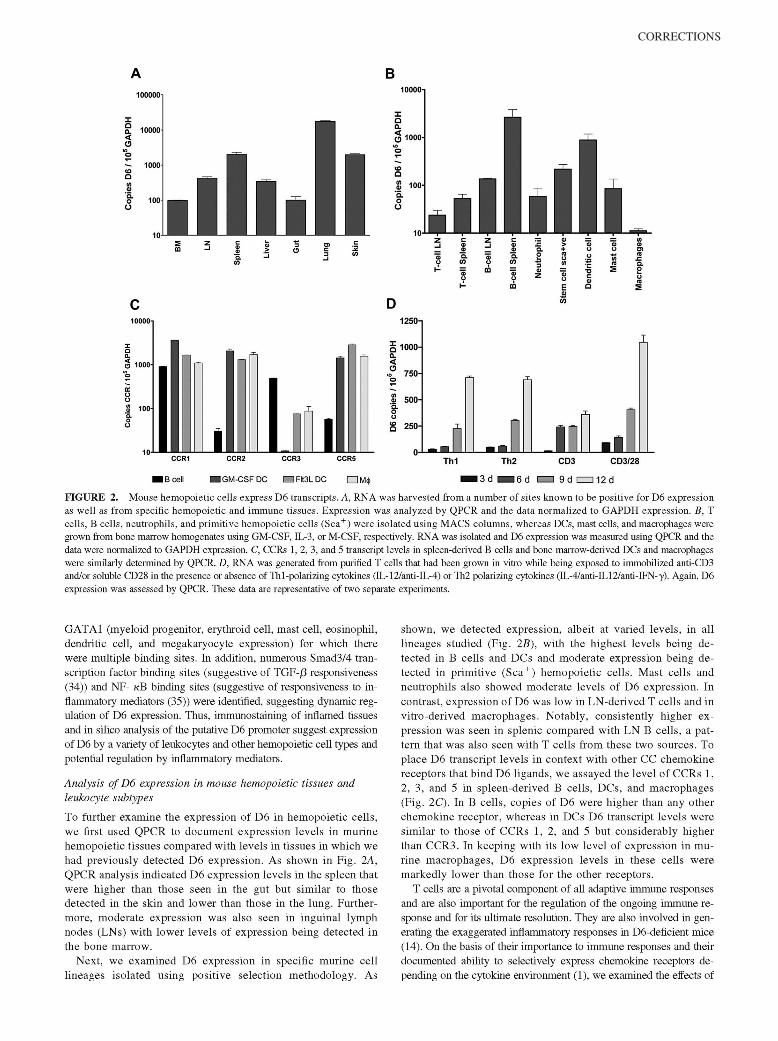
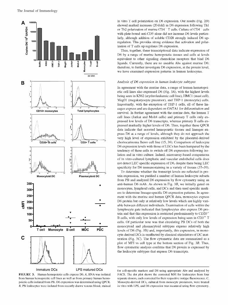
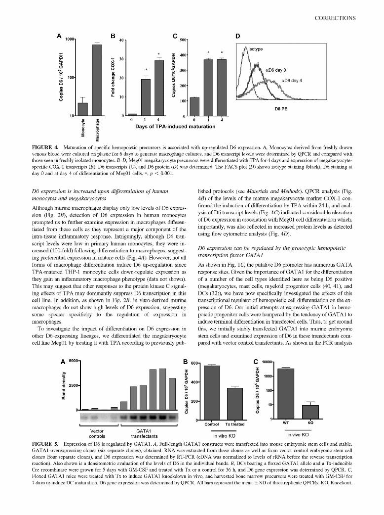
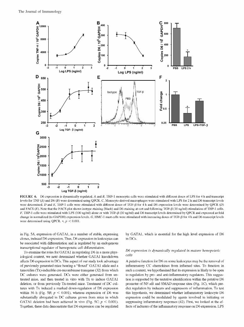
McKimmie, C. S., A. R. Fraser, C. Hansell, L. Gutierrez, S. Philipsen, L. Connell, A. Rot, M. Kurowska-Stolarska, P. Carreno, M.Pruenster, C.-C. Chu, G. Lombardi, C. Halsey, I. B. McInnes, F. Y. Liew, R. J. Nibbs, and G. J. Graham. 2008. Hemopoietic cellexpression of the chemokine decoy receptor D6 is dynamic and regulated by GATA1. J. Immunol. 181: 3353–3363.
Changes to Ref. 24 and in the numbering of the references throughout the article, requested by the authors at page proof stage, werenot made in production. This resulted in publication of the article with multiple errors in the references. The editors and staff of The Journalof Immunology apologize for this error.
The entire article is reproduced on the following pages with the correct references. The referencing errors have been corrected in theonline version, which now differs from the print version as originally published.
In addition, the authors revised the footnotes to include additional funding information. The corrected footnote is shown below.
1This study was supported by research funding from the Novartis Institute for Biomedical Research (Vienna) (to G.J.G. and R.J.N.) andfrom INNOCHEM (to G.J.G. and A.R.). S.P. and L.G. are supported by Netherlands Organization for Scientific Research-ALW Grant815-02-008. L.C. and R.J.N. received financial support from Tenovus Scotland (Grant S06/06).
Copyright © 2008 by The American Association of Immunologists, Inc. 0022-1767/08/$2.00
The Journal of Immunology
www.jimmunol.org
Corrections
www.jimmunol.org
CORRECTIONS
The Journal of Immunology
CORRECTIONS
The Journal of Immunology
CORRECTIONS
The Journal of Immunology
CORRECTIONS
The Journal of Immunology
CORRECTIONS
The Journal of Immunology