Embed Size (px)
Citation preview
Il cancro è una malattia genetica.
Differentemente da altre malattie genetiche, a trasmissione
mendeliana oppure ad eziologia multifattoriale,
1
mendeliana oppure ad eziologia multifattoriale,
esso è principalmente causato da mutazioni de novoche avvengono
nel genoma di cellule somatiche.

2

3
Figure 20-11 Molecular Biology of the Cell (© Garland Science 2008)

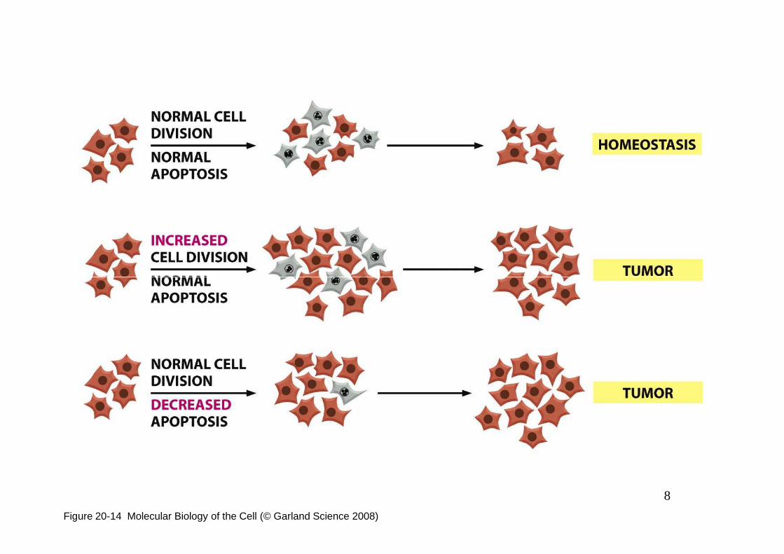
Affinché si sviluppi un tumore non è sufficiente la mutazione di un
singolo gene, bensì è necessario che si accumulino all’interno della
stesso genoma differenti mutazioni (il cui numero esatto varia in
dipendenza dello specifico tipo di neoplasia):
4
infatti, il controllo della proliferazione e del differenziamento
cellulare sono caratteristiche fenotipiche estremamente complesse e
multigeniche.
5
6
7
8
Figure 20-14 Molecular Biology of the Cell (© Garland Science 2008)
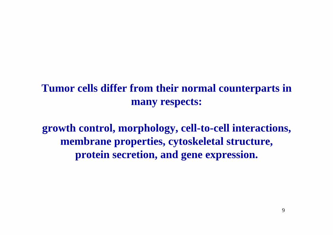
Tumor cells differ from their normal counterparts i n many respects:
9
growth control, morphology, cell-to-cell interactions, membrane properties, cytoskeletal structure,
protein secretion, and gene expression.
10

11
Figure 20-20b Molecular Biology of the Cell (© Garland Science 2008)
12
Figure 20-22 Molecular Biology of the Cell (© Garland Science 2008)

13
Figure 20-2 Molecular Biology of the Cell (© Garland Science 2008)
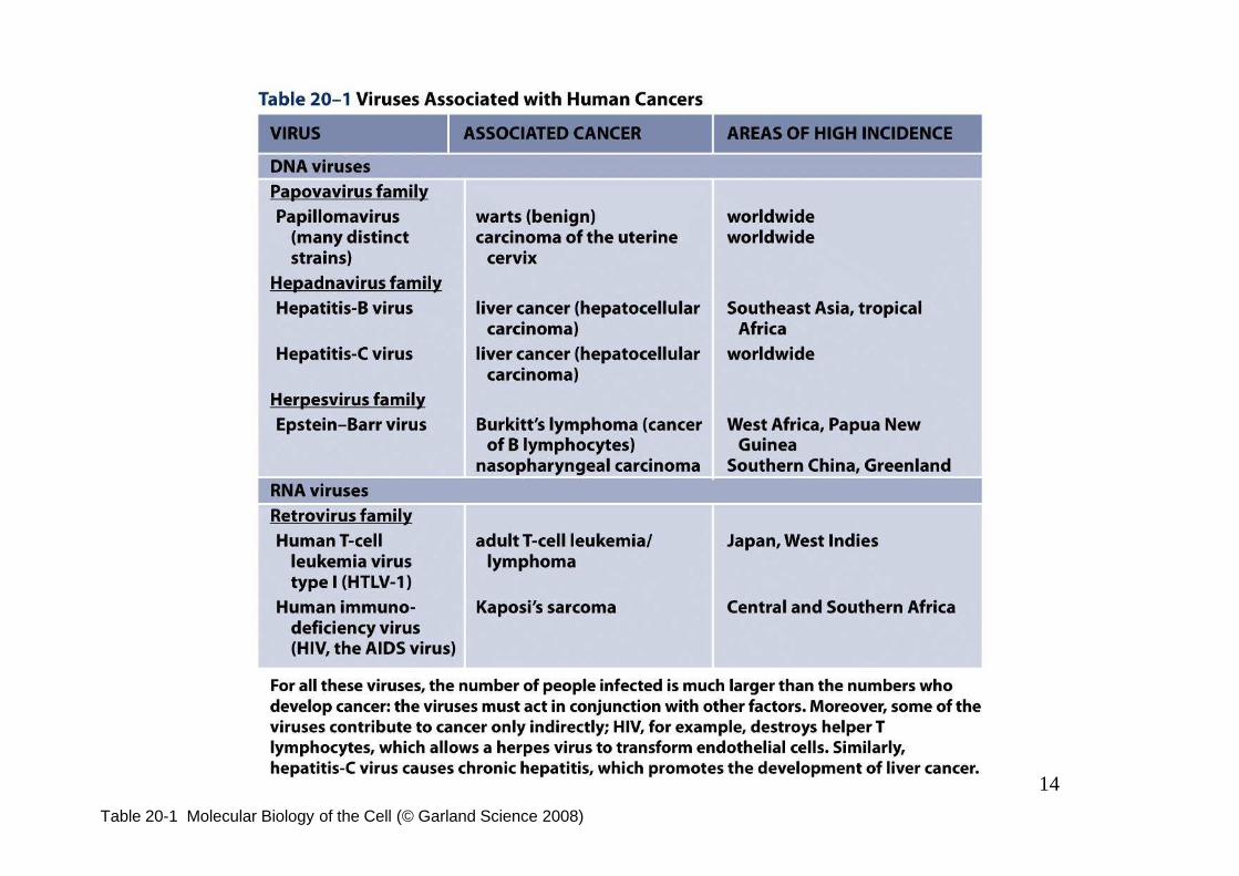
14
Table 20-1 Molecular Biology of the Cell (© Garland Science 2008)

Quali sono i geni le cui mutazioni possono
contribuire all’insorgenza del
fenotipo neoplastico ?
15

16
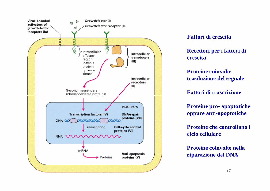
Fattori di crescita
Recettori per i fattori di crescita
Proteine coinvolte trasduzione del segnale
Fattori di trascrizione
17
Proteine pro- apoptoticheoppure anti-apoptotiche
Proteine che controllano ilciclo cellulare
Proteine coinvolte nellariparazione del DNA
18
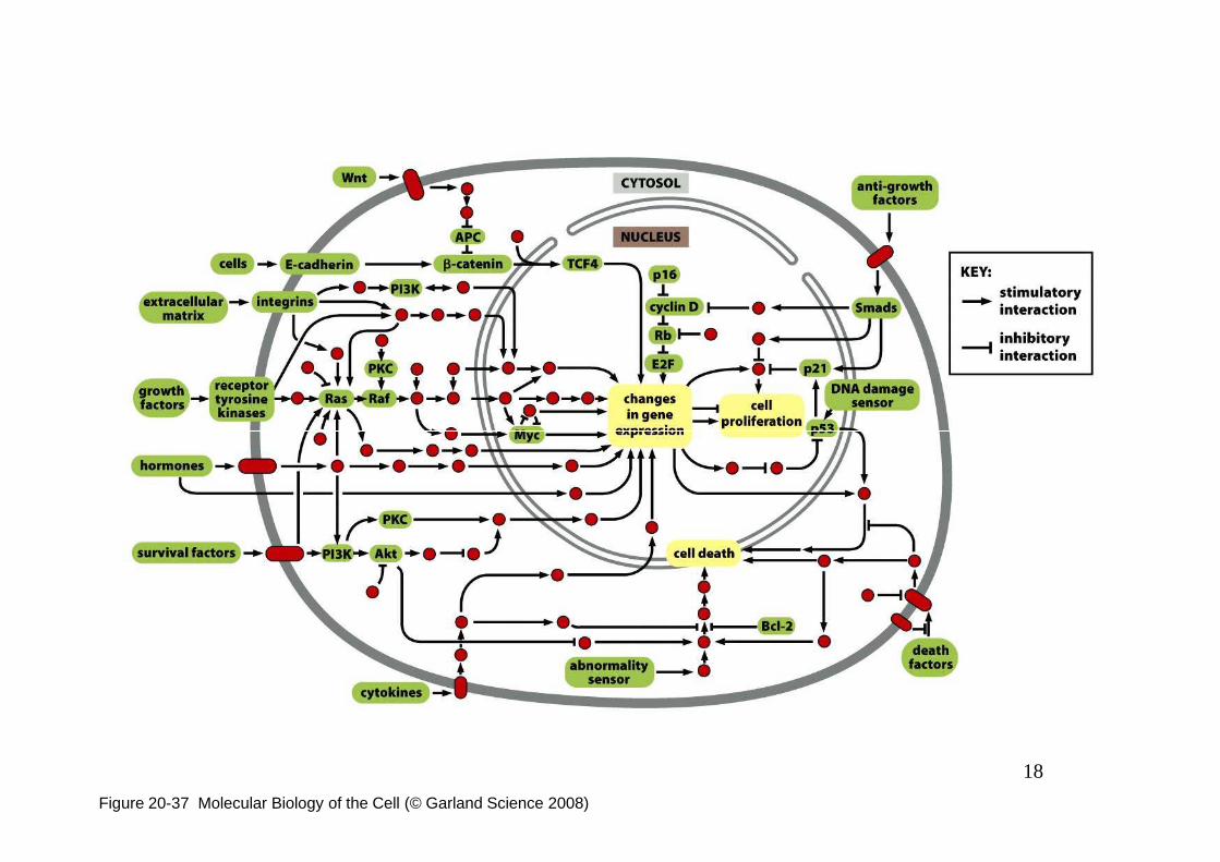
Figure 20-37 Molecular Biology of the Cell (© Garland Science 2008)
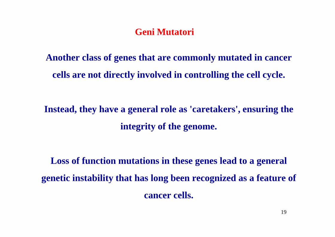
Another class of genes that are commonly mutated in cancer
cells are not directly involved in controlling the cell cycle.
Instead, they have a general role as 'caretakers', ensuring the
Geni Mutatori
19
integrity of the genome.
Loss of function mutations in these genes lead to a general
genetic instability that has long been recognized as a feature of
cancer cells.
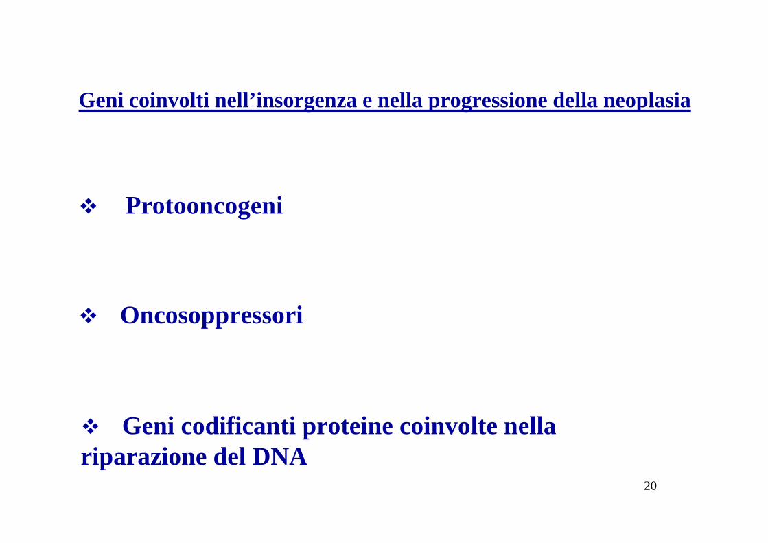
Geni coinvolti nell’insorgenza e nella progressione della neoplasia
� Protooncogeni
20
� Oncosoppressori
� Geni codificanti proteine coinvolte nella riparazione del DNA
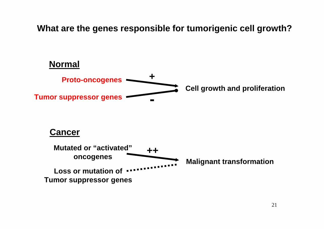
What are the genes responsible for tumorigenic cell growth?
Normal
Proto-oncogenesCell growth and proliferation
Tumor suppressor genes
+
-
21
Cancer
Mutated or “activated”oncogenes
Malignant transformationLoss or mutation of
Tumor suppressor genes
++
22

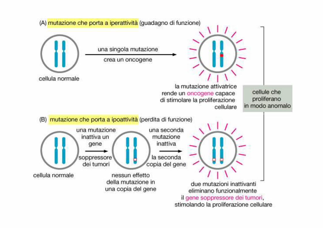
Le mutazioni dei protooncogèni(che diventano oncogèni) alterano la
struttura e le funzioni del gene, e della proteina codificata,
in modo tale da determinare un cambiamento del fenotipo anche in
23
condizione di eterozigosi (gain of function mutations):
si comportano, quindi, in maniera dominante.

D’altra parte, le mutazioni dei geni oncosoppressori(cioè, geni che
modulano negativamente la proliferazione cellulare),
24
causano perdita della funzione del gene e della proteina codificata,
si comportano in modo recessivo (loss of function mutations).
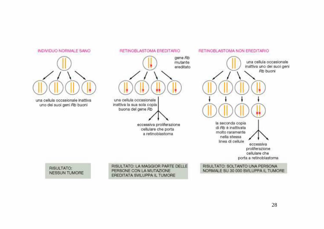
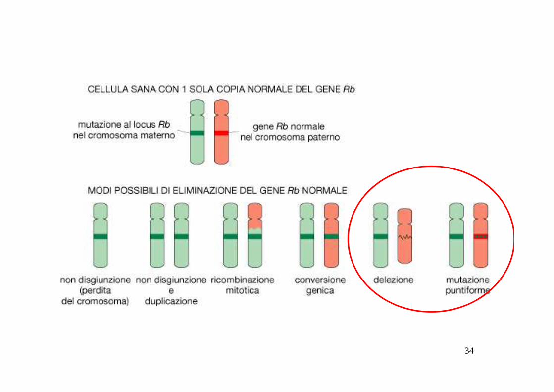
In alcuni tipi di tumori, gli individui affetti ereditano dai ge nitori
alcune delle mutazioni,
La Predisposizione familiareMutazioni germinali a carico di oncosoppresori
25
alcune delle mutazioni,
che possono contribuire all’insorgenza di un determinato fenotipo
neoplastico.

Sporadic and familial (Mendelian) forms of cancerKnudson’s two-hit hypothesis
SporadicNormal tumorsuppressorgene
Somaticmutationin one allele
26
Single tumors,unilateral,later-onset
Somaticmutationin other allele
• two mutations (two hits) are required for loss of t umor suppressor function

Sporadic and familial (Mendelian) forms of cancerKnudson’s two-hit hypothesis
FamilialTumor suppressor genecontaining a germlinemutation in one allele -heterozygous for themutation
Somaticmutation
27
Multiple tumors,bilateral,early-onset
mutationin other allele
• two mutations (two hits) are required for loss of t umor suppressor function• the first “hit” is inherited and the second “hit” is so matic
28

29

30
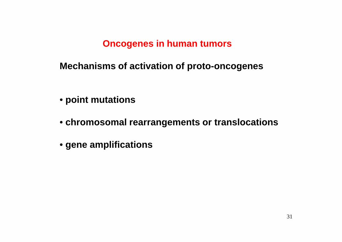
Oncogenes in human tumors
Mechanisms of activation of proto-oncogenes
• point mutations
• chromosomal rearrangements or translocations
31
• chromosomal rearrangements or translocations
• gene amplifications

1. Point mutationsin a proto-oncogene that result in a constitutively acting protein product
2. Localized reduplication (gene amplification) of a DNA segment that includes a proto-oncogene, leading to
32
segment that includes a proto-oncogene, leading to overexpression of the encoded protein
3. Chromosomal translocation that brings a growth-regulatory gene under the control of a different promoter and that causes inappropriate expression of the gene
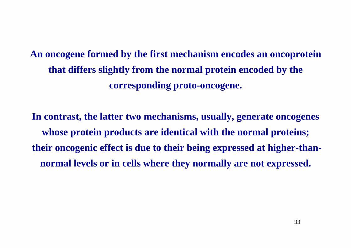
An oncogene formed by the first mechanism encodes an oncoprotein
that differs slightly from the normal protein encoded by the
corresponding proto-oncogene.
In contrast, the latter two mechanisms, usually, generate oncogenes
33
whose protein products are identical with the normal proteins;
their oncogenic effect is due to their being expressed at higher-than-
normal levels or in cells where they normally are not expressed.
34

35
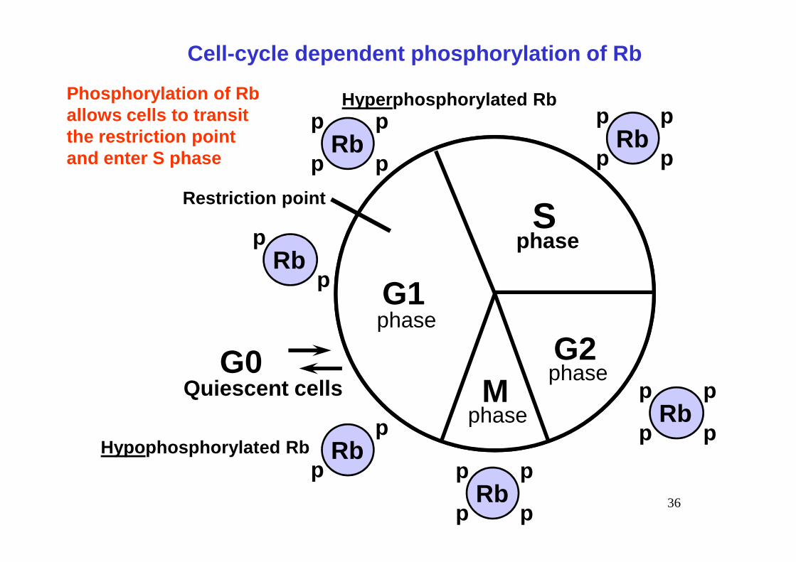
Cell-cycle dependent phosphorylation of Rb
G1
Sphase
Rbp
p
p
pRb
p
p
p
p
Rbp
p
Restriction point
Phosphorylation of Rballows cells to transitthe restriction pointand enter S phase
Hyper phosphorylated Rb
36
G1
G2M
G0Quiescent cells
phase
phase
phase
Rbp
p
p
p
Rbp
p
p
p
p
Rbp
pHypo phosphorylated Rb
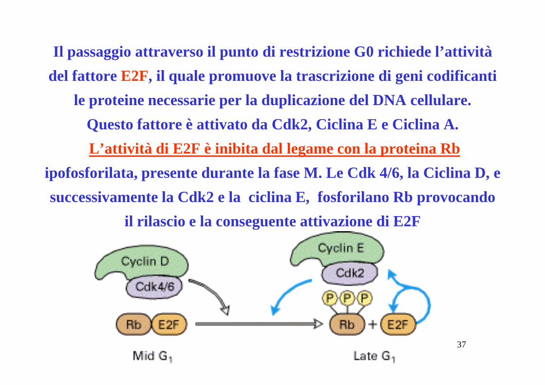
Il passaggio attraverso il punto di restrizione G0 richiede l’attività
del fattore E2F, il quale promuove la trascrizione di geni codificanti
le proteine necessarie per la duplicazione del DNA cellulare.
Questo fattore è attivato da Cdk2, Ciclina E e Ciclina A.
L’attività di E2F è inibita dal legame con la proteina Rb
ipofosforilata, presente durante la fase M. Le Cdk 4/6, la Ciclina D, e
successivamente la Cdk2 e la ciclina E, fosforilano Rb provocando
37
successivamente la Cdk2 e la ciclina E, fosforilano Rb provocando
il rilascio e la conseguente attivazione di E2F
38

39

40
Figure 20-40 Molecular Biology of the Cell (© Garland Science 2008)
41

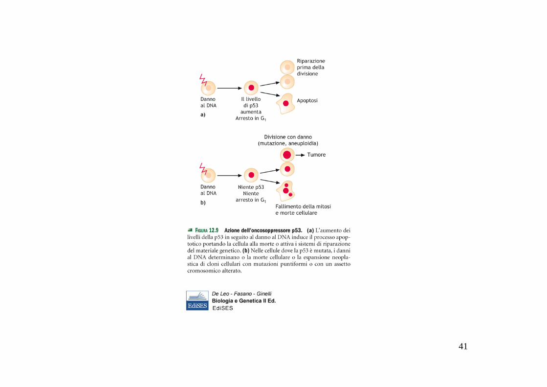
I danni subiti dal DNA nucleare sono identificati da un sistema di controllo, attivo durante G1 e G2, che si basa sulla attivazione dip53, un fattore di trascrizione che stimola l’espressione di p21CIP.
Questo cyclin-kinase inhibitor(CKI) si lega ai complessi Cdk-Ciclina e li inibisce, causando l’arresto del ciclo in G1 oppure in G2 finchè
il danno non sia stato riparato
42
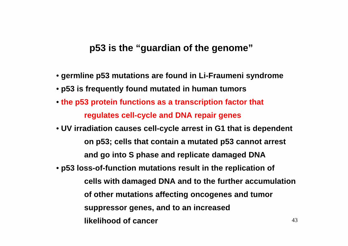
p53 is the “guardian of the genome”
• germline p53 mutations are found in Li-Fraumeni syn drome
• p53 is frequently found mutated in human tumors
• the p53 protein functions as a transcription factor that
regulates cell-cycle and DNA repair genes
43
• UV irradiation causes cell-cycle arrest in G1 that is dependent
on p53; cells that contain a mutated p53 cannot arr est
and go into S phase and replicate damaged DNA
• p53 loss-of-function mutations result in the replic ation of
cells with damaged DNA and to the further accumulat ion
of other mutations affecting oncogenes and tumor
suppressor genes, and to an increased
likelihood of cancer
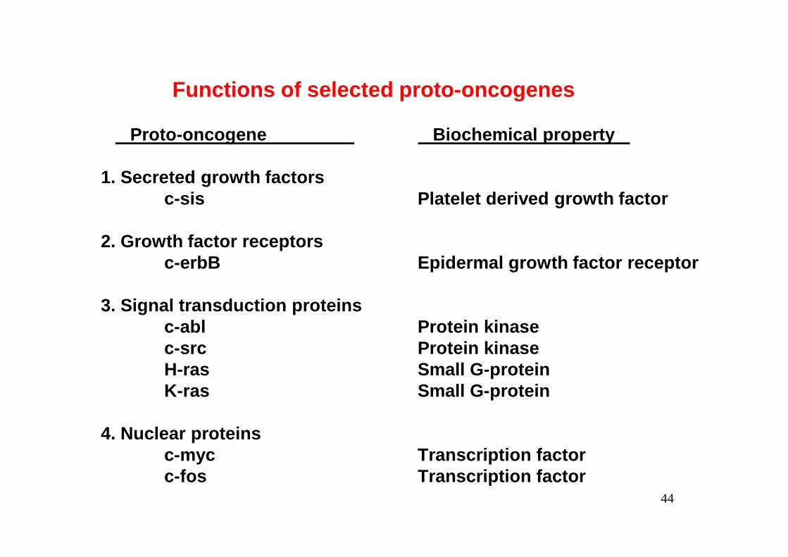
Functions of selected proto-oncogenes
Proto-oncogene Biochemical property
1. Secreted growth factorsc-sis Platelet derived growth factor
2. Growth factor receptorsc-erbB Epidermal growth factor receptor
44
3. Signal transduction proteinsc-abl Protein kinasec-src Protein kinaseH-ras Small G-proteinK-ras Small G-protein
4. Nuclear proteinsc-myc Transcription factorc-fos Transcription factor
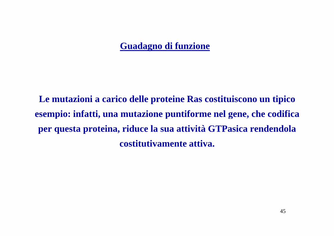
Le mutazioni a carico delle proteine Ras costituiscono un tipico
esempio: infatti, una mutazione puntiforme nel gene, che codifica
Guadagno di funzione
45
esempio: infatti, una mutazione puntiforme nel gene, che codifica
per questa proteina, riduce la sua attività GTPasica rendendola
costitutivamente attiva.
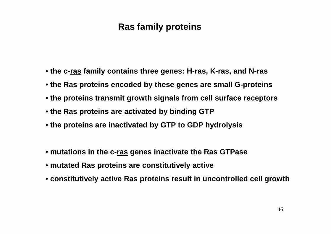
Ras family proteins
• the c-ras family contains three genes: H-ras, K-ras, and N-ra s
• the Ras proteins encoded by these genes are small G -proteins
• the proteins transmit growth signals from cell surf ace receptors
• the Ras proteins are activated by binding GTP
46
• the proteins are inactivated by GTP to GDP hydrolys is
• mutations in the c-ras genes inactivate the Ras GTPase
• mutated Ras proteins are constitutively active
• constitutively active Ras proteins result in uncont rolled cell growth

amino acid positionRas gene 12 59 61 Tumor
c-ras (H, K, N) Gly Ala Gln normal cells
H-ras Gly Ala Leu lung carcinomaVal Ala Gln bladder carcinoma
K-ras Cys Ala Gln lung carcinoma
Amino acid substitutions in Ras family proteins
47
K-ras Cys Ala Gln lung carcinomaArg Ala Gln lung carcinomaVal Ala Gln colon carcinoma
N-ras Gly Ala Lys neuroblastomaGly Ala Arg lung carcinoma
Murine sarcoma virus
H-ras Arg Thr Gln Harvey strainK-ras Ser Thr Gln Kirsten strain

48
Le ricerche sono state condotte sul cetuximab, un anticorpo monoclonale utilizzato
per il trattamento del cancro del colon-retto metastatico ed è stato individuato un
gene (KRAS) che predice l'efficacia di questa molecola sul paziente.
I risultati mostrano che il cetuximab funziona meglio nei pazienti che non
presentano mutazioni in questo marcatore.
Questa scoperta è un altro decisivo passo avanti verso la messa a punto di terapie
sempre più mirate e su misura per il paziente.

49

Una mutazione puntiforme nel gene, che codifica per la
proteina Ras, riduce la sua attività GTPasica rendendola
costitutivamente attiva.
50

Cetuximab è un anticorpo
monoclonale che blocca il
recettore dell’EGF.
La proteina Ras si trova a valle
di EGFR, quindi anche bloccando
51
di EGFR, quindi anche bloccando
il recettore non si ottiene la
risposta desiderata (blocco della
proliferazione cellulare)
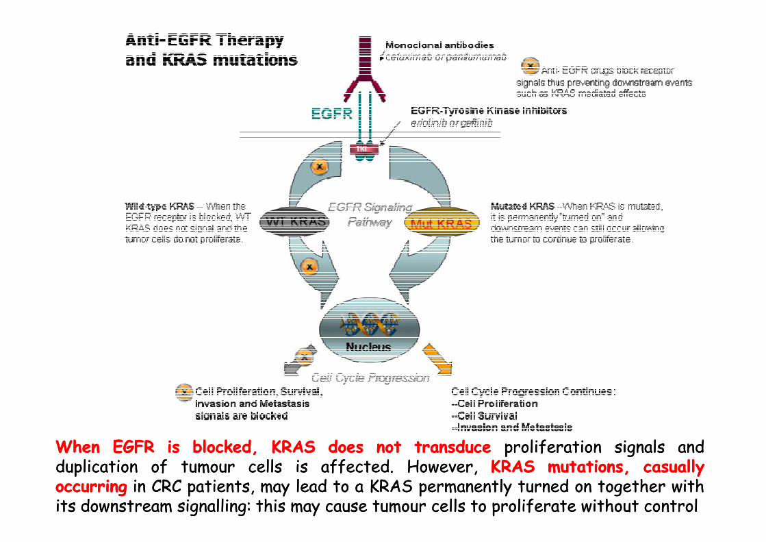
WhenWhen EGFREGFR isis blocked,blocked, KRASKRAS doesdoes notnot transducetransduce proliferationproliferation signalssignals andandduplicationduplication ofof tumourtumour cellscells isis affectedaffected.. However,However, KRASKRAS mutations,mutations, casuallycasuallyoccurringoccurring inin CRCCRC patients,patients, maymay leadlead toto aa KRASKRAS permanentlypermanently turnedturned onon togethertogether withwithitsits downstreamdownstream signallingsignalling:: thisthis maymay causecause tumourtumour cellscells toto proliferateproliferate withoutwithout controlcontrol

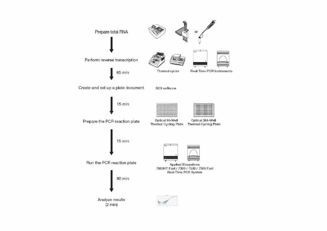
Ricerca delle mutazioni del gene K-Ras
nella biopsia del paziente
53
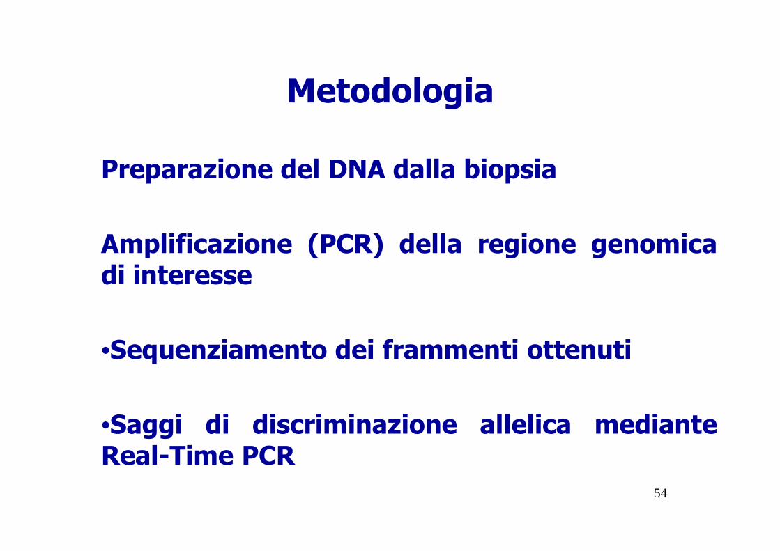
Metodologia
Preparazione del DNA dalla biopsia
Amplificazione (PCR) della regione genomicadi interesse
54
di interesse
•Sequenziamento dei frammenti ottenuti
•Saggi di discriminazione allelica medianteReal-Time PCR
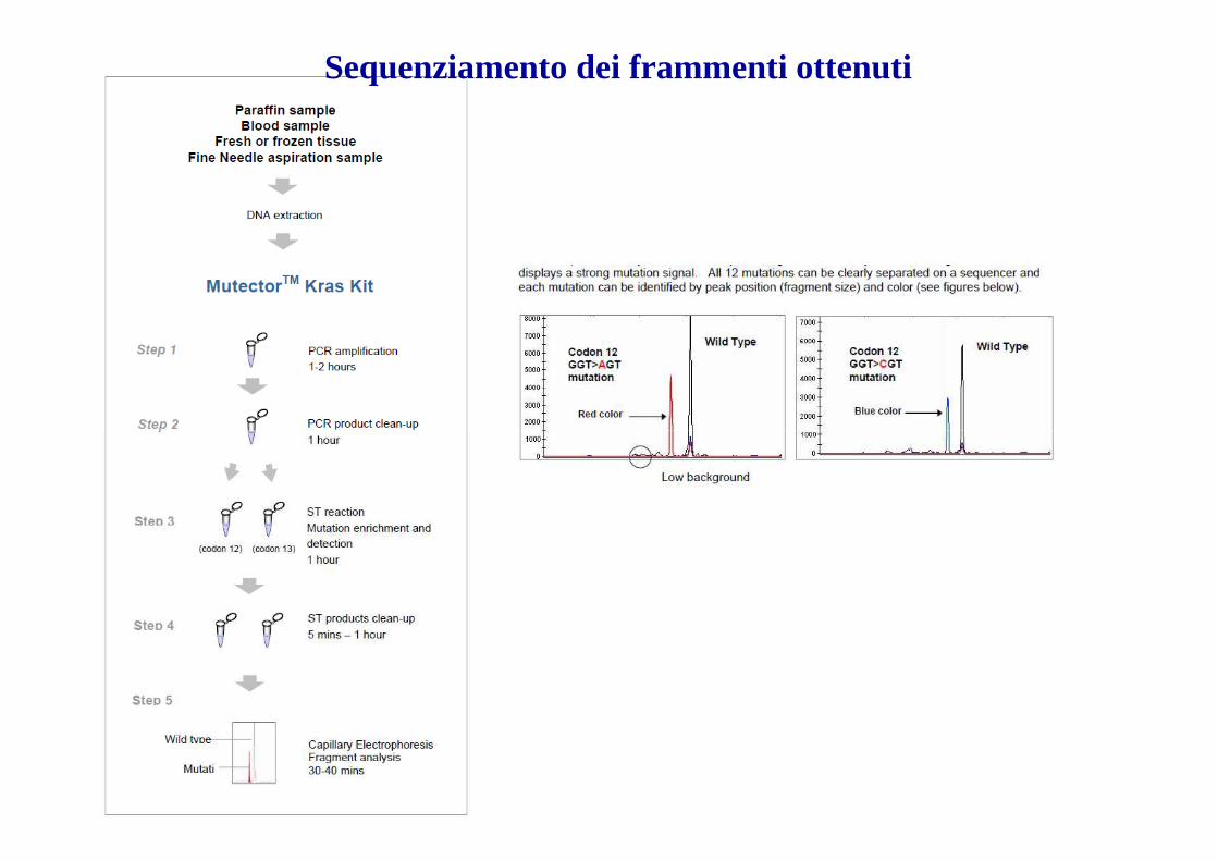
Sequenziamento dei frammenti ottenuti
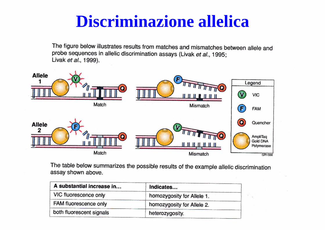
Discriminazione allelica
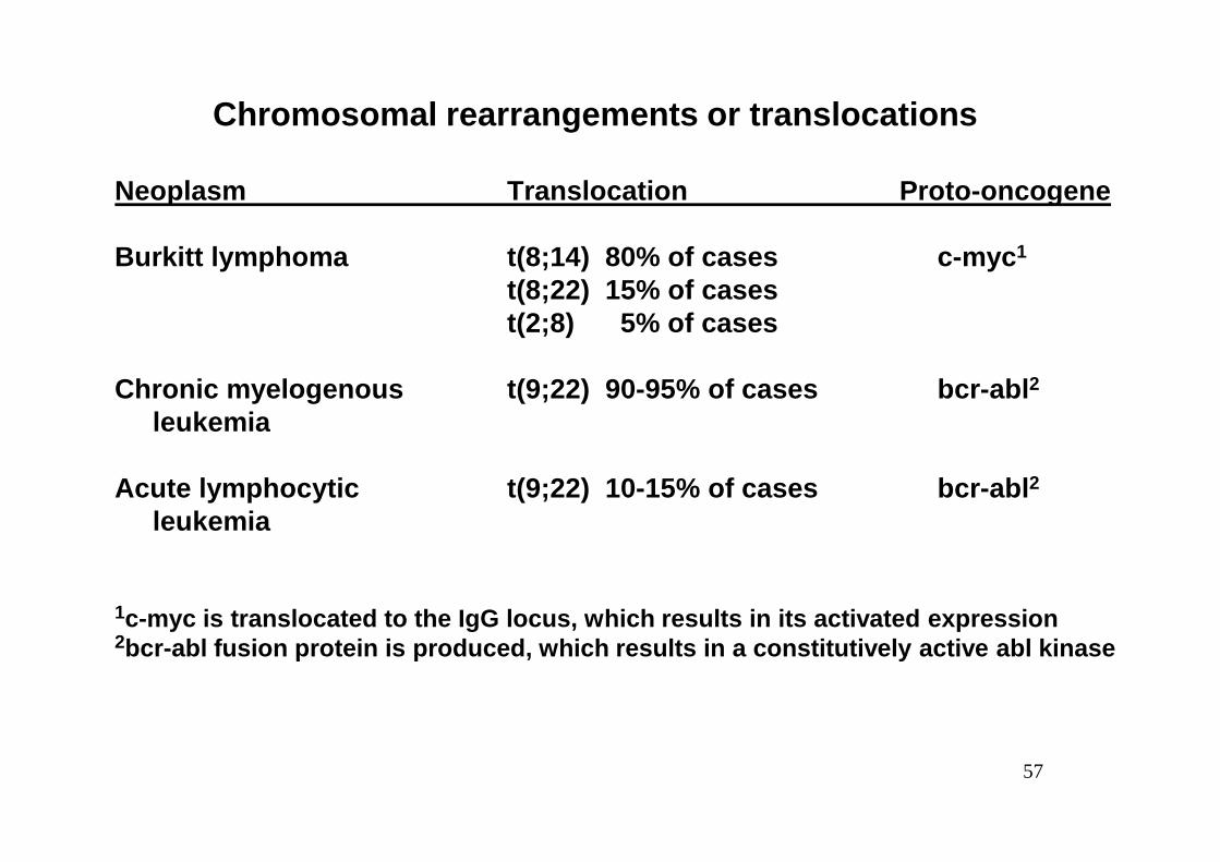
Chromosomal rearrangements or translocations
Neoplasm Translocation Proto-oncogene
Burkitt lymphoma t(8;14) 80% of cases c-myc 1
t(8;22) 15% of casest(2;8) 5% of cases
Chronic myelogenous t(9;22) 90-95% of cases bcr-abl 2
leukemia
57
leukemia
Acute lymphocytic t(9;22) 10-15% of cases bcr-abl 2
leukemia
1c-myc is translocated to the IgG locus, which resul ts in its activated expression2bcr-abl fusion protein is produced, which results i n a constitutively active abl kinase
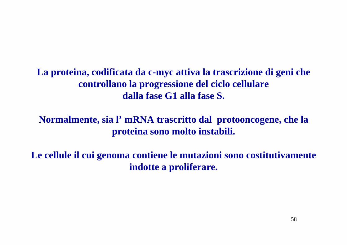
La proteina, codificata da c-myc attiva la trascrizione di geni che controllano la progressione del ciclo cellulare
dalla fase G1 alla fase S.
Normalmente, sia l’ mRNA trascritto dal protooncogene, che la
58
Normalmente, sia l’ mRNA trascritto dal protooncogene, che la proteina sono molto instabili.
Le cellule il cui genoma contiene le mutazioni sono costitutivamente indotte a proliferare.

sono tipiche le traslocazioni che interessano il cromosoma 8, dove è
localizzato il gene c-myc, ed uno dei tre cromosomi in cui sono
localizzati i geni che codificano per le catene pesanti e leggere delle
immunoglobuline.
La traslocazione più frequente è la [t (8:14)], che si riscontra nel
90% dei BL:
59
90% dei BL:
la traslocazione, spostando cMyc in prossimità dell’enhancer del
gene che codifica per una delle catene delle immunoglobuline, causa
la sua continua espressione e di conseguenza le cellule sono
costitutivamente stimolate a proliferare.
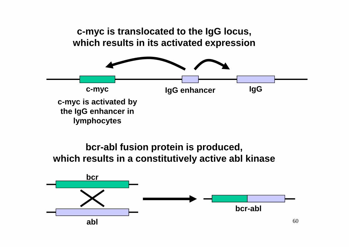
c-myc is translocated to the IgG locus,which results in its activated expression
c-myc IgGIgG enhancerc-myc is activated bythe IgG enhancer in
lymphocytes
60
bcr-abl fusion protein is produced,which results in a constitutively active abl kinase
bcr-abl
bcr
abl
lymphocytes
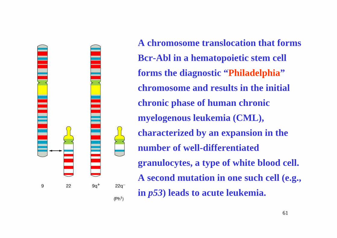
A chromosome translocation that forms
Bcr-Abl in a hematopoietic stem cell
forms the diagnostic “Philadelphia”
chromosome and results in the initial
chronic phase of human chronic
myelogenous leukemia (CML),
61
myelogenous leukemia (CML),
characterized by an expansion in the
number of well-differentiated
granulocytes, a type of white blood cell.
A second mutation in one such cell (e.g.,
in p53) leads to acute leukemia.
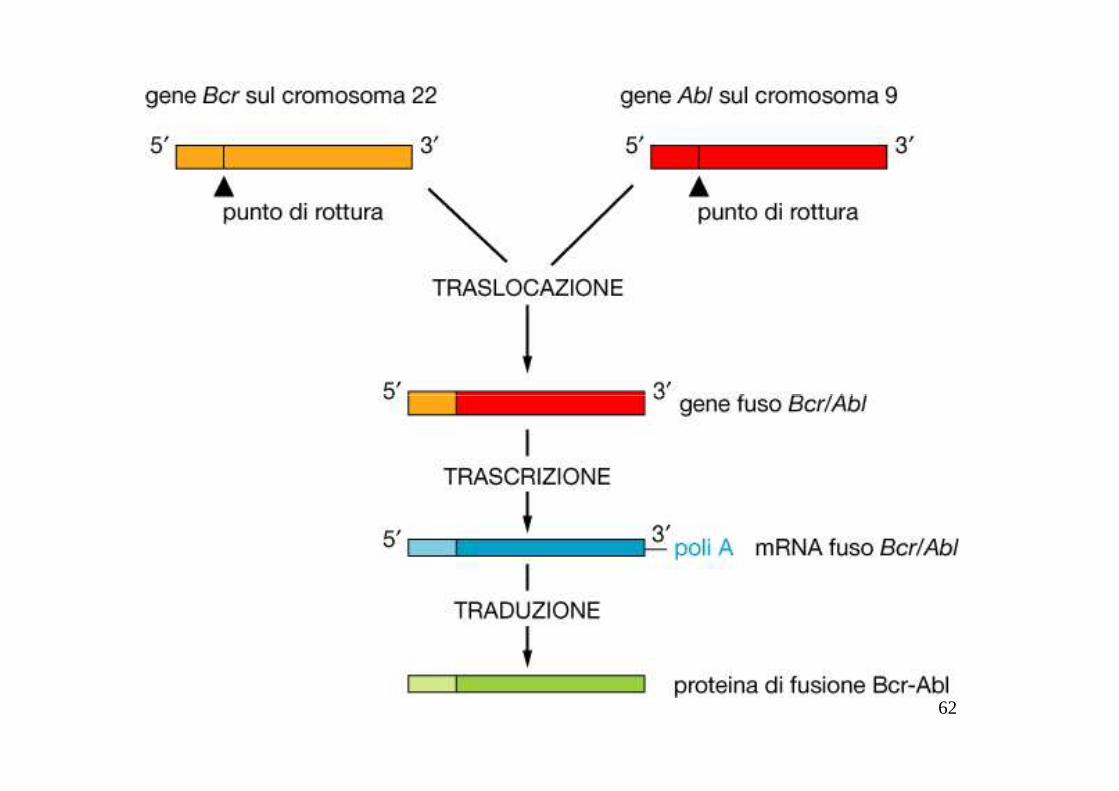
62
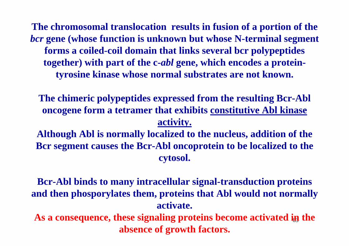
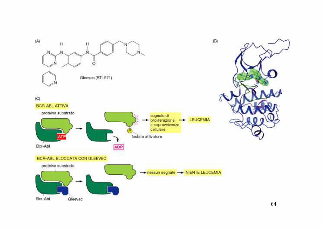
The chromosomal translocation results in fusion of a portion of the bcr gene (whose function is unknown but whose N-terminal segment
forms a coiled-coil domain that links several bcr polypeptides together) with part of the c-abl gene, which encodes a protein-
tyrosine kinase whose normal substrates are not known.
The chimeric polypeptides expressed from the resulting Bcr-Abl oncogene form a tetramer that exhibits constitutive Abl kinase
activity.
63
activity. Although Abl is normally localized to the nucleus, addition of the Bcr segment causes the Bcr-Abl oncoprotein to be localized to the
cytosol.
Bcr-Abl binds to many intracellular signal-transduction proteins and then phosporylates them, proteins that Abl would not normally
activate.As a consequence, these signaling proteins become activated in the
absence of growth factors.
64

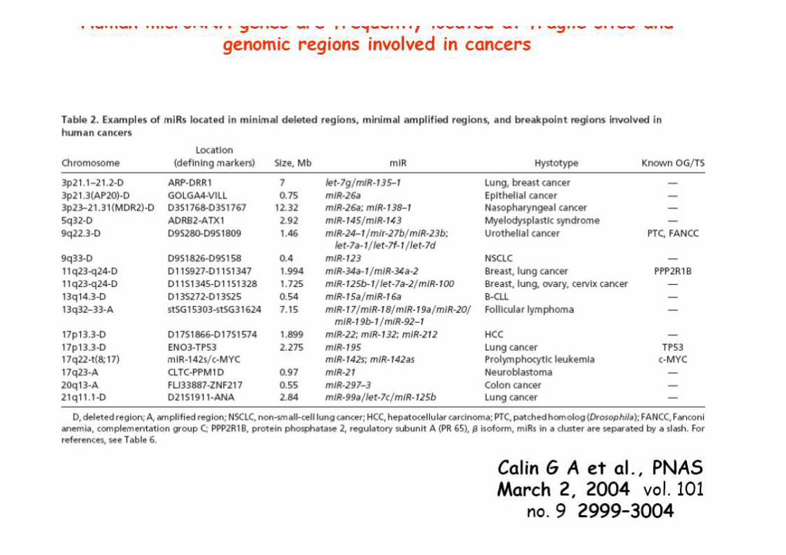
microRNAs CAN FUNCTION AS TS AND OG
65


68
74
75
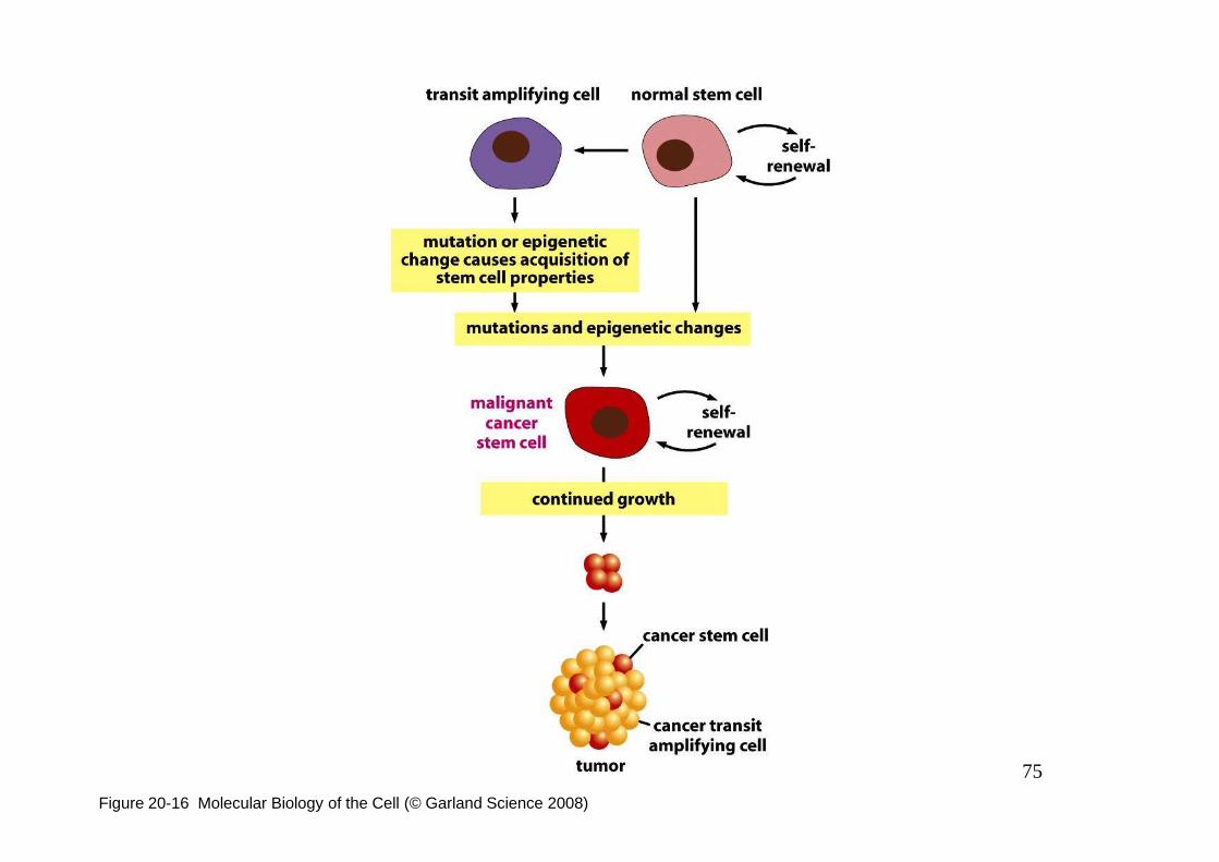
Figure 20-16 Molecular Biology of the Cell (© Garland Science 2008)