Embed Size (px)
Citation preview
Imaging of non-neoplastic liver lesions
in infants
Kristen Wrigley MD, Aruna Vade MD, and Jennifer E Lim- Dunham MD
Radiology Department Loyola University Medical Center
Maywood, IL
Non-neoplastic liver disease in infancy is rare and can present as a focal lesion or an infiltrative process.
While some liver lesions are benign and incidental findings, others may progress rapidly to end stage liver disease and eventually death.
Early recognition of these lesions may avoid prolonged morbidity and facilitate timely management.
Clinical findings seen in 16 infants with non-neoplastic liver lesions
Abdominal distentionHepatomegalyJaundiceAbnormal liver functionIncidental liver abnormality found on abdominal or renal ultrasound
ResultsNon-neoplastic liver processes were divided into
5 etiological categories:
IschemiaInfectionVascular lesionsTraumatic lesionsBiliary abnormalities
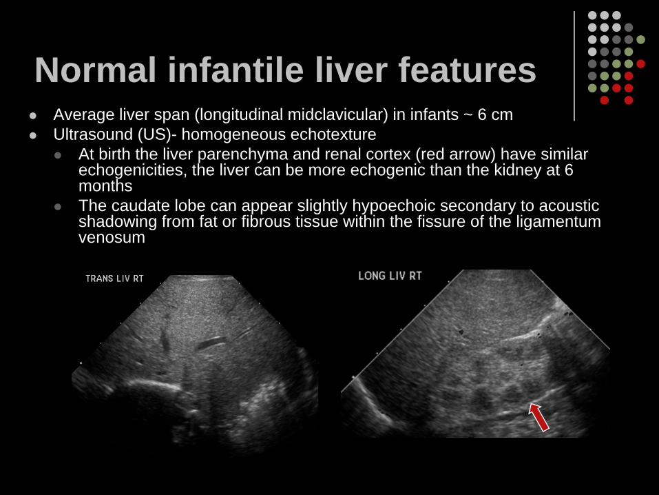
Normal infantile liver featuresAverage liver span (longitudinal midclavicular) in infants ~ 6 cmUltrasound (US)- homogeneous echotexture
At birth the liver parenchyma and renal cortex (red arrow) have similar echogenicities, the liver can be more echogenic than the kidney at 6 monthsThe caudate lobe can appear slightly hypoechoic secondary to acoustic shadowing from fat or fibrous tissue within the fissure of the ligamentumvenosum
Normal infantile liver features
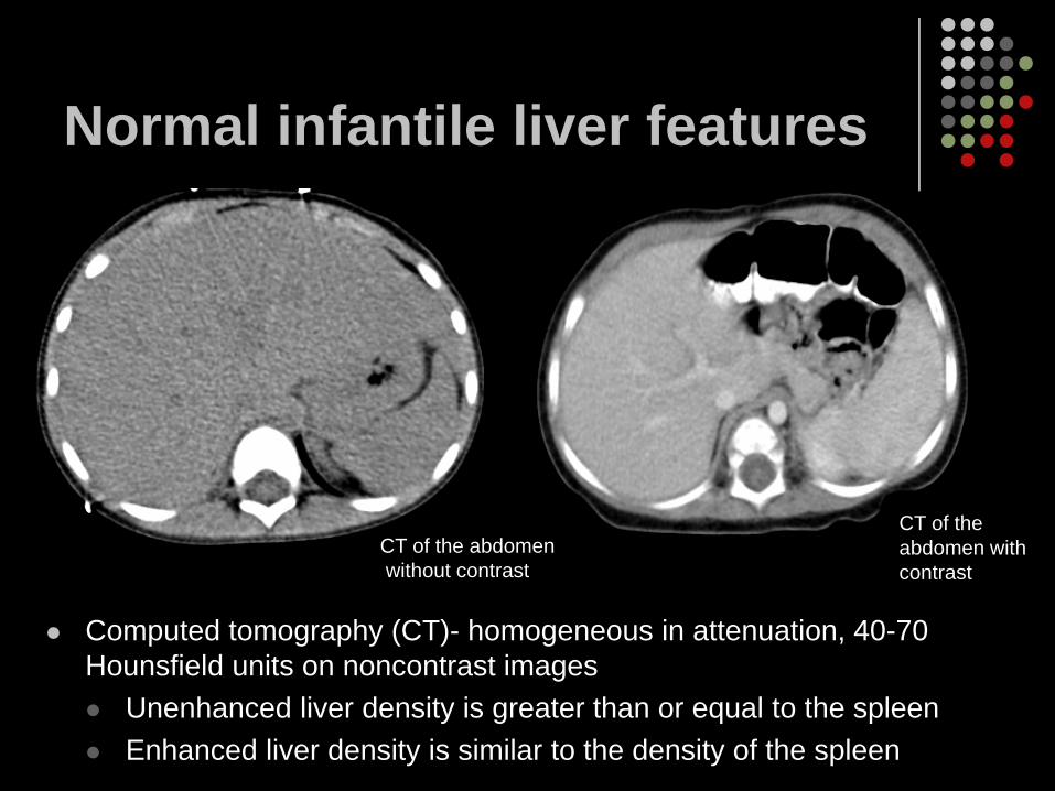
Computed tomography (CT)- homogeneous in attenuation, 40-70 Hounsfield units on noncontrast images
Unenhanced liver density is greater than or equal to the spleenEnhanced liver density is similar to the density of the spleen
CT of the abdomenwithout contrast
CT of the abdomen withcontrast
Normal infantile liver features
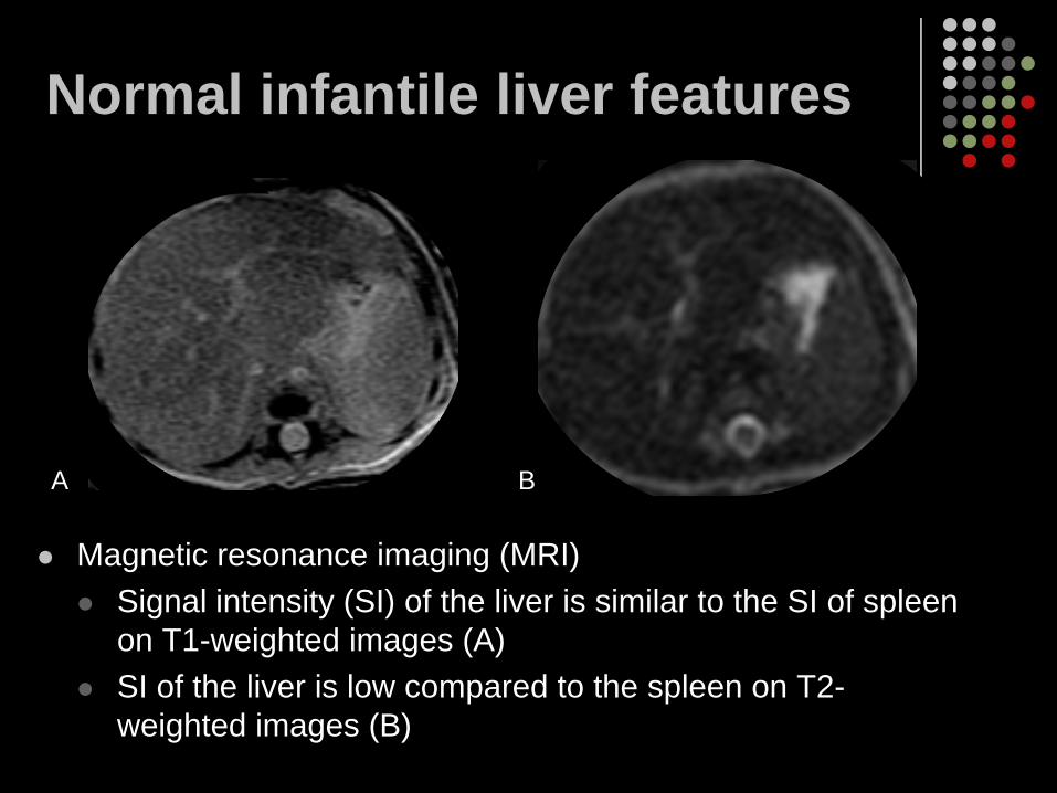
Magnetic resonance imaging (MRI)Signal intensity (SI) of the liver is similar to the SI of spleen on T1-weighted images (A)SI of the liver is low compared to the spleen on T2-weighted images (B)
A B
Case 1
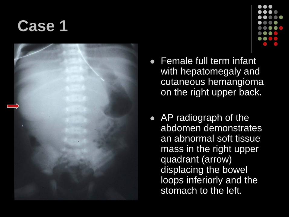
Female full term infant with hepatomegaly and cutaneous hemangioma on the right upper back.
AP radiograph of the abdomen demonstrates an abnormal soft tissue mass in the right upper quadrant (arrow) displacing the bowel loops inferiorly and the stomach to the left.
Case 1
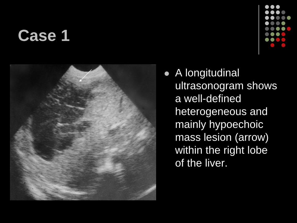
A longitudinal ultrasonogram shows a well-defined heterogeneous and mainly hypoechoic mass lesion (arrow) within the right lobe of the liver.
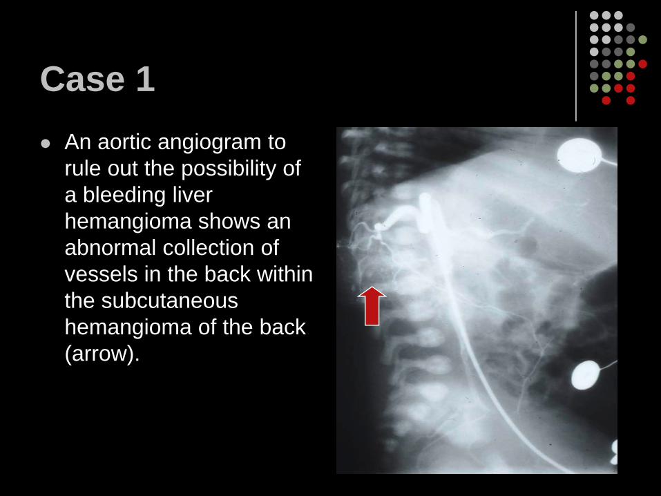
Case 1An aortic angiogram to rule out the possibility of a bleeding liver hemangioma shows an abnormal collection of vessels in the back within the subcutaneous hemangioma of the back (arrow).
Case 1
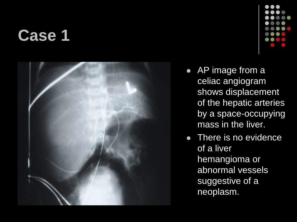
AP image from a celiac angiogram shows displacement of the hepatic arteries by a space-occupying mass in the liver. There is no evidence of a liver hemangioma or abnormal vessels suggestive of a neoplasm.
Case 1
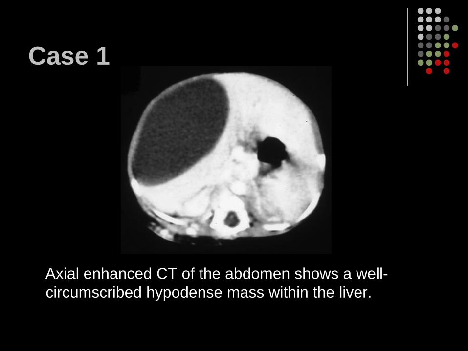
Axial enhanced CT of the abdomen shows a well-circumscribed hypodense mass within the liver.

Case 1
The liver mass (arrow) is hypointense on coronal T1-weighted (A) and axial T2-weighted images (B) compared to the normal liver (L). This is consistent with intralesional hemosiderin.
Collectively the US, CT, and MRI findings in this case are consistent with a liver hematoma.
Follow-up US after 6 months showed complete resolution of the hematoma.
A B
LL
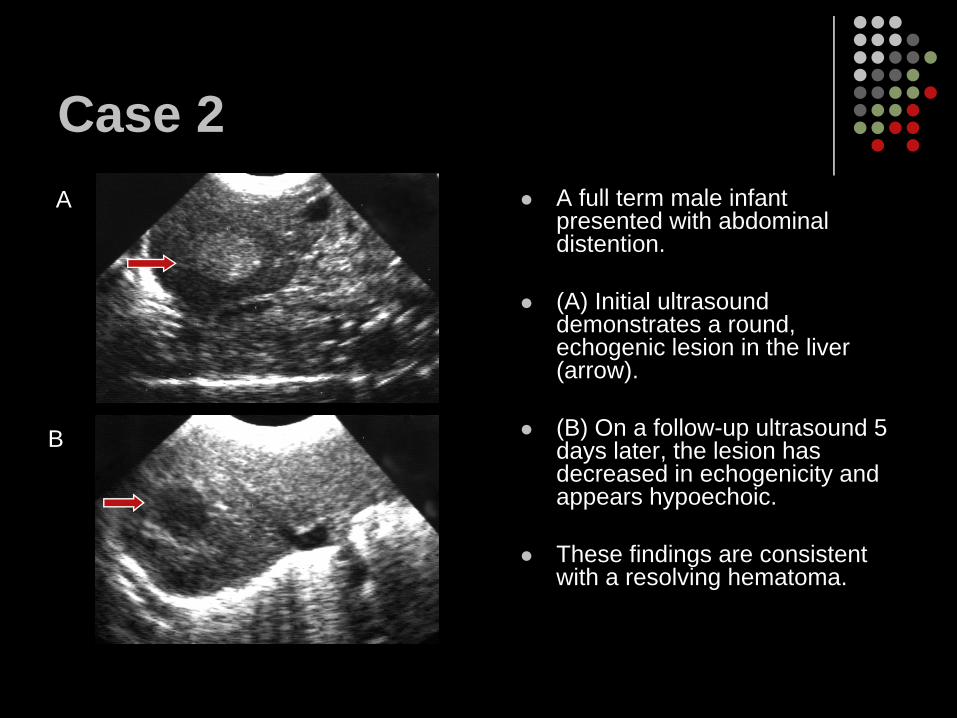
Case 2A full term male infant presented with abdominal distention.
(A) Initial ultrasound demonstrates a round, echogenic lesion in the liver (arrow).
(B) On a follow-up ultrasound 5 days later, the lesion has decreased in echogenicity and appears hypoechoic.
These findings are consistent with a resolving hematoma.
A
B
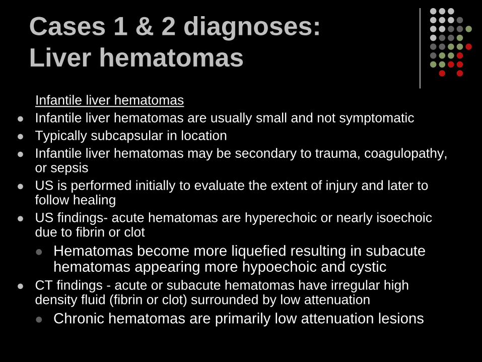
Cases 1 & 2 diagnoses:Liver hematomasInfantile liver hematomasInfantile liver hematomas are usually small and not symptomaticTypically subcapsular in locationInfantile liver hematomas may be secondary to trauma, coagulopathy, or sepsisUS is performed initially to evaluate the extent of injury and later to follow healingUS findings- acute hematomas are hyperechoic or nearly isoechoic due to fibrin or clot
Hematomas become more liquefied resulting in subacute hematomas appearing more hypoechoic and cystic
CT findings - acute or subacute hematomas have irregular high density fluid (fibrin or clot) surrounded by low attenuation
Chronic hematomas are primarily low attenuation lesions
Case 3
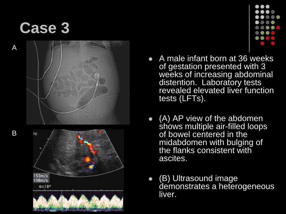
A male infant born at 36 weeks of gestation presented with 3 weeks of increasing abdominal distention. Laboratory tests revealed elevated liver function tests (LFTs).
(A) AP view of the abdomen shows multiple air-filled loops of bowel centered in the midabdomen with bulging of the flanks consistent with ascites.
(B) Ultrasound image demonstrates a heterogeneous liver.
A
B
Case 3
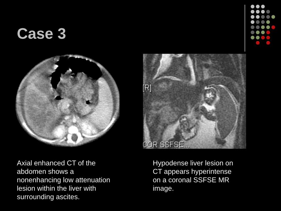
Axial enhanced CT of the abdomen shows a nonenhancing low attenuation lesion within the liver with surrounding ascites.
Hypodense liver lesion on CT appears hyperintense on a coronal SSFSE MR image.
Case 3Differential Diagnosis: metastatic neuroblastoma, diffuse hepatic necrosis from infection or ischemia. Neoplastic processes such as hepatoblastoma or hamartoma were considered less likely secondary to the diffuse appearance and lack of enhancement.
Liver biopsy confirmed multifocal parenchymal necrosis, focal portal and lobular inflammation, and moderate portal fibrosis. Mild cholestasis and proliferation of ductal structures were believed to be due to a nonspecific reactive process secondary to liver ischemia.
Case 3 diagnosis:Liver necrosis secondary to ischemia
Liver necrosisLiver ischemia in infants may be secondary to intraoperative ligation of the hepatic artery, vasculitis, or hypercoaguable statesUS findings - acute infarction appears as a peripheral hypoechoic area
Late infarction may appear as a cystic lesion of bileCT findings - typically wedge-shaped hypodense lesions, however can be centrally located with indistinct margins without significant enhancement
Case 4
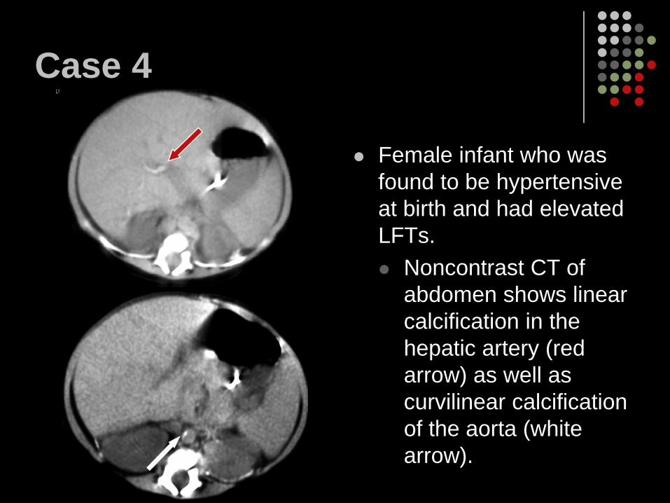
Female infant who was found to be hypertensive at birth and had elevated LFTs.
Noncontrast CT of abdomen shows linear calcification in the hepatic artery (red arrow) as well as curvilinear calcification of the aorta (white arrow).
Case 5
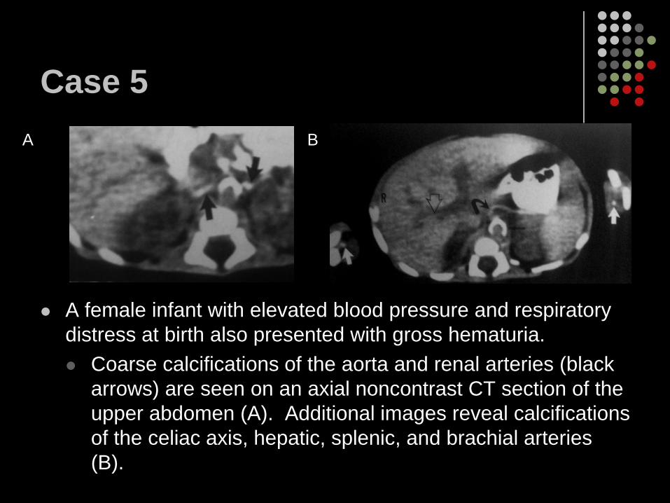
A female infant with elevated blood pressure and respiratory distress at birth also presented with gross hematuria.
Coarse calcifications of the aorta and renal arteries (black arrows) are seen on an axial noncontrast CT section of the upper abdomen (A). Additional images reveal calcifications of the celiac axis, hepatic, splenic, and brachial arteries (B).
A B
Case 5On the 19th day of life, the patient developed abdominal distention and necrotizing enterocolitis leading to death on the 21st day from extensive hemorrhagic bowel infarction with purulent peritonitis.
On autopsy the diagnosis of idiopathic infantile arterial calcification was confirmed.
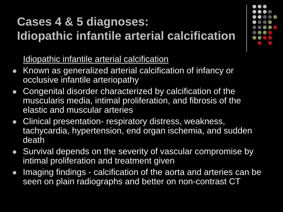
Cases 4 & 5 diagnoses:Idiopathic infantile arterial calcification
Idiopathic infantile arterial calcificationKnown as generalized arterial calcification of infancy or occlusive infantile arteriopathy Congenital disorder characterized by calcification of the muscularis media, intimal proliferation, and fibrosis of the elastic and muscular arteriesClinical presentation- respiratory distress, weakness, tachycardia, hypertension, end organ ischemia, and sudden deathSurvival depends on the severity of vascular compromise by intimal proliferation and treatment givenImaging findings - calcification of the aorta and arteries can be seen on plain radiographs and better on non-contrast CT
Case 6
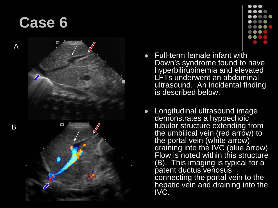
Full-term female infant with Down’s syndrome found to have hyperbilirubinemia and elevated LFTs underwent an abdominal ultrasound. An incidental finding is described below.
Longitudinal ultrasound image demonstrates a hypoechoic tubular structure extending from the umbilical vein (red arrow) to the portal vein (white arrow) draining into the IVC (blue arrow). Flow is noted within this structure (B). This imaging is typical for a patent ductus venosus connecting the portal vein to the hepatic vein and draining into the IVC.
A
B
Case 7
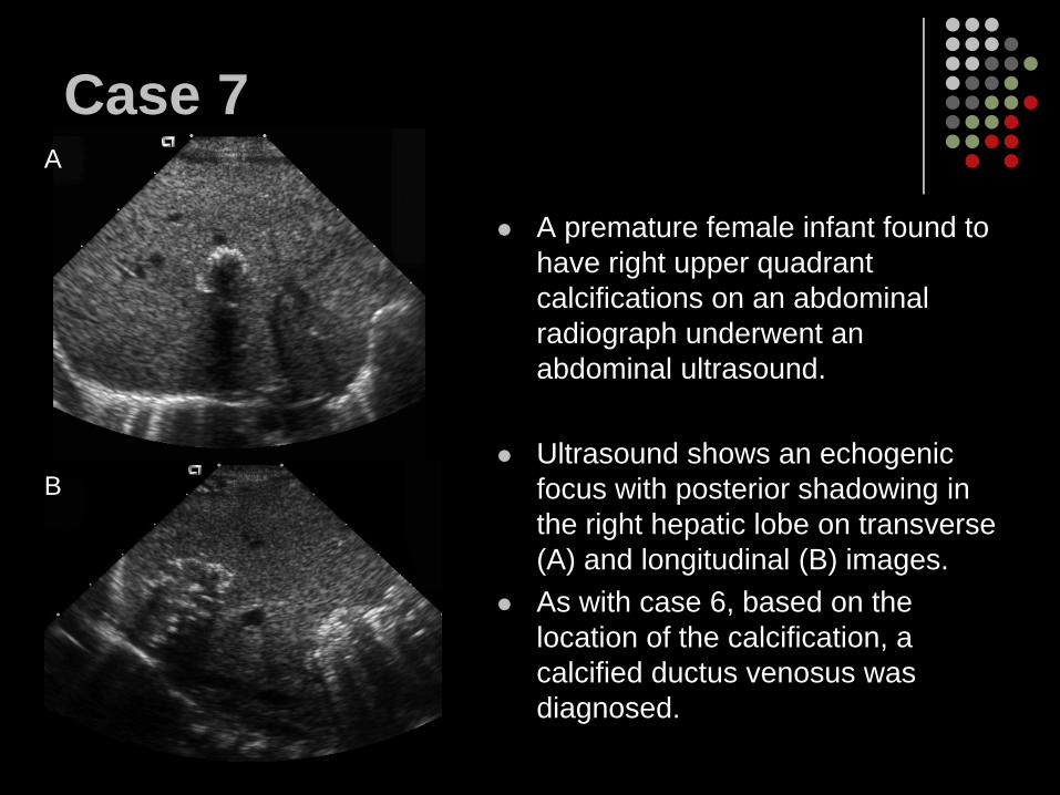
A premature female infant found to have right upper quadrant calcifications on an abdominal radiograph underwent an abdominal ultrasound.
Ultrasound shows an echogenic focus with posterior shadowing in the right hepatic lobe on transverse (A) and longitudinal (B) images. As with case 6, based on the location of the calcification, a calcified ductus venosus was diagnosed.
A
B
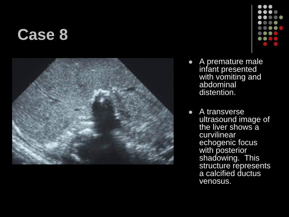
Case 8 A premature male infant presented with vomiting and abdominal distention.
A transverse ultrasound image of the liver shows a curvilinear echogenic focus with posterior shadowing. This structure represents a calcified ductus venosus.
Cases 6, 7, & 8 diagnoses:Ductus venosus remnants/calcification
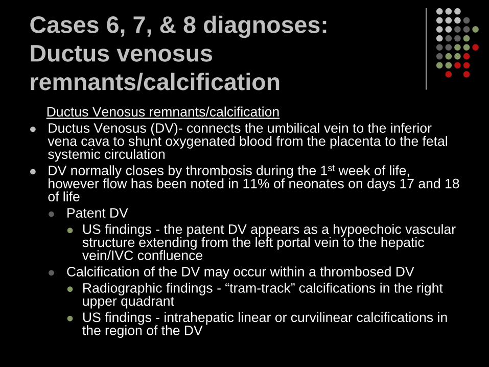
Ductus Venosus remnants/calcificationDuctus Venosus (DV)- connects the umbilical vein to the inferior vena cava to shunt oxygenated blood from the placenta to the fetal systemic circulationDV normally closes by thrombosis during the 1st week of life, however flow has been noted in 11% of neonates on days 17 and 18 of life
Patent DV US findings - the patent DV appears as a hypoechoic vascular structure extending from the left portal vein to the hepatic vein/IVC confluence
Calcification of the DV may occur within a thrombosed DVRadiographic findings - “tram-track” calcifications in the right upper quadrantUS findings - intrahepatic linear or curvilinear calcifications in the region of the DV
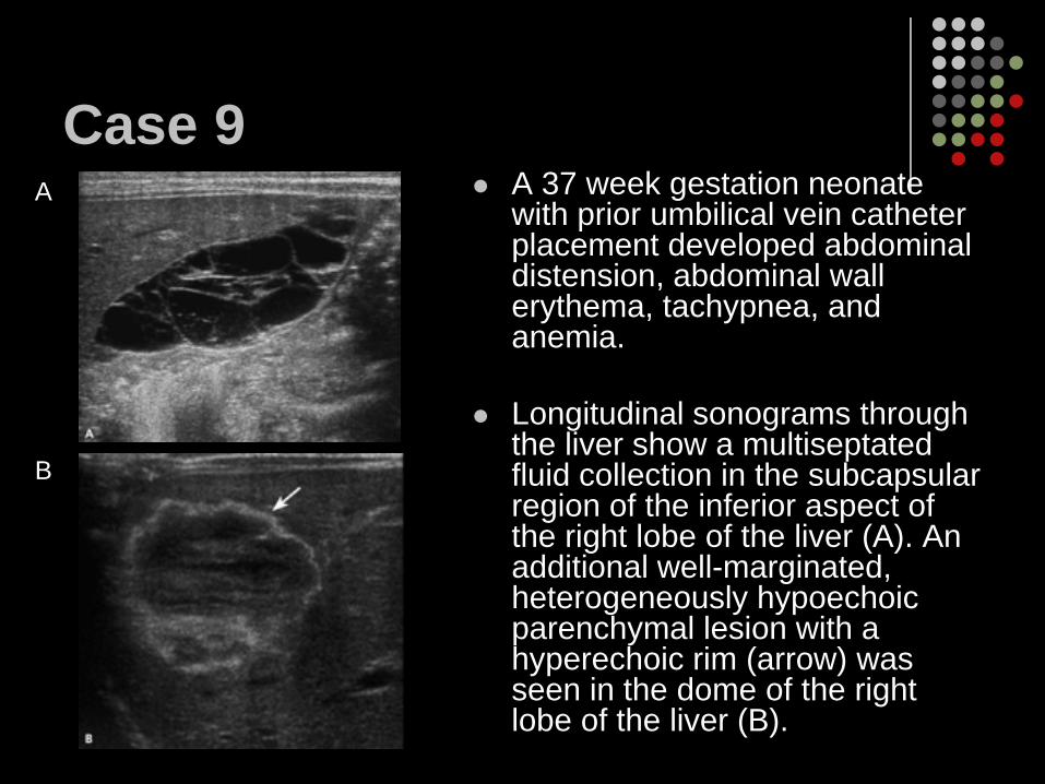
Case 9A 37 week gestation neonate with prior umbilical vein catheter placement developed abdominal distension, abdominal wall erythema, tachypnea, and anemia.
Longitudinal sonograms through the liver show a multiseptated fluid collection in the subcapsular region of the inferior aspect of the right lobe of the liver (A). An additional well-marginated, heterogeneously hypoechoic parenchymal lesion with a hyperechoic rim (arrow) was seen in the dome of the right lobe of the liver (B).
A
B
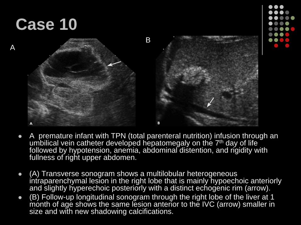
Case 10
A premature infant with TPN (total parenteral nutrition) infusion through an umbilical vein catheter developed hepatomegaly on the 7th day of life followed by hypotension, anemia, abdominal distention, and rigidity with fullness of right upper abdomen.
(A) Transverse sonogram shows a multilobular heterogeneous intraparenchymal lesion in the right lobe that is mainly hypoechoic anteriorly and slightly hyperechoic posteriorly with a distinct echogenic rim (arrow). (B) Follow-up longitudinal sonogram through the right lobe of the liver at 1 month of age shows the same lesion anterior to the IVC (arrow) smaller in size and with new shadowing calcifications.
AB
Cases 9 & 10Differential diagnosis for hepatic lesions with heterogeneously hypoechoic centers and hyperechoic rims in neonates:
AbscessesHemangioendotheliomasHematomas from birth traumaHamartomasHepatic erosion by umbilical catheter
Clinical history correlation can exclude the possibility of a hematoma from lack of birth trauma and an abscess from lack of history of necrotizing enterocolitis. Lack of peripheral hypervascularity on sonography can exclude the possibility of a hemangioendothelioma and an abscess.
Cases 9 & 10 : Hepatic Erosion by Umbilical Vein Catheters
Umbilical vein catheter traumaEarly diagnosis can prevent life-threatening complications caused by hepatic necrosis and hemorrhage Liver capsular rupture can cause TPN ascitesTherapeutic aspiration of the TPN ascites may be needed in some, but therapeutic intraperitoneal drainage tubes should be avoided Neonates with a history of umbilical vein catheter erosion do well clinically after the removal of the catheter US findings are nonspecific with lesions appearing similar to hematomas
The lesions in the acute/subacute phase can appear as a complex cystic collection within the liver or hepatic subcapsular spaceChronic lesions may have dystrophic calcifications
Follow-up sonograms can document the decrease in liver lesions and ascites
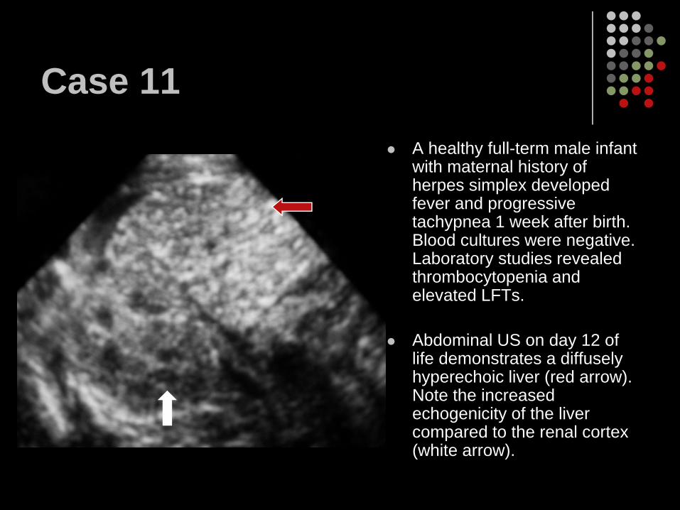
Case 11A healthy full-term male infant with maternal history of herpes simplex developed fever and progressive tachypnea 1 week after birth. Blood cultures were negative. Laboratory studies revealed thrombocytopenia and elevated LFTs.
Abdominal US on day 12 of life demonstrates a diffusely hyperechoic liver (red arrow). Note the increased echogenicity of the liver compared to the renal cortex (white arrow).
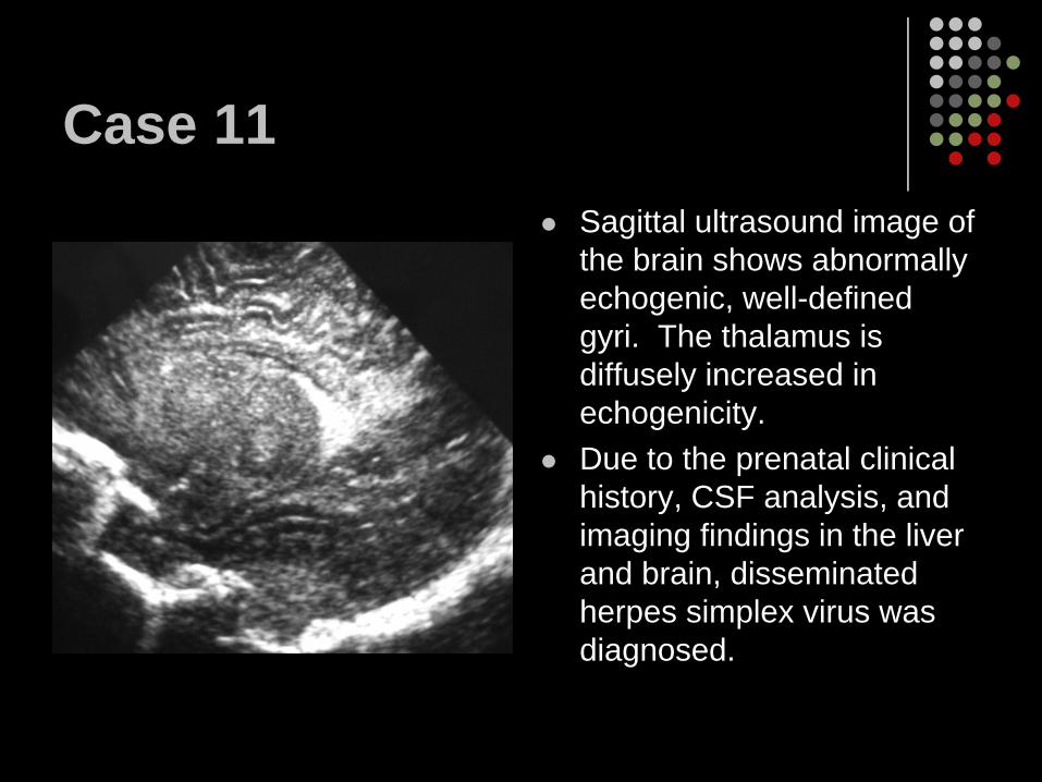
Case 11Sagittal ultrasound image of the brain shows abnormally echogenic, well-defined gyri. The thalamus is diffusely increased in echogenicity.Due to the prenatal clinical history, CSF analysis, and imaging findings in the liver and brain, disseminated herpes simplex virus was diagnosed.
Case 11 diagnosis:Disseminated Herpes Simplex Virus
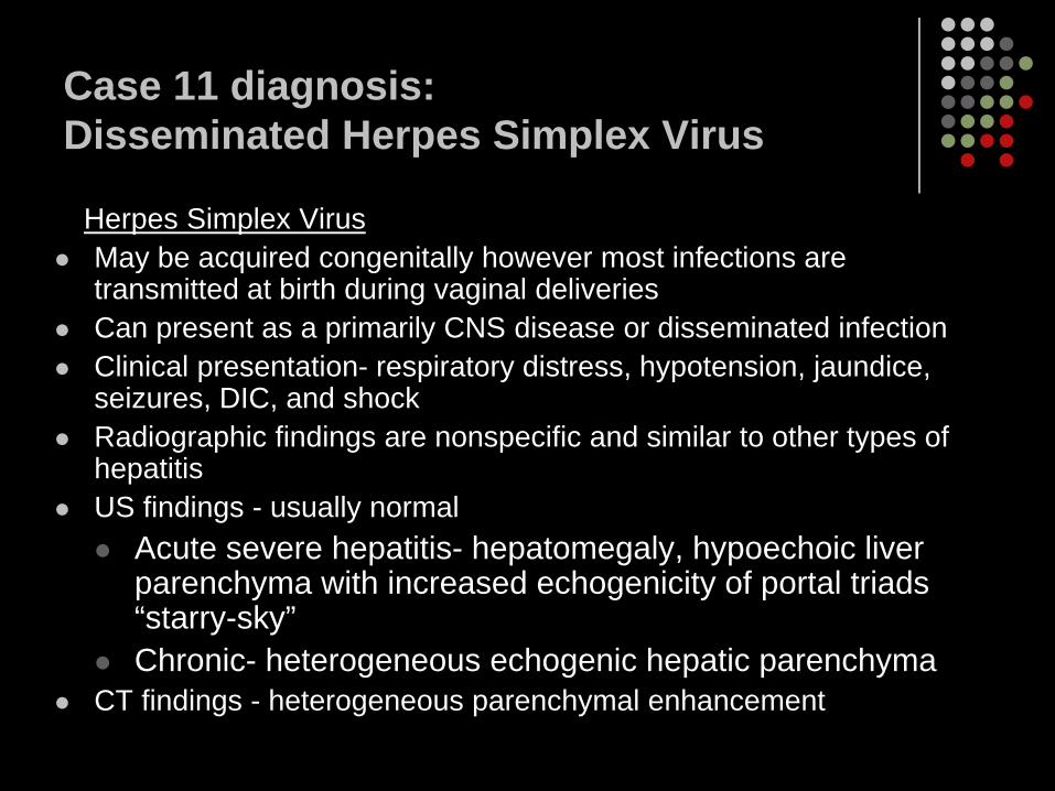
Herpes Simplex VirusMay be acquired congenitally however most infections are transmitted at birth during vaginal deliveriesCan present as a primarily CNS disease or disseminated infectionClinical presentation- respiratory distress, hypotension, jaundice, seizures, DIC, and shockRadiographic findings are nonspecific and similar to other types of hepatitisUS findings - usually normal
Acute severe hepatitis- hepatomegaly, hypoechoic liver parenchyma with increased echogenicity of portal triads “starry-sky”Chronic- heterogeneous echogenic hepatic parenchyma
CT findings - heterogeneous parenchymal enhancement
Case 12
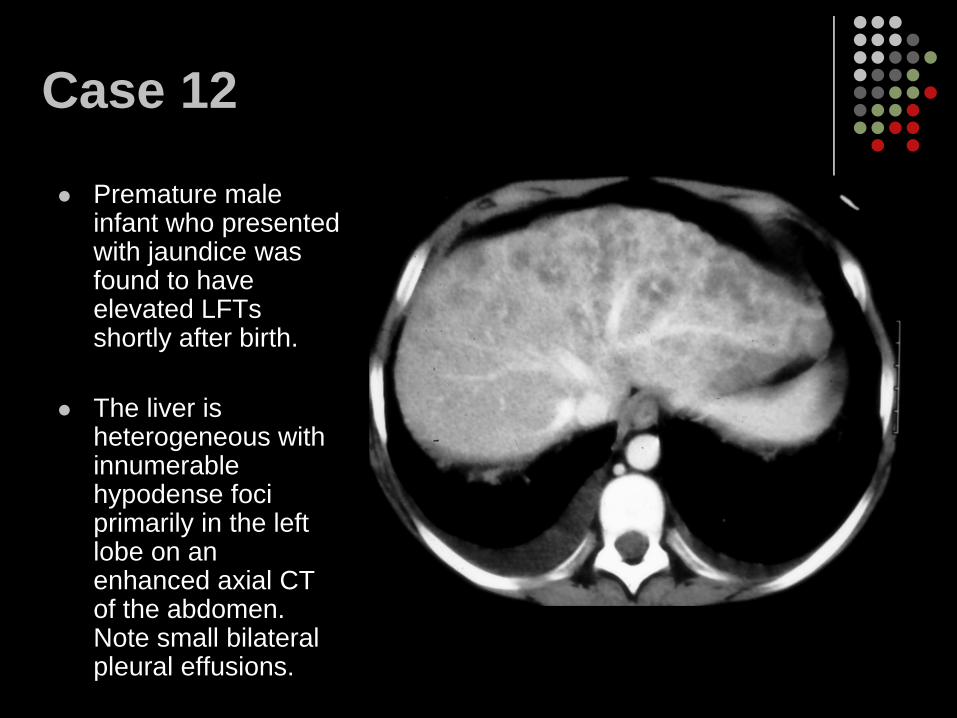
Premature male infant who presented with jaundice was found to have elevated LFTs shortly after birth.
The liver is heterogeneous with innumerable hypodense foci primarily in the left lobe on an enhanced axial CT of the abdomen. Note small bilateral pleural effusions.
Case 12Differential diagnosis for multiple nonenhancing, low attenuation lesions in the liver include:
- infectious etiologies (bacterial, viral, or fungal)- infiltrative lesions (metabolic disorders) - diffuse neoplastic processes (hepatoblastoma, metastatic neuroblastoma, congenital histiocytosis, or leukemia)
Liver biopsy was obtained which confirmed the diagnosis of hepatic cytomegalovirus.
Case 12 diagnosis:Cytomegalovirus
CytomegalovirusCytomegalovirus (CMV) is a double-stranded DNA virus in the herpes familyCMV can be transmitted transplacentally, during delivery, or via breast milk ingestionClinical Presentation - most are asymptomatic at birth
5-20% are symptomatic - jaundice, hepatosplenomegaly, petechiae
Imaging findings are generally nonspecific and similar to other viral infections that cause hepatitis
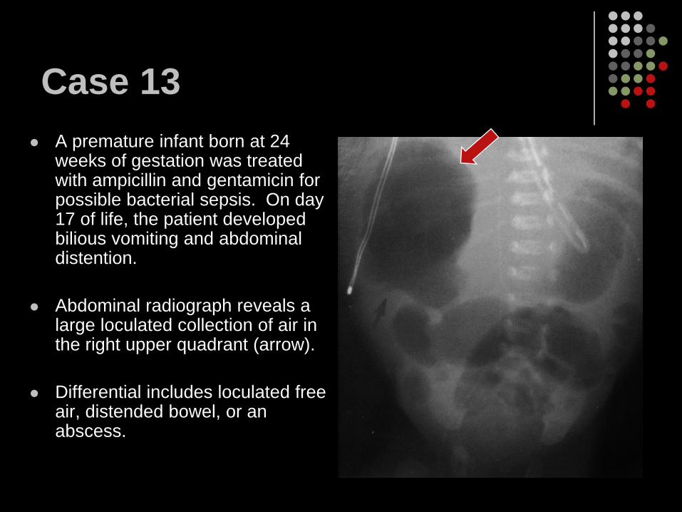
Case 13A premature infant born at 24 weeks of gestation was treated with ampicillin and gentamicin for possible bacterial sepsis. On day 17 of life, the patient developed bilious vomiting and abdominal distention.
Abdominal radiograph reveals a large loculated collection of air in the right upper quadrant (arrow).
Differential includes loculated free air, distended bowel, or an abscess.
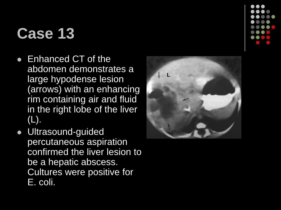
Case 13Enhanced CT of the abdomen demonstrates a large hypodense lesion (arrows) with an enhancing rim containing air and fluid in the right lobe of the liver (L).Ultrasound-guided percutaneous aspiration confirmed the liver lesion to be a hepatic abscess. Cultures were positive for E. coli.
L
Case 13 diagnosis:Liver Abscess
Liver AbscessCommon etiologic agents in infants are E. coli, Staphylococcus spp., Streptococcus spp., Pseudomonas aeruginosa, Haemophilis parainfluenzae, and KlebsiellaOccur secondary to hematogenous spread of bacteria from infection such as omphalitis, thrombophlebitis after umbilical vein catheterization, and sepsisClinical presentation- fever, RUQ pain, hepatomegaly, elevated LFTsRadiograph findings - may see an abnormal collection of gas within the abscess cavityUS findings - irregular, thick walls which may show hypervascularity and hypoechoic centers with through transmission; abscesses containing a large amount of air will demonstrate increased echogenicity with posterior shadowing CT findings - hypodense mass with thickened, enhancing walls, ±foci of gas
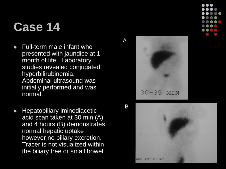
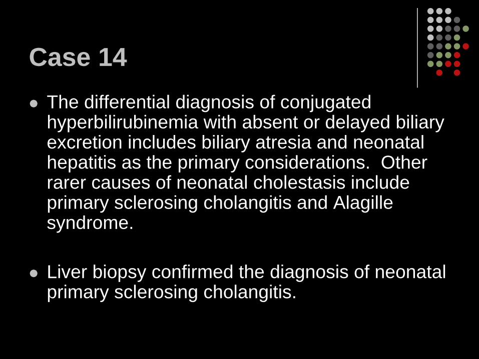
Case 14Full-term male infant who presented with jaundice at 1 month of life. Laboratory studies revealed conjugated hyperbilirubinemia. Abdominal ultrasound was initially performed and was normal.
Hepatobiliary iminodiacetic acid scan taken at 30 min (A) and 4 hours (B) demonstrates normal hepatic uptake however no biliary excretion. Tracer is not visualized within the biliary tree or small bowel.
A
B
Case 14The differential diagnosis of conjugated hyperbilirubinemia with absent or delayed biliary excretion includes biliary atresia and neonatal hepatitis as the primary considerations. Other rarer causes of neonatal cholestasis include primary sclerosing cholangitis and Alagille syndrome.
Liver biopsy confirmed the diagnosis of neonatal primary sclerosing cholangitis.
Case 14 diagnosis:Neonatal primary sclerosing cholangitis
Neonatal primary sclerosing cholangitis Neonatal primary sclerosing cholangitis is a rare entity
The cause is unknown however genetic and immunological factors may play a part in the etiology
Characterized by inflammation of the bile ducts resulting in fibrosis, obstruction, cholestasis, and biliary cirrhosisClinical presentation - cholestasis, hepatomegaly, jaundice, elevated LFTs occur in the first week of life
In all reported cases, jaundice resolved by the first year of life however the disease progressed to cirrhosis
HIDA scan findings - normal hepatic uptake of radiotracer however excretion into the intestine is severly delayed or absentCholangiography findings - irregularity of the bile ducts with areas of stenosis, dilatation, and beadingCT findings - bile duct segmental strictures with wall thickening
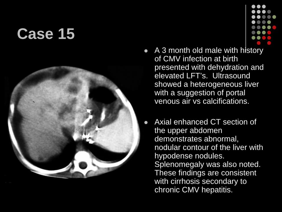
Case 15A 3 month old male with history of CMV infection at birth presented with dehydration and elevated LFT’s. Ultrasound showed a heterogeneous liver with a suggestion of portal venous air vs calcifications.
Axial enhanced CT section of the upper abdomen demonstrates abnormal, nodular contour of the liver with hypodense nodules. Splenomegaly was also noted. These findings are consistent with cirrhosis secondary to chronic CMV hepatitis.
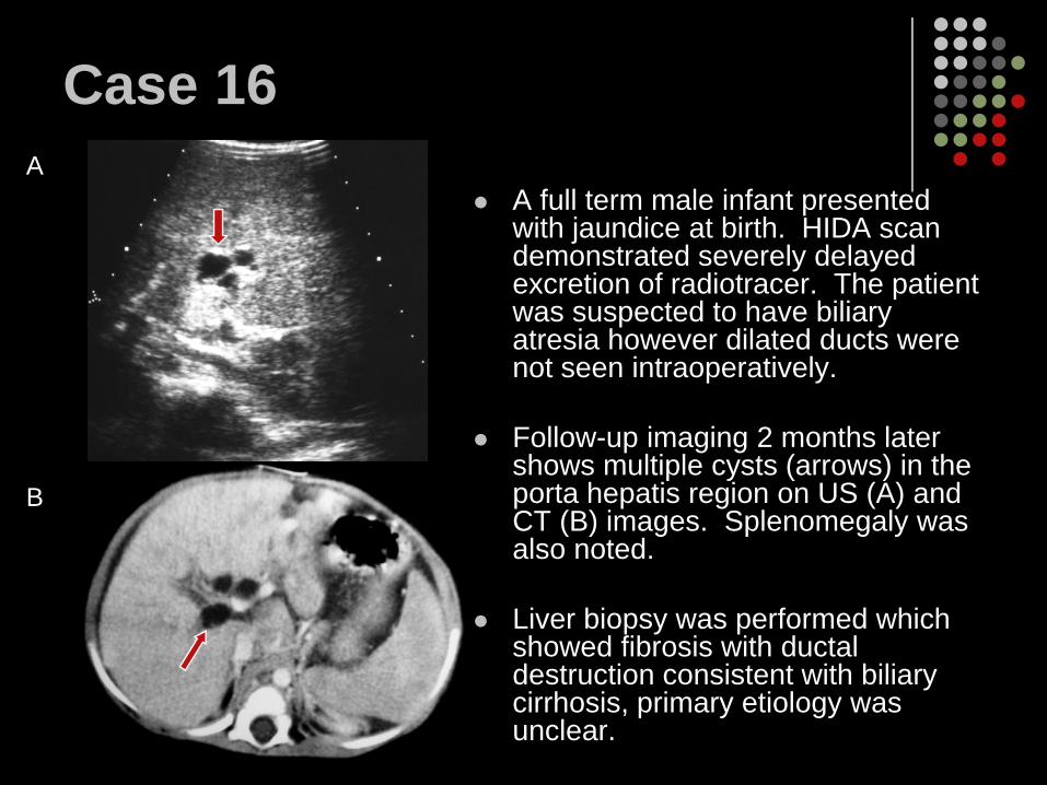
Case 16
A full term male infant presented with jaundice at birth. HIDA scan demonstrated severely delayed excretion of radiotracer. The patient was suspected to have biliary atresia however dilated ducts were not seen intraoperatively.
Follow-up imaging 2 months later shows multiple cysts (arrows) in the porta hepatis region on US (A) and CT (B) images. Splenomegaly was also noted.
Liver biopsy was performed which showed fibrosis with ductal destruction consistent with biliary cirrhosis, primary etiology was unclear.
A
B
Cases 15 & 16 diagnoses:Cirrhosis
Infantile cirrhosisEnd stage liver disease characterized by chronic destruction of the liver parenchyma with fibrotic replacement and nodular regenerationMay be secondary to chronic hepatitis, congenital hepatic fibrosis, biliary atresia, cystic fibrosis, Budd-Chiari syndrome, disorders of metabolism, medications, or TPNPrimary biliary cirrhosis- cholestatic liver disease with destruction of the interlobular bile ductsUS findings - atrophy of the right hepatic lobe and medial segment of the left lobe with hypertrophy of the caudate lobe and lateral segment of the left lobe, nodular contour, diffuse heterogeneity of the liver parenchymaCT findings - nodular contour, may see regenerating nodules, ± ascites
SummaryDifferential Diagnosis of diffuse liver lesions in infants
Viral hepatic infections Diffuse hepatic necrosis from infection or ischemiaMetastatic neuroblastomaNeoplastic processes such as hepatoblastomaCirrhosisPrimary sclerosing cholangitis
Differential diagnosis of focal liver lesions in infantsAbscessHematomaHamartomaHemangioendotheliomaVascular malformationsLymphangioma, hemangiomaLiver cystIatrogenic trauma from umbilical venous catheters
SummaryNon-neoplastic liver lesions presenting in the neonate/infant can be difficult to diagnose as they are rare and can have overlapping clinical and radiographic features.The lesions can be categorized into ischemia, infection, vascular lesions, traumatic lesions, and biliary abnormalities.While a few of these lesions can be incidental findings, others will progress to end-stage liver disease without treatment.Specific findings on ultrasound, CT, and MRI can be helpful in narrowing the differential in a timely manner.Tissue biopsy may be necessary in some cases to make the final diagnosis and determine treatment.
ReferencesAkkaya, A, Erbas, G, et al. Normal liver, spleen, and kidney dimensions in neonates, infants, and children: evaluation with sonography. AJR 1998:171:1693-1698.Anderson, B, Challapalli, M, Sajous, C, Vade, A. Neonatal hepatic abscess. Computerized Medical Imaging and Graphics 1998: 22:357-359.Balistreri, WF, Sokol, RJ, Suchy, FJ. Liver Disease in Children. Lippincott Williams & Wilkins; 2001 :444-455.Brant, WE, Major, NM, Webb, WR. Liver. In: Fundamentals of Body CT. Philadelphia, PA: Saunders; 2006: 211-231.Cohen, HL, Da Silva, MG, Haller, JO, Mulvihill, DM, Rizzo, AJ. Calcification of the ductus venosus: a cause of right upper quadrant calcification in the newborn. Radiology 1989: 173:89-90.Donnelly, LF. Gastrointestinal Tract. In: Fundamentals of Pediatric Radiology. Philadelphia, PA: WB Saunders; 2001: 123-130.Eckner, F, Rosenthal, IR, Vade, A. Computerized tomography in occlusive infantile arteriopathy. Pediatr Cardiol 1989: 10:221-224.
ReferencesHertzberg, BS, Kurtz, AB, Middleton, WD. Liver. In: the requisites, ultrasound. St. Louis, MO: Mosby; 2004:49-86.Kimberlin, DW, Lin, CY, Jacobs, RF, et al. Natural history of neonatal herpes simplex virus infections in the acyclovir era. Pediatrics 2001: 108:223.Kleinman, RE, Walker, WA. The liver. In: Walker’s Pediatric Gastrointestinal Disease. USA: PMPH; 2008: 796.Osborn, LM, Reiff, MI. Clinical estimation of liver size in newborn infants. Pediatrics 1983:71:46-48.Siegel, MJ. Liver and biliary tract. In: Pediatric Sonography. New York, NY: Raven Press; 1995: 171-237.Stagno, S, Britt, W. Cytomegalovirus infections. In: Infectious Diseases of the Fetus and Newborn Infant, 6th Ed, Remington, JS, Klein, JO, Wilson, DB, Baker, CJ (Eds), Elsevier Saunders, Philadelphia; 2006: 739-740.