Embed Size (px)
Citation preview
108
Cytological diagnosis of bancroftian filariasis in lesions clinicallyanticipated as neoplastic
A Jha,1 R Shrestha,2 G Aryal,2 AD Pant,1 RC Adhikari1 and G Sayami1
Department of Pathology, 1Institute of Medicine, Tribhuvan University Teaching Hospital, Maharajgunj Kathmandu, Nepal and 2NepalMedical College, Jorpati, Kathmandu, Nepal
Corresponding author: Dr. Abhimanyu Jha, Department of Pathology, Institute of Medicine, Tribhuvan University Teaching Hospital,Maharajgunj, Kathmandu, Nepal. P. O. Box No: 9631. e-mail: [email protected]
ABSTRACTFilariasis is a common disabling parasitic disease in this region and cytological diagnosis is often not required.
Cytology has important role in diagnosis of sub-clinical filariasis. Most cases of cytologically diagnosed filariasis
are clinically unanticipated. Microfilaria, ova and fragments of adult worm of Wuchereria bancrofti, in exfoliative
as well as aspiration cytology have been reported and are useful in cytological detection of bancroftian filariasis.
Microfilaria is frequently detected in association with neoplasm, although the role in tumorogenesis is
controversial. The objective of the study was to investigate importance of cytology in diagnosis of filariasis in
lesions clinically anticipated to be of neoplastic and to review the cytomorphology of bancroftian filaria and its
association with neoplasm. This is a retrospective study carried out in cytology department of Tribhuvan
University Teaching Hospital. 14 cases of cytological specimen out of 4291 (0.3%) showed microfilaria; 12
cases were from FNAC from different sites and 2 cases were from pleural fluid. 2 cases showed ova in addition
to microfilaria and one of them in addition showed fragment of adult worm. Microfilaria in 4 cases of FNAC
and one case of pleural fluid were associated with malignant cells.
Keywords: Filariasis, incidental cytological diagnosis, cytomorphology, association with malignancy.
INTRODUCTIONFilariasis is disabling parasitic disease prevalent
worldwide caused by various species of filarial organism.
Bancroftian filariasis is infection by the filarial worm
Wuchereria bancrofti which causes disease by blocking
lymphatic vessels.1 W. bancrofti is responsible for 90.0%
of cases of filariasis and is found throughout the tropics
and in some sub-tropical areas world-wide. Brugia
malayi is confined to South-east and Eastern Asia. B.
timori is found only in Timor and its adjacent islands.2
Certain parts of Nepal are endemic for bancroftian filaria;
58 districts of Nepal are potentially endemic for
bancroftian filariasis.3
Filariasis may produce acute as well as chronic clinical
manifestations or person may remain asymtomatic in
endemic areas.1 However, in all the cases typical clinical
manifestations of filariasis may not be seen. Pathological
findings associated with filarial lesions are chronic
inflammatory cell infiltrate consisting of lymphocytes,
histiocytes, plasma cells, and eosinophils. Epithelioid
granulomas, necrosis and acute inflammatory cell
infiltrate are also seen.1 Association of filarial parasite
with malignancy has been described but its role in
tumorogenesis is not so far explained and it could be
just a chance association.4
Common methods of diagnosis of filariasis in this
country are by demonstration of microfilaria in stained
or unstained blood films, circulating filarial antigen
detection and demonstration of organism in
histopathological sections. Fluid cytology or fine needle
aspiration cytology (FNAC) are rarely applied for routine
diagnosis of clinically suspected filariasis. But filarial
organism can be detected in cytological smears from
various sites of body in clinically unsuspected cases of
filariasis. Such lesions may be primarily caused by the
organism or it may be associated with other pathology
such as malignancy. Incidental detection of filarial
organism has been reported in cytological smears from
almost any part of body. Forms of bancroftian filaria
and background pathology, however, can vary.
Microfilaria is the most common form of filarial
organism detected in cytological smears; however ova
of the organism and fragments of adult worms can also
be detected rarely.4 Microfilariae have also been reported
in association with various benign and malignant
tumors.4 Thus role of cytology in diagnosis of fialriasis
can not be underestimated in clinically unanticipated
cases. This study is an attempt to prove importance of
cytology in diagnosis of filariasis.
MATERIALS AND METHODCytological records of the year 2004 to 2005 in
department of cytology of Tribhuvan University
Teaching Hospital (TUTH) were retrieved for diagnosed
cases of filariasis. All the cases were clinically
Original Article Nepal Med Coll J 2008; 10(2): 108-114

109
unsuspected of filariasis. In all the cases
cytological smears were stained with
Papanicolau and Giemsa stain. Slides
with filarial organisms were reviewed
and findings are tabulated. Histology
was available in only one case.
RESULTSTotal number of cytology during one
year was 4291 that included FNAC
(2289 cases) from different sites,
cervicovaginal smears, sputum
cytology, and body fluid cytology
including urine cytology (2002 cases).
Total numbers of cases with filariasis
were only 14 (0.3% of all cytological
specimens) that include 12 cases of
FNAC (0.5% of 2289 cases of FNAC)
from different sites (Table-1) and 2
cases of pleural fluid. Frequency of detection of filariasis
in FNAC was 0.5% and in other cytological specimen it
was 0.1% (2 cases of 2002 cases). In later only 2 cases
of pleural fluid (out of 97 cases) showed the organism.
Respective frequencies of detection of the organism in
FNAC from different sites are shown in table 1. Out of 4
cases with filariasis in FNAC of lymph nodes, 3 were
from axilla and only one from inguinal region. None of
the above cases had clinical filariasis. Male and female
were in 1:1 ratio (7 each) with age ranging from 11 years
to 78 years.
In all the 14 cases microfilaria was identified as
microfilaria of W. bancrofti, based on its characteristic
cytomorphology that is sheathed larvae with tail-tip free
from nuclei (Fig. 1). In the smears from breast aspirate
in one case showed numerous coiled microfilariae as
well (Fig. 2). Ova of the organism in addition to
microfilaria were seen in two cases of FNAC, one from
the breast lump and other from inguinal lymph node. In
latter fragments of adult worm packed with microfilaria
and ova of the organism was also seen (Fig. 3 and 4).
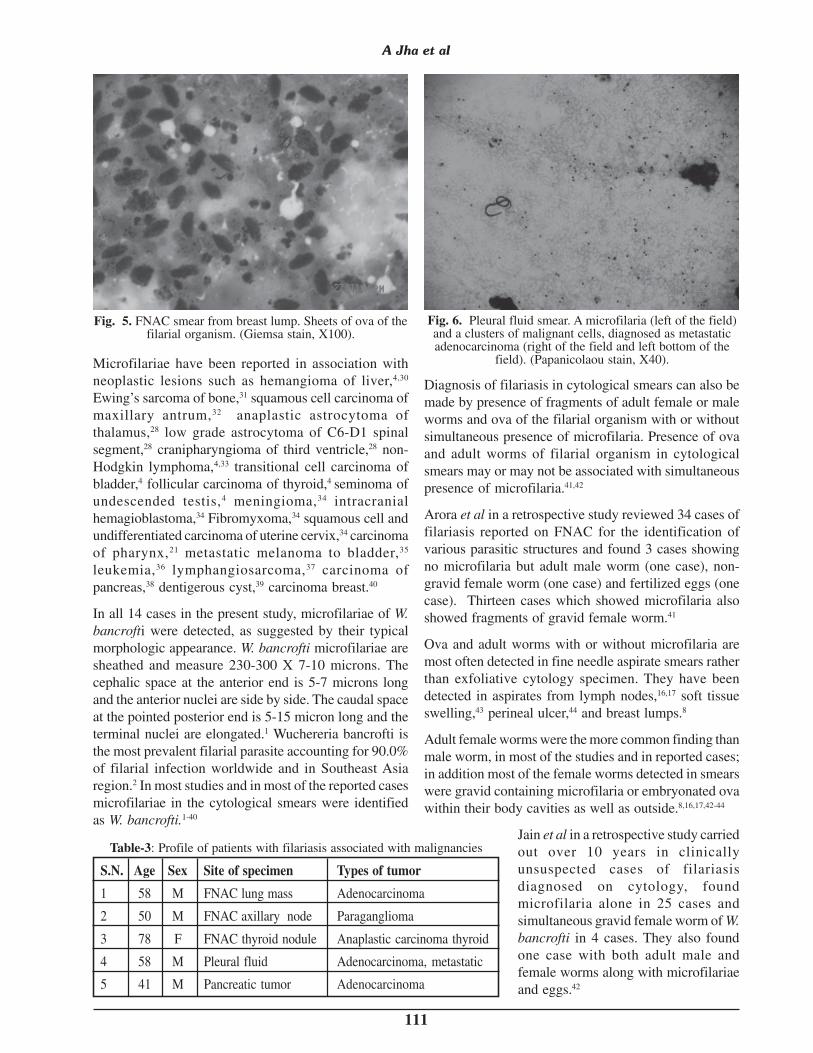
Aspirate from the breast lump also showed sheets of ova
of the organism (Fig. 5). Frequency of additional
associated cytological findings such as epithelioid cell
granulomas, histiocytes, giant cells, eosinophils, and
necrosis is shown in Table-2. All the cases showed mixed
inflammatory infiltrate composed of scattered
lymphocytes and occasional neutrophils.
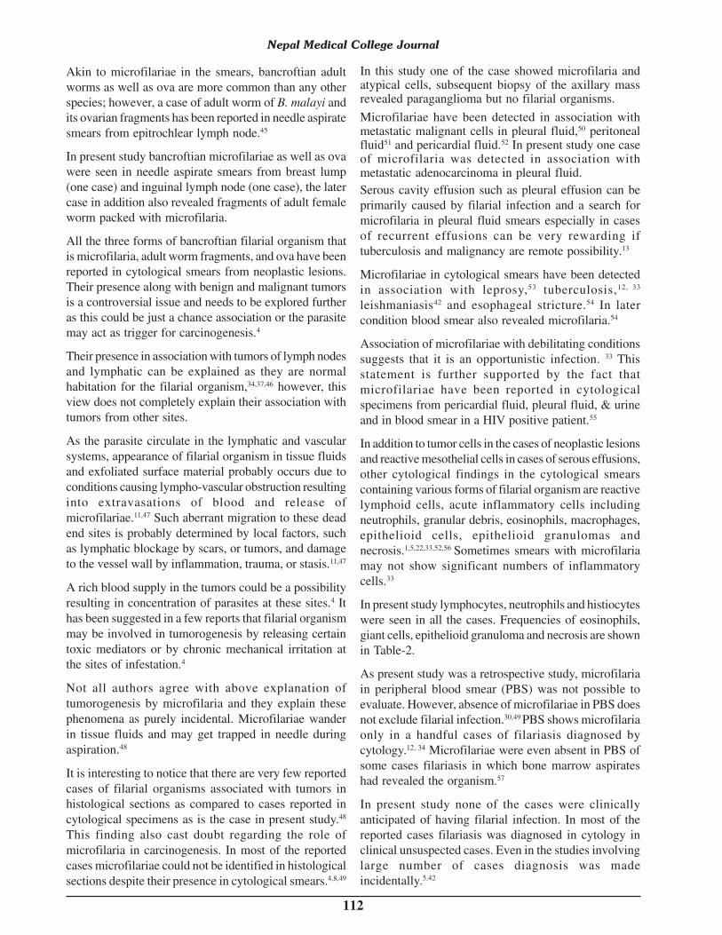
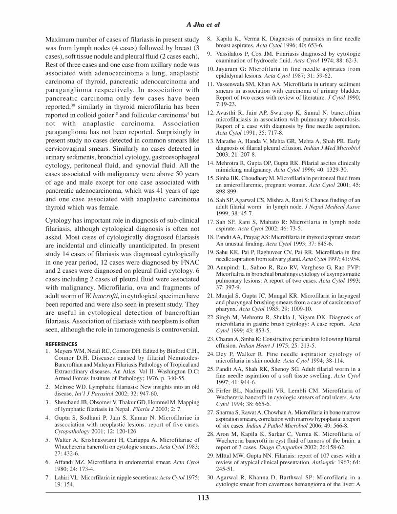
Five cases (35.7% of 14 cases) were associated with
malignancies (Fig. 6) as shown in Table-3. The second
case in the table 3 showed atypical cells clusters in
association with microfilaria which was later confirmed
as paraganglioma in the tissue biopsy. Microfilaria load
was more in the smears from non-neoplastic lesions in
comparison to smears associated with malignancy. In
later case only occasional microfilaria was seen. Patient
profile in these 5 cases is shown in Table-3.
DISCUSSIONBancroftian filariasis produce wide spectrum of clinical
manifestations. The acute phase is characterized by fever,
lymphangitis, lymphadenitis, epididymo-orchitis, and
funniculitis. Headache, backache, muscle pain, insomnia,
anorexia, urticarial rash, malaise, nausea and fatigue are
common complaints. Eosinophilia and microfilaremia
are common in acute phase. Chronic stage of bancroftian
filariasis is characterized by lymphadenopathy,
lymphadema, hydrocele, and elephantiasis.1 A significant
number of infected individuals in endemic areas remain
asymptomatic throughout their life.1 The later situation
is traditionally classified as ‘endemic normals’.2 In recent
years the traditional classification of filarial disease has
been challenged. The introduction of assays for
A Jha et alTable-2: Microscopic findings in cytological smears
S. N. Microscopic features n. of cases Sites of cytology
1 Microfilaria 14 All 8 sites. (see table 1)
2 Ova 02 Breast lump and inguinal lymph node
3 Fragments of organism 01 Inguinal lymph node
4 Malignant cells 04 See table 2
5 Necrosis 02 Breast and thyroid
6 Epithelioid granuloma 03 Breast 2 cases and inguinal lymphnode
7 Eosinophils 09 Breast 3 cases, inguinal lymph node 4cases and soft tissue 2 cases
8 Histiocytes, lymphocytes, 14 All 14 cases (see table 1)neutrophils
9 Giant cells 03 Breast 2 cases and inguinal lymphnode one case.
Table-1: Frequency of filariasis in FNAC
Total n. n. of cases
of cases with filariasis
1 Breast lump 549 3 0.5
2 Lymph nodes 320 4 1.2
3 Soft tissue 74 2 2.7
4 Lung 66 1 1.5
5 Thyroid nodule 500 1 0.2
6 Pancreatic tumor 12 1 8.3
7 Other lesions 780 00 00
Total 2289 12 0.524
S. N. Specimen %
110
circulating filarial antigen, the discovery of occult
lymphatic pathology and renal disease in ‘asymptomatic
microfilaremics’ and the recognition of the role of
bacterial infection in the pathogenesis of acute and
chronic disease suggests that the old classification based
on presence or absence of microfilaremia and/or chronic
pathology is outdated. It is no longer wise to think of
individuals as having filarial ‘infection’ without ‘filarial
disease’ for the same reason – many of the former will
have evidence of ‘covert disease’ if the studies are
rigorous enough.2 Significant numbers of patients never
undergo tests for filarial infection because they are never
included in epidemiological studies nor they present
features typical of filariasis.
In present study none of the patients were clinically
suspected of filariasis; clinically they presented with
breast lump (3 cases), lymphadenopathy (4 cases), and
soft tissue nodule (2 cases). One case of lymph node
enlargement in the axilla was also associated with
paraganglioma. Likewise 3 cases were associated
malignant tumor in lung, thyroid and pancreas, one in
each case. 2 cases presented with features of pleural
effusion, one out of that was associated with metastatic
adenocarcinoma.
A review of literature reveals detection microfilaria in
most of the commonly performed cytological specimens
and mostly they are incidental. Microfilaria have been
detected in cervicovaginal smears,5 endometrial smears,6
nipple secretions,7ovarian cyst fluid,5 breast aspirates,8
hydrocele fluid,9 epididymal aspirates, 10 urine samples,11 lung aspirates, 12 pleural fluid, 13 bronchial washings,5
ascitic fluid,14 intraoperative peritoneal fluid,15 lymph
node aspirates,16,17 thyroid aspirates,18 salivary gland
aspirates,19 bronchial brushings,20 laryngeal and
pharyngeal brushings,21 gastric brushings,22 pericardial
fluid,23 cutaneous nodule,24 soft tissue nodule,25 oral
ulcer,26 bone marrow aspirates,27 brain aspirates28 and
joint aspirates.29
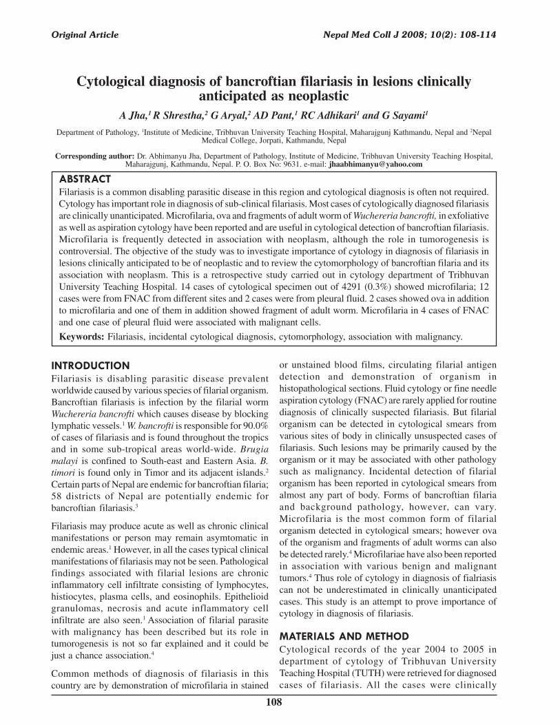
Fig. 1. FNAC smear from soft tissue nodule. microfilaria ofWuchereria bancrofti. Sheathed larva with tail tip free from
granules. ( Papanicolaou stain, X100).
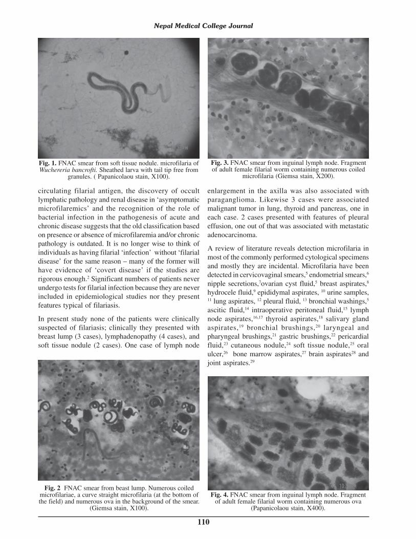
Fig. 2 FNAC smear from beast lump. Numerous coiledmicrofilariae, a curve straight microfilaria (at the bottom ofthe field) and numerous ova in the background of the smear.
(Giemsa stain, X100).
Fig. 3. FNAC smear from inguinal lymph node. Fragmentof adult female filarial worm containing numerous coiled
microfilaria (Giemsa stain, X200).
Fig. 4. FNAC smear from inguinal lymph node. Fragmentof adult female filarial worm containing numerous ova
(Papanicolaou stain, X400).
Nepal Medical College Journal
111
Microfilariae have been reported in association with
neoplastic lesions such as hemangioma of liver,4,30
Ewing’s sarcoma of bone,31 squamous cell carcinoma of
maxillary antrum,32 anaplastic astrocytoma of
thalamus,28 low grade astrocytoma of C6-D1 spinal
segment,28 cranipharyngioma of third ventricle,28 non-
Hodgkin lymphoma,4,33 transitional cell carcinoma of
bladder,4 follicular carcinoma of thyroid,4 seminoma of
undescended testis,4 meningioma,34 intracranial
hemagioblastoma,34 Fibromyxoma,34 squamous cell and
undifferentiated carcinoma of uterine cervix,34 carcinoma
of pharynx,21 metastatic melanoma to bladder,35
leukemia,36 lymphangiosarcoma,37 carcinoma of
pancreas,38 dentigerous cyst,39 carcinoma breast.40
In all 14 cases in the present study, microfilariae of W.
bancrofti were detected, as suggested by their typical
morphologic appearance. W. bancrofti microfilariae are
sheathed and measure 230-300 X 7-10 microns. The
cephalic space at the anterior end is 5-7 microns long
and the anterior nuclei are side by side. The caudal space
at the pointed posterior end is 5-15 micron long and the
terminal nuclei are elongated.1 Wuchereria bancrofti is
the most prevalent filarial parasite accounting for 90.0%
of filarial infection worldwide and in Southeast Asia
region.2 In most studies and in most of the reported cases
microfilariae in the cytological smears were identified
as W. bancrofti.1-40
Diagnosis of filariasis in cytological smears can also be
made by presence of fragments of adult female or male
worms and ova of the filarial organism with or without
simultaneous presence of microfilaria. Presence of ova
and adult worms of filarial organism in cytological
smears may or may not be associated with simultaneous
presence of microfilaria.41,42
Arora et al in a retrospective study reviewed 34 cases of
filariasis reported on FNAC for the identification of
various parasitic structures and found 3 cases showing
no microfilaria but adult male worm (one case), non-
gravid female worm (one case) and fertilized eggs (one
case). Thirteen cases which showed microfilaria also
showed fragments of gravid female worm.41
Ova and adult worms with or without microfilaria are
most often detected in fine needle aspirate smears rather
than exfoliative cytology specimen. They have been
detected in aspirates from lymph nodes,16,17 soft tissue
swelling,43 perineal ulcer,44 and breast lumps.8
Adult female worms were the more common finding than
male worm, in most of the studies and in reported cases;
in addition most of the female worms detected in smears
were gravid containing microfilaria or embryonated ova
within their body cavities as well as outside.8,16,17,42-44
Jain et al in a retrospective study carried
out over 10 years in clinically
unsuspected cases of filariasis
diagnosed on cytology, found
microfilaria alone in 25 cases and
simultaneous gravid female worm of W.
bancrofti in 4 cases. They also found
one case with both adult male and
female worms along with microfilariae
and eggs.42
Fig. 5. FNAC smear from breast lump. Sheets of ova of thefilarial organism. (Giemsa stain, X100).
Fig. 6. Pleural fluid smear. A microfilaria (left of the field)and a clusters of malignant cells, diagnosed as metastaticadenocarcinoma (right of the field and left bottom of the
field). (Papanicolaou stain, X40).
A Jha et al
Table-3: Profile of patients with filariasis associated with malignancies
S.N. Age Sex Site of specimen Types of tumor
1 58 M FNAC lung mass Adenocarcinoma
2 50 M FNAC axillary node Paraganglioma
3 78 F FNAC thyroid nodule Anaplastic carcinoma thyroid
4 58 M Pleural fluid Adenocarcinoma, metastatic
5 41 M Pancreatic tumor Adenocarcinoma
112
Akin to microfilariae in the smears, bancroftian adult
worms as well as ova are more common than any other
species; however, a case of adult worm of B. malayi and
its ovarian fragments has been reported in needle aspirate
smears from epitrochlear lymph node.45
In present study bancroftian microfilariae as well as ova
were seen in needle aspirate smears from breast lump
(one case) and inguinal lymph node (one case), the later
case in addition also revealed fragments of adult female
worm packed with microfilaria.
All the three forms of bancroftian filarial organism that
is microfilaria, adult worm fragments, and ova have been
reported in cytological smears from neoplastic lesions.
Their presence along with benign and malignant tumors
is a controversial issue and needs to be explored further
as this could be just a chance association or the parasite
may act as trigger for carcinogenesis.4
Their presence in association with tumors of lymph nodes
and lymphatic can be explained as they are normal
habitation for the filarial organism,34,37,46 however, this
view does not completely explain their association with
tumors from other sites.
As the parasite circulate in the lymphatic and vascular
systems, appearance of filarial organism in tissue fluids
and exfoliated surface material probably occurs due to
conditions causing lympho-vascular obstruction resulting
into extravasations of blood and release of
microfilariae.11,47 Such aberrant migration to these dead
end sites is probably determined by local factors, such
as lymphatic blockage by scars, or tumors, and damage
to the vessel wall by inflammation, trauma, or stasis.11,47
A rich blood supply in the tumors could be a possibility
resulting in concentration of parasites at these sites.4 It
has been suggested in a few reports that filarial organism
may be involved in tumorogenesis by releasing certain
toxic mediators or by chronic mechanical irritation at
the sites of infestation.4
Not all authors agree with above explanation of
tumorogenesis by microfilaria and they explain these
phenomena as purely incidental. Microfilariae wander
in tissue fluids and may get trapped in needle during
aspiration.48
It is interesting to notice that there are very few reported
cases of filarial organisms associated with tumors in
histological sections as compared to cases reported in
cytological specimens as is the case in present study.48
This finding also cast doubt regarding the role of
microfilaria in carcinogenesis. In most of the reported
cases microfilariae could not be identified in histological
sections despite their presence in cytological smears.4,8,49
In this study one of the case showed microfilaria andatypical cells, subsequent biopsy of the axillary massrevealed paraganglioma but no filarial organisms.
Microfilariae have been detected in association withmetastatic malignant cells in pleural fluid,50 peritonealfluid51 and pericardial fluid.52 In present study one caseof microfilaria was detected in association withmetastatic adenocarcinoma in pleural fluid.
Serous cavity effusion such as pleural effusion can be
primarily caused by filarial infection and a search for
microfilaria in pleural fluid smears especially in cases
of recurrent effusions can be very rewarding if
tuberculosis and malignancy are remote possibility.13
Microfilariae in cytological smears have been detected
in association with leprosy,53 tuberculosis,12, 33
leishmaniasis42 and esophageal stricture.54 In later
condition blood smear also revealed microfilaria.54
Association of microfilariae with debilitating conditions
suggests that it is an opportunistic infection. 33 This
statement is further supported by the fact that
microfilariae have been reported in cytological
specimens from pericardial fluid, pleural fluid, & urine
and in blood smear in a HIV positive patient.55
In addition to tumor cells in the cases of neoplastic lesions
and reactive mesothelial cells in cases of serous effusions,
other cytological findings in the cytological smears
containing various forms of filarial organism are reactive
lymphoid cells, acute inflammatory cells including
neutrophils, granular debris, eosinophils, macrophages,
epithelioid cells, epithelioid granulomas and
necrosis.1,5,22,33,52,56 Sometimes smears with microfilaria
may not show significant numbers of inflammatory
cells.33
In present study lymphocytes, neutrophils and histiocytes
were seen in all the cases. Frequencies of eosinophils,
giant cells, epithelioid granuloma and necrosis are shown
in Table-2.
As present study was a retrospective study, microfilaria
in peripheral blood smear (PBS) was not possible to
evaluate. However, absence of microfilariae in PBS does
not exclude filarial infection.30,49 PBS shows microfilaria
only in a handful cases of filariasis diagnosed by
cytology.12, 34 Microfilariae were even absent in PBS of
some cases filariasis in which bone marrow aspirates
had revealed the organism.57
In present study none of the cases were clinically
anticipated of having filarial infection. In most of the
reported cases filariasis was diagnosed in cytology in
clinical unsuspected cases. Even in the studies involving
large number of cases diagnosis was made
incidentally.5,42
Nepal Medical College Journal
113
Maximum number of cases of filariasis in present study
was from lymph nodes (4 cases) followed by breast (3
cases), soft tissue nodule and pleural fluid (2 cases each).
Rest of three cases and one case from axillary node was
associated with adenocarcinoma a lung, anaplastic
carcinoma of thyroid, pancreatic adenocarcinoma and
paraganglioma respectively. In association with
pancreatic carcinoma only few cases have been
reported,38 similarly in thyroid microfilaria has been
reported in colloid goiter18 and follicular carcinoma4 but
not with anaplastic carcinoma. Association
paraganglioma has not been reported. Surprisingly in
present study no cases detected in common smears like
cervicovaginal smears. Similarly no cases detected in
urinary sediments, bronchial cytology, gastroesophageal
cytology, peritoneal fluid, and synovial fluid. All the
cases associated with malignancy were above 50 years
of age and male except for one case associated with
pancreatic adenocarcinoma, which was 41 years of age
and one case associated with anaplastic carcinoma
thyroid which was female.
Cytology has important role in diagnosis of sub-clinical
filariasis, although cytological diagnosis is often not
asked. Most cases of cytologically diagnosed filariasis
are incidental and clinically unanticipated. In present
study 14 cases of filariasis was diagnosed cytologically
in one year period, 12 cases were diagnosed by FNAC
and 2 cases were diagnosed on pleural fluid cytology. 6
cases including 2 cases of pleural fluid were associated
with malignancy. Microfilaria, ova and fragments of
adult worm of W. bancrofti, in cytological specimen have
been reported and were also seen in present study. They
are useful in cytological detection of bancroftian
filariasis. Association of filariasis with neoplasm is often
seen, although the role in tumorogenesis is controversial.
REFERENCES1. Meyers WM, Neafi RC, Connor DH. Edited by Binford C.H.,
Connor D.H. Diseases caused by filarial Nematodes-Bancroftian and Malayan Filariasis Pathology of Tropical and
Extraordinary diseases. An Atlas. Vol II. Washington D.C:Armed Forces Institute of Pathology; 1976. p. 340-55.
2. Melrose WD. Lymphatic filariasis: New insights into an olddisease. Int’l J Parasitol 2002; 32: 947-60.
3. Sherchand JB, Obsomer V, Thakur GD, Hommel M. Mappingof lymphatic filariasis in Nepal. Filaria J 2003; 2: 7.
4. Gupta S, Sodhani P, Jain S, Kumar N. Microfilariae inasscociation with neoplastic lesions: report of five cases.Cytopathology 2001; 12: 120-126
5. Walter A, Krishnaswami H, Cariappa A. Microfilariae ofWhuchereria bancrofti on cytologic smears. Acta Cytol 1983;27: 432-6.
6. Affandi MZ. Microfilaria in endometrial smear. Acta Cytol
1980; 24: 173-4.
7. Lahiri VL: Micorfilaria in nipple secretions: Acta Cytol 1975;
19: 154.
8. Kapila K., Verma K. Diagnosis of parasites in fine needlebreast aspirates. Acta Cytol 1996; 40: 653-6.
9. Vassilakos P, Cox JM. Filariasis diagnosed by cytologicexamination of hydrocele fluid. Acta Cytol 1974; 88: 62-3.
10. Jayaram G: Microfilaria in fine needle aspirates fromepididymal lesions. Acta Cytol 1987; 31: 59-62.
11. Vassenwala SM, Khan AA. Microfilaria in urinary sedimentsmears in association with carcinoma of urinary bladder.Report of two cases with review of literature. J Cytol 1990;7:19-23.
12. Avasthi R, Jain AP, Swaroop K, Samal N. bancroftianmicrofilariasis in association wih pulmonary tuberculosis.Report of a case with diagnosis by fine needle aspiration.Acta Cytol 1991; 35: 717-8.
13. Marathe A, Handa V, Mehta GR, Mehta A, Shah PR. Earlydiagnosis of filarial pleural effusion. Indian J Med Microbiol2003; 21: 207-8.
14. Mehrotra R, Gupta OP, Gupta RK. Filarial ascites clinicallymimicking malignancy. Acta Cytol 1996; 40: 1329-30.
15. Sinha BK, Choudhary M. Microfilaria in peritoneal fluid froman amicrofilaremic, pregnant woman. Acta Cytol 2001; 45:898-899.
16. Sah SP, Agarwal CS, Mishra A, Rani S: Chance finding of anadult filarial worm in lymph node. J Nepal Medical Assoc1999; 38: 45-7.
17. Sah SP, Rani S, Mahato R: Microfilaria in lymph nodeaspirate. Acta Cytol 2002; 46: 73-5.
18. Pandit AA, Prayag AS: Microfilaria in thyroid aspirate smear:An unusual finding. Acta Cytol 1993; 37: 845-6.
19. Sahu KK, Pai P, Raghuveer CV, Pai RR. Microfilaria in fineneedle aspiration from salivary gland. Acta Cytol 1997; 41: 954.
20. Anupindi L, Sahoo R, Rao RV, Verghese G, Rao PVP:Micorfialria in bronchial brushings cytology of asymptomaticpulmonary lesions: A report of two cases. Acta Cytol 1993;37: 397-9.
21. Munjal S, Gupta JC, Mungal KR. Microfilaria in laryngealand pharyngeal brushing smears from a case of carcinoma ofpharynx. Acta Cytol 1985; 29: 1009-10.
22. Singh M, Mehrotra R, Shukla J, Nigam DK. Diagnosis ofmicrofilaria in gastric brush cytology: A case report. ActaCytol 1999; 43: 853-5.
23. Charan A, Sinha K: Constrictive pericarditis following filarialeffusion. Indian Heart J 1975; 25: 213-5.
24. Dey P, Walker R. Fine needle aspiration cytology ofmicrofilaria in skin nodule. Acta Cytol 1994; 38-114.
25. Pandit AA, Shah RK, Shenoy SG. Adult filarial worm in afine needle aspiration of a soft tissue swelling. Acta Cytol1997; 41: 944-6.
26. Firfer BL, Nadimpalli VR, Lembli CM. Microfilaria ofWuchereria bancrofti in cytologic smears of oral ulcers. ActaCytol 1994; 38: 665-6.
27. Sharma S, Rawat A, Chowhan A. Microfilaria in bone marrowaspiration smears, correlation with marrow hypoplasia: a reportof six cases. Indian J Pathol Microbiol 2006; 49: 566-8.
28. Aron M, Kapila K, Sarkar C, Verma K. Microfilaria ofWuchereria bancrofti in cyst fluid of tumors of the brain: areport of 3 cases. Diagn Cytopathol 2002; 26:158-62.
29. MIttal MW, Gupta NN. Filariais: report of 107 cases with areview of atypical clinical presentation. Antiseptic 1967; 64:245-51.
30. Agarwal R, Khanna D, Barthwal SP: Microfilaria in acytologic smear from cavernous hemangioma of the liver: A
A Jha et al

114
case report. Acta Cytol 1998; 42:781-2.
31. Ahluwalia C, Choudhury M, Bajaj P. Incidental detection ofmicrofilaria in aspirates from Ewing’s sarcoma. DiagnCytopathol 2003; 29: 31-2.
32. Mohan G, Chaturvedi S, Misra PK. Microfilaria in a fineneedle aspirate of primary solid malignant tumor of themaxillary antrum. A case report. Acta Cytol 1998; 42: 772-4.
33. Gupta K, Sehgal A, Puri MM, Sidhwa HK. Microfilariae inassociation with other diseases. A report of six cases. ActaCytol 2002; 46: 776-8.
34. Agarwal PK, Srivastava AN, Agarwal N. Microfialria inassociation with neoplasms. A report of six cases. Acta Cytol1982; 26: 480-90.
35. Kapila K, Verma K. Microfilaria in urinary sediment:coexistent metastatic melanoma cells and Wuchereriabancrofti. Acta Cytol 1986; 30: 696-7.
36. Justus PG, Kitchens CS. Secondary leukemia withMonchausen filariasis. Ann Intern Med 1976; 85: 685.
37. Devi KR, Bahuleyan CK. Lymphagiosarcoma of the lowerextremity associated with chronic lymphoedema of filarialorigin. Indian J Cancer 1977; 14: 176-8.
38. Mohan S, Andley M, Talwar N, Ravi B, Kumar A. An unusualassociation with carcinoma pancreas: a case report.Cytopathology 2005; 16: 215-6.
39. Agrawal K, Mekhala A, Chitra S, Narasimhan R, RatnakarC. Whuchereria bancrofti in dentigerous cystic fluid: anunusual presentation. Ann Plast Surg 1998; 41: 205-7.
40. Atal P, Choudhary M, Ashik S. Coexistence of carcinoma ofthe breast with microfilariasis. Diagn Cytopathol 2000; 4:259-60.
41. Arora VK, Singh N, Bhatia A. Cytomorphologic profile oflymphatic filariasis. Acta Cytol 1996; 40: 948-52.
42. Jain S, Sodhani P, Gupta S, Sakhuja P, Kumar N.Cytomorphology of filariasis revisited. Expansion of themorphologic spectrum and coexistence with other lesions.Acta Cytol 2001; 45: 186-91.
43. Kapila K, Verma K: Gravid adult female worms of Wuchereriabancrofti in fine needle aspirates of soft tissue swellings:Report of three cases. Acta Cytol 1989; 33: 390-2.
44. Sharma P, Kumar N, Jain P, Gur R, Jain S: Chronic
wuchereriasis presenting as a vaginoperineal fistula: Reportof a case with aspiration cytologic diagnosis. Acta Cytol 2005;49: 335-8.
45. Arora VK, Sen B, Bhatia A: Fine needle aspirationidentification of the adult worm of Brugia malayi and itsovarian fragment from epitrochlear lymph node. Acta Cytol1993; 37: 437-8.
46. Dey P, Radhika S, Jain A: Microfilaria of Wuchereria bancroftiin lymph node aspirate: A case report. Acta Cytol 1993; 37:745-6.
47. Walter A, Sinha K. Microfilaria of W. bancrofti in cytologicsmears. Acta Cytol 1983; 27: 435-6.
48. Wagla N, Patel MM, Pandya AN, Thakral C. Microfilariae incytology smears: real culprits or trapped accidentally by needles?A study of 10 cases. Cytopathology 2005; 16: 315-20.
49. Mehrotra R, Singh M, Javed KZ, Gupta RK: Cytodiagnosisof microfilaria of the breast from a needle aspirate. Acta Cytol1999; 43: 517-8.
50. Sivakumaran P, Wilsher ML. Microfilarial pleural effusionassociated with adenocarcinoma. Aust New Zealand J 1997;27: 341.
51. Khan AA, Vasenwala SM, Ahmad S. Coexistent metastaticadenocarcinoma and microfilaria in ascitic fluid. Acta Cytol1993; 37: 643-4.
52. Verghese R, Raghuveer CV, Pai MR, Bansal R. Microfilariaein cytologic smears: A report of six cases. Acta Cytol 1996;40: 299-301.
53. Malik A, Singh N, Arora VK and Bhatia A: Association ofmicrofilaria with leprosy and other disease. Acta Cytol 2002;46: 69-70
54. Rawal A, Hayer J, Dey P: Microfilariain esophageal stricture.Acta Cytol 2002; 46: 70-71.
55. Roy I, Mukhopadhyay C, Ayyagari A. Multisysteminvolvement of microfialria in a HIV positive patient. NepalMed Coll J 2004; 6: 64-6.
56. Pinto RW, Mandreker S: Microfilaria of Wuchereria bancroftiin fine needle aspiration smears. Acta Cytol 1996; 40: 1326-7.
57. Pradhan S. Lahiri VL, Elhence BR, Singh KN. Microfilariaof Whuchereria bancrofti in bone marrow smears. Amer JTrop Med Hyg 1976; 25: 199-200.
Nepal Medical College Journal