Embed Size (px)
Citation preview
IMAGING OF THE
JUGULAR FORAMEN
Dr. Mohamed Eid, MD
Lecturer of Diagnostic Imaging
Alexandria Faculty of Medicine
Bibliography
1. Ong CK, Chong VFH. Imaging of Jugular Foramen. NeuroimagClin N Am 2009; 19:469–82.
2. Davagnanam I, Chavda SV. Identification of the NormalJugular Foramen and Lower Cranial Nerve Anatomy: Contrast-Enhanced 3D Fast Imaging Employing Steady-StateAcquisition MR Imaging. AJNR Am J Neuroradiol 2008;29:574–76
3. Macdonald AJ et al. Primary Jugular Foramen Meningioma:Imaging Appearance and Differentiating Features. AJR 2004;182:373-7.
4. Rao AB et al. From the Archives of the AFIP: Paragangliomasof the Head and Neck: Radiologic-Pathologic Correlation.RadioGraphics 1999;19:1605-32.
INTRODUCTION
The jugular foramen is a complex crossroad of neurovascular structures deep in the skull base.
It is inaccessible to clinical examination, and safe surgical approach is often hindered by crucial surrounding structures.
Radiology plays a central role in the diagnostic evaluation and management planning of jugular foramen lesions.
INTRODUCTION
A jugular foramen lesion may originate from
its intrinsic contents, or arise from the
surrounding structures and involve the
foramen secondarily.
Normal variants and imaging artifacts are not
infrequent in this region, simulating diseases.
IMAGING ALGORITHM
Optimal assessment of jugular foramen diseases
requires both MR imaging and CT with thin-section
bone algorithm.
MR imaging shows the exact soft tissue extent of
lesions, whereas CT allows precise evaluation of the
surrounding bone changes.
Angiography outlines a vascular roadmap for
surgeons, and preoperative embolization may be of
value for certain hypervascular tumors.
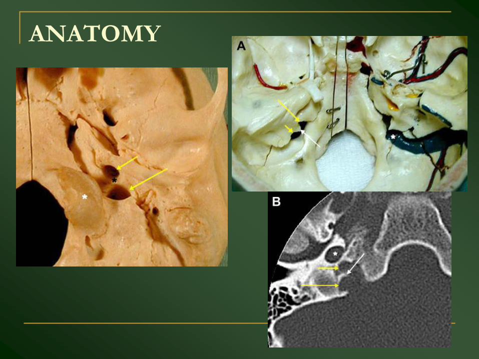
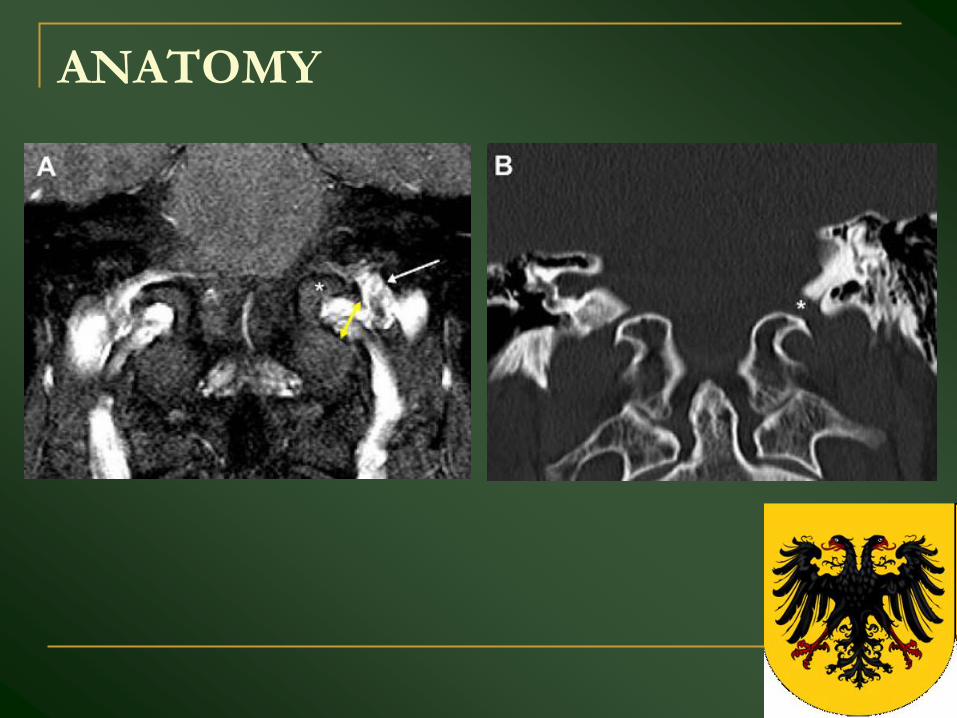
ANATOMY
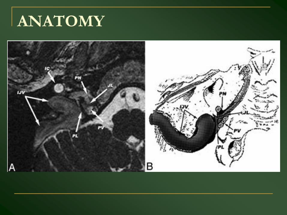
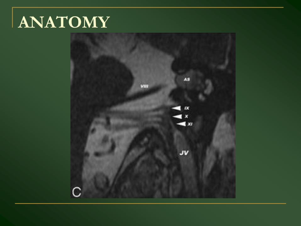
ANATOMY
ANATOMY
ANATOMY
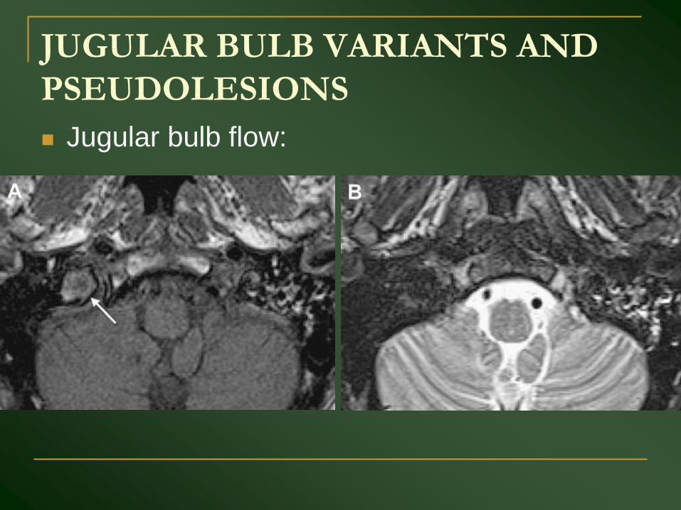
JUGULAR BULB VARIANTS AND
PSEUDOLESIONS
Jugular bulb flow:
JUGULAR BULB VARIANTS AND
PSEUDOLESIONS
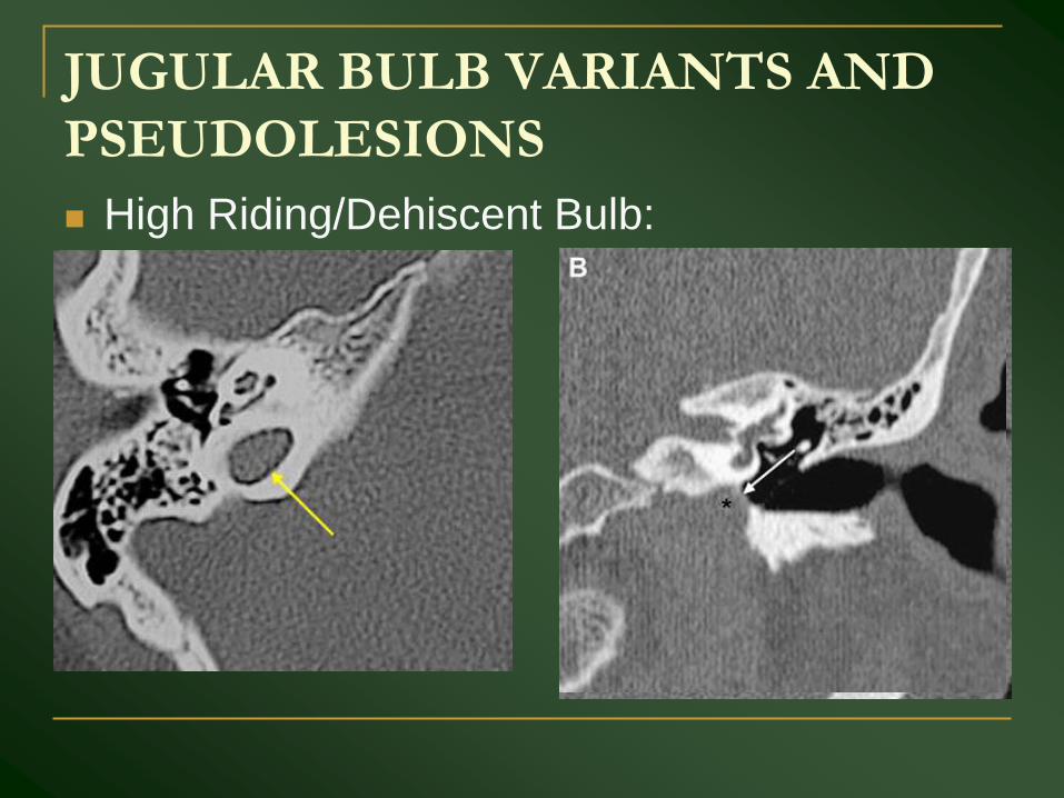
High Riding/Dehiscent Bulb:
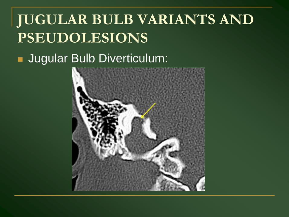
JUGULAR BULB VARIANTS AND
PSEUDOLESIONS
Jugular Bulb Diverticulum:
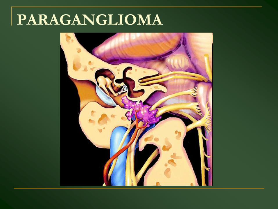
PARAGANGLIOMA
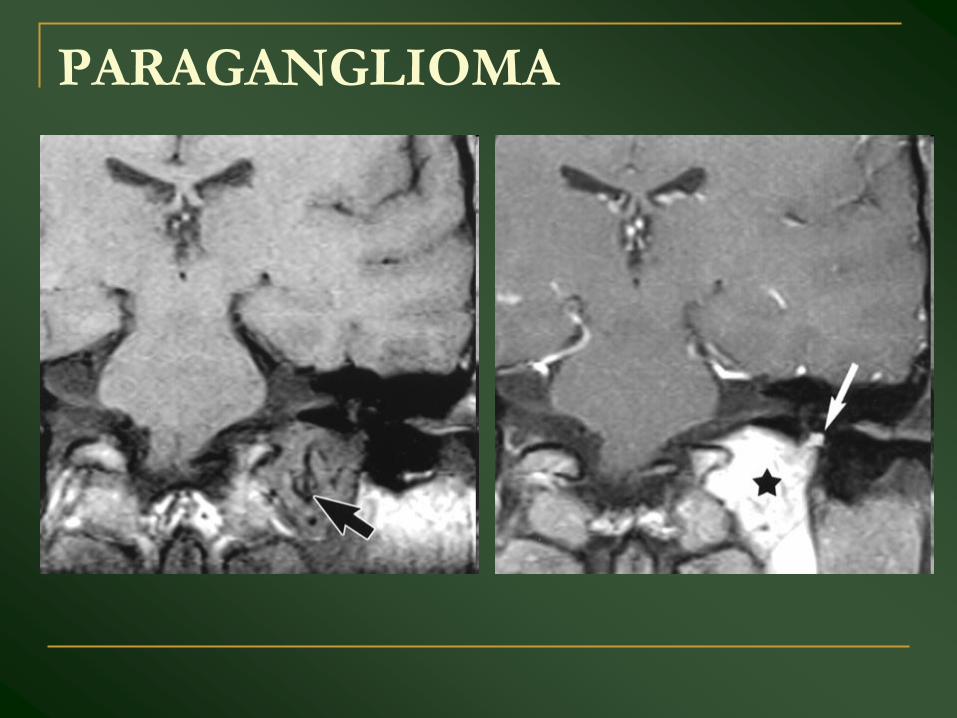
PARAGANGLIOMA
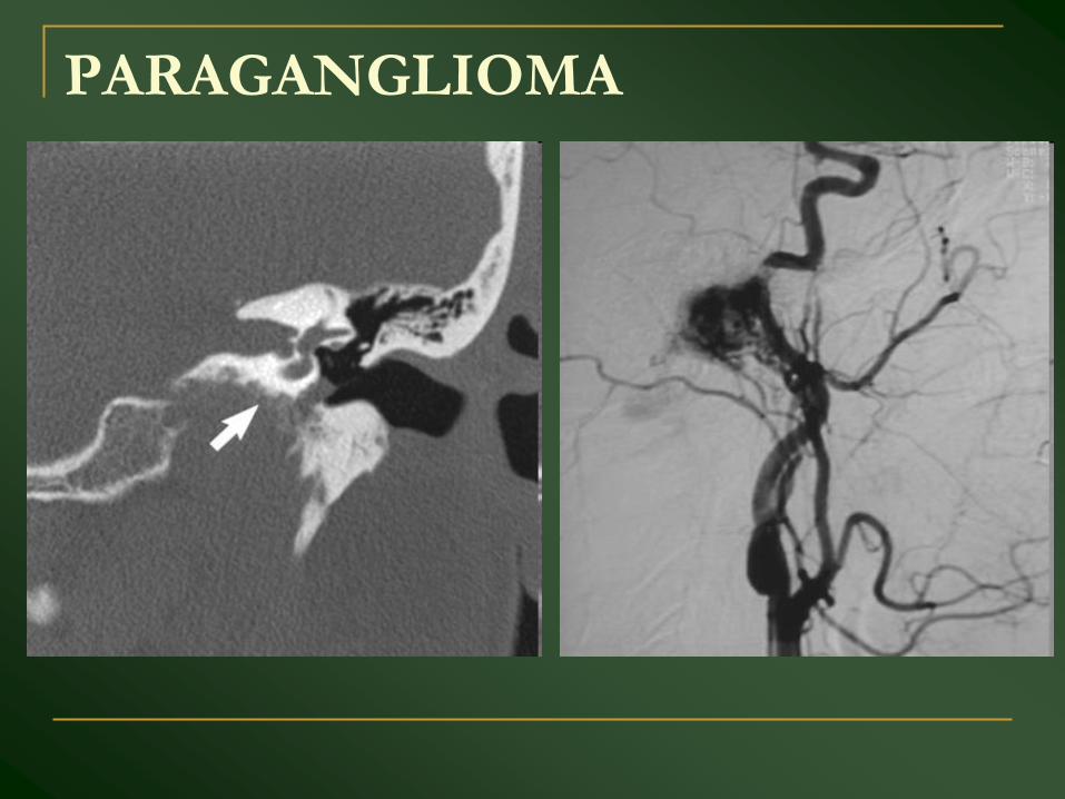
PARAGANGLIOMA
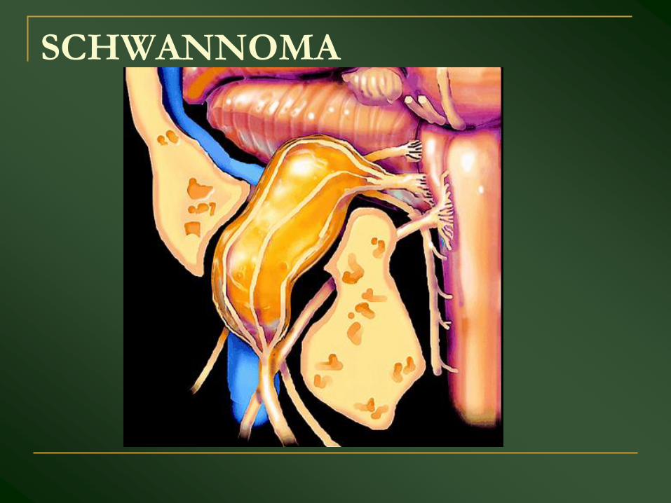
SCHWANNOMA
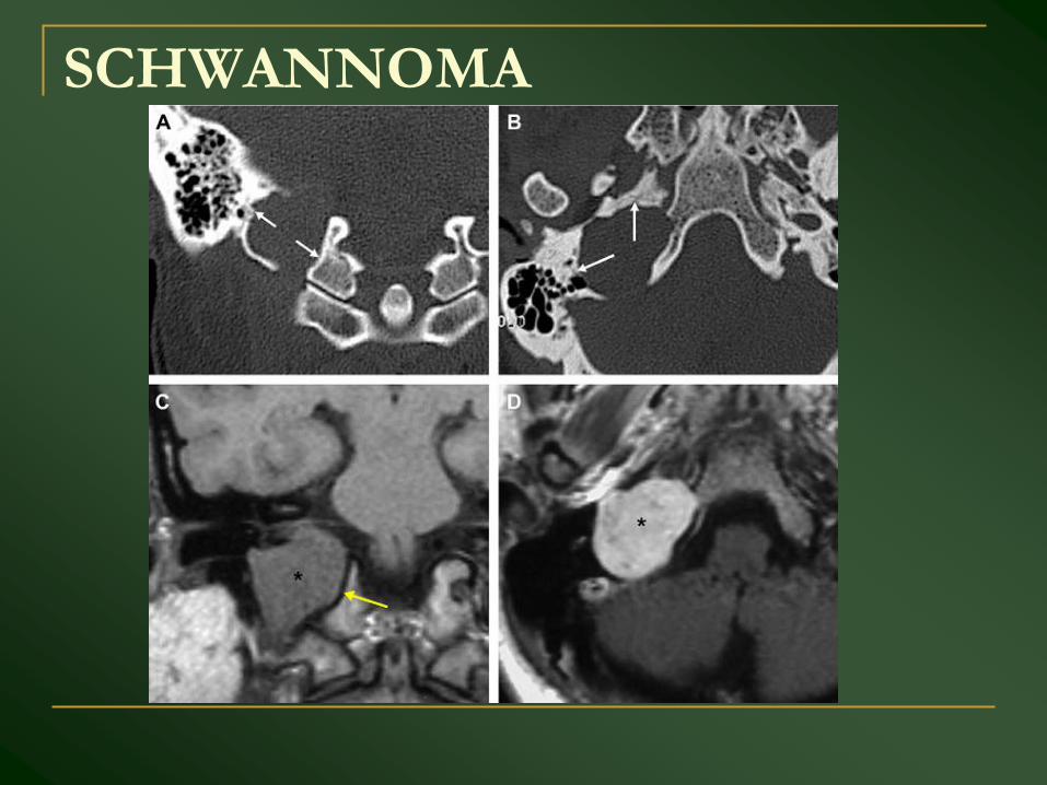
SCHWANNOMA
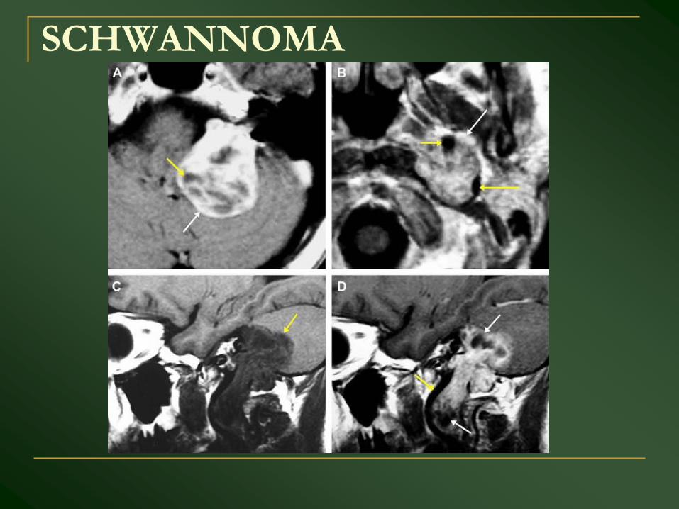
SCHWANNOMA
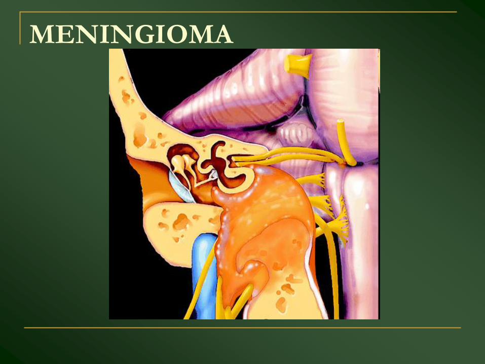
MENINGIOMA
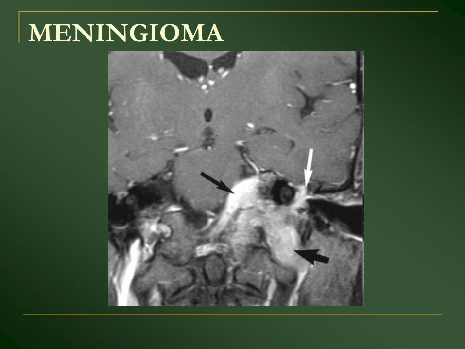
MENINGIOMA
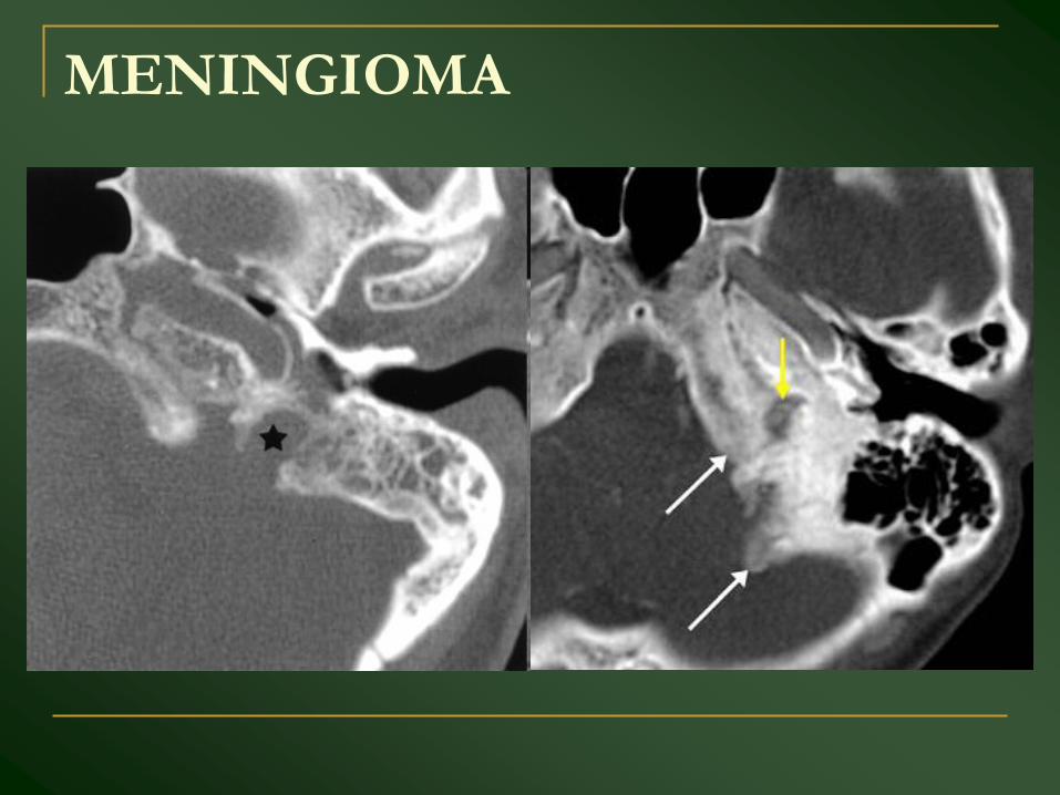
MENINGIOMA
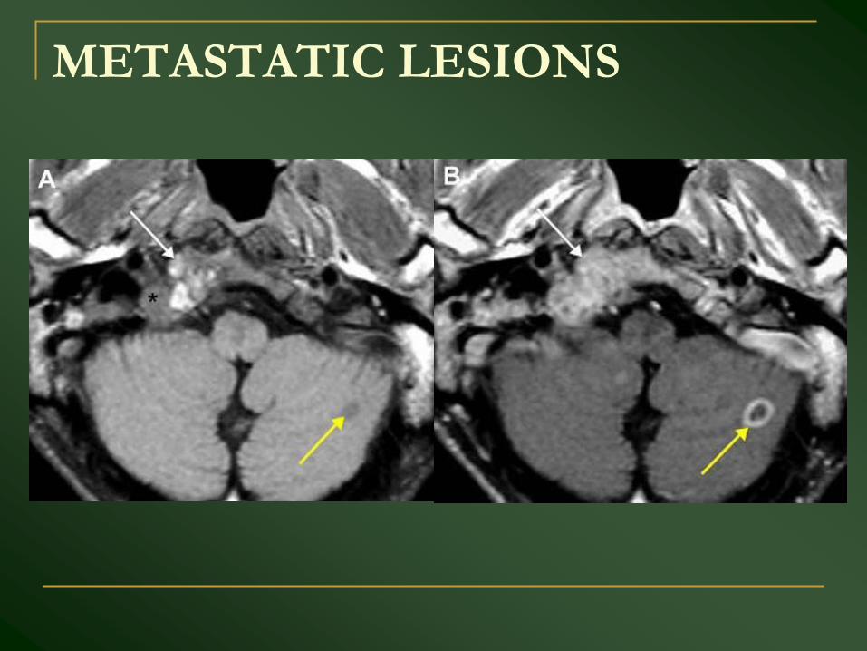
METASTATIC LESIONS
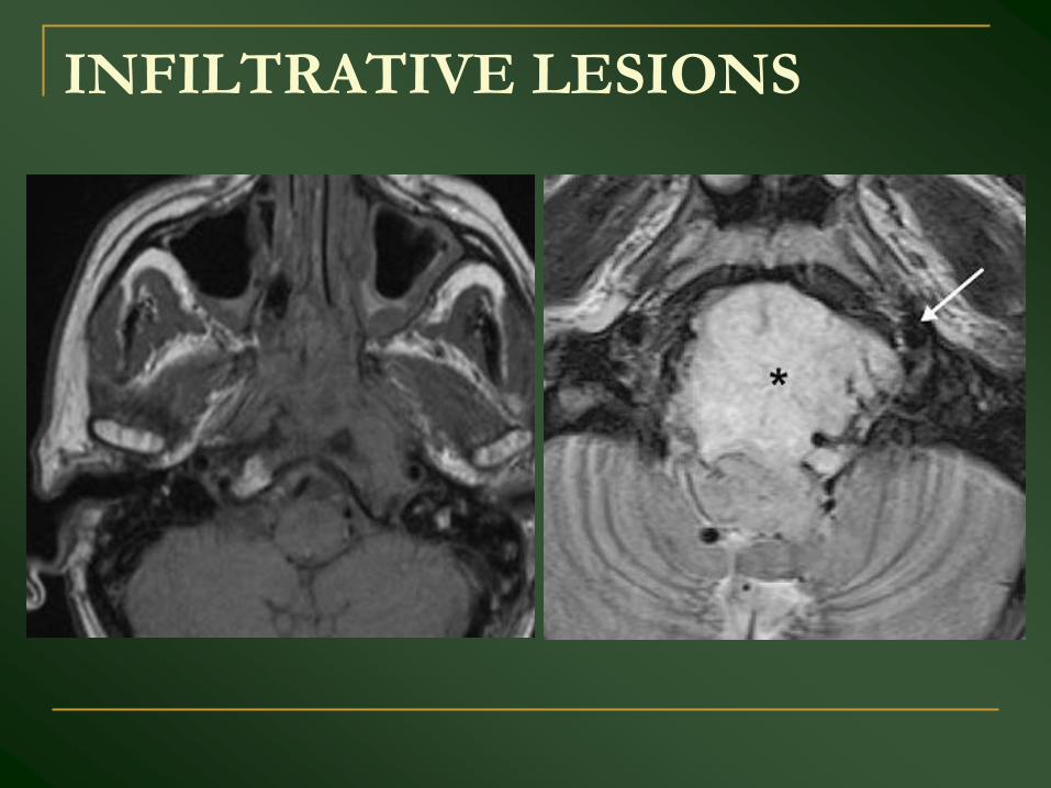
INFILTRATIVE LESIONS
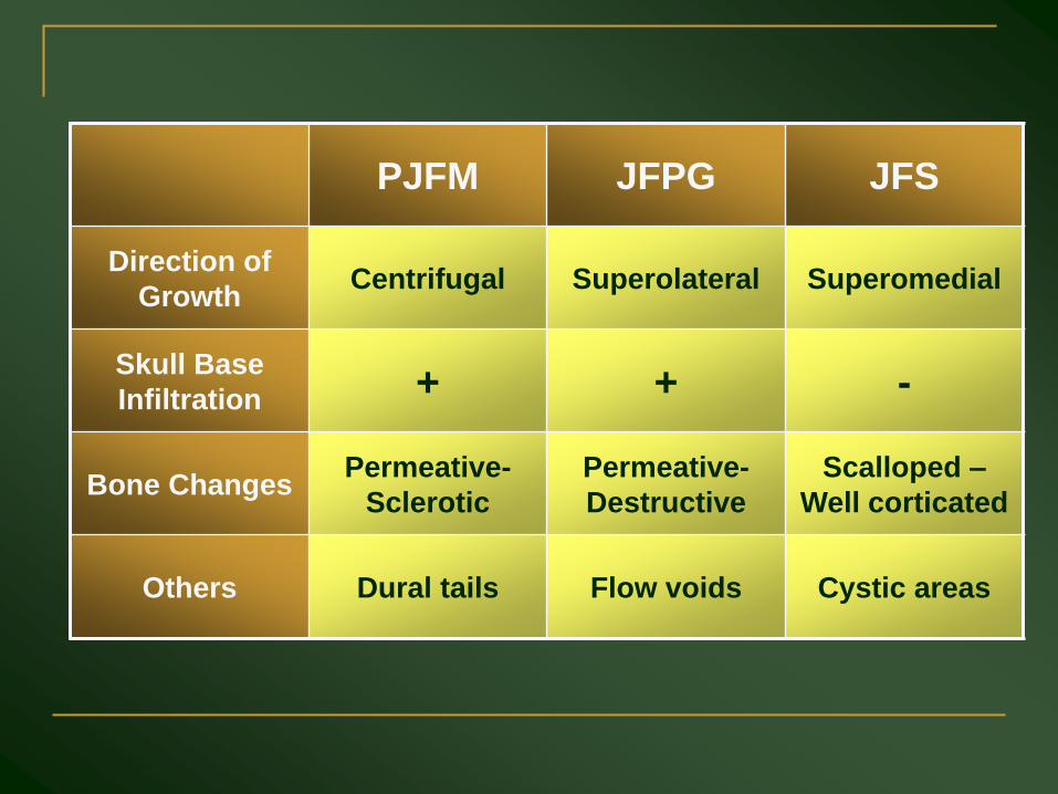
PJFM JFPG JFS
Direction of
GrowthCentrifugal Superolateral Superomedial
Skull Base
Infiltration + + -
Bone ChangesPermeative-
Sclerotic
Permeative-
Destructive
Scalloped –
Well corticated
Others Dural tails Flow voids Cystic areas

THANK YOU