Embed Size (px)
Citation preview

Imaging techniques to determine tissue perfusion in
critical limb ischemiaMarianne Brodmann, MD
Division of Angiology, Medical University Graz Austria

DisclosureSpeaker name:
Marianne Brodmann, MD
I have the following potential conflicts of interest to report:
x Consulting
Employment in industry
Stockholder of a healthcare company
Owner of a healthcare company
Other(s)
I do not have any potential conflict of interest

Tissue perfusion in CLI
▪ Diagnosis and FU of critical threshold of tissue perfusion▪ When does tissue perfusion decrease to a certain threshold which is critical
for tissue loss?
▪ Currently available hemodynamic parameters are not made for this
▪ Revascularization is the key element to restore adequate tissue perfusion in CLI▪ But how to determine if this was successfully achieved
▪ Angiogram alone not reliable

Tissue perfusion in CLI
▪ Ideal test for foot perfusion:
inexpensive
readily available
reproducible
improve the clinician’s ability to predict outcomes
provide perfusion data specific to the area of the foot with a wound
▪ Improving the ability to evaluate foot perfusion would benefit patients with CLI by
assisting with the etiology of a non-healing wound
identifying patients with poor perfusion in the angiosome of interest who might benefit from revascularization
identifying patients with seemingly adequate perfusion who may not require revascularization
selecting a target vessel for revascularization
providing insight when revascularization is sufficient
facilitating surveillance for patency
Misra S, Shishehbor MH, Takahashi EA, et al. Perfusion assessment in critical limb ischemia: Principles for under- standing and the development of evidence and evaluation of devices: A scientific statement from the American Heart Association. Circulation 2019; 140: e657–e672.

Tissue perfusion in CLI
▪ Current modalities of foot perfusion have limitations▪ Arterial non compressibility makes the ankle–brachial index (ABI) non-meaningful in
many patients with CLI
▪ Transcutaneous oximetry has a high coefficient of variation
▪ Intra- procedural measures of foot perfusion are attractive to the interventionalist to direct the target and extent of revascularization
… but there are limitations!✓ ‘Angiographic blush’ have been reported to correlate with wound healing but is
somewhat subjective and dependent on technique
✓Two-dimensional (2D) angiographic perfusion imaging is an intra-procedural method that attempts to provide objective angiographic data of perfusion but is also technique-dependent and is yet to be validated
✓………..

Tissue perfusion in CLI_Imaging techniques
Transcutaneous oximetry (TcPO2)
measures oxygen diffusion from the capillary beds to the epidermal layer of the skin
Sensor-containing electrode placed on the skin, warms the surrounding skin leading to local hyperthermia, measures the partial pressure of oxygen in the underlying tissue
Benefit▪ Can be predictive of wound healing
▪ Identify individuals with nonhealing ischemic wounds who may benefit from hyperbaric oxygen therapy (HBOT)
▪ Noninvasive, bedside tool with no radiation exposure
Limitation▪ Mechanism of action (skin perfusion): barriers to diffusion can lead to falsely low TcPO2 values
Tissue perfusion in CLI_Imaging techniques
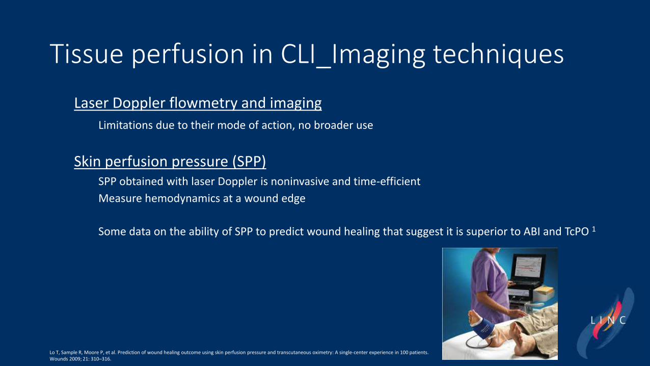
Laser Doppler flowmetry and imaging
Limitations due to their mode of action, no broader use
Skin perfusion pressure (SPP) SPP obtained with laser Doppler is noninvasive and time-efficient
Measure hemodynamics at a wound edge
Some data on the ability of SPP to predict wound healing that suggest it is superior to ABI and TcPO 1
Lo T, Sample R, Moore P, et al. Prediction of wound healing outcome using skin perfusion pressure and transcutaneous oximetry: A single-center experience in 100 patients.Wounds 2009; 21: 310–316.

Tissue perfusion in CLI_Imaging techniques
Two-dimensional angiographic perfusion imaging
software analysis package
measurement of parameters of contrast delivery to a region of interest in the foot
no additional radiation, intra-procedural time, or contrast
Two-dimensional angiographic perfusion. Right: Color map of the foot generated from a standard catheter-based angiogram. Left: Time density curve representing the time to peak contrast density in the foot. The average time to peak contrast density for this patient was 4.0 seconds.

Tissue perfusion in CLI_Imaging techniques
Two-dimensional angiographic perfusion imaging
Available evidence/3 studies• Majority of patients increase in perfusion was observed after revascularization
• ABI correlated well to time-to-peak and peak density ratios
• Improvement in 2D angiographic perfusion parameters was observed following revascularization
Benefit▪ Practical, as it can be incorporated into a standard angiogram
▪ ‘Angiosome specific’, providing information of regional foot perfusion
▪ Theoretically could guide revascularization decisions in real time
Limitation▪ No evidence on longitudinal outcomes associated with 2D perfusion parameters
▪ Modality not standardized
− Technique would need to be the same across operators and institutions so that the data generated would be generalizable to patient care.
Miguel Montero-Baker et al; Vascular Medicine2020, Vol. 25(3) 235–245

Tissue perfusion in CLI_Imaging techniquesIndocyanine green angiography
• Water-soluble contrast agent quickly bound to plasma proteins
• Visualization of local-regional blood flow by a charge-coupled camera to detect the fluorescence emitted after excitation by a laser light source of a specific wavelength
To obtain the objective fluorescence intensity curves to assess foot perfusion in the patient with CLI, a single area of the foot is analyzed and there must be virtually no movement of either the foot or camera once recording begins
However, looking in this single view may limit subjective visualization of regional perfusion variations in the entire foot that might be seen at different angles.

Tissue perfusion in CLI_Imaging techniquesIndocyanine green angiography
Available evidence/3 studies• Ingress (the increase in fluorescence intensity from baseline) and Ingress rate (the increase in intensity over the
time to maximum intensity) have shown some correlation with ABI and toe pressure1
• Able to detect severe ischemia, based on the Wound-Ischemia-foot-Infection (WIfI) classification, with reasonable accuracy 2
• Parameter of time to half-maximal intensity to detect response to revascularization 3
Benefit▪ appear to show promise for providing incremental information
Limitation▪ Need for intravenous access
▪ Contraindication in previously documented iodinated contrast allergies
▪ Specialized equipment
▪ Inability to assess perfusion deeper than 5 mm
1 Braun JD, Trinidad-Hernandez M, Perry D, et al. J Vasc Surg 2013; 57: 1213–1218. 2 Braun JD. J Vasc Surg 2014; 60: 538. 3 Colvard B, Itoga NK, Hitchner E, et al. SPY technology as . J Vasc Surg 2016; 64: 195–201.

Tissue perfusion in CLI_Imaging techniquesOxygen Microsensors
• injectable oxygen microsensors to directly monitor oxygen in the subcutaneous tissue in vivo
Soft biocompatible hydrogel sensor injected into subcutaneous space
Fluorescence chemistry on hydrogel responds based on analyte concentration Reader collects emissions and data sent to cloud
Excitation light from surface reader reaches hydrogel in tissue
Baseline Interventions End
Oxygen increase
Lum
ee
Oxyg
en
In
de
x [-
]

Tissue perfusion in CLI_Imaging techniques
Oxygen Microsensors
Available evidence/3 studies• Healthy volunteer study provided validation of the ability of oxygen microsensors to reliably detect changes in
tissue oxygen confirmed by TcPO2, with data over 3 months after sensor injection 1
• FIH/ 10 CLI: patients average tissue oxygen in the affected foot increased after EVR 2
• Post-market Registry study (OMNIA): Increases in oxygen levels assessed during endovascular revascularization procedures were associated with wound healing 3 months following the procedure 3, 4
Benefit▪ Sensitivity to both arterial insufficiency and microvascular impairment (eg diabetics)
▪ Treatment planning and long-term monitoring
▪ Prediction of wound healing
Limitation▪ Limited data available
1 Kanick SC, Schneider PA, Klitzman B, et al. Microvasc Res 2019; 124: 6–18. 2 Montero-Baker MF, Au-Yeung KY, Wisniewski NA, et al. J Vasc Surg 2015; 61: 1501–1509.e1. 3 Brodmann M. Interventional course (LINC), Leipzig, Germany, 2019

Tissue perfusion in CLI_Imaging techniquesSingle photon emission computed tomography (SPECT)
Assessment of skeletal muscle microvascular perfusion
Mitchel R. Stacy, et al Circ Cardiovasc Imaging. 2018;11:e006932. DOI: 10.1161/CIRCIMAGING.117.006932

Tissue perfusion in CLI_Imaging techniques
Impact in CLI patients
Proper diagnosis
Identification of differences related to sex and ethnicity
Reduction of major amputation
Identification of procedural failures or incomplete revascularization
Potential for telemedicine to reduce disparities in CLI
Potential cost savings in the care of patients with CLI