Embed Size (px)
Citation preview
CLINICAL MICROBIOLOGY REVIEWS,0893-8512/00/$04.0010
Jan. 2000, p. 35–54 Vol. 13, No. 1
Copyright © 2000, American Society for Microbiology. All Rights Reserved.
Immune Response to Giardia duodenalisGAETAN FAUBERT*
Institute of Parasitology, Macdonald Campus of McGill University, Ste. Anne-de-Bellevue, Quebec, Canada H9X 3V9
INTRODUCTION .........................................................................................................................................................35Giardia ........................................................................................................................................................................35Giardiasis...................................................................................................................................................................36
ANTIGENS OF GIARDIA............................................................................................................................................36Polypeptides...............................................................................................................................................................36Heat Shock Proteins.................................................................................................................................................38Lectins ........................................................................................................................................................................38Giardins......................................................................................................................................................................38Tubulin .......................................................................................................................................................................38
ANTIGENIC VARIATION...........................................................................................................................................38Antigenic Variation in Giardiasis...........................................................................................................................39Biological Significance..............................................................................................................................................39The Variant Protein VSPH7....................................................................................................................................39Immune Response in Animal Models ....................................................................................................................39
EFFECTOR MECHANISMS OF THE IMMUNE RESPONSE ............................................................................40Human Innate Immunity .........................................................................................................................................40Mechanisms of Acquired Immunity in Humans ..................................................................................................40
MOUSE MODEL..........................................................................................................................................................40Immune Response in Susceptible and Resistant Mice........................................................................................41Humoral Effector Mechanisms in Animals ...........................................................................................................41Usefulness of Specific Antibodies in Studies on Encystation.............................................................................43Cell-Mediated Effector Mechanisms in Animals ..................................................................................................43Acquired Resistance in Animals .............................................................................................................................45Passive Transfer of Immunity.................................................................................................................................47Immunosuppression in Infected Mice....................................................................................................................47
IMMUNOCOMPROMISED HOSTS.........................................................................................................................47Humans ......................................................................................................................................................................47Animals.......................................................................................................................................................................48
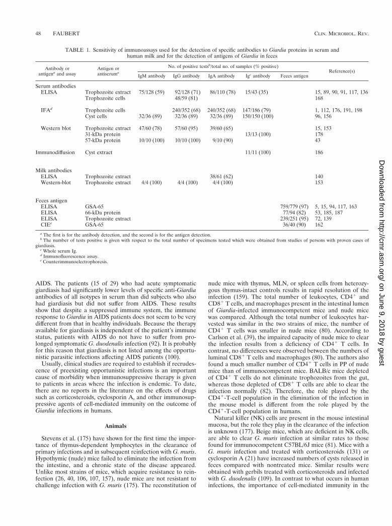
IMMUNODIAGNOSIS ................................................................................................................................................49Sensitivity of Serological Assays.............................................................................................................................49Detection of Antigens in Feces................................................................................................................................50
VACCINE.......................................................................................................................................................................50CONCLUSIONS ...........................................................................................................................................................50ACKNOWLEDGMENTS .............................................................................................................................................51REFERENCES ..............................................................................................................................................................51
INTRODUCTION
Giardia
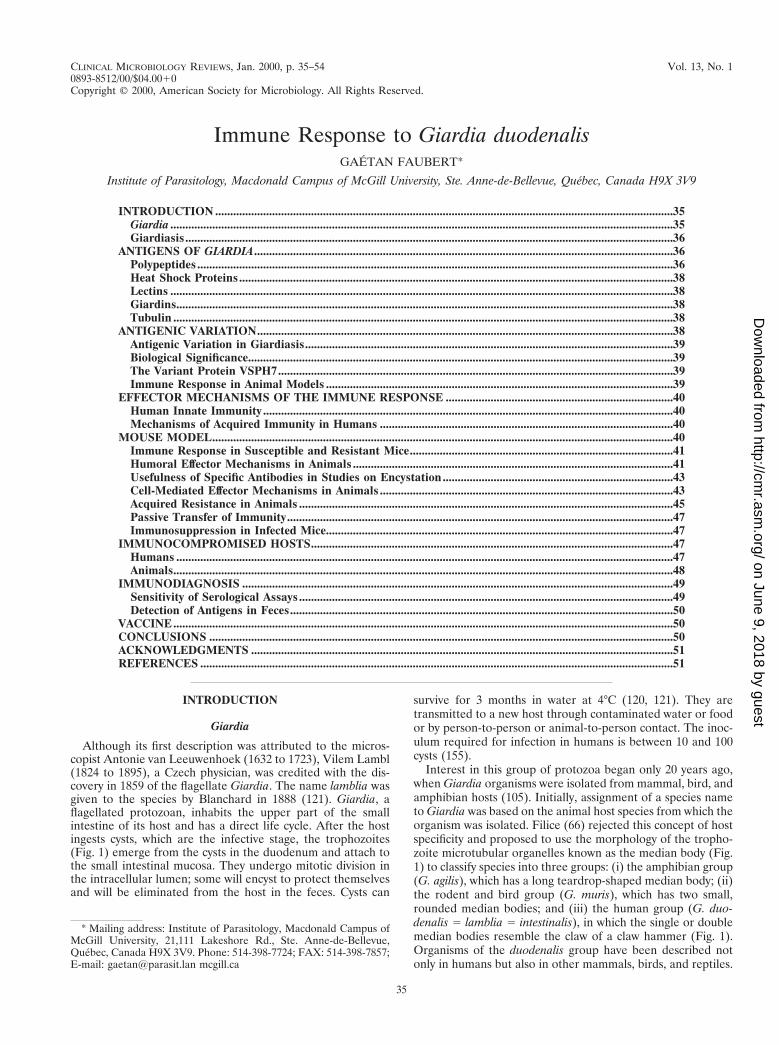
Although its first description was attributed to the micros-copist Antonie van Leeuwenhoek (1632 to 1723), Vilem Lambl(1824 to 1895), a Czech physician, was credited with the dis-covery in 1859 of the flagellate Giardia. The name lamblia wasgiven to the species by Blanchard in 1888 (121). Giardia, aflagellated protozoan, inhabits the upper part of the smallintestine of its host and has a direct life cycle. After the hostingests cysts, which are the infective stage, the trophozoites(Fig. 1) emerge from the cysts in the duodenum and attach tothe small intestinal mucosa. They undergo mitotic division inthe intracellular lumen; some will encyst to protect themselvesand will be eliminated from the host in the feces. Cysts can
survive for 3 months in water at 4°C (120, 121). They aretransmitted to a new host through contaminated water or foodor by person-to-person or animal-to-person contact. The inoc-ulum required for infection in humans is between 10 and 100cysts (155).
Interest in this group of protozoa began only 20 years ago,when Giardia organisms were isolated from mammal, bird, andamphibian hosts (105). Initially, assignment of a species nameto Giardia was based on the animal host species from which theorganism was isolated. Filice (66) rejected this concept of hostspecificity and proposed to use the morphology of the tropho-zoite microtubular organelles known as the median body (Fig.1) to classify species into three groups: (i) the amphibian group(G. agilis), which has a long teardrop-shaped median body; (ii)the rodent and bird group (G. muris), which has two small,rounded median bodies; and (iii) the human group (G. duo-denalis 5 lamblia 5 intestinalis), in which the single or doublemedian bodies resemble the claw of a claw hammer (Fig. 1).Organisms of the duodenalis group have been described notonly in humans but also in other mammals, birds, and reptiles.
* Mailing address: Institute of Parasitology, Macdonald Campus ofMcGill University, 21,111 Lakeshore Rd., Ste. Anne-de-Bellevue,Quebec, Canada H9X 3V9. Phone: 514-398-7724; FAX: 514-398-7857;E-mail: [email protected] mcgill.ca
35
on June 9, 2018 by guesthttp://cm
r.asm.org/
Dow
nloaded from

Giardia trophozoites recently isolated from the great blueheron (56) and budgerigar (58) were given the names of G.ardea and G. psittaci, respectively, because these species werefound to be distinct from G. duodenalis when examined byelectron microscopy. However, these new species share manyof the characteristics of the duodenalis organism group (58). Itis likely that new Giardia species will be described in the future.In this review, because Filice’s (66) classification is followed,the name “G. duodenalis” is used to describe the human typeof Giardia.
Giardiasis
In humans, the clinical effects of Giardia infection rangefrom the asymptomatic carrier state to a severe malabsorptionsyndrome. In fact, it was only in the late 1970s that Giardia wasrecognized to cause pathology. In a clinical study in 1978,Kulda and Nohynkova concluded that this parasite can causedisease in humans based on symptoms such as malabsorptionand the pathology observed in the upper part of the smallintestine in patients from whom the organism was isolated(105). In 1981, the World Health Organization added Giardiato its list of parasitic pathogens (197).
Factors possibly contributing to the variation in clinical man-ifestations include the virulence of the Giardia strain (8, 136),the number of cysts ingested, the age of the host, and the stateof the host immune system at the time of infection. The clinicaldiagnosis of giardiasis is difficult since symptoms are nonspe-cific and resemble those of a number of other gastrointestinalailments. Clinical features may range from diarrhea to consti-pation, nausea, headache, and flatulence (121, 199). Moreover,the symptoms observed vary with the life cycle stage of theparasite. The incubation period may last 12 to 19 days and ismarked by the first detection of cysts in the feces (97). Thisperiod is followed by the acute phase, where a variety of symp-toms signal the onset of the disease. If the immune system ofthe host is fully developed and healthy, the acute phase usuallyresolves spontaneously and the symptoms will disappear. Un-fortunately, in certain cases, in spite of a healthy and fully
developed immune system, the acute phase develops into achronic stage. In these situations, the symptoms of the diseasewill reappear for short and recurrent periods (199). There arealso some asymptomatic patients who pass cysts in their feces.In one study, it was found that between 60 and 80% of infectedchildren in day care nurseries and their household contactshave asymptomatic giardiasis (101). Asymptomatic individualsare an important reservoir for spread of the infection.
The histopathological changes occurring at the mucosal sitesrange from minimal to severe enough to cause enteropathywith enterocyte damage, villus atrophy, and crypt hyperplasia(65). The reasons for these variations are similar to thosementioned above as possible factors contributing to the varia-tion of clinical manifestation. Shortly after the trophozoitesleave the stomach of their new host in response to low pH,excystation will take place. Using their flagella and ventral disc,trophozoites released in the upper part of the small intestinemove to the microvillus-covered surface of the duodenum andjejunum, where they attach themselves (88, 116), and play arole in the onset of the pathology (22, 34, 124). The suctionforce created by this mode of attachment may damage themicrovilli and interfere with the process of food absorption(88, 116). Eventually, the rapid multiplication of the tropho-zoites by binary fission creates a physical barrier between theintestinal epithelial cells and the lumen of the intestine, inter-fering with the process of absorption of nutrients.
Since it is difficult to access the intestinal mucosa of humanswithout using invasive procedures, our knowledge of the mu-cosal pathology caused by Giardia is limited. The trophozoitesdo not usually penetrate the epithelium (65). However, whenthe conditions are favorable, trophozoites may invade tissuessuch as the gallbladder and the urinary tract (73, 122). Mucosalinvasion by trophozoites has also been observed in the mousemodel of the disease (114, 145). The migration of trophozoitesfrom the lumen of the intestine into surrounding tissues is,however, an unusual occurrence in humans and mice.
The jejunal morphology ranges from normal to subtotalvillus atrophy, and a correlation between the degree of villusdamage and malabsorption has been reported (21, 35, 124, 125,201). In humans, polymorphonuclear leukocytes and eosino-phils have been detected (202). These changes revert to normalafter treatment or when the parasite has been eliminated bythe immune system. On the other hand, Brandborg et al. foundnormal jejunal histology with absence of inflammatory cells insymptomatic patients (with diarrhea) (29). A higher incidenceof giardiasis has been reported in hypogammaglobulinemicpatients (200); it appears that more severe damage to the villusis present in the hypogammaglobulinemic patients than inthose with a normal immune system (65). Interestingly, thedegree of villus pathology observed in patients with AIDS iscomparable to that in immunocompetent patients (103), al-though AIDS patients are deficient in CD41 T cells. Further-more, AIDS patients do not appear to be more susceptiblethan healthy persons to giardiasis (166). For a review of theeffects of G. duodenalis on the structure, kinetics, and functionof absorptive intestinal cells and other epithelial cells and acorrelation with morphological injury and physiological alter-ations, the reader is referred to the review by Buret et al. (34).
ANTIGENS OF GIARDIA
Polypeptides
The identification of G. duodenalis antigens that play a rolein acquired immunity has been difficult for a variety of reasons:(i) usually the trophozoites do not invade the tissues (if there
FIG. 1. Trophozoite of the G. duodenalis type of organism.
36 FAUBERT CLIN. MICROBIOL. REV.
on June 9, 2018 by guesthttp://cm
r.asm.org/
Dow
nloaded from
is a stimulation of the immune system, it remains localized);(ii) antigenic variation on the surface membrane of trophozo-ites has been reported (see the following section); (iii) inves-tigators have used different isolates of Giardia, different anti-body reagents, and a variety of assays in studies of the immuneresponse to Giardia; and (iv) it is difficult to compare theresults obtained by different laboratories. Crude antigenic ex-tracts prepared from G. duodenalis trophozoites cultured invitro have revealed different polypeptides depending on thetechniques used to characterize them. For example, a mini-mum of 20 distinct Coomassie blue-staining bands ranging inmolecular mass from 14 to 125 kDa were obtained by sodiumdodecyl sulfate-polyacrylamide gel electrophoresis (SDS-PAGE) (126). However, high-performance liquid chromatog-raphy showed five distinct fractions, and when they were usedas antigens in an enzyme-linked immunosorbent assay(ELISA) to detect specific antibodies in the serum of immu-nized rabbits, the assay was positive only with the higher-molecular-mass fractions (126). These findings indicate thatmany polypeptides detected by SDS-PAGE are probably notplaying a role in the immune response.
On the other hand, SDS-PAGE has been useful in demon-strating similarities in the antigen profiles of G. duodenalisisolates from the same geographic area (196). Since G. duode-nalis is a ubiquitous organism, it is possible that the antigenicprofiles of isolates from different geographic areas will vary.Surprisingly, analysis of the molecular mass of polypeptidesfrom crude extracts of trophozoites obtained from differentgeographic isolates shows that there are many similarities. Forexample, similarities were reported among the proteins in iso-lates from Afghanistan, Puerto Rico, Ecuador, and Oregon.Their molecular masses ranged from 12 to 140 kDa (167). Inthis case, it is not surprising that the antigenic profiles of G.duodenalis isolates from a same geographic area have alsorevealed many similarities among them (196).
Nash and Keister (132) were able to classify 19 isolates of G.duodenalis into three groups by comparing the reactivity ofantibodies raised against excretory-secretory (ES) products re-leased in vitro in the culture medium by each isolate. Fiveisolates showed major antibody cross-reactivity, and 11 showedmoderate antibody cross-reactivity. Three isolates releasedidentical ES products. Similarities were also observed in theantigens present on the surface of the trophozoites of the 19isolates even if the patients had been infected in differentgeographic areas (132). None of these studies of antigenicprofiles in geographic areas were able to identify a single dom-inant protein among the isolates.
The identification of a G. duodenalis trophozoite major sur-face antigen that is present on all isolates will be an asset forthe development of immunodiagnostic tests or for the designof a vaccine. The existence of a dominant surface antigen onthe trophozoite of G. duodenalis was first reported by Einfeldand Stibbs (55). The characterization of this 82-kDa antigenrevealed that it was pronase and periodate modifiable and heatlabile (55). Using surface iodinated techniques, Edson et al.(54) identified an 88-kDa major trophozoite surface antigenwhich they claim is similar to the 82-kDa polypeptide reportedby Einfeld and Stibbs. Antibodies to the 88-kDa polypeptidewere detected in the sera of infected patients, but no clearcorrelation was established between the appearance of specificserum antibodies to G. duodenalis major antigens and protec-tive immunity. Unfortunately, only two anti-G. duodenalis hu-man sera were used in their study (54). The identification of amajor surface antigen of approximately 80 kDa is an interest-ing finding. It is not known if this major antigen is also presentin isolates from different geographic areas. Antigens with dif-
ferent molecular masses were identified from isolates obtainedfrom symptomatic and asymptomatic patients. By using immu-noblotting, 65- and 70-kDa antigens were identified in thefeces of gerbils infected with strains obtained from symptom-atic and asymptomatic patients, respectively (127, 128).
Clark and Holberton (44) introduced methods to study Gi-ardia molecules from pure fractions of plasma membranes.After purification of the cell membrane preparation by cen-trifugation on a Percoll gradient, a major band was found at 75kDa. The investigators concluded that the antigen corre-sponded to the iodinatable and antibody-precipitated 82-kDaantigen reported earlier by Einfeld and Stibbs (55). In addi-tion, 22-, 54-, and 58-kDa polypeptides were identified. Inter-estingly, the 54- and 58-kDa proteins comigrated with a- andb-tubulins. The authors concluded that tubulin is a constituentof Giardia membranes and appears in a different form from thetubulin found in microtubules (44). It is possible that all thesedifferent polypeptides observed on the surface membrane oftrophozoites in the early literature were in fact variant surfaceproteins described in the late 1980s by Nash et al. (137); thiswould also explain the difficulty encountered in the isolation ofdominant antigens.
Genes that encode surface membrane proteins of trophozo-ites have been cloned. Sequence analysis of a gene encoding a72.5-kDa protein revealed a single open reading frame speci-fying a hydrophilic cysteine-rich protein with an amino-termi-nal signal peptide and a postulated hydrophobic membrane-spanning anchor region near the carboxyl terminus (67). Thecysteine residues (58 of 84 residues) were in a Cys-Xaa-Xaa-Cys motif dispersed 29 times throughout the sequence. Theauthors hypothesized that the abundance of cysteine residuessuggests that the native proteins on the parasite surface maycontain numerous disulfide bonds. These bonds would conferresistance to intestinal-fluid proteases and to the detergentactivity of bile salts, thereby helping the parasite survive in thehostile environment of the intestine (67). Upcroft et al. (183)have expressed Giardia antigens in Escherichia coli by cloningG. duodenalis genomic DNA into pUC vectors. Expressed pro-teins were part of the organelles of the trophozoite. For ex-ample, a 32-kDa protein which is associated with the spiral partof the ventral disc was also found in the flagella and axonemes.Other proteins expressed by the clones covered the surface ofthe trophozoites or were associated with the coat (183).
Cyst antigens detected in human feces have a molecularmass varying between 21 and 49 kDa (71). Similar antigenswere also detected in immunoblots of parasites cultured invitro in encysting medium. These polypeptides are not found inthe trophozoites (71). Monoclonal antibodies (MAbs) raisedagainst cyst antigens were able to recognize polypeptides rang-ing from 29 to 45 kDa in immunoblot and immunofluorescenceassays. The polypeptides appeared within 8 h of exposure ofthe trophozoites to encystation medium (37, 193). These in-vestigators concluded that the molecules appearing early dur-ing encystation represent potential targets for strategies di-rected at inhibiting the process of encystation. Genes thatexpress protein components of the cyst wall have been identi-fied. One of the cloned genes expresses an acidic, leucine-rich26-kDa polypeptide (CWP1) that contains 5.3 tandemly ar-ranged copies of a degenerate 24-amino-acid repeat (129).Interestingly, the levels of the transcripts from the cyst wallprotein gene increase more than 100-fold during encystation.Cyst wall protein expression also increases dramatically duringencystation. Before CWP1 is incorporated into the nascent cystwall, it is contained within encystation-specific vesicles of en-cysting trophozoites. CWP1 was not observed in nonencystingtrophozoites (129). Another gene expressing a different cyst
VOL. 13, 2000 IMMUNE RESPONSE TO GIARDIA DUODENALIS 37
on June 9, 2018 by guesthttp://cm
r.asm.org/
Dow
nloaded from
wall protein has been cloned. The novel 39-kDa polypeptide(CWP2) is also expressed during encystation; unlike CWP1,CWP2 has a 121-residue COOH-terminal extension (113).These studies of polypeptides of G. duodenalis trophozoitesand cysts demonstrate the antigenic complexity of this intesti-nal parasite and the challenge it provides to the immune sys-tem of its host.
Heat Shock Proteins
Heat shock proteins (HSP) are synthesized by mammal,bacterium, protozoan, helminth, and even plant cells in re-sponse to stresses such as an abrupt rise in temperature, pH, orother stressful treatment. These proteins help the cell to sur-vive the stress. Giardia trophozoites live in the intestine, ahabitat where stresses are likely to occur. Few studies havebeen done on HSP in giardiasis, and the role they may play inthe immune response has yet to be defined. HSP have beendetected on the surface membrane of trophozoites. The syn-thesis of [35S]methionine-labeled proteins of 30, 70, 83, and100 kDa was increased at 43°C (110). During in vitro encysta-tion, several stage-specific proteins were recognized in immu-noblots by antisera raised against antigens of the HSP60 familyfrom Mycobacterium bovis and HSP70 from Plasmodium falci-parum (152). The detection of HSP in encysting cells is inter-esting. Giardia trophozoites have developed a way of survivingfor a certain period in the harsh environment of the host smallintestine. However, the phenomenon of encystment may rep-resent an escape mechanism for the trophozoites at the timewhen the immune system detects the presence of this invaderattaching itself to the intestinal mucosal surface. At present,little is known about how and when the trophozoites turn ongenes to build the cyst structure. Whether HSP plays a role inthe phenomenon of encystation is unknown.
Lectins
Lectins are glycoproteins that bind to specific sugars andoligosaccharides and are linked to glycoproteins or glycolipidspresent on the cell surface of eukaryotes. Trophozoites of G.duodenalis have surface membrane lectins with specificity forD-glucosyl and D-mannose residues (61). Ward et al. (194) haveidentified and characterized taglin, a mannose-6-phosphatebinding, trypsin-activated lectin from the trophozoite mem-brane. Activation of G. duodenalis lectin by proteases from thehuman duodenum has been reported (108). After activation,the lectin agglutinated intestinal cells to which the parasiteadheres in vitro. The lectin was specific for mannose-6-phos-phate and was bound to the plasma membrane of Giardia(108). A systematic analysis of G. duodenalis trophozoite sur-face carbohydrate residues with lectins and glucosidases ofknown sugar specificity has revealed that N-acetyl-D-glu-cosamine is the only detectable saccharide on the plasma mem-brane (192). The biological functions of lectins are unknown,but it appears that they play a role in the mechanisms ofattachment of the trophozoites at the site of colonization (61).The role that lectins play in the immune response to Giardiais unknown. The immunobiology of the N-acetyl-D-galac-tosamine surface lectin of Entamoeba histolytica is well known(38). This lectin binds to mucin for colonization and preventsthe trophozoites from making contact with the underlying sur-face of the epithelium (181). Taglin, a lectin present on thesurface membrane of Giardia, does not bind to mucin. It is alsounknown if taglin is able to transform the local lymphocytesinto blast cells. In this case, it is unlikely that lectins are im-portant in the immune response to giardiasis.
Giardins
Giardins are unique proteins of Giardia cells; to date, noth-ing in the literature indicates the presence of similar proteinsin the cytoskeletons of other cell types. In contrast to surfacemembrane antigens of trophozoites, structural proteins of G.duodenalis appear to be highly conserved among isolates. Forexample, analysis of the amino acid sequence of a 33-kDaprotein located in the ventral disk and axostyle revealed asingle open reading frame of 813 bp (6). The giardins aredefined as a family of ;30-kDa structural proteins found inmicroribbons attached to microtubules in the disk cytoskeletonof Giardia trophozoites (46). Using SDS-PAGE, Crossley andHolberton (47) characterized the proteins from the axonemesand disk cytoskeleton of G. duodenalis trophozoites. In addi-tion to tubulin and the 30-kDa disk protein, at least 18 minorcomponents copurify with the two major proteins in Triton-insoluble structures (47). The 30-kDa polypeptide accounts forabout 20% of the organelle proteins on gels. In continuous 25mM Tris-glycine buffer, this polypeptide migrates as a close-space doublet and was given the name of giardin. Peattie et al.(147) have studied the molecular aspects of giardins and havefound giardins at the edges of disk microribbons of the tro-phozoite; they named these particular proteins a-giardins. In asubsequent study (141), more than one giardin was present atthe edges of the disk. The giardins were renamed a1-giardin,a2-giardin, and g-giardin. Sequence analysis comparison re-vealed that the genes coding for the a-giardins had 81% iden-tity at the nucleotide level and 77% identity at the predictedamino acid level (141). The interest in giardins as primaryantigens in the immune response to Giardia stems from thefact that they form a family of proteins unique to this parasite.They also represent a large proportion of the proteins found inthe organelle of attachment (ventral disk) of the parasite to itshost. They are surface antigens, and they are probably the firstset of antigens detected by the local immune system afterattachment of the parasite to the mucosal surfaces. No studieshave been reported on the role played by giardins in immunityin giardiasis.
Tubulin
Tubulin determinants have been localized separately in thedisk cytoskeleton and flagella (180). After tubules were fixed informalin, a-tubulin was detected in the flagella, ventral disk,funis, and median body (45). However, unfixed tubules showeddifferent antigenic structures. For instance, disk microtubuleswere not stained by antitubulin antibodies. Crossley and Hol-berton (47) have identified at least five isoelectric variants ofG. duodenalis tubulin. These molecules may represent a pri-mary target for the immune system since they are found inmany organelles. The role they play in immunity has not beenstudied.
ANTIGENIC VARIATION
Antigenic variation represents a mechanism whereby se-lected viruses, bacteria, and parasites evade the immune re-sponse of the host. By the time the host has developed aprotective immune response to the antigens originally present,the latter have been replaced in a few surviving organisms bynew antigens. Antigenic variation affects the surface antigensof the infectious agents in which it occurs.
38 FAUBERT CLIN. MICROBIOL. REV.
on June 9, 2018 by guesthttp://cm
r.asm.org/
Dow
nloaded from
Antigenic Variation in Giardiasis
Nash et al. (137) were first to report the phenomenon ofantigenic variation in giardiasis. Some characteristics of thisphenomenon in giardiasis are as follows: (i) certain epitopesare reexpressed in clones, suggesting the presence of a favoredset in the repertoire of epitopes; (ii) the repertoires of variantsurface proteins (VSPs) may differ among isolates; and (iii) thesame epitope detected on the surfaces of independent isolatesis present in molecules with different molecular masses (134,135, 138). In contrast to other parasites in which the phenom-enon has been observed, antigenic variation in giardiasis wasfirst observed as a phenomenon occurring in vitro. Most of thestudies on antigenic variation were done with the WB isolateobtained from a symptomatic individual infected in Afghani-stan. Clones of the WB isolate of G. duodenalis were exposedin vitro to a cytotoxic MAb which reacts with a 170-kDa surfaceantigen (137). Analysis of progeny and clones of the progenyby different assays failed to detect the high-cysteine 170-kDaantigen. In a subsequent study, it was demonstrated that theloss of this antigen was associated with the appearance of anew 64-kDa surface antigen (3). Specific variants have beendetected after 12 generations of in vitro growth of the WBisolate (133). The abundant, highly variable VSPs which coverthe surface of trophozoites have been confirmed (204), andthese VSPs are capable of binding 65Zn in vitro. The finding ofa cysteine-rich protein(s) in Giardia trophozoites (3, 7) was notunexpected, since Giardia has a high nutritional requirementfor cysteine (69). The gene VSPA6 coding for the 170-kDasurface antigen has been cloned (3). This gene consists of threeregions: a short 59 region containing a hydrophobic leader, arepeat region comprising 4,056 nucleotides and 20.8 repeats,and a 39 region containing a region of homology to the otherVSPA6 genes (2). Antigenic variation at the surface membraneof trophozoites occurs frequently in Giardia isolates. Theseantigens are made of cysteine-rich proteins (6, 33), which arecontrolled by 20 to 184 genes (133). In contrast to Africantrypanosomiasis, where genes controlling variant surface anti-gens are expressed in telomere-associated sites, the VSP genescontrolling the VSPs in Giardia are not telomere associated(138).
Biological Significance
The importance of antigenic variation as a parameter in theimmune response to Giardia was realized when the phenom-enon was documented in vivo in humans, mice, and gerbils (10,77). Gerbils were inoculated orally with live trophozoites of G.duodenalis clone WB Cl-6E7, which expresses a major 179-kDasurface membrane protein. By day 7 postinfection, this proteinwas no longer detected on the surface of trophozoites and hadbeen replaced by a series of new antigens, including a majorprotein at 92 kDa (10). When immunocompetent BALB/cmice were infected with a cloned human isolate of G. duode-nalis, trophozoites removed from the small intestine had lost amajor surface epitope by day 22 postinfection (77). Gottsteinand Nash hypothesized that B-cell-dependent mechanisms aremost likely to be responsible for the surface antigen switch(77). In contrast, the trophozoites removed from the guts ofinfected athymic nude and scid mice still expressed the majorsurface membrane epitope at the same level on day 25 postin-fection. Interestingly, the initial antigenic surface variant re-mained unchanged after encystment and subsequent excyst-ments by infection in a new host (138). The facts that antigenicvariation was not observed in athymic mice and the initialsurface variant antigens remained unchanged after encystation
indicate that the phenomenon of antigenic variation in giardi-asis is driven by the immune system of the host.
The Variant Protein VSPH7
Neonatal ZU.ICR mice infected with trophozoites of G.duodenalis clone GS/M-83-H7 expressing the variant proteinVSPH7 transiently produced milk immunoglobulin A (IgA)antibodies against a variant-specific 314-amino-acid N-termi-nal region of VSPH7. These IgA antibodies exhibit a strongparasiticidal effect on VSPH7-type trophozoites both in vitroand in vivo. Not only are they promoting antigenic variation inclone GS/M-83-H7, but also they influence the early course ofthe infection in mice (174). VSPH7 consists of two antigeni-cally distinct fragments: a unique, variant-specific 314-amino-acid N-terminal region which elicits a low antibody responsethat is preferentially detectable during the early phase of in-fection, and a 171-amino-acid C-terminal region which elicits ahigh antibody response during the later phase or after resolu-tion of infection (130). Again, these results provide a goodexample of the complexity of the immune response to Giardiaantigens. A low antibody response was detected against a spe-cific epitope during the early phase of the infection, while ahigher antibody response was obtained against a differentepitope in the late phase of the infection. The immunogenicityof VSPH7 in adult female ZU.ICR mice was studied afterperoral immunization with a recombinant vaccine (173). Forthis purpose, the biocarrier Salmonella enterica serovar Typhi-murium strain LT2M1C was used to deliver the VSPH7 anti-gens to the mucosal site. The vaccination induced VSPH7-specific IgG1, IgG2a, and IgG2b antibodies in the serumwhereas IgA antibodies were detected from supernatants of invitro-maintained intestinal-cell conglomerates. The authorsconcluded that the live attenuated serovar Typhimurium strainLT2M1C is an ideal antigen delivery system, since the specificsystematic and local antibody responses were similar to thoseinduced by experimental or natural infections of mice with G.lamblia clone GS/M-83-H7. Unfortunately, the authors did notdetermine if the mice immunized with the biocarrier serovarTyphimurium were protected against a challenge infectionwith G. lamblia.
Immune Response in Animal Models
The variety of humoral and cellular immune responses stim-ulated during the occurrence of antigenic variation has beenstudied by using the mouse and gerbil animal models of thedisease. The predominant anti-Giardia-specific antibodies areof the IgM and IgG isotypes, whereas the CD41 T lymphocytesisolated from mouse Peyer’s patches (PP) show a predominantproliferative response to the antigens (75). On the other hand,spleen and mesenteric lymph node (MLN) cells did not showany lymphoproliferative response and no specific anti-GiardiaIgA antibodies were detected. These results show that in anatural infection the lymphoid cells responding to the anti-genic stimulation are located along the intestinal mucosal sur-faces. The variant surface antigens of G. duodenalis have beenlocalized on the surface membrane of the trophozoites, andthey are usually associated with the presence of a thick cell coat(149). The entire surface of the organism is usually covered bythe thick surface coat containing the variant surface protein,but on some trophozoites the thick surface coat is absent (149).It is not known if the absence of a thick surface membrane isassociated with an absence of antigenic variation.
VOL. 13, 2000 IMMUNE RESPONSE TO GIARDIA DUODENALIS 39
on June 9, 2018 by guesthttp://cm
r.asm.org/
Dow
nloaded from
EFFECTOR MECHANISMS OF THEIMMUNE RESPONSE
Our understanding of the mechanisms of the immune re-sponse in giardiasis comes from four sources: (i) in vitro stud-ies involving the growth of axenically grown G. duodenalistrophozoites together with immune cells from a variety ofhosts; (ii) studies of mice infected with their natural parasite,G. muris; (iii) animal models involving G. duodenalis-infectedadult gerbils or weanling mice; and (iv) studies of humansnaturally infected with Giardia or those who have volunteeredto be infected with Giardia (62).
Human Innate Immunity
In some patients, giardiasis resolves within a few days, whilein others the symptoms last for years, even in the presence ofcirculating antibodies in serum or secretory antibodies at mu-cosal sites and the cell-mediated immunity. Because of itsbiological characteristics, it is likely that nonimmune factorsplay a role in susceptibility to infection or in the duration andseverity of the disease. For example, normal human milk killsG. duodenalis trophozoites independently of specific secretoryIgA antibodies (68). A number of laboratories have demon-strated one giardiacidal factor present in milk, such as conju-gated bile salts (70), unsaturated fatty acids (160), or free fattyacids (154). When grown in vitro in the presence of humanmilk, trophozoites can be protected from its giardiacidal effectby addition of intestinal mucus to the culture medium (203). G.duodenalis trophozoites are killed by products of lipolysispresent in human duodenal and upper jejunal fluid (50). Aleyet al. (11) have also reported that human neutrophil defensinsand indolicidin have antitrophozoite activities when they areadded to the culture medium. These results demonstrate theimportance of nonimmune mechanisms in the control of theparasite population in the intestine. On the other hand, mech-anisms of innate immunity may protect the parasite from de-struction. For example, mucus has been reported to protect thetrophozoites from being killed by lipolytic products present inthe intestinal fluid (205).
Mechanisms of Acquired Immunity in Humans
Both humoral and cell-mediated immune responses havebeen reported to occur in human giardiasis (4). However, littleis known about the mechanisms involved in this immune re-sponse because most of our knowledge is based on the mousemodel of disease involving a rodent source of Giardia (G.muris). Also, studies of the immunological aspects of the host-parasite relationship with G. duodenalis types of organismswere done in vitro with culture media developed for growinglymphoid cells, not Giardia trophozoites (4, 62, 78). The cul-ture of trophozoites under inappropriate conditions has alsomade the parasite more vulnerable to immunological attack.Because of this, the interpretation of many in vitro studies ofthe effector mechanisms implicated in the immune response toG. duodenalis trophozoites is problematic.
The lethal effect of human serum for G. duodenalis tropho-zoites appears to be dependent on the presence of an intactclassical pathway of complement. Human sera containinganti-G. duodenalis antibodies killed more than 98% of theparasites in vitro (84). The killing effect of human sera wasabrogated when the sera were chelated with EDTA or heatinactivated at 56°C for 30 min, conditions known to inactivatecomplement. These results were confirmed in another study,where sera, obtained from infected humans, containing anti-G.duodenalis trophozoite antibodies of the IgM class and com-
plement lysed the trophozoites (51); these authors concludedthat the activation of the classical pathway of complementproduced the lysis. Since Giardia trophozoites reside in thelumen of the intestine, it is unlikely that the above mechanismsplay a role in controlling parasite numbers within the intestine.However, lysis of trophozoites by specific antibodies in thepresence of complement may play a role in limiting the inva-sion of tissues by trophozoites. The humoral arm of the im-mune system has been reported to play a role in infectedpatients. For example, the jejunal-plasma immune response toGiardia involves a decrease in the number of IgA cells and anincrease in the number of IgM cells (104).
The functional importance of mucosal-associated lymphoidtissue is indicated by its large population of antibody-produc-ing plasma cells that are secreting primarily IgA antibodies.However, cell-mediated immunity also plays an important roleat the mucosal sites. Lymphocytes are found in large numbersin the lamina propria, in PP, and within the epithelial layer.Many of these cells are T cells of different phenotypes. SinceGiardia antigens are T-cell-dependent antigens, the role playedby cell-mediated immunity at mucosal sites has been studied.Due to the invasive techniques required for harvesting cells atthe mucosal sites, studies of cell-mediated immunity studies inhuman giardiasis have been done with lymphocytes circulatingin the blood. Specific cellular immune responses to G. duode-nalis antigens have been reported. A lymphocyte proliferativeresponse was obtained by stimulating human peripheral bloodleukocytes with antigens obtained from homologous or heter-ologous isolates (76). As predicted, the higher stimulation in-dices were obtained with the homologous parasite antigens.Experiments designed to study the role played by humanmononuclear cells as effector mechanisms against Giardia haveproduced contradictory results. Aggarwal and Nash (9) deter-mined the cytotoxicity of mononuclear cells to Giardia by usinga thymidine assay and found that G. duodenalis trophozoitesdied spontaneously without the presence of mononuclear cellsand, surprisingly, that the presence of mononuclear cells in-creased the ability of the parasite to survive. On the otherhand, Hill and Pearson (87) reported the opposite results.They found that incubation of Giardia cells with mononuclearcells and the addition of 20% immune serum increased theingestion of parasites eightfold, indicating that opsonizationexists in giardiasis. Killing of trophozoites was attributed to theoxidative microbicidal activity of phagocytes. Human neutro-phils and monocytes are able to interfere with the in vitroattachment of Giardia trophozoites to the sides of culturetubes, demonstrating that the adherence mechanism of theparasites may be a feasible target for immunological attack(48). When trophozoites encyst, they lose their property toattach to substrate (64). Since encystation coincides with theimmune system expulsion, one can speculate about whetherneutrophils and/or other effector mechanisms of the local im-mune response play a role in the phenomenon of encystation.
MOUSE MODEL
The G. muris-mouse model of giardiasis, described by Rob-erts-Thomson et al. (157) in the mid-1970s, has provided apowerful tool to study the immune effector mechanisms thatoccur during Giardia infection. The selection of the mouseover other animal models for the study of immune mechanismsin giardiasis has considerable advantages: (i) adult mice arebeing infected with their natural parasites; (ii) a considerablevariety of reagents and technologies exists for the study of theimmune response in mice; and (iii) immunologically well-de-fined inbred strains of mice are available. The mouse model of
40 FAUBERT CLIN. MICROBIOL. REV.
on June 9, 2018 by guesthttp://cm
r.asm.org/
Dow
nloaded from
giardiasis has been useful for the understanding of not only theimmune mechanisms of giardiasis but also the immunologicalphenomena at mucosal intestinal sites. The natural habitat ofG. muris trophozoites is the mouse small intestine, where itresides in the lumen or attached to the epithelium. This pro-tozoan lives extracellularly and, like G. duodenalis, does notinvade host cells or tissues.
Immune Response in Susceptible and Resistant Mice
The first evidence of the involvement of the immune systemin the elimination of Giardia in primary infection was reportedby Roberts-Thomson et al. (157), who showed that athymicnude mice develop a prolonged Giardia infection. Reconstitu-tion of these mice with lymphocytes restored a normal patternof elimination of the parasite at 7 weeks. Among many mousestrains, some mice have been identified as being particularlysusceptible to Giardia infection, developing a prolonged elim-ination phase or even persistent infection (27). For instance, incontrast to the resistant B10.A and DBA/2 mice, the infectionin susceptible A/J and C3H/He mice is characterized by a shortlatent period, a high cyst output during the acute phase ofinfection, and a relatively long period of resolution of infec-tion. The immunological basis for prolonged or chronic infec-tion in susceptible mouse strains has not yet been elucidated. Ithas been reported that susceptible C3H/He mice recognizedifferent antigen recognition patterns from resistant BALB/cmice (60). For example, a crude trophozoite antigenic extractbound to wheat germ agglutinin used to vaccinate BALB/cmice failed to induce protection (60). On the other hand, nodifferences were observed in the giardiacidal activity of spleen,MLN, and peritoneal lymphoid cells from susceptible or resis-tant mice (17) and no apparent relationships were found be-tween this capacity to mount cell-mediated or humoral effectorimmune responses and their ability to control the infection(25). These observations highlight the complexity of the im-munological aspect of the host-parasite relationship. Nonin-fected and infected resistant mice have a greater capacity torecruit cells into the peritoneal cavity after thioglycolate injec-tion than do compared to susceptible mice (18, 165). Thequantitative differences observed in the inflammatory re-sponses in resistant infected mice were related to functionaldifferences in phagocytosis and a greater capacity to respond tochemotaxis in vitro (18). The involvement of immune systemmechanisms to explain prolonged infection became puzzlingwhen it was found that susceptible adult female C3H/He micecould protect their suckling young and develop higher antibodyresponses than resistant adult female BALB/c mice (182).Moreover, following treatment with metronidazole to elimi-nate the trophozoites from the intestine, susceptible C3H/Hemice became resistant to challenge infection (182). Recently,Venkatesan et al. (184) reported no differences in the timing,titer, or specificity of secretory or serum antibodies to G. murisbetween susceptible and resistant strains of mice. However,when serum IgG subclass responses were compared, the resis-tant strain produced IgG2a while the susceptible strain pro-duced IgG1. According to these authors, these results suggestdifferential involvement of T-helper (Th) 1 and Th 2 subsets oflymphocytes (184). When cells harvested from MLN werestimulated with concanavalin A, gamma interferon (IFN-g)and interleukin-5 (IL-5) were secreted by cells from the resis-tant strain but only IL-5 was secreted by cells from the suscep-tible strain (184). The lack of secretion of IFN-g by MLN cellsfrom the susceptible strain is interesting because it may explainwhy this intestinal parasite is particularly susceptible in thesemice. IFN-g is recognized as playing a role not only in the
proliferation of B cells but also in the switch from one Ig toanother. Furthermore, if hypersensitivity reactions are playinga role in the control of the infection at the gut level, thenonsecretion of IFN-g by MLN cells would affect this mecha-nism of defense.
Humoral Effector Mechanisms in Animals
The expulsion of G. muris from the small intestines of in-fected mice is closely associated with the appearance of anti-G.muris IgA antibody in intestinal secretions (169). Parasite-specific IgA and IgG antibodies bind to G. muris trophozoitescolonizing the small intestine (83). The percentage of tropho-zoites with adherent neutrophils increases in the presence ofanti-Giardia-specific IgG serum antibodies or immune mousemilk or secretory IgA antibodies (99). Phagocytosis of tropho-zoites by macrophages increases after incubation with immuneserum (17, 98, 99, 150) or immune mouse milk (99). On theother hand, bone marrow-derived macrophages from C3H/HeN mice pretreated with recombinant IFN-g ingest signifi-cantly larger numbers of G. duodenalis trophozoites than dountreated macrophages (23). The classical pathway of comple-ment can be activated by immune complexes containing IgM orIgG antibodies, and it appears that anti-Giardia-specific anti-bodies of the IgM or IgG isotypes support the lytic effect ofcomplement on Giardia cells. Deguchi et al. (51) have reportedthat G. duodenalis trophozoites sensitized with anti-Giardiaantibodies of the IgM class are lysed. Butscher and Faubert(36) obtained similar results with G. muris trophozoites sensi-tized to similar antibody isotypes. Moreover, an IgG1 MAbwas found to bind in vitro to the surface of trophozoites, fla-gella, and flagellar insertions (36). This MAb was able to lyseG. muris trophozoites in the presence of exogenous comple-ment, and when administered directly into the duodenum ofmice, it significantly reduced the number of trophozoites dur-ing the acute phase of the infection (24). The main target forthis MAb was a 35-kDa Triton-soluble glycoprotein located onthe surface membrane of the trophozoite (24, 36). Finally, therole of complement in lysing Giardia cells was also demon-strated with a MAb which recognized proteinaceous cyst anti-gens and was able to abolish the formation of the cyst whenadded to the culture medium together with a source of com-plement (37). All these studies show that anti-Giardia antibod-ies in the presence of an exogenous source of complement caneffectively lyse trophozoites and encysting cells in vitro. Unfor-tunately, the complement proteins are absent in the lumen ofthe intestine. The only source of complement near the intes-tinal lumen would come from the few macrophages present inthe deep invagination of the M cells which are located in themucous membrane.
Although the role played by T and B lymphocytes in thecontrol of the infection is well documented, there is only onestudy reported in the literature on the cytokines produced byCD41 T cells in response to Giardia antigenic stimulation.When Giardia trophozoite proteins were used to challenge PPand spleen cells removed from infected mice, IL-4, IL-5, andIFN-g were not detected in the culture supernatant (52). How-ever, when the cells were challenged with concanavalin A, allthree cytokines were detected. The release of IL-4 and IL-5 bythe spleen and PP cells in the culture supernatant confirms therole played by antibodies of the IgA isotype in the control ofgiardiasis. Two conclusions can be drawn from these experi-ments. First, it appears that Giardia proteins are poor immu-nogens since they were not able to stimulate lymphoid cellsadequately for the production of lymphokines. A weak lym-phocyte proliferation was observed when a G. muris crude
VOL. 13, 2000 IMMUNE RESPONSE TO GIARDIA DUODENALIS 41
on June 9, 2018 by guesthttp://cm
r.asm.org/
Dow
nloaded from
42 FAUBERT CLIN. MICROBIOL. REV.
on June 9, 2018 by guesthttp://cm
r.asm.org/
Dow
nloaded from
extract from trophozoites was used to stimulate PP cells fromnoninfected mice in vitro (86). Second, the relative success ofG. muris in completing its life cycle in a primary infectionmight be due, in part, to poor stimulation of Th1 and Th2immune responses. The Th1-type immune response is virtuallyabsent in the primary infection. In vitro studies have shown thecentral role played by macrophages and IFN-g in the killing oftrophozoites (23).
Usefulness of Specific Antibodies in Studies on Encystation
The process of encystation is a key step in the Giardia lifecycle that allows this intestinal protozoan to survive betweenhosts during person-to-person, waterborne, or food-bornetransmission. To my knowledge, the existence of serum or localantibodies at the gut level against cyst antigens in infectedpatients has never been reported. The absence of antibodiesagainst novel molecules appearing on the surface membrane ofthe encysting trophozoite is not surprising. Encystation is acomplex phenomenon occurring over a short period and isprobably not detected by the local immune system. In spite ofthe apparent absence of antibodies against encysting moleculesin a natural infection, I believe that studying the immunoge-nicity of the latter is important since they offer immunologicalstrategies for stopping, or at least decreasing, the spread of theinfection in the environment.
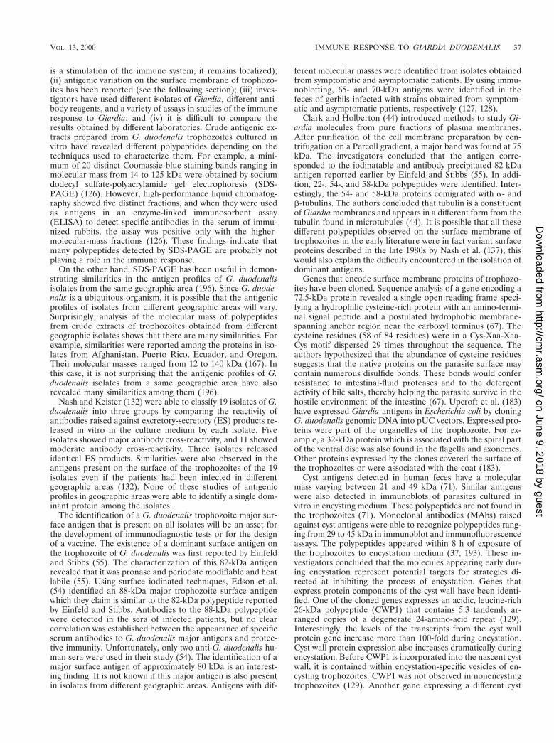
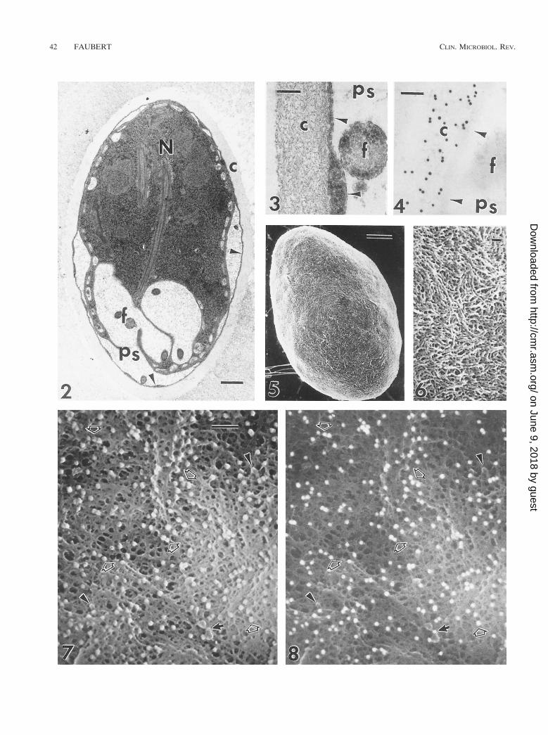
Our knowledge of the formation of the cyst structure waslimited until polyclonal antibodies and MAbs specific to cystmolecules were developed and used in studies of cyst wallformation. Using immunofluorescence and immunogold stain-ing, Erlandsen et al. (57) studied the chronological eventstaking place during encystment. The phenomenon begins withthe formation of an intracellular and extracellular phase, whichrequires a minimum of 14 h. The extracellular phase is initiatedwith the appearance of cyst wall antigens on small protusionsof the trophozoite membrane, which enlarge to form “caplikestructures” with progression to formation of the cyst wall. Cap-like structures are detected over the entire surface of the tro-phozoites, including the adherence disk and flagella (57, 59).Late stages in encystment include a “tailed” cyst, in whichsome of the flagella are not fully retracted into the cyst. Afterencystation is completed, the cyst wall is composed of filamen-tous and membranous portions and is separated from the cy-toplasm of the trophozoite by the peritrophic space (Fig. 2through 8). These observations confirm the findings of earlierinvestigators (37, 64, 71, 118, 151, 192). Using monospecificantibodies to a VSP antigen (TSA 417), which is a type 1
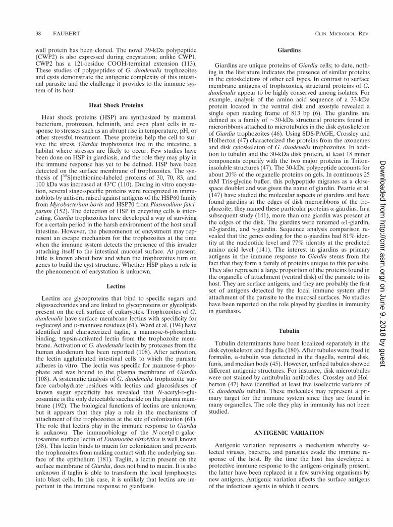
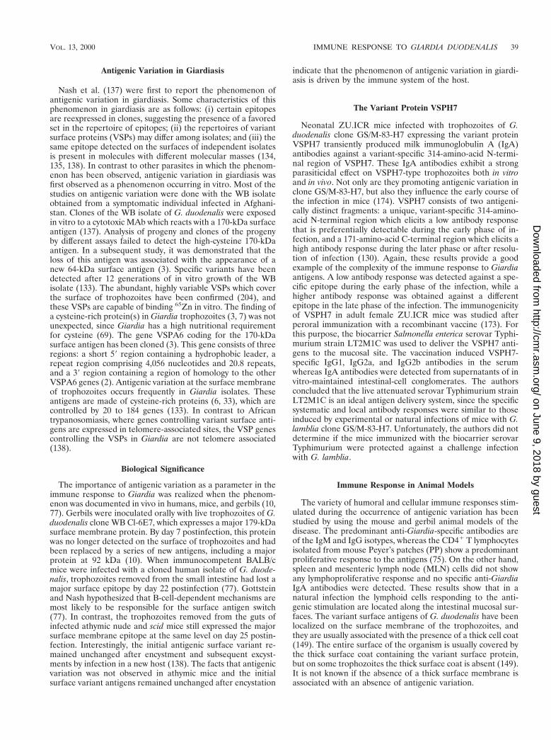
integral membrane protein that covers the entire surface of thetrophozoite, and a MAb against a cyst wall protein (8C5),McCaffery et al. (118) observed the transport of the epitopesthat bind to these two specific antibodies during encystation. Inpreencysting cells, both proteins are localized on the nuclear-envelope endoplasmic reticulum cisternae, and cytoplasmicmembrane cisternae, thereby reflecting their site of synthesis.However, only epitope 8C5 is localized on the encystation-specific vesicles (ESV). The ESV are the equivalent of caplikestructures described by Erlandsen et al. (57). These large se-cretory vesicles form only during encystation, and they trans-port cyst antigens (Fig. 9) to the nascent wall (118). In contrast,only TSA 417 was found on the outer surface of the plasma-lemma of trophozoites, encysting cells, and underlying thewalls of many cysts (Fig. 9 and 10). As encystation progresses(Fig. 10), TSA 417 disappears from the plasmalemma and itslevel in the lysosome-like peripheral vesicles and other largecytoplasmic vesicles is increased (118).
Preexposure of cysts to polyclonal rabbit antiserum againstpurified cyst wall proteins or to wheat germ agglutinin inhibitsexcystation by more than 90% (119). The investigators con-cluded that the ligand binding cyst wall epitopes inhibit encys-tation, most probably by interfering with the proteolysis of cystwall glycoproteins.
Cell-Mediated Effector Mechanisms in Animals
Many of the cellular events of the intestinal mucosal site inresponse to parasite antigens are under the complex regulationof T cells. Heyworth et al. (79) found that most of the cellsharvested from the intestinal lumen of mice infected with G.muris were lymphocytes mixed with a small number of macro-phages. When the cells were identified by immunofluorescentstaining, approximately 50% of the intraluminal leukocyteswere shown to be T lymphocytes. The kinetics of intraepitheliallymphocyte (IEL) and lamina propria lymphocyte (LPL) re-sponse during G. duodenalis infection in weanling mice havebeen studied. An increase in the numbers of suppressor andCD81 T cells in the IEL and LPL tissues was observed duringthe latent period; the numbers peaked during the acute phaseand decreased during the elimination phase. In contrast, thenumber of CD41-T-cell subsets remains small during the firsttwo phases of the infection and increases significantly duringthe elimination phase (189). Meanwhile, the number of IgA-plasma cells in the lamina propria declined during the latentand acute phases of infection and increased during the elimi-nation phase (189). Investigators concluded that induction of
FIG. 2. Transmission electron micrograph of a G. muris cyst fixed in the presence of 1% bovine serum albumin. The cyst wall is composed of filamentous (c) andmembranous (arrowheads) portions and is separated from the cytoplasm of the trophozoite by the peritrophic space (ps). N, nucleus; f, flagellum. Originalmagnification, 316,000. Bar, 0.5 mm. Reprinted from reference 59 with permission of the publisher.
FIG. 3. Transmission electron micrograph of the filamentous (c) Giardia cyst wall showing the course of individual filaments. The membranous portion of the cystwall (arrowheads) separates the filamentous portion from the underlying peritrophic space (ps). f, flagellum. Original magnification, 380,000. Bar, 0.1 mm. Reprintedfrom reference 59 with permission of the publisher.
FIG. 4. Thin section of G. muris cyst wall, comparable morphologically to that in Fig. 3 but immunostained with rabbit polyclonal antiserum (R-AGLMB) and goatanti-rabbit IgG labeled with 15-nm colloidal gold. Specific staining with immunogold is detected over the filamentous (c) portion of the cyst wall, and no labeling isseen on the membranous portion of the cyst wall (arrowheads) or in the peritrophic space (ps). f, flagellum. Original magnification, 380,000. Bar, 0.1 mm. Reprintedfrom reference 59 with permission of the publisher.
FIG. 5. Low-voltage field emission SEM of a G. muris cyst taken at 1.5 kV, illustrating the filamentous nature of the cyst wall. Original magnification, 39,500. Bar,1 mm. Reprinted from reference 59 with permission of the publisher.
FIG. 6. Higher magnification of the filamentous cyst wall of the G. muris cyst seen in Fig. 5. Individual filament populations, ranging from 7 to 20 nm, are easilydiscerned and appear to form a tightly interwoven mesh. Original magnification, 339,500. Bar, 0.1 mm. Reprinted from reference 59 with permission of the publisher.
FIG. 7 and 8. SEI (Fig. 7) and BEI (Fig. 8) of the filamentous cyst wall of G. muris immunocytochemically labeled with rabbit antiserum to the Giardia cyst wall(R-AGLMB) and goat anti-rabbit IgG coupled to 15-nm colloidal gold. A comparison of the SEI and BEI taken at 10 kV by FESEM reveals the one-to-onecorrespondence (open arrows) between the 15-nm immunogold complexes associated with the filamentous cyst wall as seen by surface topography (Fig. 7) or byatomic-number contrast (Fig. 8). In some instances, 15-nm immunogold particles were not obvious with SEI (small solid arrow) but were easily detected by BEI, as(Fig. 8). Other particles detected by SEI (arrowheads) were shown not to be immunogold by BEI. Original magnification, 380,000. Bar, 0.05 mm. Reprinted fromreference 59 with permission of the publisher.
VOL. 13, 2000 IMMUNE RESPONSE TO GIARDIA DUODENALIS 43
on June 9, 2018 by guesthttp://cm
r.asm.org/
Dow
nloaded from
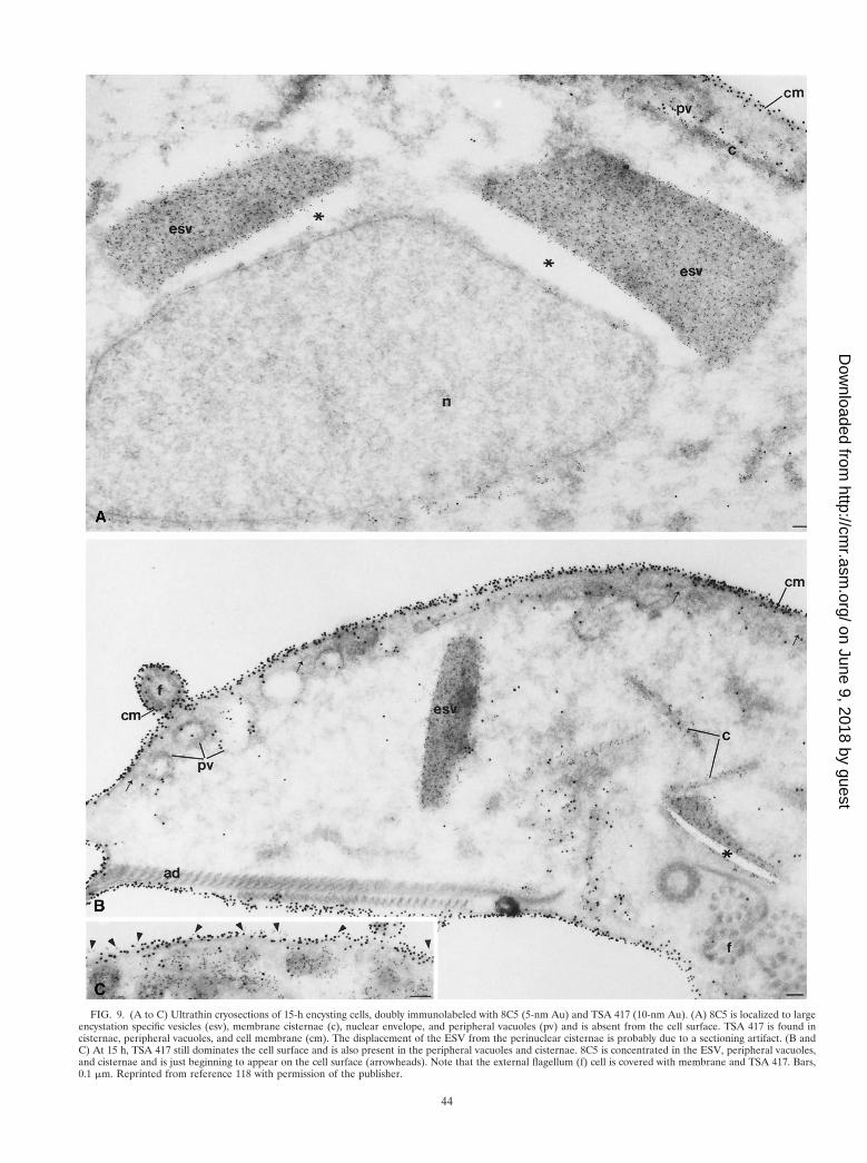
FIG. 9. (A to C) Ultrathin cryosections of 15-h encysting cells, doubly immunolabeled with 8C5 (5-nm Au) and TSA 417 (10-nm Au). (A) 8C5 is localized to largeencystation specific vesicles (esv), membrane cisternae (c), nuclear envelope, and peripheral vacuoles (pv) and is absent from the cell surface. TSA 417 is found incisternae, peripheral vacuoles, and cell membrane (cm). The displacement of the ESV from the perinuclear cisternae is probably due to a sectioning artifact. (B andC) At 15 h, TSA 417 still dominates the cell surface and is also present in the peripheral vacuoles and cisternae. 8C5 is concentrated in the ESV, peripheral vacuoles,and cisternae and is just beginning to appear on the cell surface (arrowheads). Note that the external flagellum (f) cell is covered with membrane and TSA 417. Bars,0.1 mm. Reprinted from reference 118 with permission of the publisher.
44
on June 9, 2018 by guesthttp://cm
r.asm.org/
Dow
nloaded from
CD41 T cells during the elimination phase concomitant withan increase in the number of lamina propria IgA-plasma cellsresults in the elimination of the parasite from the gut. Villusatrophy and crypt hyperplasia were observed in the duodenumof gerbils infected with G. duodenalis trophozoites (22) andmice infected with G. muris (30). Crypt mitotic rates have beenreported to double during the acute phase of Giardia infectionsin mice (115). It has been hypothesized that T lymphocytesdirectly or indirectly control the cycling time of crypt stem cellsas well as the factors that orchestrate their differentiation alongdifferent lines (14). Again, these observations reinforce therole of cell-mediated immunity in the immune response ingerbils.
PP T- and B-cell subset populations have been studied insusceptible BALB/c mice infected with G. muris. In this pe-ripheral lymphoid organ, the number of leukocytes doubledduring the course of the infection but returned to control levelsas the infection was eliminated from the intestine (40). TheCD41 and T-suppressor subsets represent 34.1 and 6.2%, re-spectively, of the total population in PP in noninfected mice;these percentages did not change after the infection with G.muris. On the other hand, the number of PP secretory IgM(sIgM) B cells increases rapidly in infected BALB/c mice toreach a maximum at the end of the latent period, whereas thenumber of sIgA B cells increases later to reach a maximumduring the acute phase (41). The switching from the IgM to theIgA isotype confirms the importance of the Th2 subset andmast cells in the self-cure phenomenon. Both types of cells arerecognized to secrete IL-5, which promotes the switching tothe IgA isotype.
The role played by macrophages in the immune response toGiardia is well documented. In the mouse model of the disease,invading G. muris trophozoites were found in the epitheliumnear dying or desquamating columnar cells (146). Macro-phages beneath the basal lamina extended pseudopods into theepithelium, trapping invading G. muris trophozoites and en-closing them in phagolysosomes. Macrophages containing di-gested trophozoites were surrounded by rosettes of lympho-blasts in the epithelium (146). On the other hand, in nude micethere was apparent hyperplasia of macrophages, which filledthe follicle domes and resulted in more frequent entrapment ofG. muris, but no contact occurred between the macrophagesand lymphoblasts in the epithelium (146). Murine mononu-clear cells isolated from collagenase-treated PP by adherenceto glass ingested a significantly larger number of G. duodenalistrophozoites when incubated with immune mouse serum thannonstimulated cells did (85). Similar results were obtained byBelosevic and Faubert (17), who reported that macrophagesisolated from the peritoneal cavities of susceptible A/J or re-sistant B10.A mice ingested a significantly larger number of G.muris trophozoites when incubated with immune mouse se-rum. Interestingly, no differences were found in the capacity ofA/J and B10.A mice to mount a cell-mediated immune re-sponse, but their efficacy in eliminating the infection was dif-ferent (17). It appears that the association of Giardia withmacrophages elicits mainly an oxidative response (85). Thecapacity of mice infected with Giardia to mount an inflamma-tory response was studied in vitro and in vivo. The B10.A miceexhibited a greater capacity to recruit cells into the peritonealcavity than did the A/J mice (18). The recruitment of inflam-matory cells by both strains of mice was higher during the acuteand elimination phases of infection. In vitro, the macrophagesfrom the B10.A mice were more phagocytically active and weremore chemotactically responsive than those of A/J mice duringthe acute and elimination phases of the infection (18). The rolethat macrophages play in acquired immunity has not been
determined with unanimity. The trophozoites inhabit the lu-men of the intestine, and the macrophages located in thepocket of the M cells are not recognized to migrate into thelumen of the intestine. Nevertheless, studies done in vitro haveshown the killing capacity of these cells. In vivo they could playa dual role: first, a role as a guardian in case the trophozoitesinvade the mucosa, and second, an indirect role by secretingIL-5.
Acquired Resistance in Animals
In the mouse model, acquired resistance was observed whenCF-1 Swiss mice were partially protected against challengewith 1,000 G. muris cysts 6, 12, and 18 weeks after the primaryinfection (158). Similar results were reported by Brett and Cox(30) with CBA mice. Underdown et al. (182) showed thatBALB/c and C3H/He mice, drug cured at 5 and 10 weeks afterprimary infection, were completely protected against a chal-lenge of 1,000 cysts. Belosevic and Faubert (26) did a temporalstudy of acquired resistance in CD-1 and inbred mice infectedwith G. muris. In the first set of experiments, these investiga-tors terminated the first infection by treating the infected micewith metronidazole on day 3, 6, 12, 24, or 48. In the second setof experiments, the first infection was allowed to last 30, 60, 90,120, or 150 days. In each case, the mice were challenged 10days later with 1,000 cysts. In all cases, a significant reductionin both cyst and trophozoite numbers in the small intestine wasobtained. The acquired resistance in inbred strains was similarto that in the outbred Swiss mice. These results show that micecan acquire significant resistance to G. muris even after a 3-dayperiod of contact with the parasite and that the resistance maylast up to 150 days.
Like many humans, most gerbils infected with G. duodenaliscysts or trophozoites undergo the self-cure phenomenon. Usu-ally, no cysts can be detected in the feces after 40 days postin-fection. The absence of cysts in stool after this period does notnecessarily means that the trophozoites have been eliminatedfrom the small intestine. It is possible that the trophozoites arepresent in small numbers; therefore, the number of cells en-cysting will also be small, not allowing their detection evenafter concentration procedures have been used to increase thesensitivity of detection by routine diagnostic methods. If this isthe case, the self-cure phenomenon in giardiasis may not rep-resent a state of sterile immunity in the infected host. Thehypothesis of nonsterile immunity in giardiasis has been testedin the laboratory. Gerbils were treated with hydrocortisoneacetate on day 50 or 70 or at 7 months postinfection. A recru-descence of the infection as evidenced by passage of cysts instool was observed in the treated gerbils (109). These resultsconfirm the hypothesis. The injection of hydrocortisone pro-voked an immunosuppression in the gerbils, as evidenced by asignificantly reduced number of plaque-forming cells in re-sponse to sheep erythrocytes (SRBC) (109). The opportunisticGiardia took advantage of the weakness of the immune systemof its host and began to multiply again.
Immunity acquired by animals experimentally infected in thelaboratory and challenged with the same isolate appears to beof long duration. Mongolian gerbils infected with 1,000 G.duodenalis trophozoites of the WB strain were protectedagainst reinfection for up to 8 months after primary infection(20, 26, 109). To date, there is no report in the literature on thelevel of resistance of humans to a secondary infection withGiardia. Nevertheless, protective immunity is suggested by theself-limiting nature of most infections and by the lower prev-alence of giardiasis in adults in areas where the disease is
VOL. 13, 2000 IMMUNE RESPONSE TO GIARDIA DUODENALIS 45
on June 9, 2018 by guesthttp://cm
r.asm.org/
Dow
nloaded from
46 FAUBERT CLIN. MICROBIOL. REV.
on June 9, 2018 by guesthttp://cm
r.asm.org/
Dow
nloaded from
endemic compared with symptomatic infections in travelers tothe same areas, who are newly exposed (13).
Passive Transfer of Immunity
Transfer of immune serum containing IgG and IgA antibod-ies against G. muris from BALB/c mice to syngeneic recipientsprior to inoculation with cysts of G. muris does not conferprotection against infection in the recipient mice. Underdownet al. (182) and Erlich et al. (60) reported failure to transferresistance to G. muris following repeated injections of a rela-tively large volume of immune serum (1.5 ml/mouse/week). Onthe other hand, antibodies directed against G. muris tropho-zoites have been used as therapeutic agents during ongoinginfections in mice. When the MAb was administered directlyinto the duodenum of the infected mice, the number of tro-phozoites in the small intestine was reduced during the late-latent and acute phases of the infection (24, 36). In vitro theactivity of the IgG1 MAb was directed against the flagella andthe surface membrane of the trophozoite. The transfer ofspleen cells from inbred NMRI mice infected with G. duode-nalis to syngeneic recipients prior to infection resulted in asignificant decrease in both the numbers of cysts released andthe numbers of trophozoites in the small intestine (190).
Immunosuppression in Infected Mice
Protozoan and metazoan parasites have the ability to de-press the immune response of their host to heterologous anti-gens (49, 63, 179). Giardia trophozoites have been associatedwith immunodepression in response to heterologous antigens.Brett (31) was the first investigator to report that G. murisinfection in mice is accompanied by a depression in the abilityof the mice to mount an immune response to the thymus-dependent antigen of SRBC but not to the thymus-indepen-dent antigen trinitrophenyl lipopolysaccharide. The number ofIgM and IgG plaque-forming cells and the hemagglutinationtiter of both IgM and IgG decreased during the acute phase ofthe infection. Interestingly, peritoneal exudate macrophagesfrom infected mice were slightly less cytostatic against tumorcells at the time of the elimination phase (19, 31). Belosevic etal. (16) reported that spleen and MLN cells isolated from miceduring the acute phase of the infection were less responsive toSRBC. The immunodepression was detected earlier and wasmore pronounced in MLN cell cultures than in spleen cellcultures. The suppressor activity was localized in the popula-tion of cells adhering to plastic. When the kinetics of anti-SRBC response in G. muris-infected A/J and B10.A mice werestudied, differences in the response were observed. The A/Jmice were significantly less responsive to SRBC antigens thanwere the B10.A mice, and the differences were not due tosuppressor T-cell activity, since both strains had a similar abil-ity to generate this T-cell subset (16). Administration of asoluble extract of G. muris trophozoites to uninfected mice alsoresulted in a depressed response to SRBC in both strains ofmice. The authors hypothesized that since G. muris causes agastrointestinal infection, the lower capacity of the MLN cellsto respond to SRBC may serve as an explanation for thesurvival of the trophozoites in the primary infection (16).
Moreover, the fact that the suppressor activity was foundamong the macrophage population may be indicative of therole played by macrophages in the control of the primaryinfection.
IMMUNOCOMPROMISED HOSTS
HumansThere are few reports in the literature regarding giardiasis in
immunocompromised hosts. Studies have shown that the prev-alence of Giardia cysts in the stools of hypogammaglobuline-mic patients is significantly higher than that in immunocom-petent hosts (12, 32, 107, 164, 195). Ament and Rubin (12)found that approximately 90% of the hypogammaglobulinemicpatients passing Giardia cysts were symptomatic (with chronicdiarrhea). Perlmutter et al. (148) have reported that whengiardiasis is present in hypogammaglobulinemic children, it isalways symptomatic. Symptomatic giardiasis has been observedin X-linked infantile congenital hypogammaglobulinemia(Bruton’s syndrome) and also in the common variable (late-onset) acquired hypogammaglobulinemia (28). In the formercongenital defect, the syndrome represents a pure B-cell defi-ciency characterized by low levels of all Igs and normal T-cellfunction, whereas in acquired hypogammaglobulinemia, onlythe IgG and IgA levels are decreased but a T-cell dysfunctionmay also occur. It is also important to underline that some ofthese hypogammaglobulinemic patients also have severe IgMdeficiency (195). No significant differences were reported be-tween the two types of hypogammaglobulinemia. These obser-vations in immunocompromised patients confirm that the de-velopment of symptomatic giardiasis cannot be associated witha particular arm of the immune system. In fact, there arecontradictory observations about the possible association ofdepressed secretory IgA and Giardia infection. Zinneman andKaplan (205) reported that hypogammaglobulinemic patientswith giardiasis had a decreased number of secretory IgA anti-Giardia-specific antibodies and that their infection was mild. Inmalnourished patients, an enhancement of giardiasis was re-ported (42). Serum antibody response in malnutrition is oftennormal, but the level of secretory IgA antibody on mucosalsurfaces is reduced (42). Since it has been demonstrated thatsecretory IgA plays a role in immunity to the infection, thismay affect the elimination of the parasite from the gut. On theother hand, Jones and Brown (95) failed to find any differencesin secretory or serum-specific IgA antibody levels betweenhypogammaglobulinemic patients with giardiasis and a controlgroup. Children with a severe T-cell deficiency due to thymicaplasia (Di George syndrome) or purine nucleoside phosphor-ylase deficiency are not more susceptible to giardiasis, andtheir morbidity is comparable to that in immunocompetentchildren (195). AIDS patients with a low CD41-T-cell count donot have persistent or severe diarrheal episodes (93). Theseresults are surprising, since in the mouse model of the disease,the CD41 T cells and other T-cell subsets play a role in theelimination of the parasite from the small intestine (159, 175,189). Using an enzyme-linked immunosorbent assay to detectIgM, IgG, and IgA specific to G. duodenalis trophozoites,Janoff et al. (92) tested sera obtained from 29 patients with
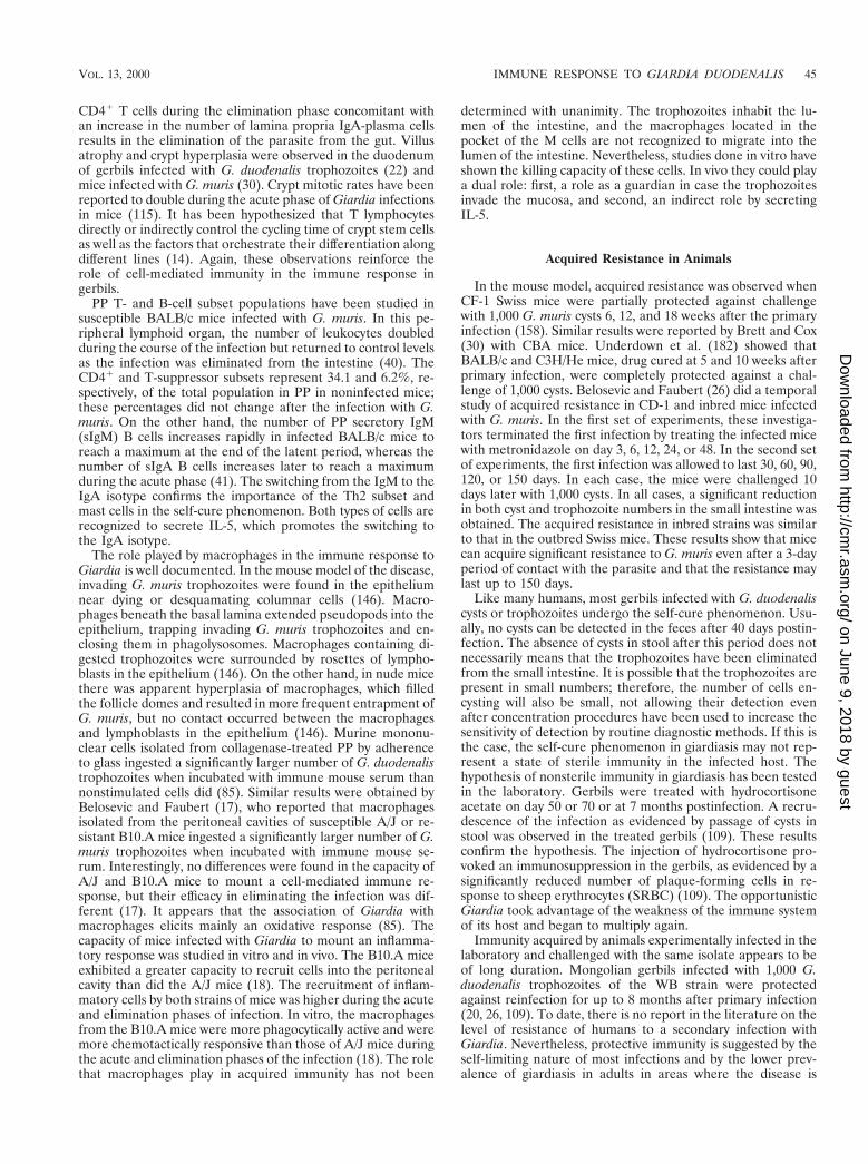
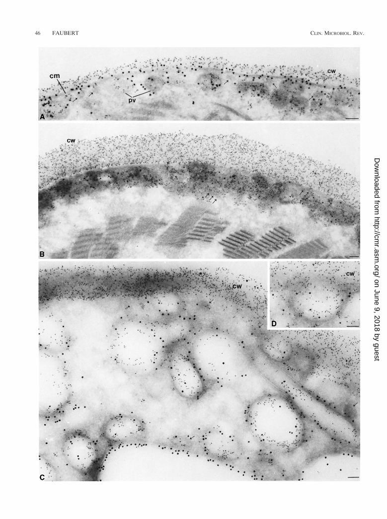
FIG. 10. Ultrathin cryosections of 24-, 48-, and 66-h cysts, doubly labeled for 8C5 (5-nm Au) and TSA 417 (10-nm Au). (A) In many 24-h encysting cells, 8C5 islocalized to the cyst wall (cw), which has been deposited over the cell membrane (cm), which is decorated with TSA 417. 8C5 and TSA are both found in peripheralvacuoles (pv). The amount of TSA 417 on the cell membrane seems somewhat reduced. (B) In 48-h cysts, the cyst wall (cw) containing 8C5 has markedly increasedin thickness and TSA 417 is completely absent from the cell membrane (cm). Note the small transport vesicles (small arrows). (C and D) In 66-h water-resistant cysts,TSA 417 is localized exclusively to the peripheral vacuoles and large internal vesicles and vacuoles, resembling the endosomal and prelysosomal compartments of highereukaryotes. 8C5 is also present in many of these vesicles. Bars, 0.1 mm. Reprinted from reference 118 with permission of the publisher.
VOL. 13, 2000 IMMUNE RESPONSE TO GIARDIA DUODENALIS 47
on June 9, 2018 by guesthttp://cm
r.asm.org/
Dow
nloaded from
AIDS. The patients (15 of 29) who had acute symptomaticgiardiasis had significantly lower levels of specific anti-Giardiaantibodies of all isotypes in serum than did subjects who alsohad giardiasis but did not suffer from AIDS. These resultsshow that despite a suppressed immune system, the immuneresponse to Giardia in AIDS patients does not seem to be verydifferent from that in healthy individuals. Because the therapyavailable for giardiasis is independent of the patient’s immunestatus, patients with AIDS do not have to suffer from pro-longed symptomatic G. duodenalis infection (92). It is probablyfor this reason that giardiasis is not listed among the opportu-nistic parasitic infections affecting AIDS patients (100).
Usually, clinical studies are required to establish if recrudes-cence of preexisting opportunistic infections is an importantcause of morbidity when immunosuppressive therapy is givento patients in areas where the infection is endemic. To date,there are no reports in the literature on the effects of drugssuch as corticosteroids, cyclosporin A, and other immunosup-pressive agents of cell-mediated immunity on the outcome ofGiardia infections in humans.
Animals
Stevens et al. (175) have shown for the first time the impor-tance of thymus-dependent lymphocytes in the clearance ofprimary infections and in subsequent reinfection with G. muris.Hypothymic (nude) mice failed to eliminate the infection fromthe intestine, and a chronic state of the disease appeared.Unlike most strains of mice, which acquire resistance to rein-fection (26, 40, 106, 107, 157), nude mice are not resistant tochallenge infection with G. muris (175). The reconstitution of
nude mice with thymus, MLN, or spleen cells from heterozy-gous thymus-intact controls results in rapid resolution of theinfection (159). The total number of leukocytes, CD41 andCD81 T cells, and macrophages present in the intestinal lumenof Giardia-infected immunocompetent mice and nude micewas compared. Although the total number of leukocytes har-vested was similar in the two strains of mice, the number ofCD41 T cells was smaller in nude mice (80). According toCarlson et al. (39), the impaired capacity of nude mice to clearthe infection results from a deficiency of CD41 T cells. Incontrast, no differences were observed between the numbers ofluminal CD81 T cells and macrophages (80). The authors alsofound a much smaller number of CD41 T cells in PP of nudemice than of immunocompetent mice. BALB/c mice depletedof CD41 T cells do not eliminate trophozoites from the gut,whereas those depleted of CD81 T cells are able to clear theinfection normally (82). Therefore, the role played by theCD41-T-cell population in the elimination of the infection inthe mouse model is different from the role played by theCD41-T-cell population in humans.
Natural killer (NK) cells are present in the mouse intestinalmucosa, but the role they play in the clearance of the infectionis unknown (177). Beige mice, which are deficient in NK cells,are able to clear G. muris infection at similar rates to thosefound for immunocompetent C57BL/6J mice (81). Mice with aG. muris infection and treated with corticosteroids (131) orcyclosporin A (21) have increased numbers of cysts released infeces compared with nontreated mice. Similar results wereobtained with gerbils treated with corticosteroids and infectedwith G. duodenalis (109). In contrast to what occurs in humaninfections, the importance of cell-mediated immunity in the
TABLE 1. Sensitivity of immunoassays used for the detection of specific antibodies to Giardia proteins in serum andhuman milk and for the detection of antigens of Giardia in feces
Antibody orantigena and assay
Antigen orantiseruma
No. of positive testsb/total no. of samples (% positive)Reference(s)
IgM antibody IgG antibody IgA antibody Igc antibody Feces antigen
Serum antibodiesELISA Trophozoite extract 75/128 (59) 92/128 (71) 86/110 (78) 15/43 (35) 15, 89, 90, 91, 117, 136
Trophozoite cells 48/59 (81) 168
IFAd Trophozoite cells 240/352 (68) 240/352 (68) 147/186 (79) 1, 112, 176, 191, 198Cyst cells 32/36 (89) 32/36 (89) 32/36 (89) 150/150 (100) 96, 156
Western blot Trophozoite extract 47/60 (78) 57/60 (95) 39/60 (65) 15, 15331-kDa protein 13/13 (100) 17857-kDa protein 10/10 (100) 10/10 (100) 9/10 (90) 43
Immunodiffusion Cyst extract 11/11 (100) 186
Milk antibodiesELISA Trophozoite extract 38/61 (62) 140Western-blot Trophozoite extract 4/4 (100) 4/4 (100) 4/4 (100) 153
Feces antigenELISA GSA-65 759/779 (97) 5, 15, 94, 117, 163ELISA 66-kDa protein 77/94 (82) 53, 185, 187ELISA Trophozoite extract 239/251 (95) 72, 139CIEe GSA-65 36/40 (90) 162
a The first is for the antibody detection, and the second is for the antigen detection.b The number of tests positive is given with respect to the total number of specimens tested which were obtained from studies of persons with proven cases of
giardiasis.c Whole serum Ig.d Immunofluorescence assay.e Counterimmunoelectrophoresis.
48 FAUBERT CLIN. MICROBIOL. REV.
on June 9, 2018 by guesthttp://cm
r.asm.org/
Dow
nloaded from
control of giardiasis in the animal models is well established(4).
The role of antibodies in immunity to G. muris has beeninvestigated with immunocompromised mice. CBA/N mice ex-pressing the xid gene have a deficient B-lymphocyte function(142). Infection of CBA/N mice with G. muris cysts leads to aprolongation of the infection compared to the duration innormal BALB/c mice (170). Interestingly, the CBA/N miceproduced high levels of IgA anti-G. muris antibodies in serumand gut secretions, while the anti-Giardia IgG antibodies in theserum were at a low level. The authors assumed that micebearing the xid gene fail to produce IgA antibodies of appro-priate specificity to Giardia antigens, whose recognition byspecific antibodies is critical for successful elimination of thetrophozoites (170). The treatment of mice from birth withrabbit anti-IgM sera results in IgM, IgA, and IgG deficienciesin the serum and gut secretions (74, 171). The effects of thistreatment on the primary infection with G. muris were studiedin BALB/c and (C57BL/6 3 C3H/He) F1 mice. The treatedmice showed no specific anti-G. muris antibodies in the serumor gut washings, and the infections became chronic, with a highload of trophozoites present in the intestine and a prolongedcyst excretion (171). These results show the importance of Bcells in the elimination of the parasite from the intestine; theyalso indicate that the nonspecific elimination of IgM antibodiesat birth has a profound effect on the outcome of giardiasis inmice.
The treatment of weanling mice with cortisone prior to in-fection with G. duodenalis results in a reduction in the numbersof CD41 T cells and IgA-producing cells in the intestine. Inspite of this immunosuppressive therapy, which should haveincreased the trophozoite load in the intestine, a significantreduction in the number of trophozoites was obtained (102).The authors concluded that control of the infection in theabsence of CD41 T cells and IgA antibodies was due to anunaltered IgM antibody response (102). The decrease in thevillus-to-crypt ratio, together with the decrease in disacchari-dase activity usually observed during the acute phase of giar-diasis, is more severe in cortisone-treated mice (22, 102). Itappears that in the animal model of the disease, immunode-pression leads to a more severe infection.
IMMUNODIAGNOSIS
The immunodiagnosis of giardiasis has received much atten-tion in the recent past. Knowledge about Giardia antigens andthe need for improved diagnostic tests are two factors thathave contributed to the increased number of publications inthis area (62). A variety of assays have been used for theserodiagnosis of giardiasis. In Table 1, the results obtained bydifferent laboratories with a variety of serological assays for thedetection of Giardia antibodies in serum of proven cases aresummarized. Proven cases of giardiasis were defined as fol-lows: “patients passing cysts in their feces and/or presentingwith one or more of the clinical symptoms of giardiasis” (15).
Sensitivity of Serological Assays
When crude extracts of trophozoites are processed for an-tigen usage in an ELISA, the sensitivity varies with the Igisotype used as the second antibody. For example, when theIgM isotype was used as the second antibody, 59% of the serafrom persons with proven cases tested positive, compared toonly 35% when the whole Ig was used (Table 1). The ELISAhas a comparable sensitivity when IgA or IgG is used as thesecond antibody. The use of intact trophozoites as the antigen
increases the sensitivity of the IgG ELISA slightly. The sensi-tivity of the immunofluorescence assay (IFA) in the detectionof anti-Giardia antibodies in the sera of persons with proveninfection is comparable to that of the ELISA. However, thesensitivities of the two essays are different depending of thetype of antigen used, since the sensitivity of the IFA increaseswhen cysts are used as the antigen (Table 1). Of note, the IFAand ELISA were able to detect antibodies of the IgA and IgGisotypes at a similar level in the serum. Since Giardia tropho-zoites stimulate the production of antibodies of the IgA isotypemainly at the gut level and do not invade the tissues, one wouldnot expect to detect anti-Giardia-specific antibodies of the IgAisotype in the serum at the same level as the IgG isotype.
The sensitivity of the Western blot assay is difficult to eval-uate since it has been used by only a few laboratories and hasbeen performed only on a limited number of sera from personswith proven cases. However, the sensitivity of the assay in-creases when purified Giardia proteins are used as antigens.Considering the variety of antigens stimulating the immunesystem of an infected patient, it is surprising that the assay isunable to detect antibodies in all the samples from the patientswith proven cases of giardiasis. Identification of a common andimmunodominant antigen for serodiagnostic purposes has notmet with success. Studies have identified several strongly reac-tive antigens whose molecular masses vary immensely. Forexample, a major 31-kDa protein was detected in the sera ofonly 11 of 16 patients passing cysts in their feces, but othermajor bands, with molecular masses ranging from 28 to 56kDa, were also detected in the 16 sera (178). Saliva samplestaken from giardiasis patients showed 24 antigen bands withmolecular masses varying between 14 and 170 kDa (161). Onlyone study reported 100% sensitivity of the ELISA, IFA, orWestern blot technique in detecting specific antibodies in per-sons with proven cases of giardiasis (172). The investigatorsreported a significantly higher titer of circulating antibodies insymptomatic patients than in asymptomatic patients; these re-sults confirmed the results of an earlier study (176).
The level of circulating anti-G. duodenalis-specific IgG, IgM,and IgA antibodies has been compared among infected per-sons living in Denver, Colo., and Soongnern, Thailand (91).Antibody levels detected by ELISA increased significantly dur-ing childhood in both geographic areas. The Giardia-specificIgA antibody levels remained elevated throughout life amongadults from Thailand but decreased among adults in Denver.On the other hand, after adolescence, Giardia-specific IgMantibodies fell steadily with increasing age in both populations.Based on these findings, the authors concluded that the levelsof G. duodenalis-specific IgM in adults may be useful to dif-ferentiate between recent and past infection (91).
By determining the levels of systemic and local antibodies toG. duodenalis in different populations, widely different immuneresponses in infected patients were recognized (123). Severalblood and milk samples were collected simultaneously fromlactating women in Texas and Mexico. Specific IgG antibodiesto G. duodenalis were present in 77% of 153 serum samplesfrom 27 Mexican mothers but in only 24% of 214 serum sam-ples from Texan mothers. Secretory IgA antibodies were de-tected in 79% of milk samples from the Mexican populationbut in only 15% of milk samples from the Texan population(123). These results highlight the difference in the immuneresponse to Giardia between infected patients in areas of en-demic infection and other areas.
The outcome of a Giardia infection and humoral antibodyresponse in humans may also vary depending on the isolate. Toillustrate, enteral inoculation of healthy volunteers with 50,000trophozoites of two distinct Giardia isolates having distinct
VOL. 13, 2000 IMMUNE RESPONSE TO GIARDIA DUODENALIS 49
on June 9, 2018 by guesthttp://cm
r.asm.org/
Dow
nloaded from
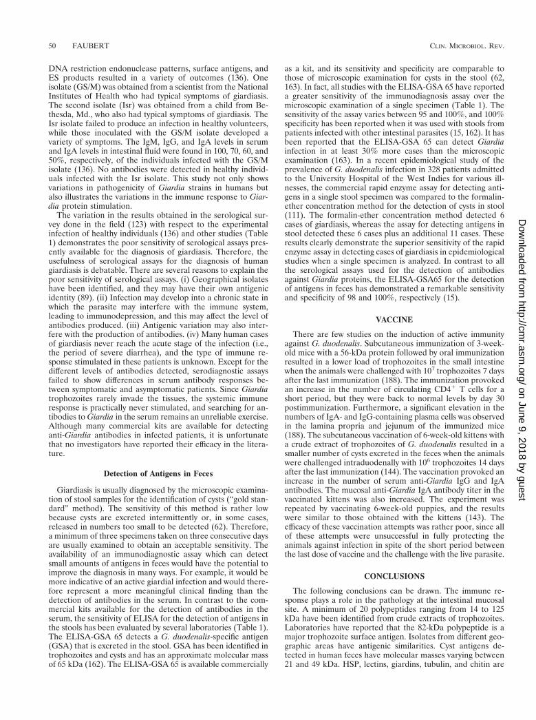
DNA restriction endonuclease patterns, surface antigens, andES products resulted in a variety of outcomes (136). Oneisolate (GS/M) was obtained from a scientist from the NationalInstitutes of Health who had typical symptoms of giardiasis.The second isolate (Isr) was obtained from a child from Be-thesda, Md., who also had typical symptoms of giardiasis. TheIsr isolate failed to produce an infection in healthy volunteers,while those inoculated with the GS/M isolate developed avariety of symptoms. The IgM, IgG, and IgA levels in serumand IgA levels in intestinal fluid were found in 100, 70, 60, and50%, respectively, of the individuals infected with the GS/Misolate (136). No antibodies were detected in healthy individ-uals infected with the Isr isolate. This study not only showsvariations in pathogenicity of Giardia strains in humans butalso illustrates the variations in the immune response to Giar-dia protein stimulation.
The variation in the results obtained in the serological sur-vey done in the field (123) with respect to the experimentalinfection of healthy individuals (136) and other studies (Table1) demonstrates the poor sensitivity of serological assays pres-ently available for the diagnosis of giardiasis. Therefore, theusefulness of serological assays for the diagnosis of humangiardiasis is debatable. There are several reasons to explain thepoor sensitivity of serological assays. (i) Geographical isolateshave been identified, and they may have their own antigenicidentity (89). (ii) Infection may develop into a chronic state inwhich the parasite may interfere with the immune system,leading to immunodepression, and this may affect the level ofantibodies produced. (iii) Antigenic variation may also inter-fere with the production of antibodies. (iv) Many human casesof giardiasis never reach the acute stage of the infection (i.e.,the period of severe diarrhea), and the type of immune re-sponse stimulated in these patients is unknown. Except for thedifferent levels of antibodies detected, serodiagnostic assaysfailed to show differences in serum antibody responses be-tween symptomatic and asymptomatic patients. Since Giardiatrophozoites rarely invade the tissues, the systemic immuneresponse is practically never stimulated, and searching for an-tibodies to Giardia in the serum remains an unreliable exercise.Although many commercial kits are available for detectinganti-Giardia antibodies in infected patients, it is unfortunatethat no investigators have reported their efficacy in the litera-ture.
Detection of Antigens in Feces
Giardiasis is usually diagnosed by the microscopic examina-tion of stool samples for the identification of cysts (“gold stan-dard” method). The sensitivity of this method is rather lowbecause cysts are excreted intermittently or, in some cases,released in numbers too small to be detected (62). Therefore,a minimum of three specimens taken on three consecutive daysare usually examined to obtain an acceptable sensitivity. Theavailability of an immunodiagnostic assay which can detectsmall amounts of antigens in feces would have the potential toimprove the diagnosis in many ways. For example, it would bemore indicative of an active giardial infection and would there-fore represent a more meaningful clinical finding than thedetection of antibodies in the serum. In contrast to the com-mercial kits available for the detection of antibodies in theserum, the sensitivity of ELISA for the detection of antigens inthe stools has been evaluated by several laboratories (Table 1).The ELISA-GSA 65 detects a G. duodenalis-specific antigen(GSA) that is excreted in the stool. GSA has been identified introphozoites and cysts and has an approximate molecular massof 65 kDa (162). The ELISA-GSA 65 is available commercially
as a kit, and its sensitivity and specificity are comparable tothose of microscopic examination for cysts in the stool (62,163). In fact, all studies with the ELISA-GSA 65 have reporteda greater sensitivity of the immunodiagnosis assay over themicroscopic examination of a single specimen (Table 1). Thesensitivity of the assay varies between 95 and 100%, and 100%specificity has been reported when it was used with stools frompatients infected with other intestinal parasites (15, 162). It hasbeen reported that the ELISA-GSA 65 can detect Giardiainfection in at least 30% more cases than the microscopicexamination (163). In a recent epidemiological study of theprevalence of G. duodenalis infection in 328 patients admittedto the University Hospital of the West Indies for various ill-nesses, the commercial rapid enzyme assay for detecting anti-gens in a single stool specimen was compared to the formalin-ether concentration method for the detection of cysts in stool(111). The formalin-ether concentration method detected 6cases of giardiasis, whereas the assay for detecting antigens instool detected these 6 cases plus an additional 11 cases. Theseresults clearly demonstrate the superior sensitivity of the rapidenzyme assay in detecting cases of giardiasis in epidemiologicalstudies when a single specimen is analyzed. In contrast to allthe serological assays used for the detection of antibodiesagainst Giardia proteins, the ELISA-GSA65 for the detectionof antigens in feces has demonstrated a remarkable sensitivityand specificity of 98 and 100%, respectively (15).
VACCINE
There are few studies on the induction of active immunityagainst G. duodenalis. Subcutaneous immunization of 3-week-old mice with a 56-kDa protein followed by oral immunizationresulted in a lower load of trophozoites in the small intestinewhen the animals were challenged with 107 trophozoites 7 daysafter the last immunization (188). The immunization provokedan increase in the number of circulating CD41 T cells for ashort period, but they were back to normal levels by day 30postimmunization. Furthermore, a significant elevation in thenumbers of IgA- and IgG-containing plasma cells was observedin the lamina propria and jejunum of the immunized mice(188). The subcutaneous vaccination of 6-week-old kittens witha crude extract of trophozoites of G. duodenalis resulted in asmaller number of cysts excreted in the feces when the animalswere challenged intraduodenally with 106 trophozoites 14 daysafter the last immunization (144). The vaccination provoked anincrease in the number of serum anti-Giardia IgG and IgAantibodies. The mucosal anti-Giardia IgA antibody titer in thevaccinated kittens was also increased. The experiment wasrepeated by vaccinating 6-week-old puppies, and the resultswere similar to those obtained with the kittens (143). Theefficacy of these vaccination attempts was rather poor, since allof these attempts were unsuccessful in fully protecting theanimals against infection in spite of the short period betweenthe last dose of vaccine and the challenge with the live parasite.
CONCLUSIONS
The following conclusions can be drawn. The immune re-sponse plays a role in the pathology at the intestinal mucosalsite. A minimum of 20 polypeptides ranging from 14 to 125kDa have been identified from crude extracts of trophozoites.Laboratories have reported that the 82-kDa polypeptide is amajor trophozoite surface antigen. Isolates from different geo-graphic areas have antigenic similarities. Cyst antigens de-tected in human feces have molecular masses varying between21 and 49 kDa. HSP, lectins, giardins, tubulin, and chitin are
50 FAUBERT CLIN. MICROBIOL. REV.
on June 9, 2018 by guesthttp://cm
r.asm.org/
Dow
nloaded from
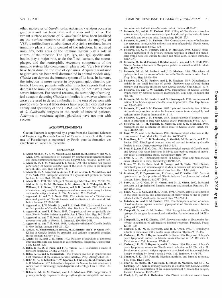
other molecules of Giardia cells. Antigenic variation occurs ingiardiasis and has been observed in vivo and in vitro. Thevariant surface antigens of G. duodenalis have been localizedon the surface membrane of trophozoites; the majority ofVSPs identified have an abundance of cysteine residues. Innateimmunity plays a role in control of the infection. In acquiredimmunity, both arms of the immune system play a role incontrol of the infection. The IgM, IgA, and IgG-specific anti-bodies play a major role, as do the T-cell subsets, the macro-phages, and the neutrophils. Accessory components of theimmune system, like complement, play a role. Very few studieshave been done on the role of cytokines. Acquired resistanceto giardiasis has been well documented in animal models only.Giardia can depress the immune system of its host. In humans,the infection is more severe in hypogammaglobulinemic pa-tients. However, patients with other infectious agents that candepress the immune system (e.g., AIDS) do not have a moresevere infection. For several reasons, the sensitivity of serolog-ical assays in detecting Giardia antibodies is low, even when theassays are used to detect antibodies in the sera of persons withproven cases. Several laboratories have reported excellent sen-sitivity and specificity of the ELISA-GSA 65 for the detectionof G. duodenalis antigens in the stools of infected patients.Attempts to vaccinate against giardiasis have not met withsuccess.
ACKNOWLEDGMENTS
Gaetan Faubert is supported by a grant from the National Sciencesand Engineering Research Council of Canada. Research at the Insti-tute of Parasitology is supported by Fonds pour la formation deschercheurs et l’aide a la recherche.
REFERENCES
1. Abdul Amid, M. Y., K. M. Mailed, A. M. Kamal, D. M. Mentally, and M. E.Abab. 1993. Serodiagnosis of giardiasis by counterimmunoelectrophoresisand indirect immunofluorescence tests. J. Egypt. Soc. Parasitol. 23:603–608.
2. Adam, R. D., Y. M. Yang, and T. E. Nash. 1992. The cysteine-rich proteingene family of Giardia lamblia: loss of the CPR170 gene in an antigenicvariant. Mol. Cell. Biol. 12:1194–1201.
3. Adam, R. D., A. Aggarwal, A. A. Lal, V. F. de la Cruz, T. McCutchan, andT. E. Nash. 1988. Antigenic variation of a cysteine-rich protein in Giardialamblia. J. Exp. Med. 167:109–118.
4. Adam, R. D. 1991. The biology of Giardia spp. Microbiol. Rev. 55:706–732.5. Addiss, D. G., H. M. Mathews, J. M. Stewart, S. P. Wahlquist, R. M.
Williams, R. J. Finton, H. C. Spencer, and D. D. Juranek. 1991. Evaluationof a commercially available enzyme-linked immunosorbent assay for Giar-dia lamblia antigen in stool. J. Clin. Microbiol. 29:1137–1142.
6. Aggarwal, A., and T. E. Nash. 1989. Characterization of a 33-kilodaltonstructural protein of Giardia lamblia and localization to the ventral disk.Infect. Immun. 57:1305–1310.
7. Aggarwal, A., J. W. Merritt, Jr., and T. E. Nash. 1989. Cysteine-rich variantsurface proteins of Giardia lamblia. Mol. Biochem. Parasitol. 32:39–48.
8. Aggarwal, A., and T. E. Nash. 1987. Comparison of two antigenically dis-tinct Giardia lamblia isolates in gerbils. Am. J. Trop. Med. Hyg. 36:325–332.
9. Aggarwal, A., and T. E. Nash. 1986. Lack of cellular cytotoxicity by humanmononuclear cells to Giardia. J. Immunol. 136:3486–3488.
10. Aggarwal, A., and T. E. Nash. 1988. Antigenic variation of Giardia lambliain vivo. Infect. Immun. 56:1420–1423.
11. Aley, S., M. Zimmerman, M. Hetsko, M. E. Selstedt, and F. D. Gillin. 1994.Killing of Giardia lamblia by cryptdins and cationic neutrophil peptides.Infect. Immun. 62:5397–5403.
12. Ament, M. E., and C. E. Rubin. 1972. Relation of giardiasis to abnormalintestinal structure and function in gastrointestinal syndrome. Gastroenter-ology 62:216–226.
13. Babb, R. R., O. C. Peck, and F. G. Vescia. 1971. Giardiasis: a cause oftravelers diarrhoea. JAMA 217:1359–1361.
14. Befus, A. D., and J. Bienenstock. 1982. Factors involved in symbiosis andhost resistance at the mucosa-parasite interface. Prog. Allergy 31:76–86.
15. Behr, M. A., E. Kokoskin, T. W. Gyorkos, L. Cedilotte, G. M. Faubert, andJ. D. MacLean. 1997. Laboratory diagnosis for Giardia lamblia infection: acomparison of microscopy, coprodiagnosis and serology. Can. J. Infect. Dis.8:33–38.
16. Belosevic, M., G. M. Faubert, and J. D. MacLean. 1985. Suppression ofprimary antibody response to sheep erythrocytes in susceptible and resis-
tant mice infected with Giardia muris. Infect. Immun. 47:21–25.17. Belosevic, M., and G. M. Faubert. 1986. Killing of Giardia muris tropho-
zoites in vitro by spleen, mesenteric lymph node and peritoneal cells fromsusceptible and resistant mice. Immunology 59:269–275.
18. Belosevic, M., and G. M. Faubert. 1986. Comparative studies of inflamma-tory responses in susceptible and resistant mice infected with Giardia muris.Clin. Exp. Immunol. 65:622–630.
19. Belosevic, M., G. M. Faubert, and J. D. MacLean. 1985. Giardia muris-induced depression of the primary immune response in spleen and mesen-teric lymph node cell culture to sheep red blood cells. Parasite Immunol.7:467–478.
20. Belosevic, M., G. M. Faubert, J. D. MacLean, C. Law, and N. A. Croll. 1983.Giardia lamblia infections in Mongolian gerbils: an animal model. J. Infect.Dis. 147:222–226.
21. Belosevic, M., G. M. Faubert, and J. D. Maclean. 1986. The effects ofcyclosporin A on the course of infection with Giardia muris in mice. Am. J.Trop. Med. Hyg. 35:496–500.
22. Belosevic, M., G. M. Faubert, and J. D. Maclean. 1989. Disaccharidaseactivity in the small intestine of gerbils (Meriones unguiculatus) duringprimary and challenge infections with Giardia lamblia. Gut 30:1213–1219.
23. Belosevic, M., and C. W. Daniels. 1992. Phagocytosis of Giardia lambliatrophozoites by cytokine-activated macrophages. Clin. Exp. Immunol. 87:304–309.
24. Belosevic, M., G. M. Faubert, and S. Dharampaul. 1994. Antimicrobialaction of antibodies against Giardia muris trophozoites. Clin. Exp. Immu-nol. 95:485–489.
25. Belosevic, M., and G. M. Faubert. 1987. Lysis and immobilization of Giar-dia muris trophozoites in vitro by immune serum from susceptible andresistant mice. Parasite Immunol. 9:11–19.
26. Belosevic, M., and G. M. Faubert. 1983. Temporal study of acquired resis-tance in infections of mice with Giardia muris. Parasitology 87:517–524.
27. Belosevic, M., G. M. Faubert, E. Skamene, and J. D. MacLean. 1984.Susceptibility and resistance of inbred mice to Giardia muris. Infect. Im-mun. 44:282–286.
28. Boyd, W. P., and B. A. Bachman. 1982. Gastrointestinal infections in thecompromised host. Med. Clin. North Am. 66:743–753.
29. Brandborg, L. L., C. B. Tankersley, S. Gottlieb, M. Barancik, and V. E.Sartor. 1967. Histological demonstration of mucosal invasion by Giardialamblia in man. Gastroenterology 52:143–150.
30. Brett, S. J., and F. E. G. Cox. 1982. Immunological aspects of Giardia murisand Spironucleus muris infections in inbred and outbred strains of labora-tory mice: a comparative study. Parasitology 85:85–99.
31. Brett, S. J. 1983. Immunodepression in Giardia muris and Spironucleusmuris infections in mice. Parasitology 87:507–515.
32. Brown, W. R., D. Butterfield, D. Savage, and T. Tada. 1972. Clinical,microbiological and immunological studies in patients with immunoglobu-lin deficiencies and gastrointestinal disorders. Gut 13:441–449.
33. Bruderer, T., P. Papanastasiou, R. Castro, and P. Kohler. 1993. Variantcysteine-rich surface proteins of Giardia isolates from human and animalsources. Infect. Immun. 61:2937–2944.
34. Buret, A., D. G. Gall, P. N. Nation, and M. E. Olson. 1990. Intestinalprotozoa and epithelial cell kinetics, structure and function. Parasitol. To-day 6:375–380.
35. Buret, A., D. G. Gall, and M. E. Olson. 1991. Growth, activities of enzymesin the small intestine, and ultrastructure of microvillous border in gerbilsinfected with G. duodenalis. Parasitol. Res. 77:109–114.
36. Butscher, W., and G. M. Faubert. 1988. The therapeutic action of mono-clonal antibodies against a surface glycoprotein of Giardia muris. Immu-nology 64:175–180.
37. Campbell, D., and G. M. Faubert. 1994. Recognition of Giardia lambliacyst-specific antigens by monoclonal antibodies. Parasite Immunol. 16:211–219.
38. Campbell, D., and K. Chadee. 1997. Survival strategies of Entamoeba his-tolytica: modulation of cell-mediated immune responses. Parasitol. Today13:184–190.
39. Carlson, J. R., M. H. Heyworth, and R. L. Owen. 1987. T-lymphocytesubsets in nude mice with Giardia muris infection. Thymus 9:189–196.
40. Carlson, J. R., M. H. Heyworth, and R. L. Owen. 1986. Response of Peyer’spatch lymphocyte subsets to Giardia muris infection in BALB/c mice. I.T-cell subsets. Cell. Immunol. 97:44–50.
41. Carlson, J. R., M. H. Heyworth, and R. L. Owen. 1986. Response of Peyer’spatch lymphocyte subsets to Giardia muris infection in BALB/c mice. II.B-cell subsets: enteric antigen exposure is associated with immunoglobulinisotype switching by Peyer’s patch B cells. Cell. Immunol. 97:51–58.
42. Chandra, R. K. 1984. Parasitic infection, nutrition, and immune response.Fed. Proc. 43:251–255.
43. Char, S., N. Shetty, M. Narasimha, E. Elliott, R. Macaden, and M. J. G.Farthing. 1991. Serum antibody response in children with Giardia lambliainfection and identification of an immunodominant 57-kilodalton antigen.Parasite Immunol. 13:329–337.
44. Clark, J. T., and D. V. Holberton. 1986. Plasma membrane isolated from
VOL. 13, 2000 IMMUNE RESPONSE TO GIARDIA DUODENALIS 51
on June 9, 2018 by guesthttp://cm
r.asm.org/
Dow
nloaded from
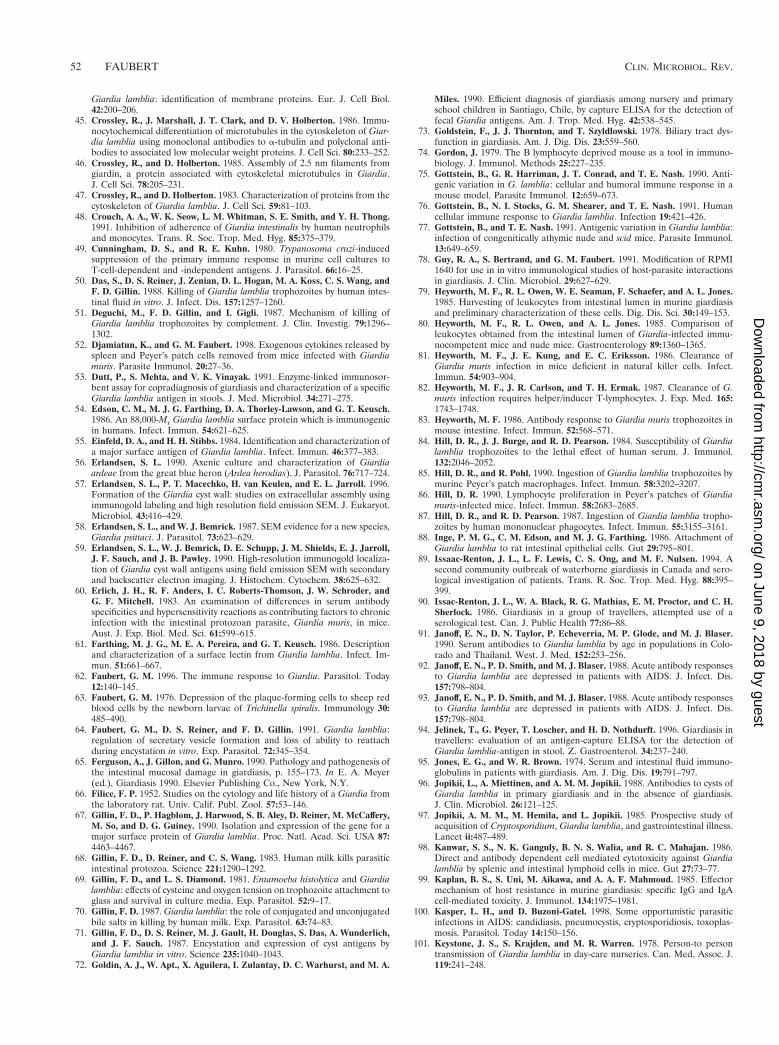
Giardia lamblia: identification of membrane proteins. Eur. J. Cell Biol.42:200–206.
45. Crossley, R., J. Marshall, J. T. Clark, and D. V. Holberton. 1986. Immu-nocytochemical differentiation of microtubules in the cytoskeleton of Giar-dia lamblia using monoclonal antibodies to a-tubulin and polyclonal anti-bodies to associated low molecular weight proteins. J. Cell Sci. 80:233–252.
46. Crossley, R., and D. Holberton. 1985. Assembly of 2.5 nm filaments fromgiardin, a protein associated with cytoskeletal microtubules in Giardia.J. Cell Sci. 78:205–231.
47. Crossley, R., and D. Holberton. 1983. Characterization of proteins from thecytoskeleton of Giardia lamblia. J. Cell Sci. 59:81–103.
48. Crouch, A. A., W. K. Seow, L. M. Whitman, S. E. Smith, and Y. H. Thong.1991. Inhibition of adherence of Giardia intestinalis by human neutrophilsand monocytes. Trans. R. Soc. Trop. Med. Hyg. 85:375–379.
49. Cunningham, D. S., and R. E. Kuhn. 1980. Trypanosoma cruzi-inducedsuppression of the primary immune response in murine cell cultures toT-cell-dependent and -independent antigens. J. Parasitol. 66:16–25.
50. Das, S., D. S. Reiner, J. Zenian, D. L. Hogan, M. A. Koss, C. S. Wang, andF. D. Gillin. 1988. Killing of Giardia lamblia trophozoites by human intes-tinal fluid in vitro. J. Infect. Dis. 157:1257–1260.
51. Deguchi, M., F. D. Gillin, and I. Gigli. 1987. Mechanism of killing ofGiardia lamblia trophozoites by complement. J. Clin. Investig. 79:1296–1302.
52. Djamiatun, K., and G. M. Faubert. 1998. Exogenous cytokines released byspleen and Peyer’s patch cells removed from mice infected with Giardiamuris. Parasite Immunol. 20:27–36.
53. Dutt, P., S. Mehta, and V. K. Vinayak. 1991. Enzyme-linked immunosor-bent assay for copradiagnosis of giardiasis and characterization of a specificGiardia lamblia antigen in stools. J. Med. Microbiol. 34:271–275.
54. Edson, C. M., M. J. G. Farthing, D. A. Thorley-Lawson, and G. T. Keusch.1986. An 88,000-Mr Giardia lamblia surface protein which is immunogenicin humans. Infect. Immun. 54:621–625.
55. Einfeld, D. A., and H. H. Stibbs. 1984. Identification and characterization ofa major surface antigen of Giardia lamblia. Infect. Immun. 46:377–383.
56. Erlandsen, S. L. 1990. Axenic culture and characterization of Giardiaardeae from the great blue heron (Ardea herodias). J. Parasitol. 76:717–724.
57. Erlandsen, S. L., P. T. Macechko, H. van Keulen, and E. L. Jarroll. 1996.Formation of the Giardia cyst wall: studies on extracellular assembly usingimmunogold labeling and high resolution field emission SEM. J. Eukaryot.Microbiol. 43:416–429.
58. Erlandsen, S. L., and W. J. Bemrick. 1987. SEM evidence for a new species,Giardia psittaci. J. Parasitol. 73:623–629.
59. Erlandsen, S. L., W. J. Bemrick, D. E. Schupp, J. M. Shields, E. J. Jarroll,J. F. Sauch, and J. B. Pawley. 1990. High-resolution immunogold localiza-tion of Giardia cyst wall antigens using field emission SEM with secondaryand backscatter electron imaging. J. Histochem. Cytochem. 38:625–632.
60. Erlich, J. H., R. F. Anders, I. C. Roberts-Thomson, J. W. Schroder, andG. F. Mitchell. 1983. An examination of differences in serum antibodyspecificities and hypersensitivity reactions as contributing factors to chronicinfection with the intestinal protozoan parasite, Giardia muris, in mice.Aust. J. Exp. Biol. Med. Sci. 61:599–615.
61. Farthing, M. J. G., M. E. A. Pereira, and G. T. Keusch. 1986. Descriptionand characterization of a surface lectin from Giardia lamblia. Infect. Im-mun. 51:661–667.
62. Faubert, G. M. 1996. The immune response to Giardia. Parasitol. Today12:140–145.
63. Faubert, G. M. 1976. Depression of the plaque-forming cells to sheep redblood cells by the newborn larvae of Trichinella spiralis. Immunology 30:485–490.
64. Faubert, G. M., D. S. Reiner, and F. D. Gillin. 1991. Giardia lamblia:regulation of secretary vesicle formation and loss of ability to reattachduring encystation in vitro. Exp. Parasitol. 72:345–354.
65. Ferguson, A., J. Gillon, and G. Munro. 1990. Pathology and pathogenesis ofthe intestinal mucosal damage in giardiasis, p. 155–173. In E. A. Meyer(ed.), Giardiasis 1990. Elsevier Publishing Co., New York, N.Y.
66. Filice, F. P. 1952. Studies on the cytology and life history of a Giardia fromthe laboratory rat. Univ. Calif. Publ. Zool. 57:53–146.
67. Gillin, F. D., P. Hagblom, J. Harwood, S. B. Aley, D. Reiner, M. McCaffery,M. So, and D. G. Guiney. 1990. Isolation and expression of the gene for amajor surface protein of Giardia lamblia. Proc. Natl. Acad. Sci. USA 87:4463–4467.
68. Gillin, F. D., D. Reiner, and C. S. Wang. 1983. Human milk kills parasiticintestinal protozoa. Science 221:1290–1292.
69. Gillin, F. D., and L. S. Diamond. 1981. Entamoeba histolytica and Giardialamblia: effects of cysteine and oxygen tension on trophozoite attachment toglass and survival in culture media. Exp. Parasitol. 52:9–17.
70. Gillin, F. D. 1987. Giardia lamblia: the role of conjugated and unconjugatedbile salts in killing by human milk. Exp. Parasitol. 63:74–83.
71. Gillin, F. D., D. S. Reiner, M. J. Gault, H. Douglas, S. Das, A. Wunderlich,and J. F. Sauch. 1987. Encystation and expression of cyst antigens byGiardia lamblia in vitro. Science 235:1040–1043.
72. Goldin, A. J., W. Apt., X. Aguilera, I. Zulantay, D. C. Warhurst, and M. A.
Miles. 1990. Efficient diagnosis of giardiasis among nursery and primaryschool children in Santiago, Chile, by capture ELISA for the detection offecal Giardia antigens. Am. J. Trop. Med. Hyg. 42:538–545.
73. Goldstein, F., J. J. Thornton, and T. Szyldlowski. 1978. Biliary tract dys-function in giardiasis. Am. J. Dig. Dis. 23:559–560.
74. Gordon, J. 1979. The B lymphocyte deprived mouse as a tool in immuno-biology. J. Immunol. Methods 25:227–235.
75. Gottstein, B., G. R. Harriman, J. T. Conrad, and T. E. Nash. 1990. Anti-genic variation in G. lamblia: cellular and humoral immune response in amouse model. Parasite Immunol. 12:659–673.
76. Gottstein, B., N. I. Stocks, G. M. Shearer, and T. E. Nash. 1991. Humancellular immune response to Giardia lamblia. Infection 19:421–426.
77. Gottstein, B., and T. E. Nash. 1991. Antigenic variation in Giardia lamblia:infection of congenitically athymic nude and scid mice. Parasite Immunol.13:649–659.
78. Guy, R. A., S. Bertrand, and G. M. Faubert. 1991. Modification of RPMI1640 for use in in vitro immunological studies of host-parasite interactionsin giardiasis. J. Clin. Microbiol. 29:627–629.
79. Heyworth, M. F., R. L. Owen, W. E. Seaman, F. Schaefer, and A. L. Jones.1985. Harvesting of leukocytes from intestinal lumen in murine giardiasisand preliminary characterization of these cells. Dig. Dis. Sci. 30:149–153.
80. Heyworth, M. F., R. L. Owen, and A. L. Jones. 1985. Comparison ofleukocytes obtained from the intestinal lumen of Giardia-infected immu-nocompetent mice and nude mice. Gastroenterology 89:1360–1365.
81. Heyworth, M. F., J. E. Kung, and E. C. Eriksson. 1986. Clearance ofGiardia muris infection in mice deficient in natural killer cells. Infect.Immun. 54:903–904.
82. Heyworth, M. F., J. R. Carlson, and T. H. Ermak. 1987. Clearance of G.muris infection requires helper/inducer T-lymphocytes. J. Exp. Med. 165:1743–1748.
83. Heyworth, M. F. 1986. Antibody response to Giardia muris trophozoites inmouse intestine. Infect. Immun. 52:568–571.
84. Hill, D. R., J. J. Burge, and R. D. Pearson. 1984. Susceptibility of Giardialamblia trophozoites to the lethal effect of human serum. J. Immunol.132:2046–2052.
85. Hill, D. R., and R. Pohl. 1990. Ingestion of Giardia lamblia trophozoites bymurine Peyer’s patch macrophages. Infect. Immun. 58:3202–3207.
86. Hill, D. R. 1990. Lymphocyte proliferation in Peyer’s patches of Giardiamuris-infected mice. Infect. Immun. 58:2683–2685.
87. Hill, D. R., and R. D. Pearson. 1987. Ingestion of Giardia lamblia tropho-zoites by human mononuclear phagocytes. Infect. Immun. 55:3155–3161.
88. Inge, P. M. G., C. M. Edson, and M. J. G. Farthing. 1986. Attachment ofGiardia lamblia to rat intestinal epithelial cells. Gut 29:795–801.
89. Issaac-Renton, J. L., L. F. Lewis, C. S. Ong, and M. F. Nulsen. 1994. Asecond community outbreak of waterborne giardiasis in Canada and sero-logical investigation of patients. Trans. R. Soc. Trop. Med. Hyg. 88:395–399.
90. Issac-Renton, J. L., W. A. Black, R. G. Mathias, E. M. Proctor, and C. H.Sherlock. 1986. Giardiasis in a group of travellers, attempted use of aserological test. Can. J. Public Health 77:86–88.
91. Janoff, E. N., D. N. Taylor, P. Echeverria, M. P. Glode, and M. J. Blaser.1990. Serum antibodies to Giardia lamblia by age in populations in Colo-rado and Thailand. West. J. Med. 152:253–256.
92. Janoff, E. N., P. D. Smith, and M. J. Blaser. 1988. Acute antibody responsesto Giardia lamblia are depressed in patients with AIDS. J. Infect. Dis.157:798–804.
93. Janoff, E. N., P. D. Smith, and M. J. Blaser. 1988. Acute antibody responsesto Giardia lamblia are depressed in patients with AIDS. J. Infect. Dis.157:798–804.
94. Jelinek, T., G. Peyer, T. Loscher, and H. D. Nothdurft. 1996. Giardiasis intravellers: evaluation of an antigen-capture ELISA for the detection ofGiardia lamblia-antigen in stool. Z. Gastroenterol. 34:237–240.
95. Jones, E. G., and W. R. Brown. 1974. Serum and intestinal fluid immuno-globulins in patients with giardiasis. Am. J. Dig. Dis. 19:791–797.
96. Jopikii, L., A. Miettinen, and A. M. M. Jopikii. 1988. Antibodies to cysts ofGiardia lamblia in primary giardiasis and in the absence of giardiasis.J. Clin. Microbiol. 26:121–125.
97. Jopikii, A. M. M., M. Hemila, and L. Jopikii. 1985. Prospective study ofacquisition of Cryptosporidium, Giardia lamblia, and gastrointestinal illness.Lancet ii:487–489.
98. Kanwar, S. S., N. K. Ganguly, B. N. S. Walia, and R. C. Mahajan. 1986.Direct and antibody dependent cell mediated cytotoxicity against Giardialamblia by splenic and intestinal lymphoid cells in mice. Gut 27:73–77.
99. Kaplan, B. S., S. Uni, M. Aikawa, and A. A. F. Mahmoud. 1985. Effectormechanism of host resistance in murine giardiasis: specific IgG and IgAcell-mediated toxicity. J. Immunol. 134:1975–1981.
100. Kasper, L. H., and D. Buzoni-Gatel. 1998. Some opportunistic parasiticinfections in AIDS: candidiasis, pneumocystis, cryptosporidiosis, toxoplas-mosis. Parasitol. Today 14:150–156.
101. Keystone, J. S., S. Krajden, and M. R. Warren. 1978. Person-to persontransmission of Giardia lamblia in day-care nurseries. Can. Med. Assoc. J.119:241–248.
52 FAUBERT CLIN. MICROBIOL. REV.
on June 9, 2018 by guesthttp://cm
r.asm.org/
Dow
nloaded from
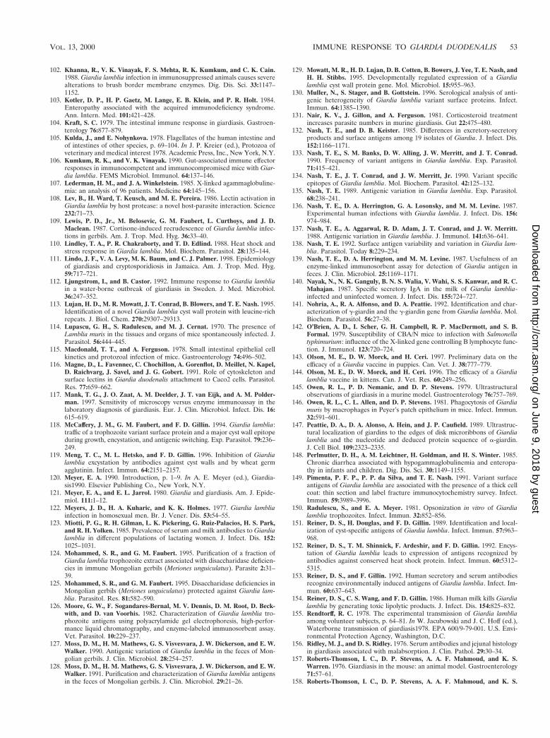
102. Khanna, R., V. K. Vinayak, F. S. Mehta, R. K. Kumkum, and C. K. Cain.1988. Giardia lamblia infection in immunosuppressed animals causes severealterations to brush border membrane enzymes. Dig. Dis. Sci. 33:1147–1152.
103. Kotler, D. P., H. P. Gaetz, M. Lange, E. B. Klein, and P. R. Holt. 1984.Enteropathy associated with the acquired immunodeficiency syndrome.Ann. Intern. Med. 101:421–428.
104. Kraft, S. C. 1979. The intestinal immune response in giardiasis. Gastroen-terology 76:877–879.
105. Kulda, J., and E. Nohynkova. 1978. Flagellates of the human intestine andof intestines of other species, p. 69–104. In J. P. Kreier (ed.), Protozoa ofveterinary and medical interest 1978. Academic Press, Inc., New York, N.Y.
106. Kumkum, R. K., and V. K. Vinayak. 1990. Gut-associated immune effectorresponses in immunocompetent and immunocompromised mice with Giar-dia lamblia. FEMS Microbiol. Immunol. 64:137–146.
107. Lederman, H. M., and J. A. Winkelstein. 1985. X-linked agammaglobuline-mia: an analysis of 96 patients. Medicine 64:145–156.
108. Lev, B., H. Ward, T. Keusch, and M. E. Pereira. 1986. Lectin activation inGiardia lamblia by host protease: a novel host-parasite interaction. Science232:71–73.
109. Lewis, P. D., Jr., M. Belosevic, G. M. Faubert, L. Curthoys, and J. D.Maclean. 1987. Cortisone-induced recrudescence of Giardia lamblia infec-tions in gerbils. Am. J. Trop. Med. Hyg. 36:33–40.
110. Lindley, T. A., P. R. Chakraborty, and T. D. Edlind. 1988. Heat shock andstress response in Giardia lamblia. Mol. Biochem. Parasitol. 28:135–144.
111. Lindo, J. F., V. A. Levy, M. K. Baum, and C. J. Palmer. 1998. Epidemiologyof giardiasis and cryptosporidiosis in Jamaica. Am. J. Trop. Med. Hyg.59:717–721.
112. Ljungstrom, I., and B. Castor. 1992. Immune response to Giardia lambliain a water-borne outbreak of giardiasis in Sweden. J. Med. Microbiol.36:247–352.
113. Lujan, H. D., M. R. Mowatt, J. T. Conrad, B. Blowers, and T. E. Nash. 1995.Identification of a novel Giardia lamblia cyst wall protein with leucine-richrepeats. J. Biol. Chem. 270:29307–29313.
114. Lupascu, G. H., S. Radulescu, and M. J. Cernat. 1970. The presence ofLamblia muris in the tissues and organs of mice spontaneously infected. J.Parasitol. 56:444–445.
115. Macdonald, T. T., and A. Ferguson. 1978. Small intestinal epithelial cellkinetics and protozoal infection of mice. Gastroenterology 74:496–502.
116. Magne, D., L. Favennec, C. Chochillon, A. Gorenflot, D. Meillet, N. Kapel,D. Raichvarg, J. Savel, and J. G. Gobert. 1991. Role of cytoskeleton andsurface lectins in Giardia duodenalis attachment to Caco2 cells. Parasitol.Res. 77:659–662.
117. Mank, T. G., J. O. Zaat, A. M. Deelder, J. T. van Eijk, and A. M. Polder-man. 1997. Sensitivity of microscopy versus enzyme immunoassay in thelaboratory diagnosis of giardiasis. Eur. J. Clin. Microbiol. Infect. Dis. 16:615–619.
118. McCaffery, J. M., G. M. Faubert, and F. D. Gillin. 1994. Giardia lamblia:traffic of a trophozoite variant surface protein and a major cyst wall epitopeduring growth, encystation, and antigenic switching. Exp. Parasitol. 79:236–249.
119. Meng, T. C., M. L. Hetsko, and F. D. Gillin. 1996. Inhibition of Giardialamblia excystation by antibodies against cyst walls and by wheat germagglutinin. Infect. Immun. 64:2151–2157.
120. Meyer, E. A. 1990. Introduction, p. 1–9. In A. E. Meyer (ed.), Giardia-sis1990. Elsevier Publishing Co., New York, N.Y.
121. Meyer, E. A., and E. L. Jarrol. 1980. Giardia and giardiasis. Am. J. Epide-miol. 111:1–12.
122. Meyers, J. D., H. A. Kuharic, and K. K. Holmes. 1977. Giardia lambliainfection in homosexual men. Br. J. Vener. Dis. 53:54–55.
123. Miotti, P. G., R. H. Gilman, L. K. Pickering, G. Ruiz-Palacios, H. S. Park,and R. H. Yolken. 1985. Prevalence of serum and milk antibodies to Giardialamblia in different populations of lactating women. J. Infect. Dis. 152:1025–1031.
124. Mohammed, S. R., and G. M. Faubert. 1995. Purification of a fraction ofGiardia lamblia trophozoite extract associated with disaccharidase deficien-cies in immune Mongolian gerbils (Meriones unguiculatus). Parasite 2:31–39.
125. Mohammed, S. R., and G. M. Faubert. 1995. Disaccharidase deficiencies inMongolian gerbils (Meriones unguiculatus) protected against Giardia lam-blia. Parasitol. Res. 81:582–590.
126. Moore, G. W., F. Sogandares-Bernal, M. V. Dennis, D. M. Root, D. Beck-with, and D. van Voorhis. 1982. Characterization of Giardia lamblia tro-phozoite antigens using polyacrylamide gel electrophoresis, high-perfor-mance liquid chromatography, and enzyme-labeled immunosorbent assay.Vet. Parasitol. 10:229–237.
127. Moss, D. M., H. M. Mathews, G. S. Visvesvara, J. W. Dickerson, and E. W.Walker. 1990. Antigenic variation of Giardia lamblia in the feces of Mon-golian gerbils. J. Clin. Microbiol. 28:254–257.
128. Moss, D. M., H. M. Mathews, G. S. Visvesvara, J. W. Dickerson, and E. W.Walker. 1991. Purification and characterization of Giardia lamblia antigensin the feces of Mongolian gerbils. J. Clin. Microbiol. 29:21–26.
129. Mowatt, M. R., H. D. Lujan, D. B. Cotten, B. Bowers, J. Yee, T. E. Nash, andH. H. Stibbs. 1995. Developmentally regulated expression of a Giardialamblia cyst wall protein gene. Mol. Microbiol. 15:955–963.
130. Muller, N., S. Stager, and B. Gottstein. 1996. Serological analysis of anti-genic heterogeneity of Giardia lamblia variant surface proteins. Infect.Immun. 64:1385–1390.
131. Nair, K. V., J. Gillon, and A. Ferguson. 1981. Corticosteroid treatmentincreases parasite numbers in murine giardiasis. Gut 22:475–480.
132. Nash, T. E., and D. B. Keister. 1985. Differences in excretory-secretoryproducts and surface antigens among 19 isolates of Giardia. J. Infect. Dis.152:1166–1171.
133. Nash, T. E., S. M. Banks, D. W. Alling, J. W. Merritt, and J. T. Conrad.1990. Frequency of variant antigens in Giardia lamblia. Exp. Parasitol.71:415–421.
134. Nash, T. E., J. T. Conrad, and J. W. Merritt, Jr. 1990. Variant specificepitopes of Giardia lamblia. Mol. Biochem. Parasitol. 42:125–132.
135. Nash, T. E. 1989. Antigenic variation in Giardia lamblia. Exp. Parasitol.68:238–241.
136. Nash, T. E., D. A. Herrington, G. A. Losonsky, and M. M. Levine. 1987.Experimental human infections with Giardia lamblia. J. Infect. Dis. 156:974–984.
137. Nash, T. E., A. Aggarwal, R. D. Adam, J. T. Conrad, and J. W. Merritt.1988. Antigenic variation in Giardia lamblia. J. Immunol. 141:636–641.
138. Nash, T. E. 1992. Surface antigen variability and variation in Giardia lam-blia. Parasitol. Today 8:229–234.
139. Nash, T. E., D. A. Herrington, and M. M. Levine. 1987. Usefulness of anenzyme-linked immunosorbent assay for detection of Giardia antigen infeces. J. Clin. Microbiol. 25:1169–1171.
140. Nayak, N., N. K. Ganguly, B. N. S. Walia, V. Wahi, S. S. Kanwar, and R. C.Mahajan. 1987. Specific secretory IgA in the milk of Giardia lamblia-infected and uninfected women. J. Infect. Dis. 155:724–727.
141. Nohria, A., R. A. Alfonso, and D. A. Peattie. 1992. Identification and char-acterization of g-giardin and the g-giardin gene from Giardia lamblia. Mol.Biochem. Parasitol. 56:27–38.
142. O’Brien, A. D., I. Scher, G. H. Campbell, R. P. MacDermott, and S. B.Formal. 1979. Susceptibility of CBA/N mice to infection with Salmonellatyphimurium: influence of the X-linked gene controlling B lymphocyte func-tion. J. Immunol. 123:720–724.
143. Olson, M. E., D. W. Morck, and H. Ceri. 1997. Preliminary data on theefficacy of a Giardia vaccine in puppies. Can. Vet. J. 38:777–779.
144. Olson, M. E., D. W. Morck, and H. Ceri. 1996. The efficacy of a Giardialamblia vaccine in kittens. Can. J. Vet. Res. 60:249–256.
145. Owen, R. L., P. D. Nemanic, and D. P. Stevens. 1979. Ultrastructuralobservations of giardiasis in a murine model. Gastroenterology 76:757–769.
146. Owen, R. L., C. L. Allen, and D. P. Stevens. 1981. Phagocytosis of Giardiamuris by macrophages in Peyer’s patch epithelium in mice. Infect. Immun.32:591–601.
147. Peattie, D. A., D. A. Alonso, A. Hein, and J. P. Caufield. 1989. Ultrastruc-tural localization of giardins to the edges of disk microribbons of Giardialamblia and the nucleotide and deduced protein sequence of a-giardin.J. Cell Biol. 109:2323–2335.
148. Perlmutter, D. H., A. M. Leichtner, H. Goldman, and H. S. Winter. 1985.Chronic diarrhea associated with hypogammaglobulinemia and enteropa-thy in infants and children. Dig. Dis. Sci. 30:1149–1155.
149. Pimenta, P. F. P., P. P. da Silva, and T. E. Nash. 1991. Variant surfaceantigens of Giardia lamblia are associated with the presence of a thick cellcoat: thin section and label fracture immunocytochemistry survey. Infect.Immun. 59:3989–3996.
150. Radulescu, S., and E. A. Meyer. 1981. Opsonization in vitro of Giardialamblia trophozoites. Infect. Immun. 32:852–856.
151. Reiner, D. S., H. Douglas, and F. D. Gillin. 1989. Identification and local-ization of cyst-specific antigens of Giardia lamblia. Infect. Immun. 57:963–968.
152. Reiner, D. S., T. M. Shinnick, F. Ardeshir, and F. D. Gillin. 1992. Encys-tation of Giardia lamblia leads to expression of antigens recognized byantibodies against conserved heat shock protein. Infect. Immun. 60:5312–5315.
153. Reiner, D. S., and F. Gillin. 1992. Human secretory and serum antibodiesrecognize environmentally induced antigens of Giardia lamblia. Infect. Im-mun. 60:637–643.
154. Reiner, D. S., C. S. Wang, and F. D. Gillin. 1986. Human milk kills Giardialamblia by generating toxic lipolytic products. J. Infect. Dis. 154:825–832.
155. Rendtorff, R. C. 1978. The experimental transmission of Giardia lambliaamong volunteer subjects, p. 64–81. In W. Jacubowski and J. C. Hoff (ed.),Waterborne transmission of giardiasis1978. EPA 600/9-79-001. U.S. Envi-ronmental Protection Agency, Washington, D.C.
156. Ridley, M. J., and D. S. Ridley. 1976. Serum antibodies and jejunal histologyin giardiasis associated with malabsorption. J. Clin. Pathol. 29:30–34.
157. Roberts-Thomson, I. C., D. P. Stevens, A. A. F. Mahmoud, and K. S.Warren. 1976. Giardiasis in the mouse: an animal model. Gastroenterology71:57–61.
158. Roberts-Thomson, I. C., D. P. Stevens, A. A. F. Mahmoud, and K. S.
VOL. 13, 2000 IMMUNE RESPONSE TO GIARDIA DUODENALIS 53
on June 9, 2018 by guesthttp://cm
r.asm.org/
Dow
nloaded from
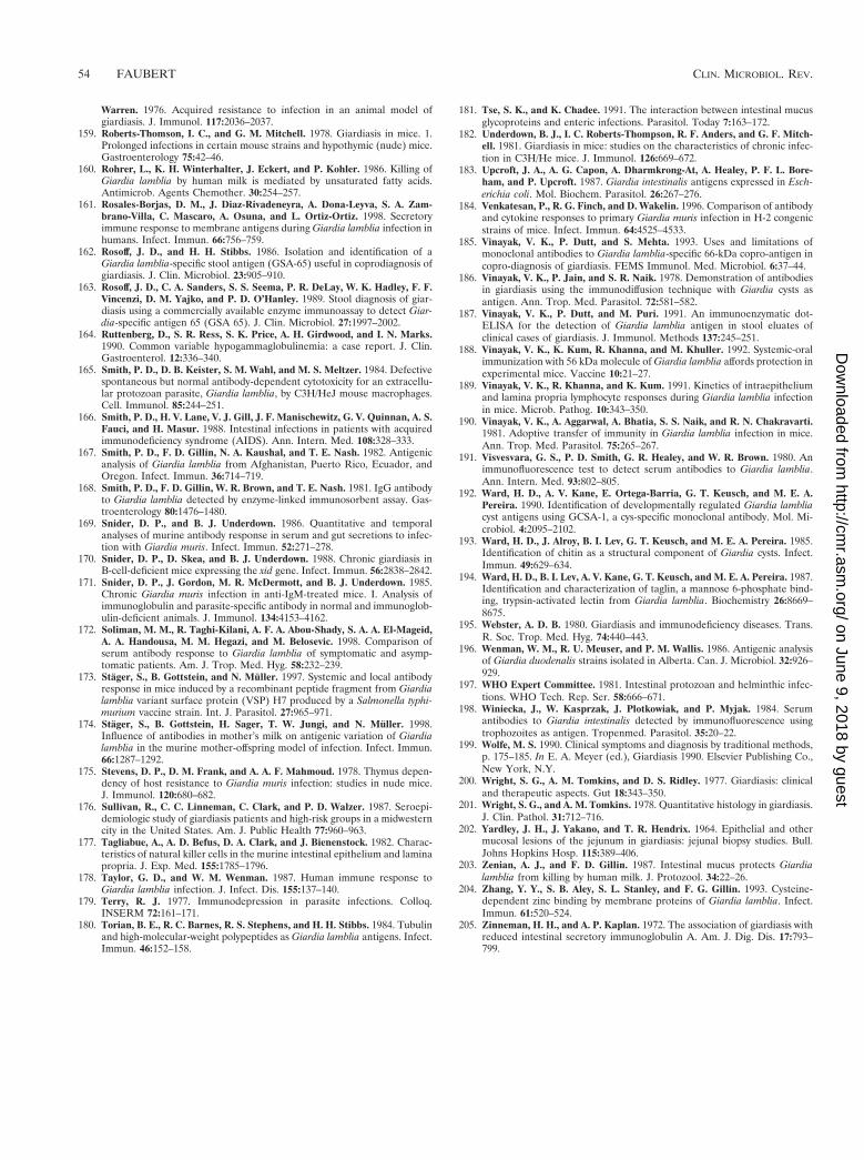
Warren. 1976. Acquired resistance to infection in an animal model ofgiardiasis. J. Immunol. 117:2036–2037.
159. Roberts-Thomson, I. C., and G. M. Mitchell. 1978. Giardiasis in mice. 1.Prolonged infections in certain mouse strains and hypothymic (nude) mice.Gastroenterology 75:42–46.
160. Rohrer, L., K. H. Winterhalter, J. Eckert, and P. Kohler. 1986. Killing ofGiardia lamblia by human milk is mediated by unsaturated fatty acids.Antimicrob. Agents Chemother. 30:254–257.
161. Rosales-Borjas, D. M., J. Diaz-Rivadeneyra, A. Dona-Leyva, S. A. Zam-brano-Villa, C. Mascaro, A. Osuna, and L. Ortiz-Ortiz. 1998. Secretoryimmune response to membrane antigens during Giardia lamblia infection inhumans. Infect. Immun. 66:756–759.
162. Rosoff, J. D., and H. H. Stibbs. 1986. Isolation and identification of aGiardia lamblia-specific stool antigen (GSA-65) useful in coprodiagnosis ofgiardiasis. J. Clin. Microbiol. 23:905–910.
163. Rosoff, J. D., C. A. Sanders, S. S. Seema, P. R. DeLay, W. K. Hadley, F. F.Vincenzi, D. M. Yajko, and P. D. O’Hanley. 1989. Stool diagnosis of giar-diasis using a commercially available enzyme immunoassay to detect Giar-dia-specific antigen 65 (GSA 65). J. Clin. Microbiol. 27:1997–2002.
164. Ruttenberg, D., S. R. Ress, S. K. Price, A. H. Girdwood, and I. N. Marks.1990. Common variable hypogammaglobulinemia: a case report. J. Clin.Gastroenterol. 12:336–340.
165. Smith, P. D., D. B. Keister, S. M. Wahl, and M. S. Meltzer. 1984. Defectivespontaneous but normal antibody-dependent cytotoxicity for an extracellu-lar protozoan parasite, Giardia lamblia, by C3H/HeJ mouse macrophages.Cell. Immunol. 85:244–251.
166. Smith, P. D., H. V. Lane, V. J. Gill, J. F. Manischewitz, G. V. Quinnan, A. S.Fauci, and H. Masur. 1988. Intestinal infections in patients with acquiredimmunodeficiency syndrome (AIDS). Ann. Intern. Med. 108:328–333.
167. Smith, P. D., F. D. Gillin, N. A. Kaushal, and T. E. Nash. 1982. Antigenicanalysis of Giardia lamblia from Afghanistan, Puerto Rico, Ecuador, andOregon. Infect. Immun. 36:714–719.
168. Smith, P. D., F. D. Gillin, W. R. Brown, and T. E. Nash. 1981. IgG antibodyto Giardia lamblia detected by enzyme-linked immunosorbent assay. Gas-troenterology 80:1476–1480.
169. Snider, D. P., and B. J. Underdown. 1986. Quantitative and temporalanalyses of murine antibody response in serum and gut secretions to infec-tion with Giardia muris. Infect. Immun. 52:271–278.
170. Snider, D. P., D. Skea, and B. J. Underdown. 1988. Chronic giardiasis inB-cell-deficient mice expressing the xid gene. Infect. Immun. 56:2838–2842.
171. Snider, D. P., J. Gordon, M. R. McDermott, and B. J. Underdown. 1985.Chronic Giardia muris infection in anti-IgM-treated mice. I. Analysis ofimmunoglobulin and parasite-specific antibody in normal and immunoglob-ulin-deficient animals. J. Immunol. 134:4153–4162.
172. Soliman, M. M., R. Taghi-Kilani, A. F. A. Abou-Shady, S. A. A. El-Mageid,A. A. Handousa, M. M. Hegazi, and M. Belosevic. 1998. Comparison ofserum antibody response to Giardia lamblia of symptomatic and asymp-tomatic patients. Am. J. Trop. Med. Hyg. 58:232–239.
173. Stager, S., B. Gottstein, and N. Muller. 1997. Systemic and local antibodyresponse in mice induced by a recombinant peptide fragment from Giardialamblia variant surface protein (VSP) H7 produced by a Salmonella typhi-murium vaccine strain. Int. J. Parasitol. 27:965–971.
174. Stager, S., B. Gottstein, H. Sager, T. W. Jungi, and N. Muller. 1998.Influence of antibodies in mother’s milk on antigenic variation of Giardialamblia in the murine mother-offspring model of infection. Infect. Immun.66:1287–1292.
175. Stevens, D. P., D. M. Frank, and A. A. F. Mahmoud. 1978. Thymus depen-dency of host resistance to Giardia muris infection: studies in nude mice.J. Immunol. 120:680–682.
176. Sullivan, R., C. C. Linneman, C. Clark, and P. D. Walzer. 1987. Seroepi-demiologic study of giardiasis patients and high-risk groups in a midwesterncity in the United States. Am. J. Public Health 77:960–963.
177. Tagliabue, A., A. D. Befus, D. A. Clark, and J. Bienenstock. 1982. Charac-teristics of natural killer cells in the murine intestinal epithelium and laminapropria. J. Exp. Med. 155:1785–1796.
178. Taylor, G. D., and W. M. Wenman. 1987. Human immune response toGiardia lamblia infection. J. Infect. Dis. 155:137–140.
179. Terry, R. J. 1977. Immunodepression in parasite infections. Colloq.INSERM 72:161–171.
180. Torian, B. E., R. C. Barnes, R. S. Stephens, and H. H. Stibbs. 1984. Tubulinand high-molecular-weight polypeptides as Giardia lamblia antigens. Infect.Immun. 46:152–158.
181. Tse, S. K., and K. Chadee. 1991. The interaction between intestinal mucusglycoproteins and enteric infections. Parasitol. Today 7:163–172.
182. Underdown, B. J., I. C. Roberts-Thompson, R. F. Anders, and G. F. Mitch-ell. 1981. Giardiasis in mice: studies on the characteristics of chronic infec-tion in C3H/He mice. J. Immunol. 126:669–672.
183. Upcroft, J. A., A. G. Capon, A. Dharmkrong-At, A. Healey, P. F. L. Bore-ham, and P. Upcroft. 1987. Giardia intestinalis antigens expressed in Esch-erichia coli. Mol. Biochem. Parasitol. 26:267–276.
184. Venkatesan, P., R. G. Finch, and D. Wakelin. 1996. Comparison of antibodyand cytokine responses to primary Giardia muris infection in H-2 congenicstrains of mice. Infect. Immun. 64:4525–4533.
185. Vinayak, V. K., P. Dutt, and S. Mehta. 1993. Uses and limitations ofmonoclonal antibodies to Giardia lamblia-specific 66-kDa copro-antigen incopro-diagnosis of giardiasis. FEMS Immunol. Med. Microbiol. 6:37–44.
186. Vinayak, V. K., P. Jain, and S. R. Naik. 1978. Demonstration of antibodiesin giardiasis using the immunodiffusion technique with Giardia cysts asantigen. Ann. Trop. Med. Parasitol. 72:581–582.
187. Vinayak, V. K., P. Dutt, and M. Puri. 1991. An immunoenzymatic dot-ELISA for the detection of Giardia lamblia antigen in stool eluates ofclinical cases of giardiasis. J. Immunol. Methods 137:245–251.
188. Vinayak, V. K., K. Kum, R. Khanna, and M. Khuller. 1992. Systemic-oralimmunization with 56 kDa molecule of Giardia lamblia affords protection inexperimental mice. Vaccine 10:21–27.
189. Vinayak, V. K., R. Khanna, and K. Kum. 1991. Kinetics of intraepitheliumand lamina propria lymphocyte responses during Giardia lamblia infectionin mice. Microb. Pathog. 10:343–350.
190. Vinayak, V. K., A. Aggarwal, A. Bhatia, S. S. Naik, and R. N. Chakravarti.1981. Adoptive transfer of immunity in Giardia lamblia infection in mice.Ann. Trop. Med. Parasitol. 75:265–267.
191. Visvesvara, G. S., P. D. Smith, G. R. Healey, and W. R. Brown. 1980. Animmunofluorescence test to detect serum antibodies to Giardia lamblia.Ann. Intern. Med. 93:802–805.
192. Ward, H. D., A. V. Kane, E. Ortega-Barria, G. T. Keusch, and M. E. A.Pereira. 1990. Identification of developmentally regulated Giardia lambliacyst antigens using GCSA-1, a cys-specific monoclonal antibody. Mol. Mi-crobiol. 4:2095–2102.
193. Ward, H. D., J. Alroy, B. I. Lev, G. T. Keusch, and M. E. A. Pereira. 1985.Identification of chitin as a structural component of Giardia cysts. Infect.Immun. 49:629–634.
194. Ward, H. D., B. I. Lev, A. V. Kane, G. T. Keusch, and M. E. A. Pereira. 1987.Identification and characterization of taglin, a mannose 6-phosphate bind-ing, trypsin-activated lectin from Giardia lamblia. Biochemistry 26:8669–8675.
195. Webster, A. D. B. 1980. Giardiasis and immunodeficiency diseases. Trans.R. Soc. Trop. Med. Hyg. 74:440–443.
196. Wenman, W. M., R. U. Meuser, and P. M. Wallis. 1986. Antigenic analysisof Giardia duodenalis strains isolated in Alberta. Can. J. Microbiol. 32:926–929.
197. WHO Expert Committee. 1981. Intestinal protozoan and helminthic infec-tions. WHO Tech. Rep. Ser. 58:666–671.
198. Winiecka, J., W. Kasprzak, J. Plotkowiak, and P. Myjak. 1984. Serumantibodies to Giardia intestinalis detected by immunofluorescence usingtrophozoites as antigen. Tropenmed. Parasitol. 35:20–22.
199. Wolfe, M. S. 1990. Clinical symptoms and diagnosis by traditional methods,p. 175–185. In E. A. Meyer (ed.), Giardiasis 1990. Elsevier Publishing Co.,New York, N.Y.
200. Wright, S. G., A. M. Tomkins, and D. S. Ridley. 1977. Giardiasis: clinicaland therapeutic aspects. Gut 18:343–350.
201. Wright, S. G., and A. M. Tomkins. 1978. Quantitative histology in giardiasis.J. Clin. Pathol. 31:712–716.
202. Yardley, J. H., J. Yakano, and T. R. Hendrix. 1964. Epithelial and othermucosal lesions of the jejunum in giardiasis: jejunal biopsy studies. Bull.Johns Hopkins Hosp. 115:389–406.
203. Zenian, A. J., and F. D. Gillin. 1987. Intestinal mucus protects Giardialamblia from killing by human milk. J. Protozool. 34:22–26.
204. Zhang, Y. Y., S. B. Aley, S. L. Stanley, and F. G. Gillin. 1993. Cysteine-dependent zinc binding by membrane proteins of Giardia lamblia. Infect.Immun. 61:520–524.
205. Zinneman, H. H., and A. P. Kaplan. 1972. The association of giardiasis withreduced intestinal secretory immunoglobulin A. Am. J. Dig. Dis. 17:793–799.
54 FAUBERT CLIN. MICROBIOL. REV.
on June 9, 2018 by guesthttp://cm
r.asm.org/
Dow
nloaded from


![multiplexing stool MD short.ppt [Compatibiliteitsmodus] · 8 0394-0527 Giardia Entamoeba coli Giardia 9 0394-0526 Giardia Entamoeba coli Giardia 10 1032-1221 Giardia negative Giardia](https://img.pdfslide.net/doc/110x75/5e4182dad3a23a3d0b082f2c/multiplexing-stool-md-shortppt-compatibiliteitsmodus-8-0394-0527-giardia-entamoeba.jpg)


![Faubert Et Al., 2010[1]](https://img.pdfslide.net/doc/110x75/577cd8e61a28ab9e78a24135/faubert-et-al-20101.jpg)