Embed Size (px)
Citation preview

Immune System
The immune system protects the body from invasion by pathogens (an entity that can cause harm to the body) What cells are found in blood?
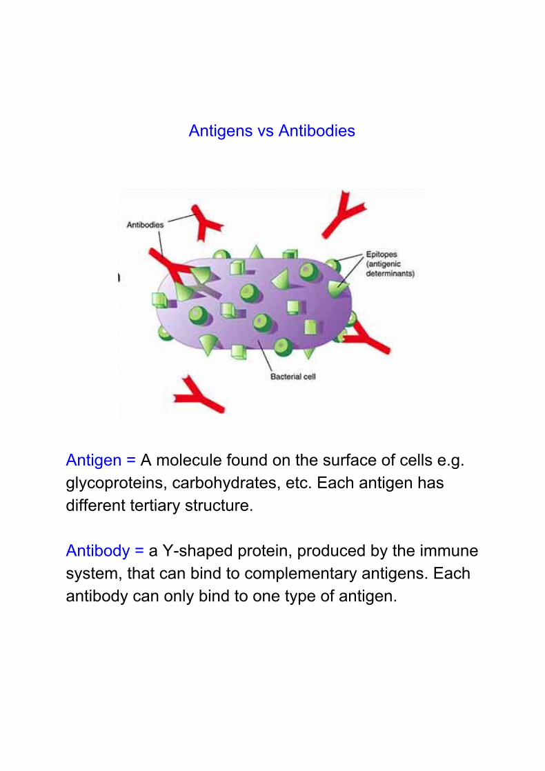
Antigens vs Antibodies
Antigen = A molecule found on the surface of cells e.g. glycoproteins, carbohydrates, etc. Each antigen has different tertiary structure. Antibody = a Y-shaped protein, produced by the immune system, that can bind to complementary antigens. Each antibody can only bind to one type of antigen.

Self -Antigens vs Non-self Antigens

How the body eliminates immune cells that recognise self-antigens
- Early on in foetal development, the immune system
contains immune cells (macrophages and lymphocytes) that recognise both self and non-self antigens
- The immune cells that recognise self-antigens are continuously inactivated (commit suicide)
- By the time the child is born, the immune system only contains immune cells that recognise non-self antigens
- The immune system also learns to tolerate bacteria that colonise the digestive system immediately after birth
- A breakdown of self-tolerance leads to autoimmune disorder (covered in more detail in the 1.15 lesson)


Non-Specific Immune Response First Line of Defence - Barriers to entry of pathogens SKIN
● Physical barrier to entry - no gaps between cells
● Waxy sebum on surface - waterproof, elastic
● Colonised by ‘normal’ flora - healthy bacteria that
prevent colonisers from gaining a foothold
● Slightly acidic pH - not tolerated by most bacteria
● Sweat contains the enzyme lysozyme, which can
destroy bacterial cell walls
(Discuss effects of antibacterial soaps/wipes - Triclosan favours antibiotic resistant bacteria)
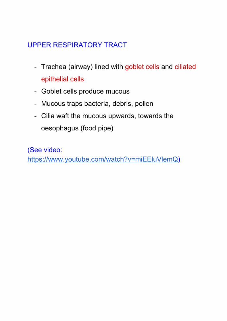
UPPER RESPIRATORY TRACT
- Trachea (airway) lined with goblet cells and ciliated
epithelial cells
- Goblet cells produce mucous
- Mucous traps bacteria, debris, pollen
- Cilia waft the mucous upwards, towards the
oesophagus (food pipe)
(See video: https://www.youtube.com/watch?v=miEEluVlemQ)

DIGESTIVE SYSTEM
- Stomach contains Hydrochloric acid
- Low pH, 1-2
- denatures proteins and enzymes in pathogens
- Small and large intestine contain normal flora, which
prevent colonisation by pathogens
TEAR DUCTS - in eyes
- tears clear dust and debris
- tears contain the enzyme lysozyme, which can kill
bacteria
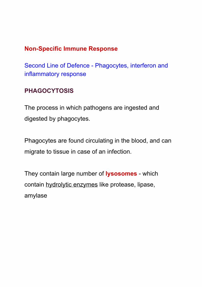
Non-Specific Immune Response Second Line of Defence - Phagocytes, interferon and inflammatory response PHAGOCYTOSIS The process in which pathogens are ingested and
digested by phagocytes.
Phagocytes are found circulating in the blood, and can
migrate to tissue in case of an infection.
They contain large number of lysosomes - which
contain hydrolytic enzymes like protease, lipase,
amylase
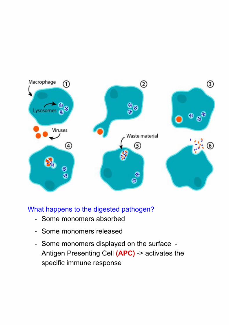
What happens to the digested pathogen?
- Some monomers absorbed
- Some monomers released
- Some monomers displayed on the surface - Antigen Presenting Cell (APC) -> activates the specific immune response

Steps in Phagocytosis - Phagocytes have many different receptors on their
surface
- When a phagocyte encounters a pathogen, it binds to
the antigen on the surface of the pathogen using its
complementary receptors
- The cell membrane folds inwards and encloses the
pathogen in a vesicle called a phagosome
- Phagosome fuses with lysosome
- Lysosome releases hydrolytic enzymes
- Which breaks down the pathogens (sugars, proteins,
lipids) releasing the monomers

INFLAMMATION
- Chemicals released by damaged cells attract white
blood cells to the site of infection.
- Mast cells release histamine, which widens the gaps in
the capillary walls, allowing white blood cells to migrate
from blood to tissue (swelling)
- Phagocytes secrete chemicals called cytokines, which
help with recruiting other blood cells and with tissue
repair
- Temperature elevated (redness) to halt bacterial
growth
INTERFERON - Proteins produced by virus-infected cells
- Binds to the surface of non-infected cells
- Triggers production of anti-viral proteins
- Prevents viral infection from spreading

Specific Immune Response Third Line of Defence - B and T cells B-cell response Step 1: Recognition of Antigen and activation B-cells use the B-cell receptor to recognise antigens on the pathogen, or antigens on the surface of APCs like phagocytes Each B-cell only displays one type of BCR
Cross-linking of receptors activates B-cells

Step 2: B-cells undergo clonal expansion by mitosis Step 3: B-cells differentiate into plasma and memory cells

Step 4: Plasma cells produce antibodies
Antibodies are a soluble form of the BCR. Memory cells are used for secondary infections.

Structure of the Antibody

● Antibodies are proteins - quaternary structure ● Made of four polypeptide chains, each folded into its
tertiary structure ● Two light chains (short) and two heavy chains (long) ● Two halves
○ Constant Region = sequence remains the same in all antibodies
○ Variable regions = sequence is unique for each type of antibody
● Each arm contains one antigen-binding pocket, formed by the variable region
● The two arms are joined together at the hinge region
● The hinge region, which is rich in disulphide bonds, allows the two arms to rotate independent of each other, so that each antibody can bind to two antigens
One antibody can bind to (is complementary to) only one antigen

How to antibodies work? - One antibody is able to bind to two antigens - This causes clumping of the pathogen = agglutination
- Agglutination prevents pathogens from attaching to surfaces - Phagocytes also find it easier to digest ‘clumps’ of pathogens - Rapid clearance of pathogen from the body

Why does the immune system produce memory cells? Memory cells are used during a secondary immune response - ie re-infection by the same pathogen
Memory exist in a state of readiness and have the ability to rapidly expand and fight off recurrence of the same disease More plasma cells produced - larger antibody response Faster response - more rapid clearance of the pathogen from the body

T-cell response Step 1: Activation of T-cells T-cells have T-cell receptors (TCR) on their surface - they can be activated by antigens presented on the surface of an APC - like a B-cell or a phagocyte
Step 2: T-cells undergo clonal expansion by mitosis Step 3: T-cells differentiate into 3 distinct cell lineages Bacterial infection: T-helper and T-memory Viral infection: T-killer and T-memory (with some T-helper)

Role of T-helper cells - Central to the immune system, have CD4 receptors - Produce chemicals called cytokines, which activate B-cells, and help them to differentiate into plasma cells
- Without T-helper cells antibody response is poor - HIV virus attacks T-helper cells, compromising the
immune system
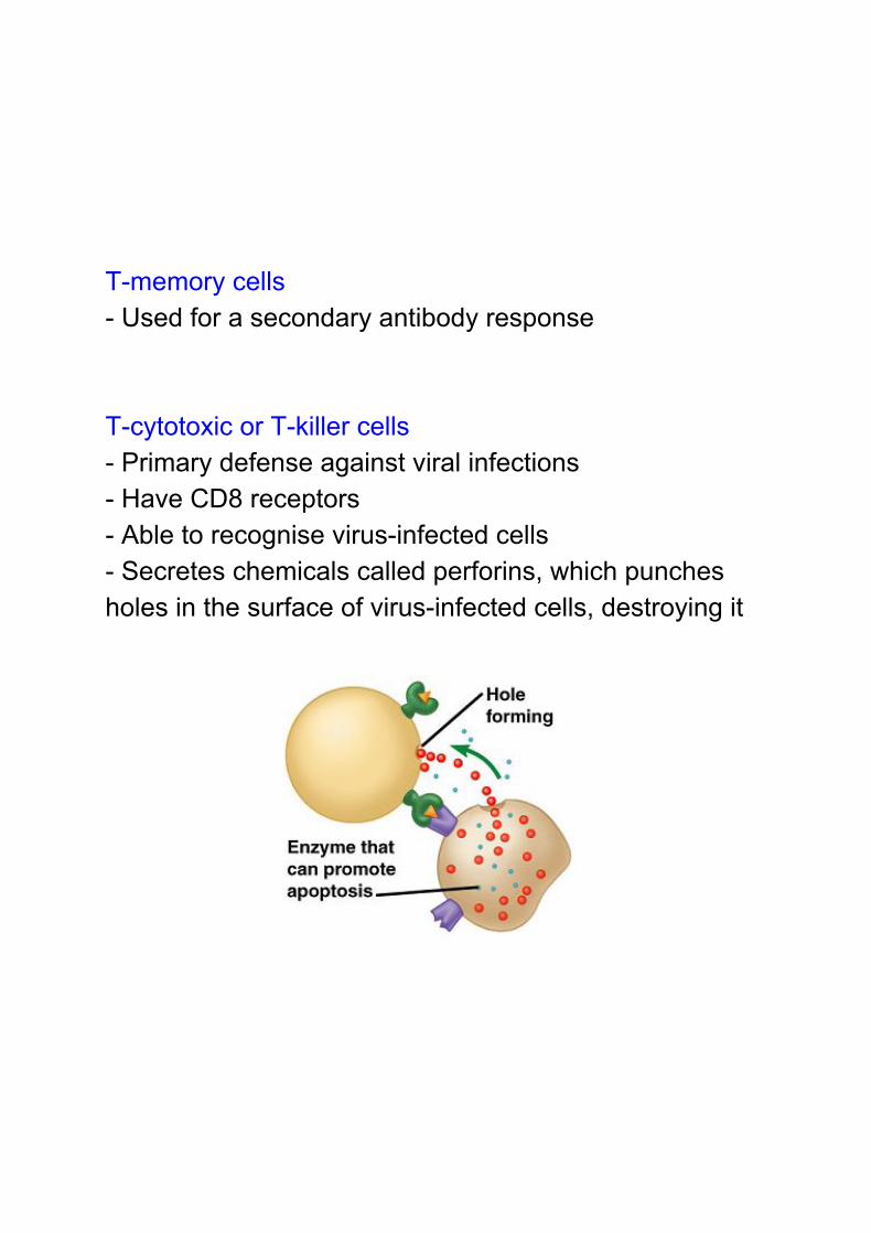
T-memory cells - Used for a secondary antibody response T-cytotoxic or T-killer cells - Primary defense against viral infections - Have CD8 receptors - Able to recognise virus-infected cells - Secretes chemicals called perforins, which punches holes in the surface of virus-infected cells, destroying it

Monoclonal antibodies - An antibody that binds to one complementary antigen - Produced by fusing Plasma Cells with malignant B-cell - Useful in research and diagnostics (Blood contains a mixture of antibodies = polyclonal)

Uses of Monoclonal Antibodies 1. Cancer treatment
- drug targeting - Using radioisotopes (targeted radiotherapy) - Blocking growth factors

2. Pregnancy testing kits
Test protein = HCG, produced by cells of the placenta Control = albumin, a protein found in blood and urine (Video: https://www.youtube.com/watch?v=aOfWTscU8YM; https://www.youtube.com/watch?v=dsX7tGDbBO4)

3. Diagnosis using ELISA - enzyme-linked assay e.g. detecting HIV antibodies in blood
1O Antibody = immobilised, and complementary to antigen 2O Antibody = complementary to antigen, attached an enzyme that can convert a colourless substrate into a coloured compound
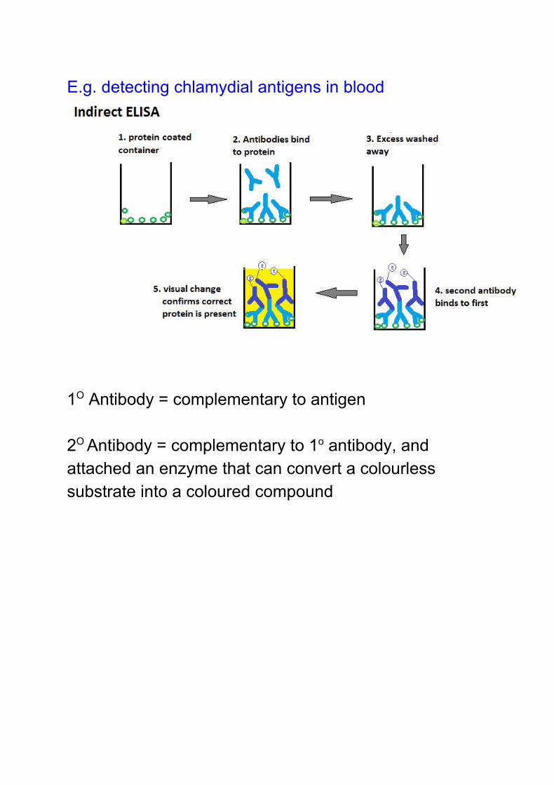
E.g. detecting chlamydial antigens in blood
1O Antibody = complementary to antigen 2O Antibody = complementary to 1o antibody, and attached an enzyme that can convert a colourless substrate into a coloured compound

TYPES OF IMMUNE RESPONSE
Natural, Active
- Pathogen enters the body naturally - Specific immune system triggered - Memory cells produced
- Long-lasting immunity - High levels of antibody over time
Natural, Passive
- Body does not encounter antigen - Instead, antibodies pass from one person to
another i.e. mother to child (via placenta or breast milk)
- No memory cells - Antibodies are proteins, so will eventually be
destroyed - Short-term immunity

Artificial, Active (Vaccines)
- Antigen artificially introduced into the body - Triggers the specific immune system - Memory cells produced
Artificial, Passive
- Monoclonal antibodies injected into body - Offers immediate protection against pathogen - No memory cells - Short term immunity
E.g. Tetanus anti-toxin Snake anti-venom Rabies vaccines Adv: Immediate effect Disadv: Short-term, expensive

Two main types of Vaccines LIVE vaccines
- Live, but weakened strain of the pathogen - Injected into body - Triggers specific immune response - Pathogen can multiply slowly, but does not cause
infection - Memory cells produced, and levels keep building up - Protects against actual infection by pathogen
Adv:
- Long-lasting immunity due to memory cells - Strong antibody response
Disadv:
- Risk of infection or side-effects in immunocompromised individuals - old, young, autoimmune disease like coeliac disease
Eg. BCG, MMR, children’s flu vaccine

INACTIVATED vaccines
- Heat-killed pathogen injected into body - Heated to temperatures that do not alter the tertiary
structure of the antigens on the surface - Antigens recognised by immune system as
non-self, triggers specific immune response - Memory cells produced - Protects against infection by actual pathogen
Adv: Low risk, minimal chance of infection Disadv: Pathogens do not multiply in the body, so levels of memory cells lower than those produced by live vaccines E.g. polio, whooping cough, adult flu vaccine

Why vaccines don’t always work:
1. Person has already been exposed to the antigen
before being given the vaccine = incubation period,
where symptoms are not apparent
2. Immune system is compromised, leading to disease
or side-effects
3. Antigenic variability: some bacteria and viruses
have the ability to constantly vary the antigens on
their surface - memory cells produced in primary
infection cannot recognise these new antigens on
reinfection
4. Everyone responds differently to vaccines - levels of
antibody vary

Herd immunity: Not possibly to immunise 100% of the
population - some people have ethical objections or are
too ill to take vaccines
Goal is to immunise 95% of the population
Minimises risk of coming into contact with an infected
individual - reduces chance of disease spreading

Flu vaccines Influenza virus shows antigenic variability - many different strains exist in the same year Epidemiologists keeps track of the strains most likely to cause infection each winter Flu vaccine is made by combining the top 5 strains likely to cause infection However, if infected with a strain that is not in the vaccine then it is still possible to get ill with flu Therefore, vaccine is given to very young or old, or pregnant women, but not normally available for free to people within the age range of 18-65 (in the UK) - as their immune system can cope with flu without any serious side-effects (cost:benefit ratio)