Embed Size (px)
Citation preview

Proc. Natl. Acad. Sci. USAVol. 81, pp. 4945-4949, August 1984Medical Sciences
Immunodeficiency with defective T-cell response to interleukin 1(T-cell deficiency/interleukin 1 receptor/interleukin 2 deficiency)
EDWARD T. CHU*, LANNY J. ROSENWASSERt, CHARLES A. DINARELLOt, FRED S. ROSEN*,AND RAIF S. GEHA**Divisions of Allergy and Immunology, Children's Hospital Medical Center, and Department of Pediatrics, Harvard Medical School, Boston, MA 02115; andtDivisions of Allergy and Experimental Medicine, Department of Medicine, Tufts-New England Medical Center, Boston, MA 02111
Communicated by Baruj Benacerraf, April 16, 1984
ABSTRACT Normal proliferation of T cells in vitro re-quires production of and response to the lymphokine interleu-kin 2 (IL-2). Optimal IL-2 production by T cells is dependenton the monokine interleukin 1 (IL-1). A 10-year-old male withrecurrent infections and failure to thrive was evaluated forpossible defects in the production and response to IL-1 and IL-2. The patient had normal levels of serum immunoglobulinsand a normal distribution of circulating T-cell subsets. Howev-er, the in vitro proliferative response of his peripheral bloodmononuclear cells (PBMC) to phytohemagglutinin was de-pressed (40% of normal) and the response of his PBMC toantigens was absent. Delayed hypersensitivity skin tests and invitro response to tetanus toxoid remained absent despite re-peated immunizations. Monocyte function in this patient wasnormal as judged by the following criteria: normal expressionof Ia antigens (77% +), normal IL-1 production, and normalcapacity to present tetanus toxoid to a maternal T-cell line spe-cific for tetanus toxoid antigen. The abnormal phytohemag-glutinin response of the patient's PBMC was corrected by theaddition of exogenous IL-2. IL-2 production by the patient'sphytohemagglutin-stimulated PBMC was severely deficientbut was corrected by the addition of phorbol 12-myristate 13-acetate, suggesting a defective response to IL-1. T-cell blastsderived from a normal subject but not T-cell blasts derivedfrom the patient absorbed out IL-1 activity from a preparationof purified human IL-1. These results indicate that the pa-tient's T-cell deficiency was due to a defective T-cell responseto IL-1 and suggest that IL-1 plays an important role in the invivo immune response.
The proliferative response of human T cells to antigen re-quires the recognition of antigen processed by Ia' accessorycells and the monokine interleukin 1 (IL-1). These two re-quirements can be separated by irradiation of the antigen-pulsed accessory cells with UV light, which inhibits the pro-duction of IL-1 (1-4). The failure of T cells to proliferate toUV-treated antigen-pulsed monocytes (Mo) is reversed bythe addition of purified IL-1 (3). The requirement for IL-1 forT-cell proliferation to mitogens such as phytohemagglutinin(PHA) and Con A is less clear. Rabbit antibody to human IL-1 does not inhibit the proliferation of peripheral blood mono-nuclear cells (PBMC) to mitogens cells but readily inhibitsthe proliferation of PBMC to antigen such as tetanus toxoid(TT) (3, 5). IL-1 induces T cells that have interacted withlectins to secrete interleukin 2 (IL-1) (6, 7). In turn, IL-2causes the proliferation of activated T cells, which expressIL-2 receptors. We now describe a patient with an inabilityto generate IL-2 due to a defect in his T-cell response to IL-1.
CASE REPORTA 10-year-old Lebanese male had a history of recurrentpneumonias and otitis media beginning at the age of 2
months. At age 3 years, he had an episode of severe herpeszoster. He has failed to grow in height and weight for severalyears. Family history revealed that three male siblings diedin infancy because of recurrent infections, whereas five oth-er siblings remain healthy. Total leukocyte count was 15,400cells per mm3 with 55% lymphocytes. Seventy-three percentof his PBMC formed rosettes with sheep erythrocytes, 67%reacted with T3, 41% with T4, 30% with T8, and 0% with T6.Serum immunoglobulin levels ,were IgG, 700 mg/dl; IgM,184 mg/dl; IgA, 260 mg/dl; and IgE, 8 international un-its/ml. Delayed hypersensitivity skin tests to tuberculin,monilia, and TT antigens were negative.
MATERIALS AND METHODSCells and Cell Cultures. The isolations of PBMC and of
highly enriched populations of Mo and of T cells were doneas described (8). Proliferation ofPBMC to antigens and mito-gens was performed as described in ref. 9.
Production and Assay of IL-2. IL-2 was generated by cul-turing nonadherent irradiated (1000 rads; 1 rad = 0.01 gray)PBMC (106 cells per ml) with PHA for 48 hr as described(10). IL-2 activity in the supernatants was assayed on Con A-induced IL-2-dependent T-cell lines as described in ref. 10.PHA-free IL-2 was a gift of A. Krensky (Dana-Farber Can-cer Institute, Boston) and was added at 2 units/ml.
TT-Specific T-Cell Lines. TT-specific T-cell lines weregenerated as described by Kurnick et al. (11). The cell lineswere resuspended every 3-4 days in fresh medium contain-ing 25% IL-2-containing supernatants and were stimulatedevery 7-10 days with autologous irradiated (5000 rads)PBMC and TT.
Generation and Testing of IL-1-Containing Supernatants.IL-i-containing supernatants were generated by incubatingadherent PBMC (2-5 x 10⁶ per ml) with Con A (20 ,ug/ml)for 18 hr, collecting the supernatants, and adding a-methyl-D-mannoside (25 mg/ml) as described in ref. 10. Such super-natants contain IL-1 activity, as assessed by a thymocytecostimulator assay (3), and can substitute for IL-i in recon-stituting the human T-cell proliferative response to TT-pulsed UV-irradiated Mo (3). IL-1 activity in the superna-tants was assayed as in ref. 3. Briefly, Mo in Petri disheswere incubated with TT (50 ,ug/ml) for 18 hr, washed, irradi-ated with UV light (1.2 joules/m2 per sec, 230-350 nm) for 60sec, and then added at a ratio of 1:10 to nylon wool-purifiedautologous T cells in the presence or absence of superna-tants to be tested for IL-1 activity.
Partially Purified Human IL-1/Leukocytic Pyrogen (LP).IL-1 was partially purified as described in ref. 12. This hu-man IL-1 copurifies with human LP down to a single band ofMr of =15,000 on 7.5% NaDodSO4/polyacrylamide gel elec-trophoresis. This material is highly active in the rabbit pyro-
Abbreviations: IL-1 and -2, interleukins 1 and 2; Mo, monocyte(s);PBMC, peripheral blood mononuclear cell(s); PHA, phytohemag-glutinin; PMA, phorbol 12-myristate 13-acetate; IT, tetanus toxoid;PWM, pokeweed mitogen; LP, leukocytic pyrogen.
4945
The publication costs of this article were defrayed in part by page chargepayment. This article must therefore be hereby marked "advertisement"in accordance with 18 U.S.C. §1734 solely to indicate this fact.

4946 Medical Sciences: Chu et al.
Table 1. Proliferative response to PHA, TT, and moniliacpm of [3H]thymidine incorporated in cultures stimulated with
Antigen
No Mitogen TTSubject stimulation PHA Con A PWM Pre Post Monilia DT
Patient 1332 ± 281 64,894 ± 9,643 25,552 ± 5,488 23,758 ± 6,975 967 ± 769 769 ± 156 1087 ± 270 1,616 ± 645Mother 512 ± 58 183,427 ± 6,502 706 ± 124 72,155 ± 8362 5107 ± 603 706 ± 261Normal
control(n = 9) 753 ± 236 238,342 ± 52,475 137,533 ± 41,320 105,094 ± 33,853 21,250 ± 8639 8458 ± 3910 11,892 ± 6485PHA was added at a concentration of 1 ,ug/mi, TT and diphtheria toxoid (DT, Massachusetts Biological Laboratories, Jamaica Plain, MA)
were added at a concentration of 20 ,ug/ml, and monilia antigen (Hollister Stier, Spokane, WA) was added at a dilution of 1:500. Valuesrepresent mean ± SEM of triplicate cultures. Mitogen-stimulated cultures were harvested at 3 days and the remainder at 6 days. Thebackground for unstimulated 6-day cultures is shown and was not significantly different from the background for 3-day cultures, which is notshown. Normal subjects ranged in age from 7 to 25 years. TT pre and post indicate before and after immunization, respectively, with TT.
gen assay (12, 13), in which one rabbit pyrogen dose is theamount of IL-1/LP that when injected intravenously in a
rabbit causes an elevation of 0.6-0.90C in body temperaturewithin 1 hr.Thymocyte Costimulator Assay. This was performed as de-
scribed in ref. 13.
RESULTSResponse to Mitogens and Antigens. PBMC from the pa-
tient had a diminished proliferative response (<3 SD belowthe mean of normal controls) to PHA, Con A, and pokeweedmitogen (PWM) and an absent response to TT, diphtheriatoxoid, and monilia antigens (Table 1). Subsequently, the pa-tient and his mother, who was not immune to TT, were givena series of three immunizations with TT antigen (5 limit floc-culation units each) over a 2-month period. Following this,PBMC from the patient remained unresponsive to TT,whereas PBMC from his healthy mother proliferated vigor-ously to TT (Table 1). After immunization, the delayed hy-persensitivity skin test with TT remained negative in the pa-tient but converted to positive in the mother.
_1()
x
C-
8
I_16
J4
T
,II
T
T
*:::... ...
*. .. .. .:
A.
...::: .:.
..:.::
:: :: ..:::
:: .:::::: ::: ::.:': ::::......':::::::::::..__ ......
T+ -t -r
AUTO Mo PT Mo ALLI() Mo
FIG. 1. Proliferative responses of maternal TT-specific T-cellblasts (T) to TT presented by Mo. Cultures contained 4 x 10⁴ T cellsand 4 x 10⁴ irradiated (5000 rads) Mo with or without soluble TT at40 tg/ml. AUTO, autologous; PT, patient; ALLO, allogeneic. Val-ues represent mean + SEM of triplicate cultures. Cultures contain-ing T-cell blasts and accessory cells always incorporated <1300 cpmof [3H]thymidine into DNA.
Antigen-Presenting Function of Mo. T-cell proliferation toantigen is dependent on antigen presentation by Ia+/DR'accessory Mo and on the secretion of IL-1 by the Mo. In twoseparate experiments, 77% and 75% of the patient's PBMCthat adhered to plastic plates (i.e., Mo) were found to ex-press Ia antigen compared to 82% ± 14% in five normal con-trols.To examine the antigen-presenting function of the pa-
tient's Mo and because no HLA identical siblings were avail-able, we examined the capacity of his Mo to support the pro-liferation of a TT-specific T-cell line derived from his HLAhaploidentical mother. The results of this experiment areshown in Fig. 1. Mo from the patient and from the mothersupported T-cell proliferation of the maternal T-cell line inthe presence of TT. Mo from an HLA-DR unrelated donorfailed to support the proliferation of the maternal T-cell linein the presence of TT.We next examined the capacity of the patient's Mo to gen-
erate IL-1 and found it to be normal. Table 2 shows thatnormal immune T cells proliferated in response to TT-pulsedautologous Mo but not in response to autologous TT-pulsedUV-irradiated Mo. This response was reconstituted by theaddition of supernatants from Con A-stimulated Mo derivedfrom both the patient and a normal donor. This effect wasnot due to residual Con A in the Mo supernatants becauseaddition of these supernatants alone did not cause prolifera-tion of T cells and Mo.
IL-2 Production and Response. Fig. 2A shows that the pa-tient's PBMC were severely deficient in their capacity togenerate IL-2 for the normal IL-2-dependent T-cell line. Al-though the patient's PBMC failed to generate IL-2, Con A-induced blasts from the patient were able to respond to IL-2,as shown in Fig. 2B, but not to supernatants of his own PHA-stimulated PBMC. Thus, it appears that the T cells of thepatient respond normally to IL-2, even though his PBMCwere unable to generate it. Mixing experiments failed to de-
Table 2. Reconstitution of the T-cell response to UV-irradiatedantigen-pulsed Mo (Morr/UV) by supernatants of Con A-stimulated adherent cells
Source ofCon A-stimulated [3H]Thymidine incorporated
Cells in culture Mo supernatant per culture, cpmT + Morr - 37,784 ± 3753T + MoTT/UV 964 ± 188T + Mo.r/UV Normal 46,8% ± 4903T + Mo-r/UV Patient 44,168 + 2150T alone Normal 620 ± 144
MoT-, autologous Mo pulsed with TT (50 gg/ml) for 18 hr. MoT-r/UV, TT-pulsed Mo irradiated with UV light. T cells and Mo wereobtained from a normal subject. Values represent mean ± SEM.
Proc. NatL Acad Sci. USA 81 (1984)
T
T
Proc. NatL Acad Sci. USA 81 (1984) 4947
E 10'
Lo G-o Normal
o---o Patient
PHA alone
104
1:2 1:4 1:8 1:2 1:4 1:8Dilution of supernatant
FIG. 2. (A) Response of normal Con A-induced T-cell blasts tosupernatants of PHA-stimullated PBMC from a normal subject ( )and from the patient (-).(B) Response of the patient's Con A-induced T-cefl blasts to supernatants of PHA-stimulated PBMCfrom a normal subject ( ) and from the patient (-).Numbersrepresent cpm. Values represent mean cpm -+ SD of [3H]thymidineincorporated in triplicate cultures of 4 x 10⁴ blasts.
tect the presence of an inhibitor of IL-2 activity in supernia-tants of the patient's PHA-stimulated PBMC (data notshown).Addition of exogenous IL-2 on day 0 to PHA-stimulated
cultures of the patient's PBMC reconstituted the prolifera-tive response after 3 days in culture to norm'al.. Furthermore,unlike the situation in normal subjects, the patient's PBMCproliferative response to PHA was not sustained for 6 daysof culture but was reconstituted to the normal level by addi-tion of exogenous IL-2 at day 3 of culture (Table 3). In con-trast to the effect of IL-2 on the PHA response, addition ofPHA-free IL-2 to cultures for the' patient's PBMC stimulatedwith TT did not result in cell proliferation (data not shown).Addition of purified IL-1 to the patient's PBMC did not cor-rect their defective proliferative responses (data not shown).Response to Phorbol 12-Myristate 13-Acetate (PM'A). We
next considered the possibility that the failure of the pa-tient's T cells to make IL-2 was due to their failure to re-spond to IL-1. In an effort to bypass the IL-1 requirementfor lectin-induced IL-2 production, we examined the capaci-ty of the patient's PBMC to produce IL-2 activity when stim-ulated with PHA in the presence of the phorbol ester PMA.Table 4 shows that when the patient's PBMC were stimulat-ed with both PHA and PMA they released substantialamounts of IL-2 activity into their supernatants. Stimulation
of the patient's PBMC with either PHA or PMA alone didnot result in the release of significant IL-2 activity as as-
sessed by blast proliferation (Table 4). The blast prolifera-tion induced by supernatants ofPBMC stimulated with PMAand PHA was not due to the action of these agents togetheron the blasts used for the assay (Table 4). PMA induced a
negligible increment or no increment in the production of IL-2 by PHA-stimulated normal T cells (data not shown).We next examined the capacity of T-cell blasts from the
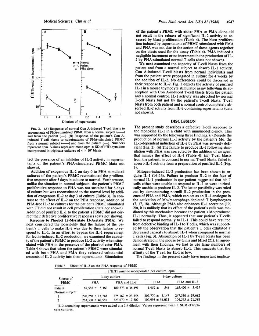
patient and from a normal subject to absorb IL-1 activity.Con A-induced T-cell blasts from normal individuals andfrom the patient were propagated in culture for 4 weeks bythe addition of IL-2. No differences could be discerned intheir response to IL-2. Fig. 3 depicts the activity of purifiedIL-1 in a mouse thymocyte stimulator assay folloWing its ab-sorption with Con A-induced T-cell blasts from the patientand a normal control. IL-1 activity was absorbed by normalT-cell blasts but not by the patient's T-cell blasts. T-cellblasts from both patient and a normal control completely ab-sorbed IL-2 activity from IL-2-containing supernatants (datanot shown).
DISCUSSIONThe present study describes a defective T-cell response tothe monokine IL-1 in a child with immunodeficiency. Thiswas supported by the following three findings. (i) Despite theproduction of normal IL-i activity by the patient's Mo, theIL-i-dependent induction of IL-2 by PHA was severely defi-cient (Fig. 2). (ii) The failure to produce IL-2 following stim-ulation with PHA was corrected, by the addition of an agentthat mimics the effect of IL-1 (Table 4). (iii) T-cell blastsfrom the patient, in contrast to normal T-cell blasts, failed toabsorb IL-1 activity from a preparation of purified IL-1 (Fig.3).Mitogen-induced IL-2 production has been shown to re-
quire IL-1 (14-16). Failure to produce IL-2 in the face ofnormal IL-1 production in our patient suggested that his Tcells either were unable to respond to IL-1 or were intrinsi-cally unable to produce IL-2. The latter possibility was ruledout by demonstrating normal IL-2 production in the pres-ence ofPHA and PMA, which can act as an IL-1 analogue inthe activation of Mo/macrophage-depleted T lymphocytes(7, 17, 18). Although PMA also enhances IL-1 secretion (19,20), it is unlikely that its effect of the patient's cells was me-diated by this mechanism because the patient's Mo producedIL-1 normally. Thus, it appeared that our patient's T cellsfailed to respond normally to IL-1. This could have resultedfrom defective binding of IL-1 to T cells, which was support-ed by the observation that the patient's T cells exhibited a
decreased capacity to absorb IL-1 when compared to normalT cells (Fig. 3). Absorption of IL-1 by T-cell blasts has beendemonstrated in the mouse by Gillis and Mizel (21). In agree-ment with their findings, we had to use large numbers ofnormal T-cell blasts to absorb IL-1. This suggests that theaffinity of the T cell for IL-1 is low.The findings in the present study have important implica-
Table 3. Effect of IL-2 on the PHA response of PBMC
[3H]Thymidine incorporated per culture, cpm
3-day culture 6-day cultureSource ofPBMC PHA PHA and IL-2 PHA PHA and IL-2
Patient 67,585 ± 5,360 180,373 ± 36,491 1,952 ± 544 165,400 ± 3,435Normal subject
1 270,250 ± 13,748 275,167 ± 23,356 207,770 ± 5,147 247,330 ± 19,6822 263,330 ± 40,581 223,070 ± 12,509 100,995 ± 54,012 104,565 ± 21,588
IL-2-containing supernatants were added at a 1:4 dilution. Values represent mean ± SEM of tripli-cate cultures.
Medical Sciences: Chu et aL

4948 Medical Sciences: Chu et al.
Table 4. PMA stimulation of IL-2 production by the patient's PBMCcpm of [3H]thymidine incorporated in the presence of supernatants
*4* * ~~~~~~~~~~~usedat dilutions' ofStimulation for generationof supernatants 1:2 1:4 1:8 1:16
PBMC + PHA-M 563 ± 52 493 ± 39 424 ± 44 463 ± 151PBMC + PMA 5,306 ± 205 4,915 ± 109 1,736 ± 109 456 ± 44PBMC + PHA-M + PMA 93,137 ± 5344 95,048 ± 5028 55,424 ± 3202 18,799 ± 6334No cells + PHA-M + PMA 9,016 ± 2600 116 ± 16 127 ± 44 186 ± 67Values represent mean ± SEM of triplicate cultures. T-cell blasts were derived from a Con A-
stimulated T-cell line obtained from a normal subject. PMA was used at 20 ng/ml and PHA-M wasused at a 1:100 dilution.
tions for the mechanism of immunodeficiency in this patientas well as for the understanding ofhuman T cell-Mo interac-tions. The PHA proliferative response of the patient'sPBMC to PHA was abnormal. It was decreased to 40% ofnormal when measured after 3 days of culture and was ab-sent when measured after 6 days in culture. These abnormal-ities were associated with a severe deficiency in IL-2 secre-tion but with normal capacity to respond to IL-2 becauseaddition of exogenous IL-2 corrected the PHA responsesmeasured both at day 3 and day 6 of culture. The early 3-dayproliferative response of the patient's T cells to PHA in theface of severely diminished IL-2 secretion suggests that IL-1may not be required for T-cell proliferation to PHA but isrequired for IL-2 secretion. Alternatively, a minimal re-sponse of the patient's T cells to IL-1 could have been suffi-cient to initiate T-cell proliferation and expression of IL-2receptors but insufficient to induce the synthesis and secre-tion of normal amounts of IL-2. The requirement for IL-1 inT-cell responses to PHA is not well established. Highly puri-fied T cells do not proliferate to PHA and this is corrected bythe addition of IL-1 (15, 16, 18). Lipsky et al. have shownthat a relative excess antibody to IL-1 does not inhibit theproliferative response ofPBMC to PHA (5). However, evena relative excess of anti-IL-1 may not have an effect on thePHA response if only very small amounts of IL-1 are neededfor this response. Such small amounts of IL-1 could be deliv-ered by direct cell-to-cell contact and may not be accessibleto anti-IL-1 antibody. It remains to be determined whetheraccessory cell-derived signals other than IL-1 could supportthe proliferation of T cells to PHA. In this regard, recentexperiments in our laboratory indicate that Epstein-Barr vi-rus-transformed B cells free ofMo can support the prolifera-tion of highly purified T cells to PHA despite the inability of
-1 Absorption
-..11 H....................................................W.=...
+ PatientT cells
Control
T cells .
1() 20
1Ill lTh vrmidille. Cp11 X1x '
FIG. 3. One-half milliliter of partially purified IL-1/LP (25 x10-2 rabbit pyrogen dose per ml) was absorbed at 40C for 2 hr with 5x 10⁷ Con A-induced T-cell blasts and the absorption was donetwice. IL-1/LP was then added at a 1:5 dilution to mouse thymo-cytes stimulated with PHA. The cultures were assayed for DNAsynthesis after 3 days. Values represent mean ± SD of [3H]thymi-dine incorporated in triplicate cultures. Mouse thymocytes alone in-corporated <300 cpm of [3H]thymidine per culture.
the Epstein-Barr virus B cells to secrete measurableamounts of IL-1.Our patient had absence of delayed hypersensitivity re-
sponses to antigens in vivo and his PBMC failed to prolifer-ate to antigens in vitro, including TT antigen with which hewas repeatedly immunized. The failure of our patient'sPBMC to proliferate to TT could have resulted from his de-fective response to IL-1. Indeed, we and others have demon-strated a requirement for IL-1 in the proliferative response ofhuman T cells to antigen (2, 3, 22). In particular, antibody tohuman IL-1 markedly inhibits the response of human PBMCto TT antigen (3). In contrast to the situation with PHA, IL-2did not correct the failure of the patient's PBMC to respondto TT antigen. This may be related to the fact that initialproliferation occurred with PHA that allowed a subsequentresponse to exogenous IL-2, while no proliferation at all oc-curred with TT antigen. In addition, it is possible that in thepresence of a defective response to IL-1 the expansion ofTT-immune T cells in vivo did not occur. Because PMA wasmitogenic for resting human lymphocytes, including our pa-tient's, we could not use PMA to substitute for the IL-1 sig-nal in antigen-driven responses.
Inability to produce normal amounts of IL-2 in the face ofnormal or increased capacity to generate IL-1 has been de-scribed in nude mice. Peritoneal macrophages from thesemice produce supranormal amounts of lymphocyte-activat-ing factor, subsequently termed IL-1 (23). Their thymocytesfail to proliferate and to generate IL-2 after mitogen stimula-tion. Addition of both mitogen and a source of exogenousIL-2 induces normal proliferative responses (24). The find-ings in the nude mouse are analogous to those made in ourpatient and suggest that he may have suffered from a T-cellmaturation defect. In this regard, a number of T-cell leuke-mias are unable to produce IL-2 in the presence ofPHA andnormal Mo as a source of IL-1 but do so in the presence ofadded phorbol ester (25). Inasmuch as T-cell leukemias rep-resent malignant transformation of T cells frozen at a partic-ular stage in their differentiation, these observations alsosuggest that our patient suffered from a T-cell maturationdefect. The history of three siblings having died in infancywith recurrent infections raises the possibility that the defectin our patient was inherited. Because of geographic con-straints only one of the patient's five healthy siblings wasstudied and was found to be normal.The authors thank Mr. Mark Tedesco and Miss Marianne Lareau
for technical assistance and Miss Melissa Smith for secretarial as-sistance. This research was supported by U.S. Public Health Ser-vice Grants RR-02172, AM31925, A120373, AI-21163, AI-07167,A117833, and A115614 and grants from the National Foundation-March of Dimes. R.S.G. is recipient of Allergic Diseases AcademicAward K07AI0440. E.T.C. is recipient of Fellowship ImmunologyTraining Grant 5T332AI07167. C.A.D. is a recipient of a Career De-velopment Award from the National Institute of Allergy and Infec-tious Diseases.1. Germain, R. N. (1981) J. Immunol. 127, 1964-1966.2. DeFreitas, E. C., Chestnut, R. W., Grey, H. M. & Chiller,
J. M. (1983) J. Immunol. 131, 23-29.
............................................................
... .X
.................................................................................
Proc. NatL Acad Sd USA 81 (1984)

Medical Sciences: Chu et al.
3. Chu, E., Rosenwasser, L. J., Dinarello, C. A., Lareau, M. &Geha, R. S. (1984) J. Immunol. 132, 1311-1316.
4. Beller, D. I. & Unanue, E. R. (1982) Lymphokines 6, 25-46.5. Lipsky, P. E., Thompson, P. A., Rosenwasser, L. J. & Dinar-
ello, C. A. (1983) J. Immunol. 130, 2708-2714.6. Mizel, S. B. (1983) Immunol. Rev. 63, 51-72.7. Farrar, J. J., Benjamin, W. R., Hilfiker, M. L., Howard, M.,
Farrar, W. L. & Fuller-Farrar, J. (1982) 63, 129-166.8. Alpert, S. D., Jonsen, M. E., Broff, M. D., Schneeberger, E.
& Geha, R. S. (1981) J. Clin. Immunol. 1, 21-29.9. Geha, R. S., Hyslop, N., Alami, S., Farah, F., Schneeberger,
E. E. & Rosen, F. S. (1979) J. Clin. Invest. 64, 385-391.10. Issekutz, T., Jebara, H. & Geha, R. S. (1982) Clin. Immunol.
Immunopathol. 23, 634-647.11. Kurnick, J. T., Altevogt, P., Lindbloom, J., Sjoberg, O., Dan-
neus, A. & Wigzell, H. (1980) Scand. J. Immunol. 11, 131-136.12. Rosenwasser, L. J., Dinarello, C. A. & Rosenthal, A. S.
(1979) J. Exp. Med. 150, 709-714.13. Rosenwasser, L. J. & Dinarello, C. A. (1981) Cell. Immunol.
63, 134-142.14. Smith, K. A., Lachman, L. B., Oppenheim, J. J. & Favata,
M. F. (1980) J. Exp. Med. 151, 1551-1556.
Proc. NatL Acad Sci. USA 81 (1984) 4949
15. Larsson, E. L., Iscove, N. N. & Coutinho, A. (1980) Nature(London) 283, 664-666.
16. Rock, K. L. (1982) J. Immunol. 129, 1360-1366.17. Koretzky, G. A., Daniele, R. P. & Nowell, P. C. (1982) J. Im-
munol. 128, 1776-1780.18. Rosenstreich, D. & Mizel, S. (1979) J. Immunol. 123, 1749-
1754.19. Orosz, C. G., Roopernian, D. C. & Bach, F. H. (1983) J. Im-
munol. 130, 1764-1769.20. Mizel, S., Rosenstreich, D. & Oppenheim, J. (1978) Cell. Im-
munol. 40, 230-235.21. Gillis, S. & Mizel, S. B. (1981) Proc. Natl. Acad. Sci. USA 78,
1133-1137.22. Scala, G. & Oppenheim, J. J. (1983) J. Immunol. 131, 1160-
1166.23. Meltzer, M. S. & Oppenheim, J. J. (1977) J. Immunol. 118, 77-
82.24. Gillis, S., Union, N. A., Baker, P. E. & Smith, K. A. (1979) J.
Exp. Med. 149, 1460-1476.25. Vyth-Dreese, F. A., van der Reijden, H. J. & deVries, J. E.
(1982) Blood 60, 1437-1446.