Embed Size (px)
Citation preview

ISSN: 1941-7632 Copyright © 2009 American Heart Association. All rights reserved. Print ISSN: 1941-7640. Online
Greenville Avenue, Dallas, TX 72514Circulation: Cardiovascular Interventions is published by the American Heart Association. 7272
DOI: 10.1161/CIRCINTERVENTIONS.108.799494 2009;2;167-177; originally published online Apr 21, 2009; Circ Cardiovasc Interv
Seong-Wook Park and for the MAIN-COMPARE Investigators Kim, Jon Suh, Sung-Cheol Yun, Cheol Whan Lee, Myeong-Ki Hong, Jae-Hwan Lee,
Seung-Jung Park, Young-Hak Kim, Duk-Woo Park, Seung-Whan Lee, Won-Jang Stenting for Unprotected Left Main Coronary Artery Stenosis
Impact of Intravascular Ultrasound Guidance on Long-Term Mortality in
http://circinterventions.ahajournals.org/cgi/content/full/2/3/167located on the World Wide Web at:
The online version of this article, along with updated information and services, is
http://www.lww.com/reprintsReprints: Information about reprints can be found online at
[email protected]. E-mail:
Fax:Kluwer Health, 351 West Camden Street, Baltimore, MD 21202-2436. Phone: 410-528-4050. Permissions: Permissions & Rights Desk, Lippincott Williams & Wilkins, a division of Wolters
http://circinterventions.ahajournals.org/subscriptions/at Subscriptions: Information about subscribing to Circulation: Cardiovascular Interventions is online
at University of Ulsan (College of Medicine) on March 6, 2010 circinterventions.ahajournals.orgDownloaded from
Impact of Intravascular Ultrasound Guidance onLong-Term Mortality in Stenting for Unprotected Left Main
Coronary Artery StenosisSeung-Jung Park, MD, PhD*; Young-Hak Kim, MD, PhD*; Duk-Woo Park, MD, PhD;
Seung-Whan Lee, MD, PhD; Won-Jang Kim, MD, PhD; Jon Suh, MD; Sung-Cheol Yun, PhD;Cheol Whan Lee, MD, PhD; Myeong-Ki Hong, MD, PhD; Jae-Hwan Lee, MD, PhD;
Seong-Wook Park, MD, PhD; for the MAIN-COMPARE Investigators
Background—Although intravascular ultrasound (IVUS) guidance has been useful in stenting for unprotected left maincoronary artery stenosis, its impact on long-term mortality is still unclear.
Methods and Results—In the MAIN-COMPARE registry, patients with unprotected left main coronary artery stenosis ina hemodynamically stable condition underwent elective stenting under the guidance of IVUS (756 patients) orconventional angiography (219 patients). Patients with acute myocardial infarction were excluded. The 3-year outcomesbetween the 2 groups were primarily compared using propensity-score matching in the entire and separate populationsaccording to stent type. In 201 matched pairs of the overall population, there was a tendency of lower risk of 3-yearmorality with IVUS guidance compared with angiography guidance (6.0% versus 13.6%, log-rank P�0.063; hazardratio, 0.54; 95% CI, 0.28 to 1.03; Cox-model P�0.061). In particular, in 145 matched pairs of patients receivingdrug-eluting stent, the 3-year incidence of mortality was lower with IVUS guidance as compared with angiographyguidance (4.7% versus 16.0%, log-rank P�0.048; hazard ratio, 0.39; 95% CI, 0.15 to 1.02; Cox model P�0.055). Incontrast, the use of IVUS guidance did not reduce the risk of mortality in 47 matched pairs of patients receivingbare-metal stent (8.6% versus 10.8%, log-rank P�0.35; hazard ratio, 0.59; 95% CI, 0.18 to 1.91; Cox model P�0.38).The risk of myocardial infarction or target vessel revascularization was not associated with the use of IVUS guidance.
Conclusions—Elective stenting with IVUS guidance, especially in the placement of drug-eluting stent, may reduce thelong-term mortality rate for unprotected left main coronary artery stenosis when compared with conventionalangiography guidance. (Circ Cardiovasc Intervent. 2009;2:167-177.)
Key Words: coronary disease � ultrasonics � imaging � stents � left main coronary artery
After the introduction of coronary stents, the feasibility ofthe percutaneous interventional approach for unpro-
tected left main coronary artery (LMCA) stenosis, in whichno graft to the left anterior descending artery and left circumflexartery is patent, was demonstrated.1 More recently, drug-elutingstent (DES), in conjunction with advances in equipment andpharmacological therapy, has improved outcomes of percuta-neous coronary intervention (PCI) for these complex coro-nary lesions.2–16 In particular, the application of intravascularultrasound (IVUS) has been useful in determining anatomicconfiguration, selecting treatment strategy, and defining op-timal stenting outcomes in PCI.17–20 Indeed, angiography haslimitations in assessing lesion morphology and the trueluminal size of LMCA because of aortic cusp opacification,
streaming of contrast agent, short vessel length, and lack of anormal reference segment.20 Therefore, IVUS assessmentbefore the procedure cannot only detect significant stenosisbut can also select the appropriate diameter and length of thestent.17 In addition, IVUS can be very helpful in optimallyexpanding the stent, with or without poststent balloon dilata-tion, to avoid under- or overstretch of the stent diameter.18
Despite this applicability, the impact of IVUS on long-termclinical outcomes in unprotected LMCA stenting is stillunclear. Therefore, using the large, multicenter registry of theMAIN-COMPARE (revascularization for unprotected leftMAIN coronary artery stenosis: COMparison of Percutane-ous coronary Angioplasty versus surgical REvascularization)study, which was designed to assess the real-world outcomes
Received June 16, 2008; accepted April 1, 2009.From the Department of Cardiology (S.-J.P., Y.-H.K., D.-W.P., S.-W.L., W.-J.K., J.S., C.W.L., M.-K.H., S.-W.P.), Asan Medical Center, University
of Ulsan College of Medicine; Seoul, Korea; Division of Biostatistics (S.-C.Y.), Center for Medical Research and Information, University of UlsanCollege of Medicine, Seoul, Korea; and Department of Cardiology (J.-H.L.), Chungnam National University Hospital, Daejeon, Korea.
*Drs Park and Kim contributed equally to this article.Correspondence to Seung-Jung Park, MD, PhD, Department of Cardiology, University of Ulsan College of Medicine, Cardiac Center, Asan Medical
Center, 388-1 Poongnap-dong, Songpa-gu, Seoul, South Korea 138-736. E-mail [email protected]© 2009 American Heart Association, Inc.
Circ Cardiovasc Intervent is available at http://circinterventions.ahajournals.org DOI: 10.1161/CIRCINTERVENTIONS.108.799494
167 at University of Ulsan (College of Medicine) on March 6, 2010 circinterventions.ahajournals.orgDownloaded from

of revascularization therapy for unprotected LMCA steno-sis,16 we compared long-term outcomes of IVUS-guidedstenting and conventional angiography-guided stenting. Inaddition, the outcomes were further stratified according tostent type to assess the differential effectiveness of IVUS inthe placement of DES and bare-metal stent (BMS).
MethodsPatientsThe protocol of MAIN-COMPARE study was described previous-ly.16 Briefly, patients with unprotected LMCA stenosis who under-went either coronary artery bypass graft surgery or PCI as the indexprocedure were enrolled at 12 major cardiac centers in Koreabetween January 2000 and June 2006. In this analysis, patients whounderwent elective stenting at the unprotected LMCA were dividedinto those undergoing stent implantation under IVUS guidance andthose undergoing stent placement under conventional angiographyguidance. The procedure was considered IVUS guided when IVUSexamination was performed during the procedure for guidance ofoptimal stenting. Patients who had prior bypass surgery, underwentconcomitant valvular or aortic surgery, or presented with cardiogenicshock or myocardial infarction (MI) were excluded. This study wasapproved by the local ethnics committee at each hospital. The authorshad full access to the data and take full responsibility for their integrity.All authors have read and agree to the manuscript as written.
ProceduresBefore March 2003, when DES became available in Korea, BMSwas used as the default stent. Beginning in March 2003, however,DES was used for most patients, with the choice of sirolimus-eluting(Cypher, Cordis Corp, Johnson & Johnson, Miami Lakes, Fla) orpaclitaxel-eluting (Taxus, Boston Scientific, Natick, Mass) stents atthe operator’s discretion. All procedures were performed withstandard interventional techniques. Use of IVUS was determined bythe operator, and IVUS images were obtained using a manual orautomatic pullback system with commercially available imagingsystems (40 MHz IVUS catheter, Boston Scientific: 20 MHz IVUScatheter, Volcano, Rancho Cordova, Calif). The use of predilation orintra-aortic balloon pump was at the discretion of the operator. Stentoverexpansion with high-pressure inflation was performed in se-lected patients with suboptimal expansion or stent inapposition, asshown by angiography or IVUS. Debulking devices, includingcutting balloon angioplasty, rotablator, or debulking coronaryatherectomy, were used in selected patients with severe calcified orfibrous plaques at the discretion of the operator.
Antiplatelet therapy and periprocedural anticoagulation followedthe standard regimen. Before or during the procedure, patients wereadministered loading doses of aspirin (200 mg) and clopidogrel (300or 600 mg) or ticlopidine (500 mg), unless they had previouslyreceived antiplatelet medications. After the procedure, patients weremaintained on aspirin (100 to 200 mg once daily) and clopidogrel (75mg once daily) or ticlopidine (250 mg twice daily) for at least 6months after DES and for at least 1 month after BMS placement,with longer treatment with clopidogrel at the operator’s discretion.Patients with high-risk clinical profiles or who underwent compli-cated procedures were also administered cilostazol (100 mg twicedaily) for 1 month at the discretion of the operator.
Primary Outcomes and DefinitionsThe primary end point of the study was mortality. All other comparisonswith regard to MI, target vessel revascularization (TVR), or compositeof events were considered secondary end points of the study.
All deaths were considered of cardiac origin unless a noncardiacorigin was established clinically or at autopsy. MI was defined ascreatine kinase-MB levels �3 times the upper limit of the normalvalue, with or without electrocardiographic changes. TVR wasdefined as any repeat revascularization in any left anterior descend-ing artery or left circumflex artery, as well as in the target segment.
For systemic risk stratification before the procedure, standard EuroSCORE was measured, with a score �6 defined as a high-risk scoreand an estimated operative mortality �10%.21
Statistical AnalysisDifferences between groups of patients undergoing IVUS andangiography guidances in baseline clinical, angiographic, and pro-cedural characteristics were compared using the t test or Wilcoxonrank sum test for continuous variables, and the �2 test or Fisher exacttest for categorical variables, as appropriate.
To reduce the impact of treatment-selection bias and potentialconfounding in an observational study, we performed rigorousadjustment for significant differences in characteristics of patients bythe use of the propensity-score matching.22,23 The propensity scoreswere estimated using multiple logistic-regression analysis. All pre-specified covariates were included in the full nonparsimoniousmodels for treatment with IVUS guidance versus angiographyguidance (Table 1). A propensity score, indicating the predictedprobability of receiving a specific treatment conditional on theobserved covariates, was then calculated from the logistic equationfor each patient. The discrimination and calibration ability of eachpropensity-score model was assessed by means of the c-statistic andthe Hosmer-Lemeshow statistic. New propensity scores were incor-porated to assess the efficacy of IVUS guidance in either BMS orDES implantations. For development of a propensity score-matchedpairs without replacement (a 1:1 match), Greedy 531 digit matchalgorithm was used as shown previously.15 Patients who did not haveclose pairs were not included in the final matched population.
After the propensity score-matched sample has been formed, weassessed the balance in baseline covariates between the 2 groups inthe propensity score-matched cohort. Continuous variables werecompared with the paired t test or the Wilcoxon signed rank test, asappropriate, and categorical variables were compared with theMcNemar’s or marginal homogeneity test, as appropriate. The effectof treatment on the outcomes and its statistical significance wasestimated by using appropriate statistical methods for matched data.In the propensity score-matched cohort, the reduction in the risk ofoutcome was compared by the use of a Cox regression model withrobust SEs that accounted for the clustering of matched pairs.24 Theproportional hazard assumptions of the model were assessed byplotting the scaled Schoenfeld residuals. In addition, to compensatethe limitation of analysis for matched population, multivariable Coxmodels were further created in all patients, and DES and BMSsubgroups, using covariates listed in Table 1 and propensity score.Cumulative incidence rates of individual clinical outcomes andcomposite outcomes were estimated by the Kaplan–Meier methodand compared by the log-rank test. To avoid bias due to differentfollow-up, the outcome was censored at a fixed point of 3 years(1080 days) in the 2 groups.
All reported P values are 2-sided, and P�0.05 were considered toindicate statistical significance. SAS software, version 9.1 (SASInstitute, Cary, NC) and the R programming language were used forstatistical analysis.
ResultsPatient Characteristics
Overall PatientsA total of 975 patients were included in this analysis: 756(77.5%) underwent IVUS-guided stenting and 219 (22.5%)underwent angiography-guided stenting. Baseline clinical,angiographic, and procedural characteristics of the 2 groupsare listed in Table 1. Patients undergoing IVUS guidancewere younger, had a lower prevalence of prior coronaryangioplasty, heart failure, peripheral disease, renal failure,and 3-vessel disease, and had higher left ventricular ejectionfraction and lower Euro SCORE. The prevalence ofbifurcation LMCA involvement was similar in the 2 groups.
168 Circ Cardiovasc Intervent June 2009
at University of Ulsan (College of Medicine) on March 6, 2010 circinterventions.ahajournals.orgDownloaded from

Table 1. Baseline Clinical, Angiographic, and Procedural Characteristics
VariableIVUS Guidance
(n�756)Angiography Guidance
(n�219) P
Patients 756 219
Demographic characteristics
Age, y 59.7�11.5 65.4�11.1 �0.001
Male gender 522 (69.0) 159 (72.6) 0.31
Coexisting conditions
Diabetes
Any type 204 (27.0) 72 (32.9) 0.09
Insulin treated 39 (5.2) 21 (9.6) 0.02
Hypertension 360 (47.6) 120 (54.8) 0.06
Hyperlipidemia 229 (30.3) 59 (26.9) 0.34
Current smoker 191 (25.3) 49 (22.4) 0.38
Family history of coronary artery disease 58 (7.7) 11 (5.0) 0.18
Previous myocardial infarction 56 (7.4) 16 (7.3) 0.96
Previous coronary angioplasty 130 (17.2) 52 (23.7) 0.03
Previous congestive heart failure 6 (0.8) 7 (3.2) 0.006
Cerebrovascular disease 50 (6.6) 22 (10.0) 0.09
Peripheral vascular disease 9 (1.2) 7 (3.2) 0.04
Chronic lung disease 15 (2.0) 4 (1.8) 0.88
Chronic renal failure 14 (1.9) 9 (4.1) 0.05
Atrial fibrillation 9 (1.2) 6 (2.7) 0.10
Acute coronary syndrome 466 (61.6) 133 (60.7) 0.81
Left ventricular ejection fraction, % 62.7�8.5 59.4�12.2 0.001
Euro SCORE
Mean 3.4�2.2 4.4�2.4 �0.001
High score�6 124 (16.4) 71 (32.4) �0.001
Angiographic characteristics
Lesion location
Ostium or shaft 392 (51.9) 104 (47.5) 0.26
Bifurcation 364 (48.1) 115 (52.5)
Extent of diseased vessel
Left main only 227 (30.0) 31 (14.2) �0.001
Left main plus single-vessel disease 184 (24.3) 47 (21.5)
Left main plus 2-vessel disease 187 (24.7) 67 (30.6)
Left main plus 3-vessel disease 158 (20.9) 74 (33.7)
Right coronary artery disease 239 (31.6) 101 (46.1) �0.001
De novo lesions 732 (96.8) 214 (97.7) 0.49
Procedural characteristics
Use of glycoprotein IIb/IIIa inhibitors 47 (6.2) 9 (4.1) 0.24
Use of intra-aortic balloon pump 28 (3.7) 4 (1.8) 0.17
Direct stenting 155 (20.5) 36 (16.4) 0.18
No. stents implanted at left main 1.2�0.4 1.2�0.5 0.66
Total stent length at left main 27.3�20.9 30.1�20.7 0.08
Average stent diameter at left main 3.6�0.5 3.4�0.4 0.002
Bifurcation treatment
Single stenting 226 (62.1) 71 (61.7) 0.95
Complex stenting (�2 stents) 138 (37.9) 44 (38.3)
Data are mean�SD or N (%).
Park et al Intravascular Ultrasound in Left Main Stenting 169
at University of Ulsan (College of Medicine) on March 6, 2010 circinterventions.ahajournals.orgDownloaded from

DES at the LMCA was similarly used in 529 patients (70.0%)undergoing IVUS guidance and 153 (69.9%) undergoingangiography guidance (P�0.98).
Propensity-Matched PatientsAfter performing propensity score matching in the entirepopulation, a total of 201 matched pairs of patients werecreated (Table 2). The patients with use of IVUS or angiog-raphy guidances were well matched with regard to baseline
clinical, angiographic, and procedural characteristics. Thec-statistic of the regression model for the propensity scorewas 0.70, and the Hosmer-Lemeshow Goodness-of-Fit was0.31. In addition, there was no significant difference in clinicalcharacteristics between IVUS-guided versus angiography-guided groups among 145 pairs of patients receiving DES and 47pairs of patients receiving BMS. The c-statistic was 0.72 and 0.82,and the Hosmer-Lemeshow Goodness-of-Fit was 0.57 and 0.27in DES and BMS lesions, respectively.
Table 2. Baseline Characteristics of the Propensity-Matched Patients
All Drug-Eluting Stent Bare-Metal Stent
IVUSGuidance
AngiographyGuidance P
IVUSGuidance
AngiographyGuidance P
IVUSGuidance
AngiographyGuidance P
Patients 201 201 145 145 47 47
Age, y 65.28�10.50 64.31�10.66 0.26 64.21�10.77 64.99�10.14 0.47 61.94�11.71 60.09�10.92 0.39
Male gender 139 (69.2) 146 (72.6) 0.52 102 (70.3) 102 (70.3) 1.00 39 (83.0) 37 (78.7) 0.82
Diabetes
Any type 70 (34.8) 63 (31.3) 0.52 49 (33.8) 49 (33.8) 1.00 9 (19.2) 11 (23.4) 0.79
Insulin treated 18 (9.0) 17 (8.5) 1.00 15 (10.3) 16 (11.0) 1.00 4 (8.51) 2 (4.26) 0.69
Hypertension 116 (57.7) 104 (51.7) 0.26 86 (59.3) 85 (58.6) 1.00 20 (42.6) 16 (34.0) 0.57
Hyperlipidemia 62 (30.9) 53 (26.4) 0.38 42 (29.0) 44 (30.3) 0.90 9 (19.2) 10 (21.3) 1.00
Current smoker 44 (21.9) 46 (22.9) 0.90 28 (19.3) 30 (20.7) 0.88 13 (27.7) 15 (31.9) 0.83
Family history of CAD 10 (5.0) 9 (4.5) 1.00 8 (5.5) 7 (4.8) 1.00 2 (4.3) 3 (6.4) 1.00
Previous myocardialinfarction
18 (9.0) 16 (8.0) 0.85 10 (6.9) 11 (7.6) 1.00 3 (6.4) 4 (8.5) 1.00
Previous coronaryangioplasty
43 (21.4) 46 (22.9) 0.80 38 (26.2) 38 (26.2) 1.00 6 (12.8) 6 (12.8) 1.00
Previous congestive heartfailure
3 (1.5) 3 (1.5) 1.00 2 (1.4) 3 (2.1) 1.00 1 (2.1) 0 1.00
Cerebrovascular disease 17 (8.5) 16 (8.0) 1.00 17 (11.7) 15 (10.3) 0.85 1 (2.1) 1 (2.1) 1.00
Peripheral vascular disease 5 (2.5) 5 (2.5) 1.00 4 (2.8) 5 (3.5) 1.00 0 0 1.00
Chronic lung disease 3 (1.5) 3 (1.5) 1.00 4 (2.8) 3 (2.1) 1.00 0 0 1.00
Chronic renal failure 7 (3.5) 5 (2.5) 0.77 7 (4.8) 6 (4.1) 1.00 0 0 1.00
Atrial fibrillation 6 (3.0) 5 (2.5) 1.00 3 (2.1) 3 (2.1) 1.00 0 0 1.00
Acute coronary syndrome 122 (60.7) 124 (61.7) 0.92 91 (62.8) 89 (61.4) 0.90 25 (53.2) 28 (59.6) 0.66
LVEF, % 61.47�10.62 61.38�10.20 0.23 60.18�10.35 61.17�10.99 0.48 63.27�6.15 62.44�8.93 0.93
LM location
Ostium or shaft 93 (46.3) 96 (47.8) 0.83 61 (42.1) 62 (42.8) 1.00 28 (59.6) 29 (61.7) 1.00
Bifurcation 108 (53.7) 105 (52.2) 84 (57.9) 83 (57.2) 19 (40.4) 18 (38.3)
Extent of diseased vessel
LM only 28 (13.9) 29 (14.4) 0.36 6 (4.1) 8 (5.5) 0.61 22 (46.8) 21 (44.7) 0.87
LM plus single-vesseldisease
53 (26.4) 45 (22.4) 35 (24.1) 32 (22.1) 10 (21.3) 10 (21.3)
LM plus 2-vesseldisease
59 (29.4) 62 (30.9) 48 (33.1) 48 (33.1) 10 (21.3) 12 (25.5)
LM plus 3-vesseldisease
61 (30.4) 65 (32.3) 56 (38.6) 57 (39.3) 5 (10.6) 4 (8.5)
RCA disease 76 (37.8) 93 (64.3) 0.082 75 (51.7) 80 (55.2) 0.63 11 (23.4) 9 (19.2) 0.82
De novo lesions 196 (97.5) 196 (97.5) 1.00 141 (97.2) 142 (97.9) 1.00 46 (97.9) 45 (95.7) 1.00
No. stents implanted at LM 1.18�0.46 1.20�0.50 0.62 1.23�0.51 1.24�0.57 0.83 1.09�0.28 1.13�0.34 0.53
Total stent length at LM 29.09�20.81 30.41�21.03 0.54 35.16�23.81 35.63�22.65 0.85 16.66�8.46 16.85�7.36 0.91
Complex stenting 45 (22.4) 45 (22.4) 1.00 39 (26.9) 42 (29.0) 0.77 1 (2.1) 2 (4.3) 1.00
Data are mean�SD or N (%). CAD indicates coronary artery disease; LVEF, left ventricular ejection fraction; LM, left main coronary artery; RCA, right coronary artery.
170 Circ Cardiovasc Intervent June 2009
at University of Ulsan (College of Medicine) on March 6, 2010 circinterventions.ahajournals.orgDownloaded from

OutcomesOverall PatientsDuring follow-up, 34 deaths (15 in BMS and 19 in DES), 56MIs, and 86 TVRs occurred in patients undergoing IVUS
guidance, and 29 deaths (11 in BMS and 18 in DES), 24 MIs,and 19 TVRs in those undergoing angiography guidanceduring the follow-up. There were 23 cardiac deaths in theIVUS-guided group and 21 in the angiography-guided group.
Figure 1. Kaplan–Meier incidence curvesof outcomes following IVUS and angiog-raphy guidances in 201 propensity-matched pairs of the overall population.Three-year incidences in the 2 groupswere presented as percent (95% CI) andwere statistically compared with a log-rank test. A, Three-year incidences ofdeath. B, Three-year incidence of deathor MI. C, Three-year incidence of death,MI, or TVR.
Park et al Intravascular Ultrasound in Left Main Stenting 171
at University of Ulsan (College of Medicine) on March 6, 2010 circinterventions.ahajournals.orgDownloaded from

Therefore, in the entire population of 975 patients, IVUSguidance was significantly associated with death (hazard ratio[HR], 0.31 [95% CI, 0.19 to 0.51] in overall; HR, 0.27 [95%CI, 0.14 to 0.52] in DES; HR, 0.36 [95% CI, 0.16 to 0.78] inBMS) and death or MI (HR, 0.470 [95% CI, 0.33 to 0.67] inoverall; HR, 0.43 [95% CI, 0.28 to 0.67] in DES; HR, 0.55[95% CI, 0.30 to 1.02] in BMS) as compared with angiogra-phy guidance. However, the risk of TVR (HR, 1.28 [95% CI,0.78 to 2.10] in overall; HR, 0.96 [95% CI, 0.51 to 1.83] inDES; HR, 1.82 [95% CI, 0.82 to 4.04] in BMS) was notdecreased by IVUS guidance. Angiographic stent thrombosisoccurred in 3 patients undergoing IVUS guidance and 1 inthose undergoing angiography guidance. Among them, latestent thrombosis beyond 1 year occurred in 1 patient under-going IVUS guidance.
Propensity-Matched PatientsFigure 1 depicts the 3-year incidence of adverse outcomesin 201 matched pairs of overall patients with 14 deaths inIVUS guidance and 24 deaths in angiography guidance. Thepropensity-matched patients did not violate the proportionalhazard assumption against time with respect to the death,TVR, and composite of death, death or MI, or death, MI, orTVR. At 3 years, 102 patients (51%) undergoing IVUSguidance and 116 patients (58%) undergoing angiographyguidance were lost to follow-up. The incidence of 3-yearmorality tended to be lower in IVUS-guided group than inangiography-guided group, but this difference was not statis-tically significant. Accordingly, there was a nonsignificanttendency of lower risk of mortality with use of IVUSguidance compared with angiography guidance as indicatedin Table 3. However, the risk of MI, TVR, or compositeoutcomes did not differ between the 2 groups.
In Figure 2, the incidence of 3-year mortality with 6 deathsin IVUS guidance and 14 deaths in angiography guidancesignificantly differed between IVUS-guided versusangiography-guided groups among 145 matched pairs ofpatients receiving DES by long-rank test. Therefore, in sucha cohort, IVUS guidance was likely to reduce the risk of3-year mortality (Table 3). In contrast, as indicated in Figure3 and Table 3, IVUS guidance was not associated with areduction of mortality in 47 matched pairs of patients receiv-ing BMS, in whom 5 and 8 deaths occurred in IVUS andangiography guidances, respectively. No association wasfound between IVUS guidance and the risk of MI or TVR inpatients receiving either DES or BMS.
In the other multivariable Cox models using covariateswith propensity score and variables listed in Table 1, IVUSguidance was significantly associated with death in overallpatients (HR, 0.46; 95% CI, 0.24 to 0.87, P�0.016) and thosereceiving BMS (HR, 0.34; 95% CI, 0.13 to 0.89, P�0.029),but not in those receiving DES (HR, 0.64; 95% CI, 0.24 to1.75, P�0.39).
DiscussionWe showed that IVUS-guided stenting may have a marginalbenefit in reducing long-term mortality rate compared withconventional angiography-guided stenting for unprotectedLMCA stenosis. In contrast to marginal improvements insurvival, the risk of repeat revascularization was not modifiedby the use of IVUS.
Although IVUS may play a fundamental role in thetreatment of complex coronary artery disease,17–20 there islittle information about the long-term clinical benefits ofIVUS-guided PCI for unprotected LMCA stenosis. In a smallstudy comparing the outcomes in 24 patients undergoingIVUS-guided PCI and 34 patients undergoing angiography-guided PCI with DES for unprotected LMCA stenosis, therewas no difference in the incidence of adverse events com-prising death, MI, or TVR.25 This study, however, waslimited by its small sample size, performance in a singlecenter, and limited follow-up. In contrast, our study is morepowered to evaluate the impact of IVUS guidance on long-term clinical benefits, because it involves a large registry ofpatients who underwent elective PCI for unprotected LMCAstenosis in multiple centers with long-term clinical observation.
The most important finding of this study was that the useof IVUS guidance, as compared with angiography guidance,in stenting for unprotected LMCA stenosis might reduce theincidence of long-term mortality. The rate of all-cause mor-tality, which was the primary end point of this study, is themost pertinent outcome to evaluate treatment effectiveness,because other clinical outcomes, such as cause-specific deathor MI, can be confounded by several factors in reporting oradjudicating the events.26 In this study, when the outcomeswere rigorously adjusted by the propensity score, we foundthat the risk of 3-year mortality for IVUS guidance was�60% lower than that for angiography-guidance in thematched population. To our knowledge, this study is the firstto demonstrate the possible benefit of IVUS guidance inreducing long-term mortality of during PCI for unprotectedLMCA disease.
Table 3. Hazard Ratios for Clinical Outcomes With Use of IVUS Guidance as Compared With Angiography Guidance AmongPropensity-Matched Patients
Outcome
All Drug-Eluting Stent Bare-Metal Stent
HR 95% CI P HR 95% CI P HR 95% CI P
Death 0.54 0.28 to 1.03 0.061 0.39 0.15 to 1.02 0.055 0.59 0.18 to 1.91 0.38
MI 0.76 0.41 to 1.40 0.38 0.83 0.43 to 1.57 0.56 0.97 0.23 to 4.16 0.97
Death or MI 0.66 0.42 to 1.04 0.071 0.61 0.35 to 1.07 0.082 0.70 0.27 to 1.8 0.46
TVR 1.33 0.72 to 2.48 0.37 0.80 0.35 to 1.86 0.62 2.31 0.68 to 7.9 0.18
Death, MI, or TVR 0.80 0.54 to 1.19 0.28 0.64 0.39 to 1.04 0.074 1.12 0.52 to 2.41 0.78
172 Circ Cardiovasc Intervent June 2009
at University of Ulsan (College of Medicine) on March 6, 2010 circinterventions.ahajournals.orgDownloaded from

The mechanism of late mortality benefit by using IVUSguidance is not certain. However, based on the clear differ-ence of mortality incidence beyond 1 year in propensity-matched patients receiving DES, not in matched-patients
receiving BMS, may provide a potential mechanism whyIVUS guidance had a long-term survival benefit as comparedwith conventional angiography guidance. Recent studies havesuggested that the risk of stent thrombosis, in particular late
Figure 2. Kaplan–Meier incidence curvesof outcomes following IVUS and angiog-raphy guidances in 145 propensity-matched pairs of patients receiving drug-eluting stent. Three-year incidences in the2 groups were presented as percent(95% CI) and were statistically comparedwith a log-rank test. A, Three-year inci-dences of death. B, Three-year incidenceof death or MI. C, Three-year incidence ofdeath, MI, or TVR.
Park et al Intravascular Ultrasound in Left Main Stenting 173
at University of Ulsan (College of Medicine) on March 6, 2010 circinterventions.ahajournals.orgDownloaded from
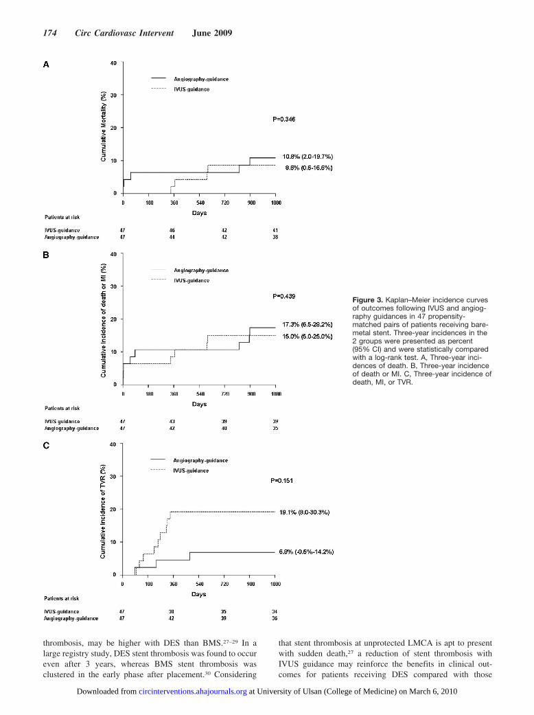
thrombosis, may be higher with DES than BMS.27–29 In alarge registry study, DES stent thrombosis was found to occureven after 3 years, whereas BMS stent thrombosis wasclustered in the early phase after placement.30 Considering
that stent thrombosis at unprotected LMCA is apt to presentwith sudden death,27 a reduction of stent thrombosis withIVUS guidance may reinforce the benefits in clinical out-comes for patients receiving DES compared with those
Figure 3. Kaplan–Meier incidence curvesof outcomes following IVUS and angiog-raphy guidances in 47 propensity-matched pairs of patients receiving bare-metal stent. Three-year incidences in the2 groups were presented as percent(95% CI) and were statistically comparedwith a log-rank test. A, Three-year inci-dences of death. B, Three-year incidenceof death or MI. C, Three-year incidence ofdeath, MI, or TVR.
174 Circ Cardiovasc Intervent June 2009
at University of Ulsan (College of Medicine) on March 6, 2010 circinterventions.ahajournals.orgDownloaded from

receiving BMS. A temporal pattern of survival difference inthe DES group supports our hypothesis, in that the survivalcurves between IVUS and angiography guidance started toseparate and progressively diverged after 1 year, when verylate stent thrombosis might occur. These findings, togetherwith those of previous studies, indicate that use of IVUS mayimprove long-term survival by reducing the risk of stentthrombosis in DES treatment.
Several previous studies proposed a possibility that IVUSguidance during PCI may reduce stent thrombosis of DES.Compared with angiography, IVUS has a unique ability toassess suboptimal results of LMCA stenting, which may beassociated with the occurrence of stent thrombosis. IVUSevaluations of stent underexpansion, incomplete lesion cov-erage, small stent area, large residual plaque, and inappositionhave been found to predict stent thrombosis after DESplacement.31–35 Alternatively, the appropriate selection ofstenting strategy by IVUS guidance may play a role inimproving outcomes. Systemic use of a 2-stent strategy,compared with a single-stent strategy, may increase the riskof stent thrombosis as well as repeat revascularization inbifurcation LMCA lesions.7,9,35–37 A better insight into plaqueconfiguration with IVUS can diminish the unnecessary use of2-stent procedures by distinguishing true stenosis versuspseudostenosis caused by various artifacts, including thedevice, coronary spasm, or calcification at the side branch.17
However, the superior benefit of IVUS guidance in DEStreatment was not consistently observed in the other Coxmodel using propensity score as a covariate for all patients.Therefore, further researches with a careful follow-up protocolshould be performed to provide more confirmative information.
Our finding, regarding the influence of IVUS on repeatrevascularization rate, conflicts with those of previous studiesshowing the benefit of IVUS guidance in reducing restenosisof BMS.38,39 We found that IVUS guidance did not reduce theincidence of repeat revascularization following either BMS orDES treatments. Although the mechanism is not clear, it maybe partly due to the low incidence of repeat revascularizationobserved in our study. We found that the 3-year cumulativerate of TVR after DES implantation was within a single digit,ranging from 7.1% to 9.1% with angiography- or IVUS-guidances in the matched population. Alternatively, an inher-ent limitation of a nonrandomized study design may havecontributed to the outcomes. Because the use of IVUS orangiography was at the discretion of the operator, IVUSguidance might be selected for lesions with more complexcoronary anatomy, in which ultrasound examination seemedto be necessary. Moreover, this study did not have anyprespecified target of optimal stenting for IVUS guidance.Thus, sufficient luminal gain enough to reduce restenosis maynot have been achieved with IVUS guidance compared withangiography guidance.
Study LimitationsOur study had several limitations, including its use of anonrandomized registry. Therefore, despite rigorous statisti-cal adjustment, unmeasured confounders may have influ-enced the outcomes. Although patients presenting with car-diogenic shock or acute MI were retrieved for fair
comparison, IVUS-guided stenting may be preferred forpatients in stable hemodynamic condition. In addition, com-parisons in the propensity-matched subgroups of DES andBMS patients might be seriously impaired by underpoweredstudy population and low incidence of events to clearly detectthe differential risk of death, MI, or revascularization. In fact,none of the 15 Cox models reported in Table 3 produced aP�0.05. Second, using a significance threshold of 0.05 maylead to high type I error rate among multiple comparisons.Third, participating centers were high-volume tertiary insti-tutions and adopted IVUS as a routine ancillary practice inpatients undergoing LMCA stenting. Therefore, the outcomesobserved in this study may not be applicable to institutionswith a restricted indication for the use of IVUS. In fact,studies in such centers may underestimate the role of IVUS.Fourth, this study may be underpowered to compare theeffectiveness of IVUS versus angiography after propensity-score matching. Finally, quantitative IVUS or angiographicassessment was not performed. Therefore, the relationshipbetween the quantitative results of imaging parameters andclinical outcomes could not be assessed. Given the aforemen-tioned limitations, our study is truly exploratory to providethe clinical insight and warrants future randomized studieshaving enough sample size and prespecified protocol toassess the efficacy of IVUS-guided PCI in DES placement forLMCA lesions.
ConclusionsUsing a large registry, we found that long-term mortality afterunprotected LMCA stenting was reduced by IVUS guidanceas compared with conventional angiography guidance. Thisresult indicates that the routine use of IVUS is generallyrecommended while performing elective PCI for unprotectedLMCA stenosis.
AcknowledgmentsWe thank the staff members of the MAIN-COMPARE registry, the othermembers of the cardiac catheterization laboratories and the cardiacsurgery departments of the participating centers, and the studycoordinators for their efforts to collect the clinical data that made thisstudy possible and to ensure the accuracy and completeness of theregistry data.
Sources of FundingAll authors received grant support from the Korean Society ofInterventional Cardiology, Health 21 R&D Project, Ministry ofHealth & Welfare, Korea (0412-CR02-0704-0001), and the Cardio-vascular Research Foundation, as part of participation in the MAIN-COMPARE Registry, from which the study data were derived.
DisclosuresNone.
References1. Park SJ, Mintz GS. Left Main Stem Disease. 1st ed. Seoul, Korea: Informa
Healthcare; 2006.2. Biondi-Zoccai GGL, Lotrionte M, Moretti C, Meliga E, Agostoni P,
Valgimigli M, Migliorini A, Antoniucci D, Carrie D, Sangiorgi G,Chieffo A, Colombo A, Price MJ, Teirstein PS, Christiansen EH, AbbateA, Testa L, Gunn JPG, Burzotta F, Laudito A, Trevi GP, Sheiban I. Acollaborative systematic review and meta-analysis on 1278 patientsundergoing percutaneous drug-eluting stenting for unprotected left maincoronary artery disease. Am Heart J. 2008;155:274–283.
Park et al Intravascular Ultrasound in Left Main Stenting 175
at University of Ulsan (College of Medicine) on March 6, 2010 circinterventions.ahajournals.orgDownloaded from
3. Park SJ, Kim YH, Lee BK, Lee SW, Lee CW, Hong MK, Kim JJ, MintzGS, Park SW. Sirolimus-eluting stent implantation for unprotected leftmain coronary artery stenosis: comparison with bare metal stent implan-tation. J Am Coll Cardiol. 2005;45:351–356.
4. Chieffo A, Stankovic G, Bonizzoni E, Tsagalou E, Iakovou I, MontorfanoM, Airoldi F, Michev I, Sangiorgi MG, Carlino M, Vitrella G, ColomboA. Early and mid-term results of drug-eluting stent implantation in unpro-tected left main. Circulation. 2005;111:791–795.
5. Valgimigli M, van Mieghem CA, Ong AT, Aoki J, Granillo GA,McFadden EP, Kappetein AP, de Feyter PJ, Smits PC, Regar E, Van derGiessen WJ, Sianos G, de Jaegere P, Van Domburg RT, Serruys PW.Short- and long-term clinical outcome after drug-eluting stent implan-tation for the percutaneous treatment of left main coronary artery disease:insights from the Rapamycin-Eluting and Taxus Stent Evaluated at Rot-terdam Cardiology Hospital registries (RESEARCH and T-SEARCH).Circulation. 2005;111:1383–1389.
6. Migliorini A, Moschi G, Giurlani L, Valenti R, Vergara R, Parodi G,Carrabba N, Dovellini EV, Antoniucci D. Drug-eluting stent supportedpercutaneous coronary intervention for unprotected left main disease.Catheter Cardiovasc Interv. 2006;68:225–230.
7. Lee MS, Kapoor N, Jamal F, Czer L, Aragon J, Forrester J, Kar S, DohadS, Kass R, Eigler N, Trento A, Shah PK, Makkar RR. Comparison ofcoronary artery bypass surgery with percutaneous coronary interventionwith drug-eluting stents for unprotected left main coronary artery disease.J Am Coll Cardiol. 2006;47:864–870.
8. Kim YH, Park SW, Hong MK, Park DW, Park KM, Lee BK, Song JM,Han KH, Lee CW, Kang DH, Song JK, Kim JJ, Park SJ. Comparison ofsimple and complex stenting techniques in the treatment of unprotectedleft main coronary artery bifurcation stenosis. Am J Cardiol. 2006;97:1597–1601.
9. Palmerini T, Marzocchi A, Marrozzini C, Ortolani P, Saia F, Savini C,Bacchi-Reggiani L, Gianstefani S, Virzi S, Manara F. Comparisonbetween coronary angioplasty and coronary artery bypass surgery for thetreatment of unprotected left main coronary artery stenosis (the BolognaRegistry). Am J Cardiol. 2006;98:54–59.
10. Price MJ, Cristea E, Sawhney N, Kao JA, Moses JW, Leon MB, CostaRA, Lansky AJ, Teirstein PS. Serial angiographic follow-up of sirolimus-eluting stents for unprotected left main coronary artery revascularization.J Am Coll Cardiol. 2006;47:871–877.
11. Sheiban I, Meliga E, Moretti C, Biondi-Zoccai GGL, Rosano G, Sciuto F,Marra WG, Omede P, Gerasimou A, Trevi GP. Long-term clinical andangiographic outcomes of treatment of unprotected left main coronaryartery stenosis with sirolimus-eluting stents. Am J Cardiol. 2007;100:431–435.
12. Sanmartin M, Baz JA, Claro R, Asorey V, Duran D, Pradas G, Iniguez A.Comparison of drug-eluting stents versus surgery for unprotected leftmain coronary artery disease. Am J Cardiol. 2007;100:970–973.
13. Chieffo A, Park SJ, Valgimigli M, et al. Favorable long-term outcomeafter drug-eluting stent implantation in nonbifurcation lesions that involveunprotected left main coronary artery. A multicenter registry. Circulation.2007;116:158–162.
14. Buszman PE, Kiesz SR, Bochenek A, Peszek-Przybyla E, Szkrobka I,Debinski M, Bialkowska B, Dudek D, Gruszka A, Zurakowski A,Milewski K, Wilczynski M, Rzeszutko L, Buszman P, Szymszal J, MartinJL, Tendera M. Acute and late outcomes of unprotected left main stentingin comparison with surgical revascularization. J Am Coll Cardiol. 2008;51:538–545.
15. Kim YH, Dangas GD, Solinas E, Aoki J, Parise H, Kimura M,Franklin-Bond T, Dasgupta NK, Kirtane AJ, Moussa I, Lansky AJ,Collins M, Stone GW, Leon MB, Moses JW, Mehran R. Effectiveness ofdrug-eluting stent implantation for patients with unprotected left maincoronary artery stenosis. Am J Cardiol. 2008;101:801–806.
16. Seung KB, Park DW, Kim YH, Lee SW, Lee CW, Hong MK, Park SW,Yun SC, Gwon HC, Jeong MH, Jang Y, Kim HS, Kim PJ, Seong IW,Park HS, Ahn T, Chae IH, Tahk SJ, Chung WS, Park SJ. Stents versuscoronary-artery bypass grafting for left main coronary artery disease.N Engl J Med. 2008;358:1781–1792.
17. Nissen SE, Yock P. Intravascular ultrasound: novel pathophysiologicalinsights and current clinical applications. Circulation. 2001;103:604–616.
18. Mintz GS. Features and parameters of drug-eluting stent deployment discov-erable by intravascular ultrasound. Am J Cardiol. 2007;100(supplement 2):S26–S35.
19. Nicholls SJ, Tuzcu EM, Sipahi I, Schoenhagen P, Nissen SE. Intra-vascular ultrasound in cardiovascular medicine. Circulation. 2006;114:e55–e59.
20. Sano K, Mintz GS, Carlier SG, de Ribamar Costa J Jr, Qian J, Missel E,Shan S, Franklin-Bond T, Boland P, Weisz G, Moussa I, Dangas GD,Mehran R, Lansky AJ, Kreps EM, Collins MB, Stone GW, Leon MB,Moses JW. Assessing intermediate left main coronary lesions using intra-vascular ultrasound. Am Heart J. 2007;154:983–988.
21. Nashef SA, Roques F, Michel P, Gauducheau E, Lemeshow S, SalamonR. European system for cardiac operative risk evaluation (EuroSCORE).Eur J Cardiothorac Surg. 1999;16:9–13.
22. Rosenbaum PR, Rubin DB. The central role of the propensity score inobservational studies for causal effects. Biometrika. 1983;70:41–55.
23. D’Agostino RB Jr. Propensity scores in cardiovascular research. Circulation.2007;115:2340–2343.
24. Therneau TM, Grambsch PM. Modeling Survival Data: Extending TheCox Model. New York: Springer-Verlag; 2000.
25. Agostoni P, Valgimigli M, Van Mieghem CAG, Rodriguez-Granillo GA,Aoki J, Ong ATL, Tsuchida K, McFadden EP, Ligthart JM, Smits PC, deJaegere P, Sianos G, Van der Giessen WJ, De Feyter P, Serruys PW.Comparison of early outcome of percutaneous coronary intervention forunprotected left main coronary artery disease in the drug-eluting stent erawith versus without intravascular ultrasonic guidance. Am J Cardiol.2005;95:644–647.
26. Camenzind E, Steg PG, Wijns W. Stent thrombosis late after implantationof first-generation drug-eluting stents: a cause for concern. Circulation.2007;115:1440–1455.
27. Pfisterer M, Brunner-La Rocca HP, Buser PT, Rickenbacher P, HunzikerP, Mueller C, Jeger R, Bader F, Osswald S, Kaiser C. Late clinical eventsafter clopidogrel discontinuation may limit the benefit of drug-elutingstents: an observational study of drug-eluting versus bare-metal stents.J Am Coll Cardiol. 2006;48:2584–2591.
28. Daemen J, Wenaweser P, Tsuchida K, Abrecht L, Vaina S, Morger C,Kukreja N, Juni P, Sianos G, Hellige G, van Domburg RT, Hess OM,Boersma E, Meier B, Windecker S, Serruys PW. Early and late coronarystent thrombosis of sirolimus-eluting and paclitaxel-eluting stents inroutine clinical practice: data from a large two-institutional cohort study.Lancet. 2007;369:667–678.
29. Stone GW, Moses JW, Ellis SG, Schofer J, Dawkins KD, Morice MC,Colombo A, Schampaert E, Grube E, Kirtane AJ, Cutlip DE, Fahy M,Pocock SJ, Mehran R, Leon MB. Safety and efficacy of sirolimus- andpaclitaxel-eluting coronary stents. N Engl J Med. 2007;356:998–1008.
30. Stettler C, Wandel S, Allemann S, Kastrati A, Morice MC, Schomig A,Pfisterer ME, Stone GW, Leon MB, de Lezo JS, Goy JJ, Park SJ, SabateM, Suttorp MJ, Kelbaek H, Spaulding C, Menichelli M, Vermeersch P,Dirksen MT, Cervinka P, Petronio AS, Nordmann AJ, Diem P, Meier B,Zwahlen M, Reichenbach S, Trelle S, Windecker S, Juni P. Outcomesassociated with drug-eluting and bare-metal stents: a collaborativenetwork meta-analysis. Lancet. 2007;370:937–948.
31. Okabe T, Mintz GS, Buch AN, Roy P, Hong YJ, Smith KA, Torguson R,Gevorkian N, Xue Z, Satler LF, Kent KM, Pichard AD, Weissman NJ,Waksman R. Intravascular ultrasound parameters associated with stentthrombosis after drug-eluting stent deployment. Am J Cardiol. 2007;100:615–620.
32. Cook S, Wenaweser P, Togni M, Billinger M, Morger C, Seiler C, VogelR, Hess O, Meier B, Windecker S. Incomplete stent apposition and verylate stent thrombosis after drug-eluting stent implantation. Circulation.2007;115:2426–2434.
33. Sonoda S, Morino Y, Ako J, Terashima M, Hassan AHM, Bonneau HN,Leon MB, Moses JW, Yock PG, Honda Y, Kuntz RE, Fitzgerald PJ.Impact of final stent dimensions on long-term results following sirolimus-eluting stent implantation: serial intravascular ultrasound analysis fromthe sirius trial. J Am Coll Cardiol. 2004;43:1959–1963.
34. Fujii K, Carlier SG, Mintz GS, Yang Y-m, Moussa I, Weisz G, DangasG, Mehran R, Lansky AJ, Kreps EM, Collins M, Stone GW, MosesJW, Leon MB. Stent underexpansion and residual reference segmentstenosis are related to stent thrombosis after sirolimus-eluting stentimplantation: an intravascular ultrasound study. J Am Coll Cardiol.2005;45:995–998.
35. Costa RA, Mintz GS, Carlier SG, Lansky AJ, Moussa I, Fujii K, Take-bayashi H, Yasuda T, Costa JR Jr, Tsuchiya Y, Jensen LO, Cristea E,Mehran R, Dangas GD, Iyer S, Collins M, Kreps EM, Colombo A, StoneGW, Leon MB, Moses JW. Bifurcation coronary lesions treated with thecrush technique: an intravascular ultrasound analysis. J Am Coll Cardiol.2005;46:599–605.
36. Iakovou I, Schmidt T, Bonizzoni E, Ge L, Sangiorgi GM, Stankovic G,Airoldi F, Chieffo A, Montorfano M, Carlino M, Michev I, Corvaja N,
176 Circ Cardiovasc Intervent June 2009
at University of Ulsan (College of Medicine) on March 6, 2010 circinterventions.ahajournals.orgDownloaded from

Briguori C, Gerckens U, Grube E, Colombo A. Incidence, predictors, andoutcome of thrombosis after successful implantation of drug-elutingstents. J Am Med Assoc. 2005;293:2126–2130.
37. Alfonso F, Suarez A, Perez-Vizcayno MJ, Moreno R, Escaned J,Banuelos C, Jimenez P, Bernardo E, Angiolillo DJ, Hernandez R, MacayaC. Intravascular ultrasound findings during episodes of drug-eluting stentthrombosis. J Am Coll Cardiol. 2007;50:2095–2097.
38. Fitzgerald PJ, Oshima A, Hayase M, Metz JA, Bailey SR, Baim DS,Cleman MW, Deutsch E, Diver DJ, Leon MB, Moses JW, Oesterle SN,
Overlie PA, Pepine CJ, Safian RD, Shani J, Simonton CA, Smalling RW,Teirstein PS, Zidar JP, Yeung AC, Kuntz RE, Yock PG. Final results ofthe Can Routine Ultrasound Influence Stent Expansion (CRUISE) study.Circulation. 2000;102:523–530.
39. Mudra H, di Mario C, de Jaegere P, Figulla HR, Macaya C, Zahn R,Wennerblom B, Rutsch W, Voudris V, Regar E, Henneke KH,Schachinger V, Zeiher A. Randomized comparison of coronary stentimplantation under ultrasound or angiographic guidance to reduce stentrestenosis (OPTICUS Study). Circulation. 2001;104:1343–1349.
Park et al Intravascular Ultrasound in Left Main Stenting 177
at University of Ulsan (College of Medicine) on March 6, 2010 circinterventions.ahajournals.orgDownloaded from
![Ultrasound guidance versus anatomical landmarks for ...€¦ · [Intervention Review] Ultrasound guidance versus anatomical landmarks for internal jugular vein catheterization Patrick](https://img.pdfslide.net/doc/110x75/5f9beef95154c7333f47d212/ultrasound-guidance-versus-anatomical-landmarks-for-intervention-review-ultrasound.jpg)