Embed Size (px)
Citation preview

IOSR Journal Of Pharmacywww.iosrphr.org
(e)-ISSN: 2250-3013, (p)-ISSN: 2319-4219
Volume 7, Issue 11 Version. 1 (November 2017), PP. 30-42
30
Impact of Quinalphos on Neurosecretory Cells of Fresh Water
Field Crab, Spiralothelphusa hydrodroma
S. Pandiammal*, J. Manju Bashini, P. Senthilkumaar
P.G. and Research Department of Zoology, Sir Theagaraya College, Chennai, India
Corresponding Author: S. Pandiammal
Abstract: The extensive use of pesticides to control agricultural pests poses a serious threat to organisms of the
aquatic environment. Chemicals entering the aquatic ecosystem through human activities, either accidentally or
by design may cause adverse effects on the aquatic biota, including deleterious changes which disrupt metabolic
activity at the biochemical levels. In the present study the impact of pesticide quinalphos on neurosecretory cells
(brain, thoracic ganglion and eye stalk) of Spiralothelphusa hydrodroma was determined. Histological
alterations and biochemical changes such as succinate dehydrogenase (SDH), lactate dehydrogenase (LDH),
acid phosphatase (ACP) and alkaline phosphatase (ALP), activities in neurosecretory cells had been carried
out. Overall work concluded that histological biomarkers provide reliable data to discriminate the usage of
pesticides which had direct influence on loss of aquatic animals.
Keywords: Quinalphos, neurosecretory cells, Spiralothelphusa hydrodroma.
----------------------------------------------------------------------------------------------------------------------------- ----------
Date of Submission: 10-11-2017 Date of acceptance: 23-11-2017
----------------------------------------------------------------------------------------------------------------------------- ----------
I. Introduction Pesticides have different modes of action to aquatic inhabitants. As a result release of pollutants from
industrial areas, and agricultural runoff into the environment severely mixes into water bodies (Tyagi, 2000).
Histological changes not only give an early indication of pollution hazard, but also provide useful data on nature
and degree of damage to cells and tissues (Shaikh et al., 2010). Environmental pollution found to be undesirable
side effect of industrialization and an important aspect of environmental degradation (Jothinarendiran, 2012).
Histological studies have a way for understanding the pathological conditions of the animal by helping in
diagnosing the abnormalities or damages of the tissues exposed to toxic stress of heavy metals (Sprague, 1971;
Andhale et al., 2011 and Maryam, et al., 2013). Aquatic ecosystems are more sensitive to the release of
industrial wastewater (Pálaez-Cid et al., 2013). Stress exerted by exposure of freshwater crabs to pesticide
drained into waterbodies had altered activity of enzyme constituents, which indicated significant influence of
toxic nature of this insecticide to crab as an important species of aquatic ecosystem (Patil et al.,2014).
Freshwater crabs are often exposed to biopesticide in their aquatic habitats through the agricultural runoff;
generally most of the pest organisms belong to the lower trophic level of the food chain in an ecosystem.
However, no attention has been paid to small invertebrates such as crabs, prawns, gastropods, bivalves, etc,
which are also used as food. Hence, further study is warranted to understand the extent of such undesirable
effects of the biopesticides on various economically and ecologically important fauna of the aquatic ecosystem
(Mintu Deyashi et al., 2016). Thus, it is important that toxic effects be determined and interpreted in
biochemical terms (Sneha Verma and Anurag Rawat et al., 2017).
II. Materials and methods The freshwater field crab, Spiralothelphusa hydrodroma was collected from Neithavoyal village,
Thiruvallur District, Tamil Nadu. The freshwater field crab, Spiralothelphusa hydrodroma was chosen for the
present study because of its presence in the rice fields in the study area. The crabs were collected from the rice
fields in early morning hours or late evening hours by hand picking and stored in plastic containers and brought
alive to the laboratory. The crabs were immediately transferred into experimental containers. Quinalphos is
an organothiophosphate chemical chiefly used as a pesticide. Ranked 'moderately hazardous' in World Health
Organization's (WHO) acute hazard ranking, use of quinalphos is either banned or restricted in most nations.
Quinalphos, which is classified as a yellow label (highly toxic) pesticide in India, is widely used in the
following crops: wheat, rice, coffee, sugarcane, and cotton.
The acute toxicity tests were conducted in duplicates using 5L experimental containers. The duration of
the test was 96h and during the study the experimental crabs were fed. A minimum of 1L water was added for
10 crabs, so that the crabs were half immersed. The experiment was carried out for finding the range of
concentrations for confirmatory evaluation. The mortality was recorded for Spiralothelphusa hydrodroma at 24,

Impact of Quinalphos on Neurosecretory Cells of Fresh Water Field....
31
48 72 and 96h exposure to pesticides were corrected for natural response by Abbott’s formula (Abbott, 1925).
The LC50 values were obtained by probit regression line, taking test concentration and corresponding percent
mortalities on log value and probit scales respectively. Straight line (regression line) was drawn between the
points which represent the survival percentage verses concentration (APHA, 1989). Sublethal studies are helpful
to assess the response of the test organism to stress caused by pesticides. Chronic time course study on the
effects of pesticide on Spiralothelphusa hydrodroma were conducted by exposing to sublethal safe
concentrations for 24 hours. At the end of the treatment period the control and treated crabs were dissected and
neurosecretory cells (brain, thoracic ganglion and eye stalk) were collected for biochemical studies. The protein
content in the tissue extracts was estimated by Bradford (1976) method using Coomassie Brilliant blue (CCB).
The carbohydrate content in the extracts was estimated as per the method of Roe (1955). The lipid content was
estimated as per the method of Folch et al., (1957).
Histological and Histopathological studies
To study the effect of pesticide on the histology of the test organism, the control and experimental
crabs treated with Quinalphos were dissected at the end of the experimental period (24 hours) and the
neurosecretory cells viz., brain, thoracic ganglion and eye stalk were fixed in Bouin’s fluid, processed and
embedded in paraffin wax. Section of 4-6 m thickness were cut and stained in hematoxylin and eosin. The
neurosecretory cells were stained in chrome-alum-hematoxylin phloxine (CHP) and haematoxylin and eosin.
The slides were observed under the light microscope for histological details and subsequently photomicrographs
were taken using a Nikon micro photographic unit. The slides were observed under the light microscope and
photomicrographs were taken using a Nikon micro photographic unit (Maharajan et al., 2015).
Biochemical analysis
The effect of pesticides on mitochondrial enzymes such as LDH, SDH was analyzed by following King
(1965) and Nachlas et al. (1960) Protocols. Acid and alkaline phosphatases were assayed following the
procedure adopted by Tenniswood et al. (1976).
Statistical Analysis The data collected was statistical analyzed using SPSS software (Version 15.0). Regression and
Analysis of variance (ANOVA) were used to determine the significance of difference among the pesticides. The
data was entered in 15.0 SPSS software for statistical analysis.
III. RESULTS In the present investigation, an attempt was made to identify the staining reactions of the cytoplasmic
contents of the neurosecretory cells found in the brain, thoracic ganglion and eye stalk between the control and
the experimental groups.
Median lethal concentration (LC50) of Quinalphos:
Median lethal concentration (LC50) of Quinalphos for S. hydrodroma was observed for 96 hrs. The
logarithm of 50% lethal concentration was obtained by finding the value on the abscissa for straight line which
assumes the probit value 5. The concentrations resulting in 50% mortality and slope of the probit line were
calculated for specific period of exposure as described by Finney (1971). The percent mortality data were
subjected to probit analysis and plotted against log of dose concentrations resulting in a straight line. The values
of LC50, upper and lower confidence limits, slope function, correlations co-efficient square and regression
results of Quinalphos on S. hydrodroma were given (Table: 1). The LC50 values for 24, 48, 72 and 96 h of
exposure periods were estimated at 2.015, 1.672, 1.372 and 1.305 ppm respectively (Graph: 1).
Table: 1 The LC50 values and regression equations for S. hydrodroma treated with Quinalphos
Exposure periods
(hours)
LC50
(ppm)
Upper confidence limits
(UCL) (ppm)
Lower
confidence limits
(LCL) (ppm)
Regression results
Slope
function
(SF)
r2
24 2.015 2.451 1.728 Y=-0.932X + 0.468 2.971 0.99
48 1.672 1.627 1.335 Y=-0.658X + 0.281 3.263 0.98
72 1.372 1.772 1.126 Y=-0.724X + 0.391 4.120 0.99
96 1.305 1.753 1.117 Y=-0.611X + 0.324 4.963 0.99

Impact of Quinalphos on Neurosecretory Cells of Fresh Water Field....
32
24 hour 48 hour
72 hour 96 hour
Graph: 1 LC50 values of Quinalphos in Spiralothelphusa hydrodroma
Impact of Quinalphos on Neurosecretory Cells of Fresh Water Field....
33
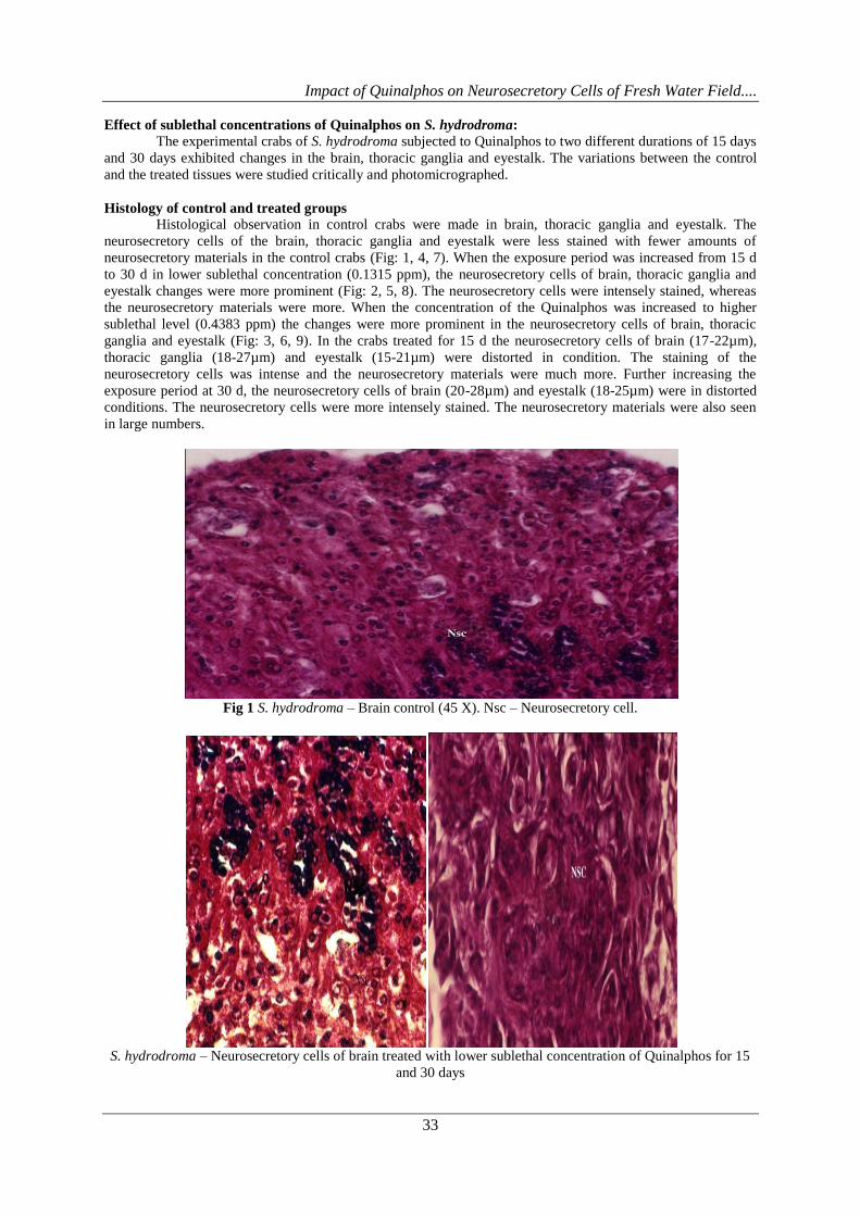
Effect of sublethal concentrations of Quinalphos on S. hydrodroma:
The experimental crabs of S. hydrodroma subjected to Quinalphos to two different durations of 15 days
and 30 days exhibited changes in the brain, thoracic ganglia and eyestalk. The variations between the control
and the treated tissues were studied critically and photomicrographed.
Histology of control and treated groups
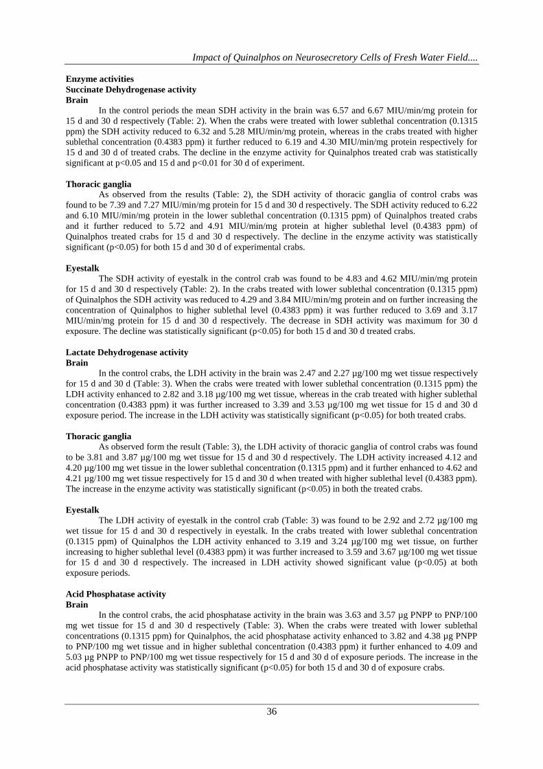
Histological observation in control crabs were made in brain, thoracic ganglia and eyestalk. The
neurosecretory cells of the brain, thoracic ganglia and eyestalk were less stained with fewer amounts of
neurosecretory materials in the control crabs (Fig: 1, 4, 7). When the exposure period was increased from 15 d
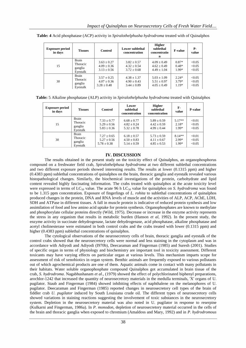
to 30 d in lower sublethal concentration (0.1315 ppm), the neurosecretory cells of brain, thoracic ganglia and
eyestalk changes were more prominent (Fig: 2, 5, 8). The neurosecretory cells were intensely stained, whereas
the neurosecretory materials were more. When the concentration of the Quinalphos was increased to higher
sublethal level (0.4383 ppm) the changes were more prominent in the neurosecretory cells of brain, thoracic
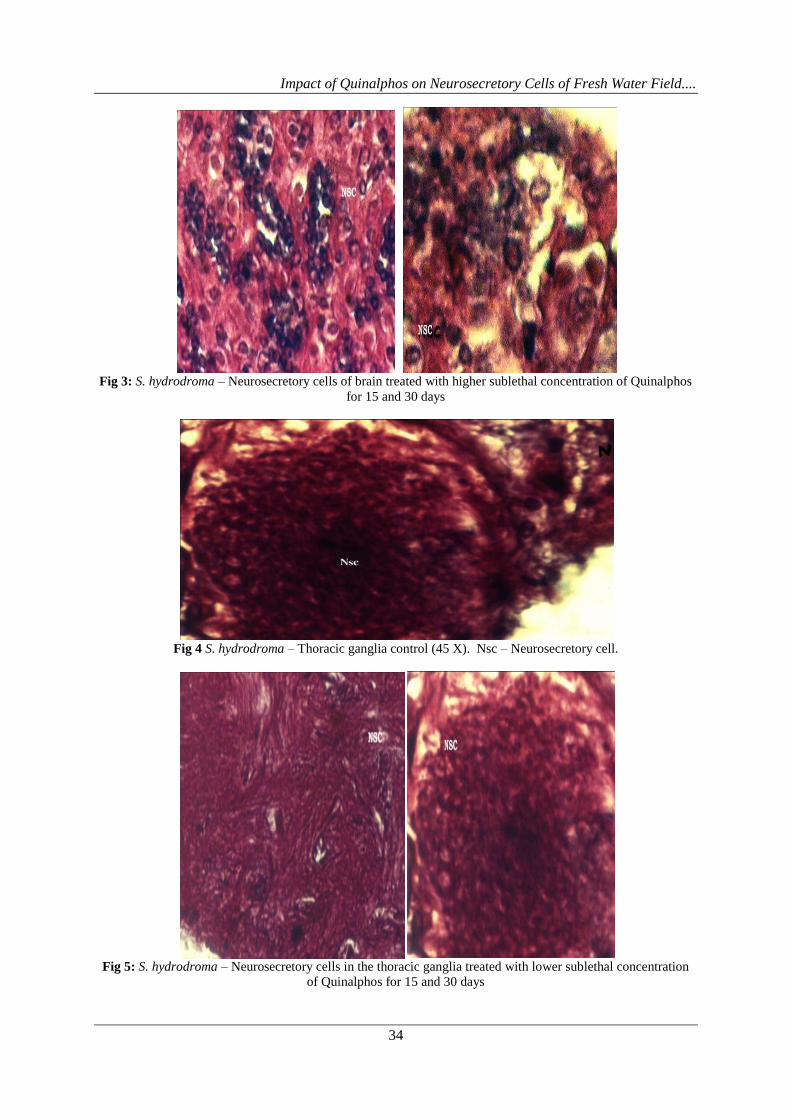
ganglia and eyestalk (Fig: 3, 6, 9). In the crabs treated for 15 d the neurosecretory cells of brain (17-22µm),
thoracic ganglia (18-27µm) and eyestalk (15-21µm) were distorted in condition. The staining of the
neurosecretory cells was intense and the neurosecretory materials were much more. Further increasing the
exposure period at 30 d, the neurosecretory cells of brain (20-28µm) and eyestalk (18-25µm) were in distorted
conditions. The neurosecretory cells were more intensely stained. The neurosecretory materials were also seen
in large numbers.
Fig 1 S. hydrodroma – Brain control (45 X). Nsc – Neurosecretory cell.
S. hydrodroma – Neurosecretory cells of brain treated with lower sublethal concentration of Quinalphos for 15
and 30 days
Impact of Quinalphos on Neurosecretory Cells of Fresh Water Field....
34
Fig 3: S. hydrodroma – Neurosecretory cells of brain treated with higher sublethal concentration of Quinalphos
for 15 and 30 days
Fig 4 S. hydrodroma – Thoracic ganglia control (45 X). Nsc – Neurosecretory cell.
Fig 5: S. hydrodroma – Neurosecretory cells in the thoracic ganglia treated with lower sublethal concentration
of Quinalphos for 15 and 30 days

Impact of Quinalphos on Neurosecretory Cells of Fresh Water Field....
35
Fig 6: S. hydrodroma – Neurosecretory cells in the thoracic ganglia treated with higher sublethal concentration
of Quinalphos for 15 and 30 days
Fig 7 S. hydrodroma – Eye Stalk control (45 X). Nsc – Neurosecretory cell.
Fig 8: S. hydrodroma – Neurosecretory cells in the eyestalk treated with lower sublethal concentration of
Quinalphos for 15 and 30 days
Fig 9: S. hydrodroma – Neurosecretory cells in the eyestalk treated with higher sublethal concentration of
Quinalphos for 15 and 30 days
Impact of Quinalphos on Neurosecretory Cells of Fresh Water Field....
36
Enzyme activities
Succinate Dehydrogenase activity
Brain
In the control periods the mean SDH activity in the brain was 6.57 and 6.67 MIU/min/mg protein for
15 d and 30 d respectively (Table: 2). When the crabs were treated with lower sublethal concentration (0.1315
ppm) the SDH activity reduced to 6.32 and 5.28 MIU/min/mg protein, whereas in the crabs treated with higher
sublethal concentration (0.4383 ppm) it further reduced to 6.19 and 4.30 MIU/min/mg protein respectively for
15 d and 30 d of treated crabs. The decline in the enzyme activity for Quinalphos treated crab was statistically
significant at p<0.05 and 15 d and p<0.01 for 30 d of experiment.
Thoracic ganglia
As observed from the results (Table: 2), the SDH activity of thoracic ganglia of control crabs was
found to be 7.39 and 7.27 MIU/min/mg protein for 15 d and 30 d respectively. The SDH activity reduced to 6.22
and 6.10 MIU/min/mg protein in the lower sublethal concentration (0.1315 ppm) of Quinalphos treated crabs
and it further reduced to 5.72 and 4.91 MIU/min/mg protein at higher sublethal level (0.4383 ppm) of
Quinalphos treated crabs for 15 d and 30 d respectively. The decline in the enzyme activity was statistically
significant (p<0.05) for both 15 d and 30 d of experimental crabs.
Eyestalk
The SDH activity of eyestalk in the control crab was found to be 4.83 and 4.62 MIU/min/mg protein
for 15 d and 30 d respectively (Table: 2). In the crabs treated with lower sublethal concentration (0.1315 ppm)
of Quinalphos the SDH activity was reduced to 4.29 and 3.84 MIU/min/mg protein and on further increasing the
concentration of Quinalphos to higher sublethal level (0.4383 ppm) it was further reduced to 3.69 and 3.17
MIU/min/mg protein for 15 d and 30 d respectively. The decrease in SDH activity was maximum for 30 d
exposure. The decline was statistically significant (p<0.05) for both 15 d and 30 d treated crabs.
Lactate Dehydrogenase activity
Brain
In the control crabs, the LDH activity in the brain was 2.47 and 2.27 µg/100 mg wet tissue respectively
for 15 d and 30 d (Table: 3). When the crabs were treated with lower sublethal concentration (0.1315 ppm) the
LDH activity enhanced to 2.82 and 3.18 µg/100 mg wet tissue, whereas in the crab treated with higher sublethal
concentration (0.4383 ppm) it was further increased to 3.39 and 3.53 µg/100 mg wet tissue for 15 d and 30 d
exposure period. The increase in the LDH activity was statistically significant (p<0.05) for both treated crabs.
Thoracic ganglia
As observed form the result (Table: 3), the LDH activity of thoracic ganglia of control crabs was found
to be 3.81 and 3.87 µg/100 mg wet tissue for 15 d and 30 d respectively. The LDH activity increased 4.12 and
4.20 µg/100 mg wet tissue in the lower sublethal concentration (0.1315 ppm) and it further enhanced to 4.62 and
4.21 µg/100 mg wet tissue respectively for 15 d and 30 d when treated with higher sublethal level (0.4383 ppm).
The increase in the enzyme activity was statistically significant (p<0.05) in both the treated crabs.
Eyestalk
The LDH activity of eyestalk in the control crab (Table: 3) was found to be 2.92 and 2.72 µg/100 mg
wet tissue for 15 d and 30 d respectively in eyestalk. In the crabs treated with lower sublethal concentration
(0.1315 ppm) of Quinalphos the LDH activity enhanced to 3.19 and 3.24 µg/100 mg wet tissue, on further
increasing to higher sublethal level (0.4383 ppm) it was further increased to 3.59 and 3.67 µg/100 mg wet tissue
for 15 d and 30 d respectively. The increased in LDH activity showed significant value (p<0.05) at both
exposure periods.
Acid Phosphatase activity
Brain
In the control crabs, the acid phosphatase activity in the brain was 3.63 and 3.57 µg PNPP to PNP/100
mg wet tissue for 15 d and 30 d respectively (Table: 3). When the crabs were treated with lower sublethal
concentrations (0.1315 ppm) for Quinalphos, the acid phosphatase activity enhanced to 3.82 and 4.38 µg PNPP
to PNP/100 mg wet tissue and in higher sublethal concentration (0.4383 ppm) it further enhanced to 4.09 and
5.03 µg PNPP to PNP/100 mg wet tissue respectively for 15 d and 30 d of exposure periods. The increase in the
acid phosphatase activity was statistically significant (p<0.05) for both 15 d and 30 d of exposure crabs.

Impact of Quinalphos on Neurosecretory Cells of Fresh Water Field....
37
Thoracic ganglia
As observed from the results (Table: 3), the acid phosphatase activity of thoracic ganglia of control
crabs was found to be 4.09 and 4.07 µg PNPP to PNP/100 mg wet tissue 15 d and 30 d respectively. The acid
phosphatase activity enhanced to 4.32 and 4.90 µg PNPP to PNP/100 mg wet tissue in the lower sublethal
concentration (0.1315 ppm), and in higher sublethal level (0.4383 ppm) of quinalphos it increased to 4.62 and
5.51 µg PNPP to PNP/100 mg wet tissue for 15 d and 30 d of exposure periods. The increase in the enzyme
activity was significant (p<0.05) for both 15 d and 30 d experimental crabs.
Eyestalk
The acid phosphatase activity of the control crab (Table: 3) was found to be 3.15 and 3.28 µg PNPP to
PNP/100 mg wet tissue for 15 d and 30 d respectively. In the crabs treated with lower sublethal concentrations
(0.1315 ppm) of Quinalphos, the acid phosphatase activity enhanced to 3.72 and 3.44 µg PNPP to PNP/100 mg
wet tissue. On further increasing to higher sublethal level (0.4383 ppm), it further increased to 4.49 and 4.05 µg
PNPP to PNP/100 mg wet tissue for 15 d and 30 d respectively. The increase in enzyme activity showed
significant values at p<0.05 for both exposure periods.
Alkaline Phosphatase (ALP) activity
Brain
In the control crabs, the enzyme activity in the brain was 7.33 and 7.27 µg PNPP to PNP/100 mg wet
tissue for 15 d and 30 d respectively (Table: 4). When the crabs were treated with lower sublethal concentration
(0.1315 ppm) of quinalphos, the enzyme activity reduced to 6.68 and 6.18 µg PNPP to PNP/mg wet tissue,
whereas in higher sublethal concentration (0.4383 ppm) it further reduced 5.89 and 5.73 µg PNPP to PNP/100
mg wet tissue for 15 d and 30 d exposure periods respectively. The decrease in the alkaline phosphatase activity
was statistically significant (p<0.01) in both 15 d and 30 d treated crabs.
Thoracic ganglia
As observed from the results (Table: 4), the alkaline phosphatase activity of thoracic ganglia of control
crabs was found to be 5.29 and 5.22 µg PNPP to PNP/100 mg wet tissue 15 d and 30 d respectively. The
enzyme activity reduced to 4.82 and 4.50 µg PNPP to PNP/100 mg wet tissue in the lower sublethal
concentration of Quinalphos (0.1315 ppm) and the enzyme activity further reduced in higher sublethal level
(0.4383 ppm) to 4.42 and 4.11 µg PNPP to PNP/100 mg wet tissue respectively for 15 to 30 d. The decline in
the enzyme activity was statistically significant (p<0.05) for both 15 d and 30 d in Quinalphos treated crabs.
Eyestalk
The eyestalk of control crab was tested for alkaline phosphatase activity (Table: 4) was found as 5.83
and 5.78 µg PNPP to PNP/100 mg wet tissue for 15 d and 30 d respectively. In the crabs treated with lower
sublethal concentration (0.1315 ppm) of Quinalphos the enzyme activity reduced to 5.32 and 5.14 µg PNPP to
PNP/100 mg wet tissue. On further increasing the concentration of Quinalphos to higher sublethal level (0.4383
ppm) it was further reduced to 4.99 and 4.85 µg PNPP to PNP/100 mg wet tissue for 15 d and 30 d respectively.
The decrease in alkaline phosphatase activity was statistically significant (p<0.05) in both the treated crab.
Table: 2 Succinate dehydrogenase (SDH) activity in Spiralothelphusha hydrodroma treated with Quinalphos
Table: 3 Lactate dehydrogenase (LDH) activity in Spiralothelphusha hydrodroma treated with Quinalphos
Impact of Quinalphos on Neurosecretory Cells of Fresh Water Field....
38
Table: 4 Acid phosphatase (ACP) activity in Spiralothelphusha hydrodroma treated with of Quinalphos
Table: 5 Alkaline phosphatase (ALP) activity in Spiralothelphusha hydrodroma treated with Quinalphos
IV. DISCUSSION The results obtained in the present study on the toxicity effect of Quinalphos, an organophosphorus
compound on a freshwater field crab, Spiralothelphusa hydrodroma at two different sublethal concentrations
and two different exposure periods showed interesting results. The results at lower (0.1315 ppm) and higher
(0.4383 ppm) sublethal concentrations of quinalphos on the brain, thoracic ganglia and eyestalk revealed various
histopathological changes. Similarly, the biochemical investigations of the protein, carbohydrate and lipid
content revealed highly fascinating information. The crabs treated with quinalphos at the acute toxicity level
were expressed in terms of LC50 value. The acute 96 h LC50 value for quinalphos on S. hydrodroma was found
to be 1.315 ppm concentration. Exposure of fingerlings of L. rohita to sublethal concentrations of quinalphos
produced changes in the protein, DNA and RNA levels of muscle and the activities of ALP, ACP, AChE, LDH,
SDH and ATPase in different tissues. A fall in muscle protein is indicative of reduced protein synthesis and low
assimilation of food and low amino acid uptake for protein synthesis. Organophosphates are known to methylate
and phosphorylate cellular proteins directly (Wild, 1975). Decrease or increase in the enzyme activity represents
the stress in any organism that results in metabolic burden (Hanson et al, 1992). In the present study, the
enzyme activity in succinate dehydrogenase, lactate dehydrogenase, acid phosphatase, alkaline phosphatase and
acetyl cholinesterase were estimated in both control crabs and the crabs treated with lower (0.1315 ppm) and
higher (0.4383 ppm) sublethal concentrations of quinalphos.
The cytological observations of the neurosecretory cells of brain, thoracic ganglia and eyestalk of the
control crabs showed that the neurosecretory cells were normal and less staining in the cytoplasm and was in
accordance with Adiyodi and Adiyodi (I970b), Deecaraman and Fingerman (1985) and Suresh (2001). Studies
of specific organ in terms of physiology and biochemistry are important tool in toxicity assessment. Different
toxicants may have varying effects on particular organ at various levels. This mechanism imparts scope for
assessment of risk of xenobiotics in organ system. Benthic animals are frequently exposed to various pollutants
out of which agrochemical products are one of them. Aquatic animals come in contact with many pollutants in
their habitats. Water soluble organophosphate compound Quinalphos got accumulated in brain tissue of the
crab, S. hydrodroma. Nagabhushanam et al., (1979) showed the effect of polychlorinated biphenyl preparations,
arochlor-1242 that increased the quantity of neurosecretory materials in the medulla terminals, 'X' organs of U.
pugilator. Staub and Fingerman (1984) showed inhibiting effects of naphthalene on the melanophores of U.
pugilator. Deecaraman and Fingerman (1985) reported changes in neurosecretory cell types of the brain of
fiddler crab U. pugilator induced by South Louisiana crude oil. The different types of neurosecretory cells
showed variations in staining reactions suggesting the involvement of toxic substances in the neurosecretory
system. Depletion in the neurosecretory material was also noted in U. pugilator in response to reserpine
(Kulkarni and Fingerman, 1986a). In P. monodon, depletion of neurosecretory material occurred in the cells of
the brain and thoracic ganglia when exposed to chromium (Amaldoss and Mary, 1992) and in P. hydrodromous
Exposure period
in days Tissues Control
Lower sublethal
concentration
Higher
sublethal
concentratio
n
F-value P-
value
15
Brain Thoracic
ganglia
Eyestalk
3.63 ± 0.27
4.09 ± 0.36 3.13 ± 0.56
3.82 ± 0.57
4.32 ± 0.54 3.72 ± 0.68
4.09 ± 0.49
4.62 ± 0.49 4.49 ± 1.04
0.87*
0.48* 1.99*
<0.05
<0.05 <0.05
30
Brain Thoracic
ganglia
Eyestalk
3.57 ± 0.25
4.07 ± 0.36 3.28 ± 0.48
4.38 ± 1.37
4.90 ± 0.43 3.44 ± 0.89
5.03 ± 1.09
5.51 ± 0.97 4.05 ± 0.49
2.24*
3.79* 1.19*
<0.05
<0.05 <0.05
Exposure period
in days Tissues Control
Lower
sublethal
concentration
Higher
sublethal
concentration
F-
value P-value
15
Brain
Thoracic
ganglia Eyestalk
7.33 ± 0.77 5.29 ± 0.56
5.83 ± 0.36
6.68 ± 0.77 4.82 ± 0.24
5.32 ± 0.78
5.89 ± 0.59 4.42 ± 0.59
4.99 ± 0.44
5.17** 2.18*
1.99*
<0.01 <0.05
<0.05
30
Brain
Thoracic ganglia
Eyestalk
7.27 ± 0.65
5.27 ± 0.56
5.78 ± 0.38
6.18 ± 0.57
4.50 ± 0.83
5.14 ± 0.59
5.73 ± 0.59
4.11 ± 0.67
4.85 ± 0.53
8.14**
2.99*
1.99*
<0.01
<0.05
<0.05
Impact of Quinalphos on Neurosecretory Cells of Fresh Water Field....
39
exposed to reserpine and chlorpromazine (Ragunathan et al., 1998). The crabs treated with lower and higher
sublethal concentrations of quinalphos showed changes in the neurosecretory cells of brain, thoracic ganglia and
eyestalk. The neurosecretory cells of treated crabs were intensely stained and the neurosecretory materials were
more in amount.
Present investigation, clearly showed decreased succinate dehydrogenase activity. Since the succinate
dehydrogenase enzyme is an important enzyme in TCA cycle, its inhibition suggests that the metabolic pathway
might have turned anaerobic to meet the increased energy demand during pollution stress. The results of the
present study are also in conformity with those of the earlier observations. Succinate dehydrogenase enzyme
plays an important role in regulating osmoregulation and any change in its activity would disrupt the
osmoregulatory mechanism (Sreenivasan et al., (2011). Similar observations were noted in the same crab in
response to (Endosulfan, Chlorpyrifos and Carbary) (Sangeetha and Deepa Rani, 2015). On the contrary lactate
dehydrogenase activity increased in the hepatopancreas in the fiddler crab, U. pugilator and decrease d in the
abdominal muscle when exposed to cadmium (Devi et al., 1993) and in S. serrata in response to cadmium
(Reddy et al., 1994). The increased lactate dehydrogenase activity in the abdominal muscle reflects anaerobic
carbohydrate metabolism when exposed to heavy metal. Reduction in the enzyme activity in fishes was
observed in response to heavy metals. Chandravathy and Reddy (1994; 1995) studied the effect of lead on
Anabas scandens and found that there was increase in the activity of lactate dehydrogenase and decrease in the
succinate dehydrogenase activity. The lactate dehydrogenase activity increased in the U. annulipes treated with
sublethal concentrations of cadmium and mercury (Suresh, 2001) and in S. hydrodroma in response to copper
and zinc (Jayakumar, 2002). The results of the present study are well in accordance with that of previous
investigations in the increased activity of lactate dehydrogenase in quinalphos treated crabs.
Decreased succinate dehydrogenase activity and increased lactate dehydrogenase activity was reported by many
workers namely in O. senex senex in response to sumithion (Reddy et al., 1983; Bhagyalakshmi et al., 1984).
Narra et al. (2012) reported alterations in enzyme activity (increased LDH and decreased SDH) in nervous
tissues of crab. Similar observations were noted in the same crab in response to (Endosulfan, Chlorpyrifos and
Carbary) (Sangeetha and Deepa Rani, 2015).
Generally, the increased activity of acid phosphatase was attributed to the activation of the enzyme
which was kept in a latent state inside the membrane of lysosomes, due to disruption of the membrane (Deduve
et al., 1955). Phosphatases play an important role in carbohydrate metabolism (Goodman and Rothstein, 1957).
Norseth (1967) reported increase in acid phosphatase activity due to accumulation of mercury in the lysosome
and blockage in the release of enzymes and carbohydrate forms the major reserve of many crustaceans
accumulated in the hepatopancreas (O'Connor and Gilbert, 1968). Bhatia et al., (1972) were of the opinion that
degradation and necrosis induced by toxicants in hepatopancreas causes release of acid phosphatase. Since
hepatopancreas was an important site of intermediary metabolism in crustaceans (Kulkarni and
Nagabhushanam, 1979) higher acid phosphatase activity was noted in hepatopancreas. Dutta et al. (1983)
concluded that both induction and inhibition of phosphatase take place depending on the concentration of
metals. Reddy et al. (1984) concluded that sensitization of cell tissues may induce proliferation of smooth
endoplasmic reticulum in hepatopancreas and resulted in increased production and liberation of acid
phosphatase. Increased acid phosphatase activity suggested glycogenolysis during metal toxicity and enhanced
breakdown of phosphatase to release energy in view of impaired ATPase system during metal stress (Reddy et
al., 1994; 1996). The acid phosphatase activity increased in the copper and zinc treated crabs as reported by
Jayakumar (2002). Increased acid phosphatase activity suggested glycogenolysis during metal toxicity and
enhanced breakdown of phosphate to release energy in view of impaired ATPase system during metal stress
(Reddy and Bhagyalakshmi, 1994). The results of Kavitha et al. (2013) reported that ACp and ALP activities
was reduced in Cypermethrin, Treated Fresh Water Female Field Crab, Spiralothelphusa hydrodroma (Herbst).
Alkaline phosphatase is a brush border enzyme that splits various phosphorus esters at an alkaline pH
and mediates membrane transport (Goldfisher et al., 1964). It is also involved in synthesis of certain enzymes
(Sumner, 1965), active transport (Denielli, 1972), protein synthesis (Pilo et al, 1972), glycogen metabolism
(Gupta and Rao, 1974) and secretory activity (Ibrahim et al., 1974). Any alteration in the activity of alkaline
phosphatase affects the organisms in a variety of ways. Bhatnagar et al. (1995) studied the effect of pyrethroid
and mortality on the fish Clarias bactrachus and found that alkaline phosphatase decreased in response to the
toxicant. Ahmed et al. (1997) studied the effect of copper on oxygen consumption and phosphatase in S. serrata
and concluded that there was decrease in alkaline phosphatase activity in muscle, hepatopancreas and
haemolymph. Similar observations were noted by Elumalai et al. (1998) in the same crab in response to
naphthalene. In the present investigation, the activity of alkaline phosphatase was found to decrease in the
experimental crabs when compared with that of the control crabs. Organophosphates inhibit acid phosphatase
and alkaline phosphatase activity in different tissues of fishes which may adversely affect nucleic acid synthesis
(Sastry and Sharma, 1981). In the present study, we noticed an increase of ACP and decrease of ALP. The
reports of Mayekar et al. (2012) showed that Exposure of Sub-Lethal Dose of Nickel produced increased Acid
Impact of Quinalphos on Neurosecretory Cells of Fresh Water Field....
40
phosphatase (ACP) and alkaline phosphatase (ALP) activities. Similar observations were noted in the same crab
in response to (Endosulfan, Chlorpyrifos and Carbary) (Sangeetha and Deepa Rani, 2015).
V. CONCLUSION
Hence, the present investigation clearly showed that the quinalphos caused damages to the tissues at
higher sublethal concentrations. There was a marked decrease in the succinate dehydrogenase, alkaline
phosphatase activities and increase in lactate dehydrogenase and acid phosphatase activities clearly indicate that
the quinalphos caused metabolic stress in the experimental crabs. High levels of accumulation of quinalphos in
the present investigation indicated that the intake was exponential in an environment where the quinalphos
routinely used as biocides and fertilizers which is highly toxic was concluded.
REFERNCES [1]. Abbot, W.S. (1925) A method of computing the effectiveness of an insecticide. J. Econ. Entomo., 18:
265-267.
[2]. Adiyodi, K.G. and R.G. Adiyodi. (1970b) Endocrine control or reproduction in decapod crustaceans.
Biol. Rev., 45: 121-165.
[3]. Ahmed, M.R., Elumalai, M., Balasubramanian, S.E. and Balasubramanian, M.P. (1997) Individual and
combined effect of copper and chromium on oxygen consumption and phosphatases of a marine edible
crab, Scylla serrata. Biochemical Letter., 55:147-152.
[4]. Amaldoss, G. and A. Mary. (1992) Chromium effects on neurosecretion of the shrimp, Penaeus
monodon. J. Environ. Biol., 13: 239-246.
[5]. Andhale, A.V., Bhosale, P.A. and Zambare, S.P. (2011) Histopathological study of nickel induced
alterations in the fresh water bivalve, Lammellidens marginalis. Journal of Experimental Sciences., 2(4):
01-03.
[6]. APHA. (1989) Standard methods for the examination of water and waste water. American Public Health
Association, Washington, D.C. 17th
Ed.
[7]. Bhagyalakshmi, A., Sreenivasulu Reddy, P. and Ramamurthi, R. (1984) Subacute stress induced by
sumithion on certain biochemical parameters in Oziotelphusa senex senex the freshwater field crab.
Toxicol. Lett., 21: 127-134.
[8]. Bhatia, S.C., S.C. Sharma and T.A. Venkatasubramanian. (1972) In vivo subacute physiological stress
induced by sumithion on the hepatopancreatic acid phosphatase activity in the freshwater crab,
Oziotelphusa senex senex. Water Air Soil Pollut., 22: 299-302.
[9]. Bhatnagar, M.C., Tyagi, M. and Tamata, S. (1995) Pyrethroid induced toxicity to phosphatases in Clarias
bactrachus (Linn). J Environ Biol., 16(1): 11-14.
[10]. Bradford, M.M. (1976) A rapid sensitive method for the quantification of microgram quantities of protein
utilizing principle of protein dye binding. Anal. Biochem., 72: 248-254.
[11]. Chandravathy, V.M. and S.L.N. Reddy. (1994) Enzymological and biochemical alterations in the
freshwater fish, Anabas scandens during lead nitrate exposure. Ecotoxicol. Environ. Monit., 4(3&4):
1631-1637.
[12]. Chandravathy, V.M. and S.L.N. Reddy. (1995). In vivo effects of lead acetate on dehydrogenase
activities and metabolites in the freshwater fish. J. Ecotoxicol. Environ. Monit., 5(2): 107-111.
[13]. Chandravathy, V.M. and Reddy, S.L.N. (1995) In vivo effects of lead acetate on dehydrogenase activities
and metabolites in the fresh water fish. Journal of Ecotoxicology and Environmental Monitoring., 5(2):
107-111.
[14]. Deduve, C., Pressman, B.C., Gianetto, R., Wattiaux, R. and Appelmans, F. (1985), Intracellular
distribution patterns of enzymes in rat-liver tissue. Biochemical Journal., 60: 604-617.
[15]. Deecaraman, M. and M. Fingerman. (1985) Changes in the neurosecretory cells of the brain of the fiddler
crab Uca pugilator induced by exposure to the water soluble fraction of South Louisiana crude oil or its
aromatic components. J. Reprod. Biol. Comp. Endocrinol., 5 (2): 89-86.
[16]. Denielli, J.F. (1972) Structural factors in cell permeability and secretion. Symp. Soc. Exp. Biol., 6: 1-15.
[17]. Devi, M., Reddy, P.S. and Fingerman, M. (1993) Effect of cadmium exposure on lactate-Dehydrogenase
activity in hepatopancreas and abdominal muscle in fiddler crab, Uca pugilator. Comp Biochem Physiol.,
106-C: 739-742.
[18]. Dutta, H.S., B. Lall. and A.Z. Haghighi. (1983) Mercuric chloride induced changes in serum protein of
bluegill, Lepomis macrochirus. Ohio. J. Sci., 83: 119-120.
[19]. Elumalai, M., Balasubramanian, S.E. and Balasubramanian, M.P. (1998) Influence of Naphthalene on
protein, carbohydrate and phosphatases system during the vitellogenesis in marine edible crab, Scylla
serrata. Bulletin of Environmental Contamination and Toxicology., 60: 25-29.
Impact of Quinalphos on Neurosecretory Cells of Fresh Water Field....
41
[20]. Folch, J., S.M. Lee and G.H. Slone-Stanley. (1957) A simple method for isolation and purification of
total lipids from animal tissues. J. Biol. Chem., 226: 497-508.
[21]. Goldfisher, S.E., Esser, E., Novikoff, A.B. (1964) Use of histological and histochemical assessment in
the prognosis of the effects of aquatic pollutants (ed.) D.E.Hinton, M.W.Kendall and B. B. Silver. Sect.
528, ASTM, Philadelphia, pp 194-208.
[22]. Goodman, J. and Rothstein, A. (1957) The active transport of phosphate into the yeast cell. J. Genet.
Physiol., 40: 915-925.
[23]. Gupta, V. and G. Rao. (1974) Histological studies on the chloride plexes of the goat embryos II.
Histological distribution of acid and alkaline phosphatase. Acta Histochem., 49: 60-63.
[24]. Hanson, J.I., Mustafa, T. and Depledge, M. (1992) Mechanism of copper toxicity in the shore crab,
Carcinus maenus: Effects Na, K-ATPase activity, haemolymph electrolyte concentrations and tissue
water contents. Marine. Biology., 114(2): 253-257.
[25]. Ibrahim, A.M., M.G. Higazi and E.S. Demian. (1974) Histochemical localization of alkaline phosphatase
activity in the alimentary tract of the snail Marisa carnvarietus (L). Zool. Soc. Egypt. Bull., 26: 94-105.
[26]. Jayakumar, S. (2002) Effects of copper and zinc toxicity on a fresh water crab, Spiralothelphusa
hydrodroma, Ph. D. Thesis, University of Madras, India.
[27]. Jothinarendiran, N. (2012) Effect of dimethoate pesticide on oxygen consumption and gill histology of
the fish, Channa punctatus. Current Biotica., 5(4): 500-507.
[28]. Kavitha, R., Deepa Rani, S., Sivagnanam, S. and Padmaja, M. (2013) Cadmium Nanoparticle Induced
Histological and Biochemical changes in Hepatopancreas of Mud Crab Scylla olivacea (Herbst, 1796).
Journal of Academia and Industrial Research (JAIR)., 2(3): 205-209.
[29]. King, J. (1965) In: Practical clinical enzymology. (Eds.), D. Van Norstand Co London.
[30]. Kulkarni, G.K. and M. Fingerman. (1986a) Effects of two tranquilizers, reserpine and chlorpromazine on
neurosecretory cells and the ovary of the fiddler crab. Uca pugilator. Gen. Pharmac., 17(6): 617-683.
[31]. Kulkarni, G.K. and R. Nagabushanam. (1979) Mobilization of organic reserves during the ovarian
development in a marine penaeid prawn, Parapenaeopsis hardwickii (Miers). Aquaculture., 18: 373-377.
[32]. Maharajan, A., Narayanasamy, Y., Ganapiriya, V. and Shanmugavel, K. (2015) Histological alterations
of a combination of Chlorpyrifos and Cypermethrin (Nurocombi) insecticide in the fresh water crab,
Paratelphusa jacquemontii (Rathbun). The Journal of Basic & Applied Zoology., 72: 104-112.
[33]. Maryam, S.M., Khalili, V., Abbasi, A. and Hedayati (2013) Sublethal effect of copper toxicity on liver
lesions of Roach (Rutilus rutilus caspicus) Juveniles. JNASCI., 2(4): 119-123.
[34]. Mayekar, V., Pathare, M., Afonso, G., Mohite, V.T., Lahir, Y.K. and Raut, P.D. (2012) Effects of sub-
lethal dose of nickel on the biochemical parameters in the tissues of female crab Scylla serrata from
Mumbai coast. Journal of Ecophysiology and Occupational Health., 12: 69-76.
[35]. Mintu Deyashi, Kamales Kumar Misra, Siddhartha Sankar Bhattachary and Suman Bhusan Chakraborty
(2016) Acute Toxicity of a Neem Seed Kernel based Biopesticide, Nimbecidine Plus on an Edible Fresh
Water Crab, Varuna litterata (Fabricius, 1798). Int. J. Adv. Res. Biol. Sci., 3(10): 122-130.
[36]. Nachlas, M.M., Margulius, S.I. and Selligman, A.M. (1960) A colorimetric method for the estimation of
SDH. The Journal of Biological Chemistry., 235: 499-503.
[37]. Nagabhushanam, R., S.W. Fingerman and M. Fingerman. (1979) Effects of the polychlorinated biphenyl
preparation, aroclor 1242, on the quantity of neurosecretory material in the medulla terminalis X-organ of
the fiddler crab, Uca pugilator. Experietntia., 35: Bikhauser Verlag., Basel (Scheweiz).
[38]. Narra, M.R., Regatte, R.R. and Kodimyala, R. Effects of chlorpyrifos on enzymes as biomarkers of
toxicity in Fresh water field crab Barytelphusa guerini. International Journal of environmental sciences.,
2(4): 2015-2023.
[39]. Norseth, T. (1967) The intracellular distribution of mercury in rat liver after single injection of mercuric
chloride. Biochem Pharmacol., 17: 581-593.
[40]. O'Connor, J.D. and Gilbert, L.I. (1968) Aspects of lipid metabolism in crustaceans. American Zoology.,
8: 529-539.
[41]. Pálaez-Cid, A.A., Velazquez-Ugalde, I., Herrera- González, A.M. and García-Serrano, J. (2013) Textile
dyes removal from aqueous solution using Opuntia ficus-indica fruit waste as adsorbant and its
characterization. Journal of Environment Management., 130(1): 90–97
[42]. Patil, V.N., Kadam, G.H. and Raut, P.D. (2014) Enzymological alterations produced by chronic
malathion exposure in freshwater crab, Paratelphusa (barytelphusa) jacquemontii. International Journal
of Science, Environment and Technology., 3(1): 303–313.
[43]. Pilo, B., M.V. Ansari. and R.V. Shah. (1972) Studies of wound healing and repair in pigeon liver III.
Histochemical studies on acid and alkaline phosphatase activities during the process. J. Anim. Morphol.
Physiol., 9: 205-212.

Impact of Quinalphos on Neurosecretory Cells of Fresh Water Field....
42
[44]. Ragunathan, M.G., Sharmila Jadhav. and M. Deecaraman. (1998) Impact of two tranquilizers, reserpine
and chlorpromazine on certain neurosecretory cells and reproductive organs of eyestalk ablated female
crab, Paratelphusa hydrodromous (Herbst). Indian J. Exp. Biol., 36: 980-984.
[45]. Reddy, P.S. and A. Bhagyalakshmi. (1994) Changes in oxidative metabolism in selected tissues of the
crab, Scylla serrata in response to cadmium toxicity. Ecotoxicol. Environ. Saf., 29(3): 255-264.
[46]. Reddy, P.S., A. Bhagyalakshmi and R. Ramamurthi. (1984) In vivo subacute physiological stress induced
by sumithion on the hepatopancreas acid phosphatase activity in the freshwater crab. Toxic. Lett., 22:
299-309.
[47]. Reddy, P.S., Bhagyalakshmi, A. and Ramamoorthy, R., (1983) Chronic sumithion toxicity effect on
carbohydrate metabolism in crab muscle. Toxicol Letters., 22: 299-309.
[48]. Reddy, P.S., M. Devi, R. Sarojini, R. Nagabhushanam. and M. Fingerman. (1994) Cadmium chloride
induced hyperglycemia in the red swamp crayfish, Procambarus clarkia: Possible role of crustacean
hyperglycemic hormone. Comp. Biochem. Physiol., 107(C): 57-61.
[49]. Reddy, P.S., R.V. Katyayani. and M. Fingerman. (1996) Cadmium and naphthalene-induced
hyperglycemia in fiddler crab, Uca pugilator. Differential modes of action on the neuroendocrine
system. Bull. Environ. Contam. Toxicol., 56: 425-431.
[50]. Roe, J.R. (1955) The determination of sugar in blood and spinal fluid with anthrone reagent. J. Biol.
Chem., 20: 335-343.
[51]. Sangeetha S. and Deepa Rani S (2015) Histological and biochemical changes in hepatopancreas of fiddler
crab, Uca triangularis exposed to endosulfan, chlorpyrifos and carbaryl pesticides. International Journal
of Multidisciplinary Research and Development., 2(12): 421-426.
[52]. Sastry, K.V. and K. Sharma. (1981) Effect of mercuric chloride on the activities of brain enzymes in
Heteropneustes fossilis. Matsya., 7: 66-69.
[53]. Shaikh, F.I., Ustad, I.R. and Ansari, N.T. (2010): Effect of heavy metal on the ovary of freshwater crab,
Barytelphusa cunicularis (Westwood). Bioscan. 5(2): 335-338.
[54]. Sneha Verma and Anurag Rawat (2017) Effect of chlorpyrifos on protein and carbohydrate content of
Heteropneustes fossilis (Bloch, 1794). International Journal of Fisheries and Aquatic Studies., 5(1): 463-
466.
[55]. Sprague, J.B. (1971): The measurement of pollutant toxicity to fish III, sub lethal effect and safe
concentration. Water Res., 5: 245-266.
[56]. Sreenivasan, R,S., Krishna Moorthy, P. and Deecaraman, M. (2011) Cypermethrin Induced Toxicity to
phosphatases and dehydrogenases in gills and hemolymph of fresh water crab, Spiralothelphusa
hydrodroma (Herbst). International Journal of Biology & Medical Research., 2(3): 784-788.
[57]. Staub, G.C. and Fingerman, M. (1984) Effect of napthalene on color changes of the sand fiddler crab,
Uca pugliator. Compo Biochem. Physiol., 77C: 7-12
[58]. Sumner, A.T. (1965) The cytology and the histochemistry of the digestive gland cells of Helis. Quart. J.
Microsc. Sci., 106: 173-192.
[59]. Suresh, V. (2001) A study on the effects of heavy metals toxicity on a brackish water crab, Uca (celuca)
lacteal annulipes (Crane, 1975) of Pulicat Lake, Tamil Nadu, Ph.D. Thesis, University of Madras, Tamil
Nadu, India.
[60]. Tenniswood, M., Bind, C.E. and Clark, A.F. (1976) Acid phosphatases androgen dependent markers of
rat prostate. Canadian Journal of Biochemistry., 54: 340-343.
[61]. Tyagi, P., Dharam, B., Choudhary, R. and Sawhney, B. L. (2000) Physico chemical quality of Ground
water in industrial areas of India. A review. Poll. Res., 19(3): 443-445.
[62]. Wild, D. (1975) Mutagenicity studies on organophosphorus insecticides. Mutat. Res., 32: 135–150.
S. Pandiammal Impact of Quinalphos on Neurosecretory Cells of Fresh Water Field Crab,
Spiralothelphusa hydrodroma.” IOSR Journal of Pharmacy (IOSRPHR), vol. 7, no. 11, 2017, pp.
30-42
IOSR Journal of Pharmacy (IOSR-PHR) is UGC approved Journal with Sl. No. 5012