Embed Size (px)
Citation preview

RESEARCH ARTICLE
Impaired Air Conditioning within the NasalCavity in Flat-Faced HomoTakeshi Nishimura1*, Futoshi Mori2, Sho Hanida3, Kiyoshi Kumahata4, Shigeru Ishikawa5,Kaouthar Samarat6, Takako Miyabe-Nishiwaki1, Misato Hayashi1, Masaki Tomonaga1,Juri Suzuki1, Tetsuro Matsuzawa1, Teruo Matsuzawa6
1 Primate Research Institute, Kyoto University, Inuyama, Aichi, Japan, 2 Institute for Biomedical Sciences,Iwate Medical University, Yahaba, Iwate, Japan, 3 Kanazawa Institute of Technology, Nonoichi, Ishikawa,Japan, 4 RIKEN Advanced Institute for Computational Science, Kobe, Hyogo, Japan, 5 KanazawaMunicipal Hospital, Kanazawa, Ishikawa, Japan, 6 Japan Advanced Institute of Science and Technology,Nomi, Ishikawa, Japan
AbstractWe are flat-faced hominins with an external nose that protrudes from the face. This feature
was derived in the genus Homo, along with facial flattening and reorientation to form a high
nasal cavity. The nasal passage conditions the inhaled air in terms of temperature and
humidity to match the conditions required in the lung, and its anatomical variation is believed
to be evolutionarily sensitive to the ambient atmospheric conditions of a given habitat. In
this study, we used computational fluid dynamics (CFD) with three-dimensional topology
models of the nasal passage under the same simulation conditions, to investigate air-condi-
tioning performance in humans, chimpanzees, and macaques. The CFD simulation showed
a horizontal straight flow of inhaled air in chimpanzees and macaques, contrasting with the
upward and curved flow in humans. The inhaled air is conditioned poorly in humans com-
pared with nonhuman primates. Virtual modifications to the human external nose topology,
in which the nasal vestibule and valve are modified to resemble those of chimpanzees,
change the airflow to be horizontal, but have little influence on the air-conditioning perfor-
mance in humans. These findings suggest that morphological variation of the nasal pas-
sage topology was only weakly sensitive to the ambient atmosphere conditions; rather, the
high nasal cavity in humans was formed simply by evolutionary facial reorganization in the
divergence of Homo from the other hominin lineages, impairing the air-conditioning perfor-
mance. Even though the inhaled air is not adjusted well within the nasal cavity in humans, it
can be fully conditioned subsequently in the pharyngeal cavity, which is lengthened in the
flat-faced Homo. Thus, the air-conditioning faculty in the nasal passages was probably
impaired in early Homomembers, although they have survived successfully under the fluc-
tuating climate of the Plio-Pleistocene, and then they moved “Out of Africa” to explore the
more severe climates of Eurasia.
PLOS Computational Biology | DOI:10.1371/journal.pcbi.1004807 March 24, 2016 1 / 18
OPEN ACCESS
Citation: Nishimura T, Mori F, Hanida S, KumahataK, Ishikawa S, Samarat K, et al. (2016) Impaired AirConditioning within the Nasal Cavity in Flat-FacedHomo. PLoS Comput Biol 12(3): e1004807.doi:10.1371/journal.pcbi.1004807
Editor: Robert L Jernigan, Iowa State University,UNITED STATES
Received: August 11, 2015
Accepted: February 11, 2016
Published: March 24, 2016
Copyright: © 2016 Nishimura et al. This is an openaccess article distributed under the terms of theCreative Commons Attribution License, which permitsunrestricted use, distribution, and reproduction in anymedium, provided the original author and source arecredited.
Data Availability Statement: All relevant data arewithin the paper and its Supporting Information files.
Funding: This work was supported by funds from theAsahi Glass Foundation, Japan (http://www.af-info.or.jp/en/index.html, to TN), by a Strategic YoungResearcher Overseas Visits Program for AcceleratingBrain Circulation (S2501, to TN, PI: Hirohisa Hirai)and Grants-in-Aid for Scientific Research (KAKENHI,24687030 to TN, 24000001 to TetM) from the JapanSociety for the Promotion of Science (http://www.jsps.go.jp/english/index.html), and by a SPIRITS programfrom the Kyoto University (to TN). The funders had no
Author Summary
This is the first investigation of nasal air conditioning in nonhuman hominoids based oncomputational fluid dynamics with digital topological models of the nasal passage madeusing medical imaging. Our comparative results of humans, chimpanzees, and macaquesshow that the inhaled air is conditioned poorly in humans compared with nonhuman pri-mates. We also show that our protruding external nose has little effect on improving airconditioning. The nasal anatomy inHomo was weakly sensitive to the ambient atmo-sphere conditions in evolution, but was formed passively by facial reorganization in thisgenus. Even though the inhaled air is not adjusted well within the nasal cavity in humans,it can be fully conditioned subsequently in the pharyngeal cavity, which is lengthened inflat-faced Homo. Thus, despite an impaired air-conditioning conformation in the nasalpassages, Homomembers must have survived successfully under the fluctuating climate ofthe Plio-Pleistocene, and then they moved “Out of Africa” in the Early Pleistocene toexplore the more severe climates and ecological environments of Eurasia.
IntroductionA flat, short face is one of the legacies of the genus Homo [1, 2]. The facial component remainsshort and fully below the expanded forehead in this genus, and this contrasts with earlier andcontemporary hominins such as the australopithecines, which possessed a long face that pro-truded away from the brain case in a manner analogous to nonhuman hominids, e.g., chimpan-zees [1–3]. Consequently, the external nose protrudes from the face [4], the nasal cavity withinthe facial cranium is high and quadrangular in a lateral view, and the vertically oriented nasalvestibule is connected close to the floor of the tall nasal cavity in humans [2, 5]. This patterncontrasts with that found in nonhuman primates, which possess a long and triangular nasalcavity, and a horizontally oriented vestibule that is connected vertically with the middle of thecavity [2, 5]. However, the subsequent pharyngeal cavity is much longer in humans than innonhuman primates [6–10]. The nasal passage, including the nasal vestibule and cavity, condi-tions the inhaled air, as well as performing other functions such as olfactory sensing, dust filter-ing, and voice resonance [11, 12]. The pharyngeal cavity also participates in conditioning theair that enters from the nasal cavity [11]. Insufficient conditioning can damage the mucosal tis-sues in the respiratory system and impair respiratory performance, thereby undermininghealth and increasing the likelihood of death [11, 12]. Thus, despite the evolutionary modifica-tions in the nasal anatomy in the phyletic divergence ofHomo from the other hominin lineages,adequate air conditioning must have been maintained, particularly to ensure their successfulsurvival in the severely fluctuating climate of the Pleistocene and their subsequent spread fromAfrica to Eurasia [1, 13, 14].
In this study, we compared the principles and performance of air conditioning in humans,chimpanzees, and macaques by using a computational fluid dynamics (CFD) model [15] to sim-ulate the airflow and heat and water exchanges over the mucosal surface in the nasal passage.The human CFDmodel used here simulates the predicted airflow and air-conditioning perfor-mance reliable for humans [15]. Three-dimensional models of the nasal passage topology wereproduced based on tomography scans of the three genera. The models include no paranasalsinuses. We compared the air-conditioning performance in the three genera using the same sim-ulation conditions: heat and water exchange were predicted with a simulation model based onthe histological compositions of the mucosal layers and the average surface temperature of thehuman nasal passage, i.e., 100% relative humidity (% RH) at 34°C (3.34% of the mass fraction of
Impaired Air Conditioning inHomo
PLOS Computational Biology | DOI:10.1371/journal.pcbi.1004807 March 24, 2016 2 / 18
role in study design, data collection and analysis,decision to publish, or preparation of the manuscript.
Competing Interests: The authors have declaredthat no competing interests exist.
water; % MF) [15]. This means that this study examines the differences in performance that arecaused by the anatomical differences of the nasal passage in the three genera, but does not simu-late a real performance in nonhuman primates. Whereas similar CFD analyses performed in thesame subjects of macaques showed a minor contribution of the maxillary sinus to air condition-ing [16], here we examine them again to compare the air-conditioning performance of the threegenera using the same simulation conditions. To evaluate the effects of the human externalnose, we also produced two virtual topology models: a “no-valve”model where the nasal valve—a narrow slit-like channel between the nasal vestibule and cavity—was removed virtually; anda “horizontal”model where the vertically oriented vestibule was made horizontal, as seen inchimpanzees.
We evaluated the performance among the three species in varied ambient atmospheric con-ditions, and discuss the evolutionary modifications in air-conditioning performance in thedivergence ofHomo from the other hominin lineages lineage, using nonhuman primates as amodel for the latter hominins.
Materials and Methods
Ethics statementThis study for animals was performed in strict accordance with the recommendations in thethird edition of the Guidelines for the Care and Use of Laboratory Primates at the PrimateResearch Institute of the Kyoto University (KUPRI), Inuyama, Japan. The protocol wasapproved by the Animal Welfare and Animal Care Committee at KUPRI (Permit Numbers:2009–075, 2010–027, 2011–067, and 2012–075). The chimpanzees were anesthetized intramus-cularly with 3.5 mg ketamine hydrochloride (Sankyo-Parke-Davis & Co., Inc.) and 0.035 mgmedetomidine hydrochloride (Meiji Seika Pharma Co., Ltd., Tokyo, Japan) per kilogram ofbody weight. The anesthesia was maintained with sevoflurane (Dainippon Sumitomo PharmaCo., Ltd., Osaka, Japan) delivered in oxygen through a precision vaporizer and a rebreathingcircuit. The macaques were anesthetized intramuscularly with 2.5 mg ketamine hydrochlorideand 0.1 mg medetomidine hydrochloride per kilogram of body weight. Every effort was madeto minimize suffering. The daily care and housing facilities strictly conformed to the recom-mendations in the third edition of the Guidelines for the Care and Use of Laboratory Primatesat the KUPRI. To ensure the animals' health and welfare, their general appearance was dailymonitored and recorded, along with their daily food and fluid intake.
This study for humans was performed in strict accordance with the recommendations inthe Declaration of Helsinki, Ethical Principle of Medical Research Involving Human Subjectsprepared by the World Medical Association. All subjects gave an informed consent. The proto-col was approved by the Human Research Ethics Committee of KUPRI (Permit Number:H2011-06).
Subjects and tomographic scanningTen non-human primates—four chimpanzees, Pan troglodytes [17], four Japanese macaques,Macaca fuscata, and two Rhesus macaques,M.mulatta—which were reared at KUPRI,Inuyama, Japan, were scanned using a computed tomography scanner (Asteion Premium 4,Toshiba Medical Systems Co., Otawara, Japan) at the KUPRI (S1 Table). The two species ofmacaques are here regarded as subjects of a same genusMacaca along with a genus Pan. All ofthe CT scans obtained in this study came from subjects without any history of surgery and hadfew abnormal traits in their heads, and few artifacts distorted the images of the nasal region.The scans were registered under PRICT # (S1 Table) and are available via the website of the
Impaired Air Conditioning inHomo
PLOS Computational Biology | DOI:10.1371/journal.pcbi.1004807 March 24, 2016 3 / 18
Digital Morphology Museum of KUPRI (dmm.pri.kyoto-u.ac.jp/archive/). The scans ofmacaques used here were also used for another CFD study [16].
Six human volunteers were scanned with a magnetic resonance imaging scanner (Magne-tom Verio, Siemens AG, Munich, Germany) at the Brain Activity Imaging Center, ATR-Pro-motions, Seika, Japan (S1 Table). Among them, the following CFD simulation analyses usedthe scans of the five subjects who had no surgery, few abnormal traits, and few artifacts distort-ing the images in the nasal region.
Analyses of computational fluid dynamicsThe CFD simulations with heat and water exchange were performed as described [15, 18]. Thephysiological model used here, developed based on the previous models [18], reflects humanrespiratory physiology and histology, including latent heat, to simulate well real performancesof the airflow, heat and water exchanges for humans [15]. The following procedures of the nasaltopological models and CFD analyses are the same as those used by Hanida et al. [15], excludingthose for generating the virtual topological models. It is slightly different from the study of Moriet al. [16] using the same macaques, in simulation model and boundary conditions.
Models of the nasal passage anatomyThe voxel data of the nasal passage anatomy were reconstructed from computed tomographyscans for nonhuman primates and magnetic resonance imaging scans for human volunteers.The black area representing the air filling the nasal passage was extracted first by using athreshold of brightness with Avizo 7 (FEI), and then the voxel data were reconstructed. Afterconverting the voxel data to STereo Lithography (STL) data, these were modified into data rep-resenting the smooth surface using Magics 9.5 (Materialize Inc., Leuven, Belgium). Finally, atetrahedron mesh with the mesh size of Δx = 2.10 to 3.65×10⁻⁴ mm, depending on the size ofthe subjects, was generated from the modified STL data using Gambit 2.4 (ANSYS Inc.,Canonsburg, PA, USA; S1 Table). The computational meshes had 2.66 to 3.70 million tetrahe-dral cells (S1 Table). The present solutions are evaluated independently to the mesh size: therewere few differences between the solutions by the present mesh size and the minimum meshsize (Δx = 2.00 to 3.30×10⁻⁴ mm, depending on the size of the subjects) in each subject; i.e., upto 1.6% of the flow velocity for the same frontal contour.
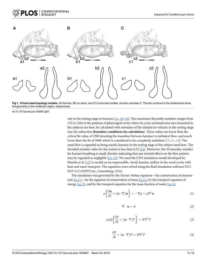
Virtual models of the nasal passage anatomyThe no-valve and horizontal nasal vestibular topology models were generated from the originalsmoothed STL data using Rhinoceros (AppliCraft Co., Ltd, Tokyo, Japan). We defined the basalplane for making the modifications as almost parallel to the narrow channel between the nasalvestibule and the nasal cavity, which approximately corresponded to the nasal valve (Fig 1A).Using the loft function in Rhinoceros, we generated a straight surface between the basal plane tothe nostril on each side, thereby removing the effects of the nasal valve (i.e., the no-valve model;Fig 1B). Next, we tilted the modified vestibule with its straight surface upward, thereby makingits lower surface horizontal relative to the floor of the nasal cavity, as seen in the vestibule ofchimpanzees (i.e., the horizontal model; Fig 1C; tilting angle: vol.1, 52 degrees; vol.2, 32 degrees;vol.3, 40 degrees; vol.5, 46 degrees, vol.6, 36 degrees).
Simulation model of inhaled airflowWe performed steady-state analyses to examine the airflow, where the turbulence model wasnot employed. A steady simulation is reasonable under a normal breathing frequency and flow
Impaired Air Conditioning inHomo
PLOS Computational Biology | DOI:10.1371/journal.pcbi.1004807 March 24, 2016 4 / 18
rate in the resting stage in humans [15, 18–24]. The maximum Reynolds numbers ranges from135 to 1264 at the position of pharyngeal cavity where its cross-sectional area was measured inthe subjects use here, by calculated with estimates of the inhaled air velocity in the resting stage(see the subsection Boundary conditions for calculation). These values are lower than thecritical Re value of 2300 denoting the transition between laminar to turbulent flow, and muchlower than the Re of 5000 which is considered to be completely turbulent [19, 21, 24]. Thenasal flow is regarded as being mostly laminar in the resting stage of the subject used here. TheStrouhal number value for the system is less than 0.25 [24]. Moreover, the Womersley numberfor human breathing is small, thereby indicating that any inertial effects on the flow patternmay be regarded as negligible [24, 25]. We used the CFD simulation model developed byHanida et al. [15] to model an incompressible, viscid, laminar airflow in the nasal cavity withheat and water transport. The equations were solved using the fluid simulation software FLU-ENT 6.3 (ANSYS Inc., Canonburg, USA).
The simulation was governed by the Navier–Stokes equation—the conservation of momen-tum Eq (1)—by the equation of conservation of mass Eq (2), by the transport equation ofenergy Eq (3), and by the transport equation for the mass fraction of water Eq (4).
r@u
@tþ ðu � rÞu
� �¼ �rpþ mr2u ð1Þ
r � u ¼ 0 ð2Þ
rCp@T@t
þ ðu � rÞT� �
¼ Kr2T ð3Þ
@F@t
þ ðu � rÞF ¼ Dr2F ð4Þ
Fig 1. Virtual nasal topologymodels. (A) Normal, (B) no-valve, and (C) horizontal models. Human volunteer 2. The two contours in the dotted lines showthe geometry in the vestibular region, respectively.
doi:10.1371/journal.pcbi.1004807.g001
Impaired Air Conditioning inHomo
PLOS Computational Biology | DOI:10.1371/journal.pcbi.1004807 March 24, 2016 5 / 18
Here t, u, p, ρ, v, K, T, Cp, F, and D denote time, velocity, pressure, density, kinematic viscos-ity, thermal conductivity, temperature, specific heat, mass fraction of water, and mass diffusioncoefficient, respectively. We regarded a solution as being of steady-state, after T (time)advanced substantially: we repeated the steps of calculation sufficiently until the values of sys-tem parameters reach the criterions of convergence, i.e, continuity, X,Y,Z-velocity, energy, andH2O reach 1×10⁻⁴, 1×10⁻⁵, 1×10⁻⁷, and 1×10⁻⁴, respectively, by the FLUENT 6.3.
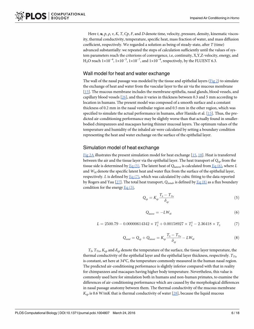
Wall model for heat and water exchangeThe wall of the nasal passage was modeled by the tissue and epithelial layers (Fig 2) to simulatethe exchange of heat and water from the vascular layer to the air via the mucous membrane[15]. The mucous membrane includes the membrane epithelia, nasal glands, blood vessels, andcapillary blood vessels [26], and thus it varies in thickness between 0.3 and 5 mm according tolocation in humans. The present model was composed of a smooth surface and a constantthickness of 0.2 mm in the nasal vestibular region and 0.5 mm in the other region, which wasspecified to simulate the actual performance in humans, after Hanida et al. [15]. Thus, the pre-dicted air-conditioning performance may be slightly worse than that actually found in smaller-bodied chimpanzees and macaques having thinner mucosal layers. The optimum values of thetemperature and humidity of the inhaled air were calculated by setting a boundary conditionrepresenting the heat and water exchange on the surface of the epithelial layer.
Simulation model of heat exchangeFig 2A illustrates the present simulation model for heat exchange [15, 18]. Heat is transferredbetween the air and the tissue layer via the epithelial layer. The heat transport of Qep from thetissue side is determined by Eq (5). The latent heat of Qlatent is calculated from Eq (6), where LandWbl denote the specific latent heat and water flux from the surface of the epithelial layer,respectively. L is defined by Eq (7), which was calculated by cubic fitting to the data reportedby Rogers and Yau [27]. The total heat transport, Qtotal, is defined by Eq (8) as a flux boundarycondition for the energy Eq (3).
Qep ¼ Kep
TS � TTis
depð5Þ
Qlatent ¼ �LWbl ð6Þ
L ¼ 2500:79� 0:00000614342� T3S þ 0:00158927� T2
S � 2:36418� TS ð7Þ
Qtotal ¼ Qep þ Qlatent ¼ Kep
TS � TTis
dep� LWbl ð8Þ
TS, TTis, Kep and δep denote the temperature of the surface, the tissue layer temperature, thethermal conductivity of the epithelial layer and the epithelial layer thickness, respectively. TTis
is constant, set here at 34°C, the temperature commonly measured in the human nasal region.The predicted air-conditioning performance is slightly inferior compared with that in realityfor chimpanzees and macaques having higher body temperature. Nevertheless, this value iscommonly used here for simulation both in humans and non-human primates, to examine thedifferences of air-conditioning performance which are caused by the morphological differencesin nasal passage anatomy between them. The thermal conductivity of the mucous membraneKep is 0.6 W/mK that is thermal conductivity of water [28], because the liquid mucous
Impaired Air Conditioning inHomo
PLOS Computational Biology | DOI:10.1371/journal.pcbi.1004807 March 24, 2016 6 / 18
membrane is assumed in the model used here [15, 18]. TS is determined by Qtotal, which com-prises Qep and Qlatent.
Simulation model of water exchangeFig 2B illustrates the wall model, implemented with a boundary layer to define the boundarycondition of species transport for water exchange [15,18]. The model is based on Fick’s law inthat the flux diffusion is proportional to the concentration gradient of water. Here, we used theDirichlet-type boundary condition (i.e., fixed transport) in the FLUENT software. The two-film theory was used here to evaluate the mass of species transport between a liquid phase anda gas phase across a boundary. The thickness of the boundary layer was set at 0.5 mm [15].Wbl
is the water flux from the boundary layer, which is determined from Eq (9), and this was used
Fig 2. Wall models of the nasal passage. (A) Model for heat exchange.Qep,Qlatent, andWbl indicate theheat flux of the nasal wall’s function, the heat flux of latent heat, and the water flux from the boundary layers in(B). TS, TTis, and δep are the temperature of the surface, the tissue layer temperature, and the epithelial layerthickness, respectively. (B) Model for water exchange.Wbl andWep are the water flux from the boundarylayer and the water flux from the tissue layer. F, FS, FTis, and δbl are the water fraction in the boundary layer,the water fraction on the epithelial surface, the water fraction on the tissue layer, and the boundary layerthickness, respectively.
doi:10.1371/journal.pcbi.1004807.g002
Impaired Air Conditioning inHomo
PLOS Computational Biology | DOI:10.1371/journal.pcbi.1004807 March 24, 2016 7 / 18
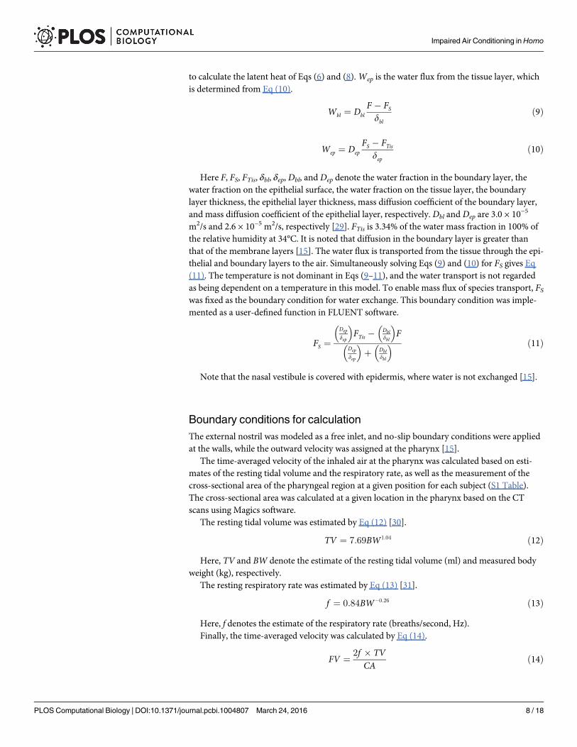
to calculate the latent heat of Eqs (6) and (8).Wep is the water flux from the tissue layer, whichis determined from Eq (10).
Wbl ¼ Dbl
F � FS
dbl
ð9Þ
Wep ¼ Dep
FS � FTis
depð10Þ
Here F, FS, FTis, δbl, δep, Dbl, and Dep denote the water fraction in the boundary layer, thewater fraction on the epithelial surface, the water fraction on the tissue layer, the boundarylayer thickness, the epithelial layer thickness, mass diffusion coefficient of the boundary layer,and mass diffusion coefficient of the epithelial layer, respectively. Dbl and Dep are 3.0 × 10⁻⁵
m2/s and 2.6 × 10⁻⁵ m2/s, respectively [29]. FTis is 3.34% of the water mass fraction in 100% ofthe relative humidity at 34°C. It is noted that diffusion in the boundary layer is greater thanthat of the membrane layers [15]. The water flux is transported from the tissue through the epi-thelial and boundary layers to the air. Simultaneously solving Eqs (9) and (10) for FS gives Eq(11). The temperature is not dominant in Eqs (9–11), and the water transport is not regardedas being dependent on a temperature in this model. To enable mass flux of species transport, FSwas fixed as the boundary condition for water exchange. This boundary condition was imple-mented as a user-defined function in FLUENT software.
FS ¼Dep
dep
� �FTis � Dbl
dbl
� �F
Dep
dep
� �þ Dbl
dbl
� � ð11Þ
Note that the nasal vestibule is covered with epidermis, where water is not exchanged [15].
Boundary conditions for calculationThe external nostril was modeled as a free inlet, and no-slip boundary conditions were appliedat the walls, while the outward velocity was assigned at the pharynx [15].
The time-averaged velocity of the inhaled air at the pharynx was calculated based on esti-mates of the resting tidal volume and the respiratory rate, as well as the measurement of thecross-sectional area of the pharyngeal region at a given position for each subject (S1 Table).The cross-sectional area was calculated at a given location in the pharynx based on the CTscans using Magics software.
The resting tidal volume was estimated by Eq (12) [30].
TV ¼ 7:69BW1:04 ð12ÞHere, TV and BW denote the estimate of the resting tidal volume (ml) and measured body
weight (kg), respectively.The resting respiratory rate was estimated by Eq (13) [31].
f ¼ 0:84BW�0:26 ð13ÞHere, f denotes the estimate of the respiratory rate (breaths/second, Hz).Finally, the time-averaged velocity was calculated by Eq (14).
FV ¼ 2f � TVCA
ð14Þ
Impaired Air Conditioning inHomo
PLOS Computational Biology | DOI:10.1371/journal.pcbi.1004807 March 24, 2016 8 / 18
Here, FV and CA denote the time-averaged flow velocity (m/s) and the measured cross-sec-tional area at a given location of the pharynx (mm2), respectively.
The CFD simulations were performed in three ambient atmospheric air conditions: cold–dry, 10% RH at 5°C (0.05% MF); hot–dry, 5% RH at 40°C (0.23% MF); and warm–wet, 60%RH at 30°C (1.58%MF).
VisualizationsThe resulting spatial pattern of the vector quantity representing the velocity and direction ofthe airflow is illustrated using streamlines in different colors that was computed from thepoints on the plane of external nostrils [15, 18]. The number of streamlines is decided depen-dent on the area of the plane of nostrils, and it reflects the relative airflow volume for a givensubject, allowing us to examine where the air mainly flows. Those of the scalar quantity repre-senting the temperature and water vapor volume are illustrated using contours in different col-ors [15, 18].
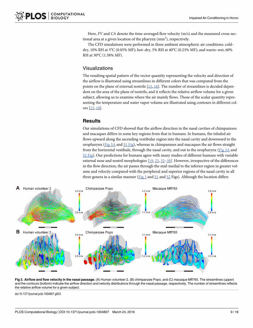
ResultsOur simulations of CFD showed that the airflow direction in the nasal cavities of chimpanzeesand macaques differs in some key regions from that in humans. In humans, the inhaled airflows upward along the ascending vestibular region into the nasal cavity and downward to theoropharynx (Fig 3A and S1 Fig), whereas in chimpanzees and macaques the air flows straightfrom the horizontal vestibule, through the nasal cavity, and out to the oropharynx (Fig 3A andS2 Fig). Our predictions for humans agree with many studies of different humans with variableexternal nose and nostril morphologies [19, 23, 32–38]. However, irrespective of the differencesin the flow direction, the air passes through the mid-medial to the inferior region in greater vol-ume and velocity compared with the peripheral and superior regions of the nasal cavity in allthree genera in a similar manner (Fig 3 and S1 and S2 Figs). Although the location differs
Fig 3. Airflow and flow velocity in the nasal passage. (A) Human volunteer 2, (B) chimpanzee Popo, and (C) macaqueMff765. The streamlines (upper)and the contours (bottom) indicate the airflow direction and velocity distributions through the nasal passage, respectively. The number of streamlines reflectsthe relative airflow volume for a given subject.
doi:10.1371/journal.pcbi.1004807.g003
Impaired Air Conditioning inHomo
PLOS Computational Biology | DOI:10.1371/journal.pcbi.1004807 March 24, 2016 9 / 18

slightly, this major flow passage was also determined in many previous experimental and CFDsimulation studies in humans [19, 20, 23, 32–38] and macaques [39]. Thus, the major flow pas-sage through the nasal cavity in humans is almost the same as in chimpanzees and macaques,although humans have an upward and curved airflow.
In our simulations, the inhaled air was conditioned well in chimpanzees and macaques,even in severe ambient conditions (Figs 4 and 5; Table 1), where the inhaled air was adjusted toapproximately 34°C and saturated to almost 100% RH before reaching the nasopharyngealregion, i.e, the most posterior frontal contour (Figs 4 and 5; Table 1). However, the air-
Fig 4. Distribution of temperature in the nasal passage. (A) Warm and wet; (B) cold and dry; and (C) hot and dry conditions. The contours represent thedistribution of temperature at each level from the nares to the nasopharynx. (D) The lowest temperatures in each contour were in the warm/wet and cold/dryconditions, and the highest temperatures were in the hot/dry conditions. The values indicate the performance when adjusting the temperature toward 34°C.
doi:10.1371/journal.pcbi.1004807.g004
Impaired Air Conditioning inHomo
PLOS Computational Biology | DOI:10.1371/journal.pcbi.1004807 March 24, 2016 10 / 18
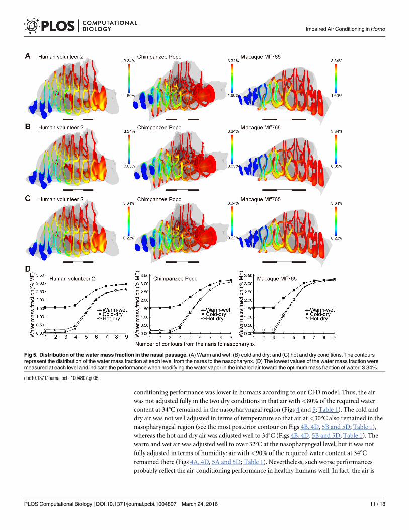
conditioning performance was lower in humans according to our CFD model. Thus, the airwas not adjusted fully in the two dry conditions in that air with<80% of the required watercontent at 34°C remained in the nasopharyngeal region (Figs 4 and 5; Table 1). The cold anddry air was not well adjusted in terms of temperature so that air at<30°C also remained in thenasopharyngeal region (see the most posterior contour on Figs 4B, 4D, 5B and 5D; Table 1),whereas the hot and dry air was adjusted well to 34°C (Figs 4B, 4D, 5B and 5D; Table 1). Thewarm and wet air was adjusted well to over 32°C at the nasopharyngeal level, but it was notfully adjusted in terms of humidity: air with<90% of the required water content at 34°Cremained there (Figs 4A, 4D, 5A and 5D; Table 1). Nevertheless, such worse performancesprobably reflect the air-conditioning performance in healthy humans well. In fact, the air is
Fig 5. Distribution of the water mass fraction in the nasal passage. (A) Warm and wet; (B) cold and dry; and (C) hot and dry conditions. The contoursrepresent the distribution of the water mass fraction at each level from the nares to the nasopharynx. (D) The lowest values of the water mass fraction weremeasured at each level and indicate the performance when modifying the water vapor in the inhaled air toward the optimummass fraction of water: 3.34%.
doi:10.1371/journal.pcbi.1004807.g005
Impaired Air Conditioning inHomo
PLOS Computational Biology | DOI:10.1371/journal.pcbi.1004807 March 24, 2016 11 / 18

often only conditioned up to 30°C or to 80–90% RH in the nasopharyngeal region of humans,even in comfortable ambient conditions [40–42]. Thus, inhaled air is not well conditioned inthe nasal cavity of humans compared with nonhuman primates.
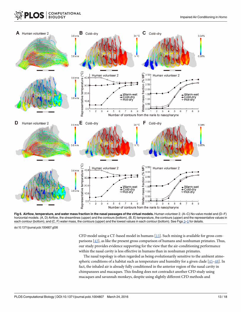
The no-valve model resulted in a few changes in the airflow direction and air-conditioningperformance for each human subject (Fig 6A–6C; S3 Fig; Table 2). The normal model exhibiteda fast and diffusive flow through the nasal valve in all three genera (Fig 3, S3 Fig), but this flowwas not found in the human no-valve model (Fig 6A; S3 Fig). In the horizontal model, the air-flow direction was changed to be slightly horizontal and straight, as seen in chimpanzees, but ithad only a minor effect on the air-conditioning performance: the temperature and humidity atthe nasopharyngeal level did not differ from those in the normal and no-valve models for eachsubject (Fig 6D–6F; S3 Fig; Table 2). Thus, the vertically oriented vestibule contributes to theupward airflow, but the topology of the nasal valve and vestibule makes little contribution toimproving the air-conditioning performance in humans.
DiscussionIn this CFD study, we used the same simulation conditions to compare the air-conditioningperformance in humans and non-human primates. It should be noted that MRI scans wereused for reconstructing the nasal passage models in humans, while CT scans were used in non-human primates. A same type of tissue is resolved differently between the two modalities. In aprecise sense, mixing CT and MRI scans potentially alters the thickness of the airway and flowvelocities for a same subject. Nevertheless, the present results using MRI scans also show an air-flow pattern, velocities, and air-conditioning performance that are similar to those by a same
Table 1. Temperature, water mass fraction, and relative humidity at the nasopharyngeal level.
Species Subjects Warm and wet Cold and dry Hot and dry
T (°C) MF (RH34) (%) T (°C) MF (RH34) (%) T (°C) MF (RH34) (%)
Humans, Homo sapiens Volunteer 1 32.6 2.68 (80.3) 26.7 2.12 (63.4) 34.2 2.18 (65.2)
Volunteer 2 33.1 2.96 (88.5) 30.9 2.64 (78.9) 34.0 2.66 (79.6)
Volunteer 3 33.6 3.14 (94.1) 32.8 2.98 (89.2) 34.0 2.99 (89.6)
Volunteer 5 32.7 2.75 (88.2) 27.5 2.24 (67.1) 34.0 2.29 (68.7)
Volunteer 6 32.9 2.82 (84.3) 28.6 2.37 (71.1) 34.0 2.42 (72.3)
Average 33.0 2.87 (85.9) 29.3 2.47 (74.0) 34.0 2.51 (75.1)
Chimpanzees, Pan troglodytes Mari 33.7 3.22 (96.4) 33.2 3.13 (93.7) 34.0 3.13 (93.7)
Pendesa 33.8 3.24 (97.1) 33.3 3.16 (94.8) 34.0 3.17 (94.8)
Popo 33.7 3.21 (96.2) 33.1 3.11 (93.2) 34.0 3.11 (93.2)
Reiko 33.6 3.19 (95.5) 32.8 3.07 (92.1) 34.0 3.08 (92.1)
Average 33.7 3.21 (96.3) 33.1 3.12 (93.3) 34.0 3.08 (93.5)
Japanese macaques, Macaca fuscata Mff765 33.9 3.27 (98.0) 33.7 3.22 (96.5) 34.0 3.23 (96.6)
Mff963 33.8 3.21 (96.1) 33.3 3.10 (92.9) 34.0 3.11 (93.2)
Mff1859 34.0 3.33 (99.8) 34.0 3.33 (99.7) 34.0 3.33 (99.7)
Mff2115 34.0 3.33 (99.6) 33.9 3.32 (99.4) 34.0 3.32 (99.4)
Rhesus macaques, Macaca mulatta Mm1701 34.0 3.34 (99.9) 34.0 3.34 (99.9) 34.0 3.34 (99.9)
Mm1715 34.0 3.34 (99.9) 34.0 3.34 (99.9) 34.0 3.34 (99.9)
Macaques, Macaca Average 34.0 3.30 (98.9) 33.8 3.28 (98.1) 34.0 3.28 (98.2)
T, the lowest values of temperature in the nasopharyngeal level (the most posterior contour in figures for a given subject) were for the warm-humid and
cold-dry conditions, and the highest value was for the hot-dry condition; MF, the lowest values of the water mass fraction in the nasopharyngeal level;
RH34, percentage of the water mass fraction to 3.34%, the water mass fraction at which the air of 34°C is fully saturated.
doi:10.1371/journal.pcbi.1004807.t001
Impaired Air Conditioning inHomo
PLOS Computational Biology | DOI:10.1371/journal.pcbi.1004807 March 24, 2016 12 / 18
CFD model using a CT-based model in humans [15]. Such mixing is available for gross com-parisons [43], as like the present gross comparison of humans and nonhuman primates. Thus,our study provides evidence supporting for the view that the air-conditioning performancewithin the nasal cavity is less effective in humans than in nonhuman primates.
The nasal topology is often regarded as being evolutionarily sensitive to the ambient atmo-spheric conditions of a habitat such as temperature and humidity for a given clade [45–48]. Infact, the inhaled air is already fully conditioned in the anterior region of the nasal cavity inchimpanzees and macaques. This finding does not contradict another CFD study usingmacaques and savannah monkeys, despite using slightly different CFD methods and
Fig 6. Airflow, temperature, and water mass fraction in the nasal passages of the virtual models.Human volunteer 2. (A–C) No-valve model and (D–F)horizontal models. (A, D) Airflow, the streamlines (upper) and the contours (bottom), (B, E) temperature, the contours (upper) and the representative values ineach contour (bottom), and (C, F) water mass, the contours (upper) and the lowest values in each contour (bottom). See Figs 3–5 for details.
doi:10.1371/journal.pcbi.1004807.g006
Impaired Air Conditioning inHomo
PLOS Computational Biology | DOI:10.1371/journal.pcbi.1004807 March 24, 2016 13 / 18
conditions [16]. These findings mean that the morphology of the nasal cavity can accept somemorphological evolutionary modifications that might impair air-conditioning in nonhumanprimates. The earlier hominins other than the genus Homo have a nasal passage in a manneranalogous to chimpanzees rather than humans, suggesting that they probably show the effec-tive air-conditioning performance as seen in non-human primates. However, the characteristicfacial reorganization in Homo has precluded the developmental elongation of the oral andnasal cavities [7–10] and has impaired their air-conditioning performance. The nasal cavityanatomy is believed to vary with an advantage to the climate conditions of the habitat of agiven population in modern humans [47, 48], but the present finding supports the idea thatmorphological modification in the nasal region is only a weak evolutionary response to air-conditioning needs in the divergence of Homo from the other hominin lineages. Rather, thenasal region is regarded just as a buffering module for facial reorganization, in contrast withother modules, including the jaw, eye, and braincase, which have been modified yet have main-tained their independent functions. Thus, human nasal topology was probably modified pas-sively by evolutionary facial reorganization in early Homo, and such an evolutionarymodification was not prevented by impaired the air-conditioning performance.
The unique external nose in Homo is believed to confer some functional advantages duringair conditioning, such as retaining the water vapor from expired air [4] or generating a vortexairflow with inhaled air to improve air conditioning [2, 44]. The nasal vestibule within theexternal nose is coated with epidermis, including vestibular hairs, and it only exchanges heatwith the air, which means that the nasal vestibule itself makes a limited contribution to the airconditioning that occurs within it. Our study confirmed that the nasal valve also has little effecton the air-conditioning performance in humans. In fact, the topography of the nasal valve hasspecific effects on the local airflow pattern within the nasal cavity such as vortices in the supe-rior meatus [34], but there are limited effects on the gross airflow pattern, including turbulence[34, 44]. Further, our study also showed that the vertically oriented vestibule makes a majorcontribution to the generation of an upward airflow in the nasal cavity in humans, but theinhaled air is still well conditioned mainly in the mid-medial to inferior regions of the nasal
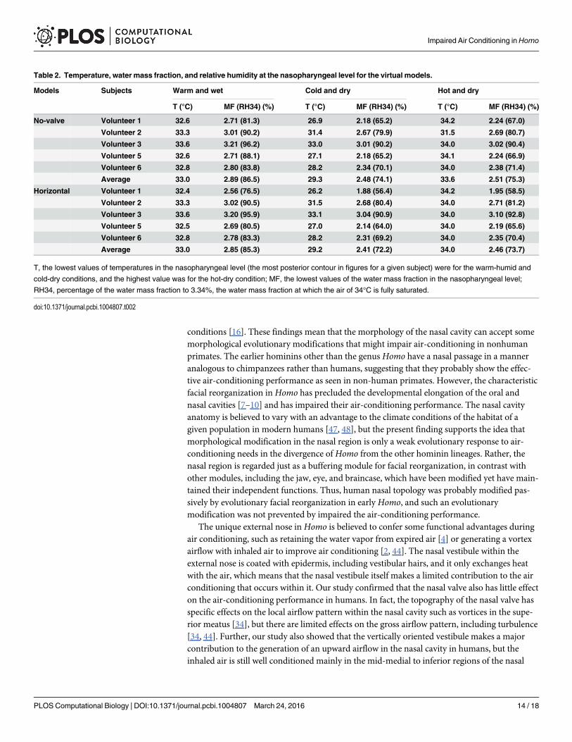
Table 2. Temperature, water mass fraction, and relative humidity at the nasopharyngeal level for the virtual models.
Models Subjects Warm and wet Cold and dry Hot and dry
T (°C) MF (RH34) (%) T (°C) MF (RH34) (%) T (°C) MF (RH34) (%)
No-valve Volunteer 1 32.6 2.71 (81.3) 26.9 2.18 (65.2) 34.2 2.24 (67.0)
Volunteer 2 33.3 3.01 (90.2) 31.4 2.67 (79.9) 31.5 2.69 (80.7)
Volunteer 3 33.6 3.21 (96.2) 33.0 3.01 (90.2) 34.0 3.02 (90.4)
Volunteer 5 32.6 2.71 (88.1) 27.1 2.18 (65.2) 34.1 2.24 (66.9)
Volunteer 6 32.8 2.80 (83.8) 28.2 2.34 (70.1) 34.0 2.38 (71.4)
Average 33.0 2.89 (86.5) 29.3 2.48 (74.1) 33.6 2.51 (75.3)
Horizontal Volunteer 1 32.4 2.56 (76.5) 26.2 1.88 (56.4) 34.2 1.95 (58.5)
Volunteer 2 33.3 3.02 (90.5) 31.5 2.68 (80.4) 34.0 2.71 (81.2)
Volunteer 3 33.6 3.20 (95.9) 33.1 3.04 (90.9) 34.0 3.10 (92.8)
Volunteer 5 32.5 2.69 (80.5) 27.0 2.14 (64.0) 34.0 2.19 (65.6)
Volunteer 6 32.8 2.78 (83.3) 28.2 2.31 (69.2) 34.0 2.35 (70.4)
Average 33.0 2.85 (85.3) 29.2 2.41 (72.2) 34.0 2.46 (73.7)
T, the lowest values of temperatures in the nasopharyngeal level (the most posterior contour in figures for a given subject) were for the warm-humid and
cold-dry conditions, and the highest value was for the hot-dry condition; MF, the lowest values of the water mass fraction in the nasopharyngeal level;
RH34, percentage of the water mass fraction to 3.34%, the water mass fraction at which the air of 34°C is fully saturated.
doi:10.1371/journal.pcbi.1004807.t002
Impaired Air Conditioning inHomo
PLOS Computational Biology | DOI:10.1371/journal.pcbi.1004807 March 24, 2016 14 / 18
cavity as seen in intact humans and non-human primates. Although the location differsslightly, such a major flow passage was confirmed in humans with variable external nose andnostril morphologies in modern humans [38]. Irrespectively of varied nostrils, the verticallyoriented vestibule had little effect on improving the air-conditioning performance, althoughthe external nose morphologies including this feature could improve the transport of odorantsto the superior olfactory slit in the upper nasal cavity. Thus, the unique external nose has littleeffect to improving the air-conditioning performance, and the impaired performance is morelikely to be a consequence of modifications in the shape of the nasal cavity itself inHomo line-age since it was diversified from the other hominins, including australopithecines, in the begin-ning of the Early Pleistocene.
Inhaled air can be adjusted through the pharyngeal cavity to be fully conditioned in humans,even though it is not fully adjusted in the nasal cavity. In the phyletic divergence ofHomo fromthe other hominin lineages, facial flattening and reorganization has reduced the dimensions ofthe horizontal oral cavity along with the nasal cavity and pushed the tongue down toward thepharynx, thereby lengthening the vertical pharyngeal cavity [8, 9]. Although the actual lengthsof the pharyngeal cavity are unknown for each previous form ofHomo [49, 50], the long pha-ryngeal cavity in extant humans contributes greatly to the sophisticated and flexible modifica-tions of the topology of the supralaryngeal vocal tract from the glottis to the lips, whichunderlies human speech production [6, 51]. This feature is also believed to provide a disadvan-tage for the other physiological functions of the pharynx such as swallowing, increasing the riskof accidental aspiration during deglutition of food and liquid boluses [52–54]. However, thelong pharyngeal cavity could in part compensate for the impaired air-conditioning performancewithin the short nasal cavity. The Late Pliocene to Early Pleistocene periods were characterizedby a highly fluctuating climate and a gradual transition from warm and humid to cool and aridenvironments, especially in the northern hemisphere [13, 55]. These linked changes in the nasaland pharyngeal regions would in part have contributed to how flat-faced hominins, i.e.,Homomembers, must have survived such fluctuations in climate, before they moved “Out of Africa”in the Early Pleistocene to explore the more severe climates and ecological environments ofEurasia.
Supporting InformationS1 Table. Subjects, scans, and estimated parameters of respiration. abbreviations: MRI,magnetic resonance imaging; CT, Computed Tomography; CA, cross-sectional area at the oro-pharyngeal level; TV, estimated tidal volume; f, estimated frequency of breath; FV, flow velocityat the pharyngeal level. �CT scans of chimpanzees and macaques used here are deposited andreleased at the Digital Morphology Museum, KUPRI (dmm.pri.kyoto-u.ac.jp/archive/), underPRICT #.†estimate.(DOCX)
S1 Fig. Airflows and flow velocity in the nasal passage in humans. (A) Human volunteer 1;(B) human volunteer 3; (C) human volunteer 5; (D) human volunteer 6. The streamlines(upper) and contours (bottom) indicate the airflow direction and velocity distributions throughthe nasal passage, respectively. The streamline number reflects the relative airflow volume for agiven subject.(TIF)
S2 Fig. Airflows and flow velocity in the nasal passage in nonhuman primates. (A) chim-panzee Mari; (B) chimpanzee Pendesa; (C) chimpanzee Reiko; (D) macaque Mff963; (D)macaque Mff1859; (F) macaque Mff2115; (G) macaque Mm1701; and (H) macaque Mm1715.
Impaired Air Conditioning inHomo
PLOS Computational Biology | DOI:10.1371/journal.pcbi.1004807 March 24, 2016 15 / 18
The streamlines (upper) and contours (bottom) indicate the airflow direction and velocity dis-tributions through the nasal passage, respectively. The streamline number reflects the relativeairflow volume for a given subject.(TIF)
S3 Fig. Airflow and flow velocity in the nasal passage of the virtual topology models. (A, B)Human volunteer 1, (C, D) human volunteer 3, (E, F) human volunteer 5, (G, H) human vol-unteer 6. (A, C, E, G) No-valve model and (B, D, F, H) horizontal models. The streamlines(upper) and contours (bottom) indicate the airflow direction and velocity distributions throughthe nasal passage, respectively. The streamline number reflects the relative airflow volume for agiven subject.(TIF)
AcknowledgmentsThe authors thank A. Watanabe, A. Kaneko, K. Kumazaki, N. Maeda, S. Watanabe, S. Funaha-shi, H. Koda, and A. Kato for help with CT scanning of chimpanzees and macaques; Y. Shi-mada, I. Fujimoto, and BAIC staff for help with MRI scanning of human volunteers.
Author ContributionsConceived and designed the experiments: TN TerM. Performed the experiments: TN TMN JSMHMT TetM FM SH SI. Analyzed the data: TN FM SH KK SI KS TerM. Contributedreagents/materials/analysis tools: FM SH KK TerM. Wrote the paper: TN FM SH TerM.
References1. Klein RG. The human career. 3rd ed. Chicago: University of Chicago Press; 2009.
2. Lieberman D. The evolution of the human head. Cambridge: Belknap Press; 2011.
3. Asfaw B, White T, Lovejoy O, Latimer B, Simpson S, Suwa G. Australopithecus garhi: A new species ofearly hominid from Ethiopia. Science. 1999; 284: 629–635. PMID: 10213683
4. Franciscus RG, Trinkaus E. Nasal morphology and the emergence of Homo erectus. Am J PhysAnthropol. 1988; 75: 517–527. PMID: 3133950
5. Losken A, Mooney MP, Siegel MI. Comparative cephalometric study of nasal cavity growth patterns inseven animal models. Cleft Palate Craniofac J. 1994; 31: 17–23. doi: 10.1597/1545-1569(1994)031<0017:CCSONC>2.3.CO;2 PMID: 8130238
6. Fitch WT. The evolution of speech: a comparative review. Trends Cogn Sci. 2000; 4: 258–267. PMID:10859570
7. Nishimura T, Mikami A, Suzuki J, Matsuzawa T. Descent of the larynx in chimpanzee infants. Proc NatlAcad Sci U S A. 2003; 100: 6930–6933. doi: 10.1073/pnas.1231107100 PMID: 12775758
8. Nishimura T, Mikami A, Suzuki J, Matsuzawa T. Descent of the hyoid in chimpanzees: Evolution of faceflattening and speech. J Hum Evol. 2006; 51: 244–54. doi: 10.1016/j.jhevol.2006.03.005 PMID:16730049
9. Nishimura T, Oishi T, Suzuki J, Matsuda K, Takahashi T. Development of the supralaryngeal vocaltract in Japanese macaques: Implications for the evolution of the descent of the larynx. Am J PhysAnthropol. 2008; 135: 182–194. doi: 10.1002/ajpa.20719 PMID: 17960727
10. Lieberman DE, McCarthy RC, Hiiemae KM, Palmer JB. Ontogeny of postnatal hyoid and larynx descentin humans. Arch Oral Biol. 2001; 46: 117–128. PMID: 11163319
11. Cole P. Modification of inspired air. In: Proctor DF, Andersen I, editors. The nose: upper airway physiol-ogy and atmospheric environment. Amsterdam, Netherlands: Elsevier Biomedical Press; 1982. pp.350–375.
12. Elad D, Wolf M, Keck T. Air-conditioning in the human nasal cavity. Respir Physiol Neurobiol. 2008;163: 121–127. Epub 2008/06/21. S1569–9048(08)00126–2 [pii]doi: 10.1016/j.resp.2008.05.002 PMID:18565805
Impaired Air Conditioning inHomo
PLOS Computational Biology | DOI:10.1371/journal.pcbi.1004807 March 24, 2016 16 / 18
13. Vrba ES. The fossil record of African antelopes (Mammalia, Bovidae) in relation to human evolutionand paleoclimate. In: Vrba ES, Denton GH, Partridge TC, Burckle LH, editors. Paleoclimate and evolu-tion, with emphasis on human origins. New Haven, CT: Yale University Press; 1995. pp. 385–424.
14. Potts R. Evolution and climate variability. Science. 1996; 273: 922–923.
15. Hanida S, Mori F, Kumahata K, Watanabe M, Ishikawa S, Matsuzawa T. Influence of latent heat in thenasal cavity. J Biomech Sci Engineer. 2013; 8: 209–224.
16. Mori F, Hanida S, Kumahata K, Miyabe–Nishiwaki T, Suzuki J, Matsuzawa T, et al. Minor contributionsof the maxillary sinus to the air-conditioning performance in macaque monkeys. J Exp Biol. 2015; 218:2394–2401. doi: 10.1242/jeb.118059 PMID: 26034122
17. Matsuzawa T, Tomonaga M, Tanaka M, editors. Cognitive development in chimpanzees. Tokyo:Springer; 2006.
18. Kumahata K, Mori F, Ishikawa S, Matsuzawa T. Nasal flow simulation using heat and humidity models.J Biomech Sci Engineer. 2010; 5: 565–577.
19. Swift DL, Proctor DF. Access of air to the respiratory tract. In: Brain JD, Proctor DF, Reid LM, editors.Respiratory defense mechanisms. 5. New York: Marcel Dekker; 1977. pp. 63–93.
20. Hornung DE, Leopold DA, Youngentob SL, Sheehe PR, Gagne GM, Thomas FD, et al. Airflow patternsin a human nasal model. Arch Otolaryngol Head Neck Surg. 1987; 113: 169–172. PMID: 3801173
21. Hahn I, Scherer PW, Mozell MM. Velocity profiles measured for airflow through a large-scale model ofthe human nasal cavity. J Appl Physiol. 1993; 75: 2273–2287. PMID: 8307887
22. Schreck S, Sullivan K, Ho C, Chang H. Correlations between flow resistance and geometry in a modelof the human nose. J Appl Physiol. 1993; 75: 1767–1775. PMID: 8282630
23. Keyhani K, Scherer PW, Mozell MM. Numerical simulation of airflow in the human nasal cavity. J Bio-mech Eng. 1995; 117: 429–441. PMID: 8748525
24. Doorly DJ, Taylor DJ, Schroter RC. Mechanics of airflow in the human nasal airways. Respir PhysiolNeurobiol. 2008; 163: 100–110. doi: 10.1016/j.resp.2008.07.027 PMID: 18786659
25. Spence CJT, Buchmann NA, Jermy MC. Unsteady flow in the nasal cavity with high flow therapy mea-sured by stereoscopic PIV. Experiments Fluids. 2012; 52: 569–579. doi: 10.1007/S00348-011-1044-Z
26. Lang J. Clinical anatomy of the nose, nasal cavity, and paranasal sinuses. New York: George ThiemeVerlag; 1989.
27. Rogers RR, Yau MK. A short course in cloud physics. 3rd edition. Oxford: Pergamon Press; 1989.
28. Lervik A, Bresme F, Kjelstrup S, Bedeaux D, Miguel Rubi J. Heat transfer in protein–water interfaces.Phys Chem Chem Phys. 2010; 12: 1610–1617. doi: 10.1039/b918607g PMID: 20126777
29. Lee CY, Wilke CR. Measurements of vapor diffusion coefficient. Ind Eng Chem. 1954; 46: 2381–2387.doi: 10.1021/ie50539a046
30. Worthington J, Young IS, Altringham JD. The relationship between body mass and ventilation rate inmammals. J Exp Biol. 1991; 161: 533–536. PMID: 1757778
31. Stahl WR. Scaling of respiratory variables in mammals. J Appl Physiol. 1967; 22: 453–460. PMID:6020227
32. Elad D, Liebenthal R, Wenig BL, Einav S. Analysis of air flow patterns in the human nose. Med Biol EngComput. 1993; 31: 585–592. doi: 10.1007/Bf02441806 PMID: 8145584
33. Kelly JT, Prasad AK, Wexler AS. Detailed flow patterns in the nasal cavity. J Appl Physiol. 2000; 89:323–337. PMID: 10904068
34. Zhao K, Scherer PW, Hajiloo SA, Dalton P. Effect of anatomy on human nasal air flow and odoranttransport patterns: Implications for olfaction. Chem Senses. 2004; 29: 365–379. doi: 10.1093/Chemse/Bjh033 PMID: 15201204
35. Naftali S, Rosenfeld M, Wolf M, Elad D. The air-conditioning capacity of the human nose. Ann BiomedEng. 2005; 33:545–553. Epub 2005/05/25. PMID: 15909660.
36. Segal RA, Kepler GM, Kimbell JS. Effects of differences in nasal anatomy on airflow distribution: A com-parison of four individuals at rest. Ann Biomed Eng. 2008; 36: 1870–1882. doi: 10.1007/s10439-008-9556-2 PMID: 18777212
37. Wen J, Inthavong K, Tu J, Wang S. Numerical simulations for detailed airflow dynamics in a humannasal cavity. Respir Physiol Neurobiol. 2008; 161: 125–135. doi: 10.1016/j.resp.2008.01.012 PMID:18378196
38. Zhu JH, Lee HP, Lim KM, Lee SJ, Wang DY. Evaluation and comparison of nasal airway flow patternsamong three subjects from Caucasian, Chinese and Indian ethnic groups using computational fluiddynamics simulation. Respir Physiol Neurobiol. 2011; 175: 62–69. doi: 10.1016/J.Resp.2010.09.008PMID: 20854936
Impaired Air Conditioning inHomo
PLOS Computational Biology | DOI:10.1371/journal.pcbi.1004807 March 24, 2016 17 / 18
39. Kepler GM, Richardson RB, Morgan KT, Kimbell JS. Computer simulation of inspiratory nasal airflowand inhaled gas uptake in a rhesus monkey. Toxicol Appl Pharmacol. 1998; 150: 1–11. doi: 10.1006/taap.1997.8350 PMID: 9630447
40. Primiano FP Jr., Saidel GM, Montague FW Jr., Kruse KL, Green CG, Horowitz JG. Water vapour andtemperature dynamics in the upper airways of normal and CF subjects. Eur Respir J. 1988; 1: 407–414.PMID: 3169211
41. Rouadi P, Baroody FM, Abbott D, Naureckas E, Solway J, Naclerio RM. A technique to measure theability of the human nose to warm and humidify air. J Appl Physiol. 1999; 87: 400–406. PMID:10409601
42. Keck T, Leiacker R, Heinrich A, Kuhnemann S, Rettinger G. Humidity and temperature profile in thenasal cavity. Rhinology. 2000; 38: 167–171. PMID: 11190750
43. Chen XB, Lee HP, Chong VF, Wang de Y. Assessment of septal deviation effects on nasal air flow: acomputational fluid dynamics model. Laryngoscope 2009; 119: 1730–6. doi: 10.1002/lary.20585 PMID:19572266
44. Churchill SE, Shackelford LL, Georgi JN, Black MT. Morphological variation and airflow dynamics inthe human nose. Am J Hum Biol. 2004; 16: 625–638. PMID: 15495233
45. Rae TC, Hill RA, Hamada Y, Koppe T. Clinal variation of maxillary sinus volume in Japanesemacaques(Macaca fuscata). Am J Primatol. 2003; 59: 153–158. PMID: 12682923
46. Franciscus RG, Long JC. Variation in human nasal height and breadth. Am J Phys Anthropol. 1991; 85:419–427. doi: 10.1002/ajpa.1330850406 PMID: 1928315
47. Yokley TR. Ecogeographic variation in human nasal passages. Am J Phys Anthropol. 2009; 138: 11–22. doi: 10.1002/ajpa.20893 PMID: 18623075
48. Weiner JS. Nose shape and climate. Am J Phys Anthropol. 1954; 12: 615–8. PMID: 14350081
49. Lieberman P, Crelin ES. On the speech of Neanderthal man. Linguist Inq. 1971; 2: 203–222.
50. Laitman JT, Heimbuch RC. The basicranium of Plio-Pleistocene hominids as an indicator of their upperrespiratory systems. Am J Phys Anthropol. 1982; 59: 323–343. doi: 10.1002/Ajpa.1330590315 PMID:6818861
51. Lieberman PH, Klatt DH, WilsonWH. Vocal tract limitations on the vowel repertoires of rhesus monkeyand other nonhuman primates. Science. 1969; 164: 1185–1187. PMID: 4976883
52. Lieberman P, Crelin ES, Klatt DH. Phonetic ability and related anatomy of the newborn and adulthumans, Neanderthal man and the chimpanzee. American Anthropologist. 1972; 74: 187–307.
53. Laitman JT, Crelin ES, Conlogue GJ. The function of the epiglottis in monkey and man. Yale J BiolMed. 1977; 50: 43–48. PMID: 403687
54. Nishimura T. Comparative morphology of the hyo-laryngeal complex in anthropoids: Two steps in theevolution of the descent of the larynx. Primates. 2003; 44: 41–49. doi: 10.1007/s10329-002-0005-9PMID: 12548333
55. Wynn JG. Influence of Plio-Pleistocene aridification on human evolution: Evidence from paleosols ofthe Turkana Basin, Kenya. Am J Phys Anthropol. 2004; 123: 106–118. PMID: 14730645
Impaired Air Conditioning inHomo
PLOS Computational Biology | DOI:10.1371/journal.pcbi.1004807 March 24, 2016 18 / 18


![Sample Nursing Assessment Form · [ ] nasal feeding tube [ ] impaired swallow reflex [ ] absent or hyperactive gag reflex [ ] reduced level consciousness [ ] infectious saliva from](https://img.pdfslide.net/doc/110x75/5d5a696b88c99355448be1e0/sample-nursing-assessment-form-nasal-feeding-tube-impaired-swallow-reflex.jpg)