Embed Size (px)
Citation preview
S H O R T C O M M U N I C A T I O N S 1099
Acta Cryst. (1995). D51, 1099-1102
Improvements in lysozyme protein crystal perfection through microgravity growth. By E. H. SNELL,
S. WEISGERBER and J. R. HELLIWELL,* Chemistry Department, University of Manchester, Manchester M13 9PL, England, and E. WECKERT, K. H(gLZER and K. SCHROER, Institiit fiir Kristallographie, University of Karlsruhe (TH), Kaiserstrasse 12, Postfach 6980, D-76128 Karlsruhe, Germany
(Received 28 July 1995; accepted 25 August 1995)
Abstract Microgravity offers an environment for protein crystallization where there is an absence of convection and sedimentation. We have investigated the effect of microgravity conditions on the perfection of protein crystals. The quality of crystals for X-ray diffraction studies is characterized by a number of factors, namely size, mosaicity and the resolution limit. By using tetragonal lysozyme crystals as a test case we show, with crystal growth in two separate Space Shuttle missions, that the mosaicity is improved by a factor of three to four over earth-grown ground control values. These microgravity- grown protein crystals are then essentially perfect diffraction gratings. As a result the peak to background of individual X-ray diffraction reflections is enhanced by a similar factor to the reduction in the mosaicity. This then offers a particularly important opportunity for improving the measurement of weak reflections such as occur at high diffraction resolution. These microgravity results set a benchmark for all future microgravity and earth-based protein crystallography procedures.
Introduction The European Space Agency (ESA) have developed the Advanced Protein Crystallization Facility (APCF) (Snyder, Fuhrmann & Walter, 1991; Bosch, Lautenschlager, Potthast & Stapelmann, 1992) as a standard tool for microgravity crystallization experiments aboard the NASA Space Shuttle. We have utilized dialysis liquid diffusion for crystallization within the APCF. The dialysis reactors each consist of two quartz glass blocks containing two chambers separated by a dialysis membrane. The upper chamber contains the protein solution, the lower chamber the salt solution. The salt and protein solution are separated by a cylindrical quartz glass plug, which also contains salt solution. To activate the reactor this plug is rotated by 90 ° , once orbit is reached, so that all volumes then come into contact. Likewise the reactor is deactivated before descent to earth.
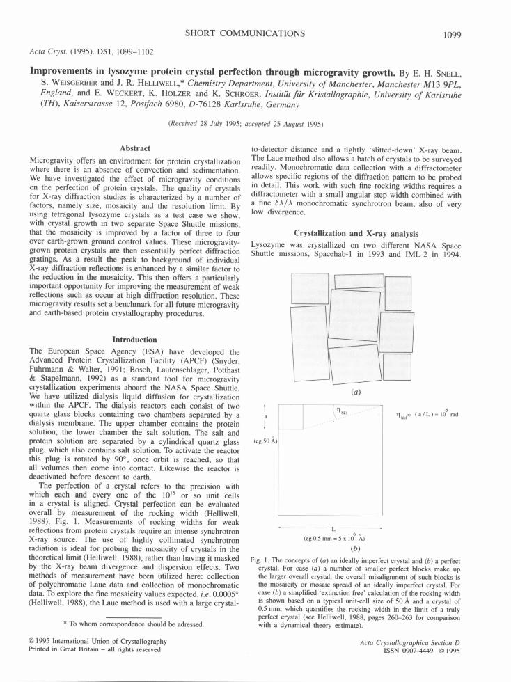
The perfection of a crystal refers to the precision with which each and every one of the 1015 or so unit cells in a crystal is aligned. Crystal perfection can be evaluated overall by measurement of the rocking width (Helliwell, 1988), Fig. 1. Measurements of rocking widths for weak reflections from protein crystals require an intense synchrotron X-ray source. The use of highly collimated synchrotron radiation is ideal for probing the mosaicity of crystals in the theoretical limit (Helliwell, 1988), rather than having it masked by the X-ray beam divergence and dispersion effects. Two methods of measurement have been utilized here: collection of polychromatic Laue data and collection of monochromatic data. To explore the fine mosaicity values expected, i.e. 0.0005 ° (Helliwell, 1988), the Laue method is used with a large crystal-
* To whom correspondence should be adressed.
© 1995 International Union of Crystallography Printed in Great Britain - all rights reserved
to-detector distance and a tightly 'slitted-down' X-ray beam. The Laue method also allows a batch of crystals to be surveyed readily. Monochromatic data collection with a diffractometer allows specific regions of the diffraction pattern to be probed in detail. This work with such fine rocking widths requires a diffractometer with a small angular step width combined with a fine ~SA/A monochromatic synchrotron beam, also of very low divergence.
Crystallization and X-ray analysis Lysozyme was crystallized on two different NASA Space Shuttle missions, Spacehab-1 in 1993 and IML-2 in 1994.
t
(eg 50 ~,)
(a)
.--;" Ilk! . . . . . . . . . . . . . i" ]]hkl: ( a / L ) = 1 0 rad
L 6 o
( e g 0 . 5 m m = 5 x l 0 A)
(b)
Fig. 1. The concepts of (a) an ideally imperfect crystal and (b) a perfect crystal. For case (a) a number of smaller perfect blocks make up the larger overall crystal; the overall misalignment of such blocks is the mosaicity or mosaic spread of an ideally imperfect crystal. For case (b) a simplified 'extinction free' calculation of the rocking width is shown based on a typical unit-cell size of 50 A and a crystal of 0.5 mm, which quantifies the rocking width in the limit of a truly perfect crystal (see HeUiwell, 1988, pages 260-263 for comparison with a dynamical theory estimate).
Acta Crystallographica Section D ISSN 0907-4449 © 1995
1100 S H O R T C O M M U N I C A T I O N S
Table 1. Crystal mosaicities estimated from Laue spot sizes at the SRS (converted to FWHM i.e. 2.3tr values)
For examples of the actual spots see Fig. 2 [space 3 is case (a) and earth 1 is case (b) i.e. the extremes observed]. Note also that the best observed here (space 3) is still larger than the theoretical limit (Helliwell, 1988)
by a factor of two or so.
Crystal Space 1 Space 2 Space 3 Earth 1 Earth 2 (~/) (o) 0.0012 0.0022 0.0010 0.0062 0.0032 cr((~))( ° ) 0.0002 0.0001 0.0001 0.0006 0.0001 Spots 3 14 7 7 7
Crystallization took place in the APCF at a constant temperature of 293 4- 0.1 K over periods of 7.5 and 12.5 d, respectively. The crystallization recipe consisted of 15.8 mg lysozyme dissolved in 188 lal of 0.04 M acetate buffer (pH 4.7). The precipitant was altered slightly for each mission due to the different durations; 1.35 and 1.26M NaC1 solutions for Spacehab-1 and IML-2, respectively. Spacehab-1 produced crystals of average size 0.7 mm, comparable with the ground control crystals grown in an identical APCF unit on earth. The longer IML-2 mission produced crystals of 1.8 mm average, compared with 0.8 mm for the ground-control crystals.
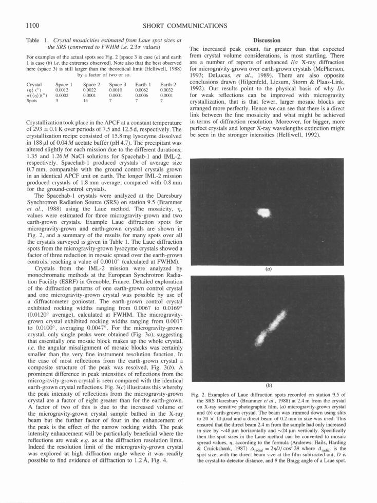
The Spacehab-1 crystals were analyzed at the Daresbury Synchrotron Radiation Source (SRS) on station 9.5 (Brammer et al., 1988) using the Laue method. The mosaicity, r/, values were estimated for three microgravity-grown and two earth-grown crystals. Example Laue diffraction spots for microgravity-grown and earth-grown crystals are shown in Fig. 2, and a summary of the results for many spots over all the crystals surveyed is given in Table 1. The Laue diffraction spots from the microgravity-grown lysozyme crystals showed a factor of three reduction in mosaic spread over the earth-grown controls, reaching a value of 0.0010 ° (calculated at FWHM).
Crystals from the IML-2 mission were analyzed by monochromatic methods at the European Synchrotron Radia- tion Facility (ESRF) in Grenoble, France. Detailed exploration of the diffraction patterns of one earth-grown control crystal and one microgravity-grown crystal was possible by use of a diffractometer goniostat. The earth-grown control crystal exhibited rocking widths ranging from 0.0067 to 0.0169 ° (0.0120 ° average), calculated at FWHM. The microgravity- grown crystal exhibited rocking widths ranging from 0.0017 to 0.0100 °, averaging 0.0047 °. For the microgravity-grown crystal, only single peaks were obtained (Fig. 3a), suggesting that essentially one mosaic block makes up the whole crystal, i.e. the angular misalignment of mosaic blocks was certainly smaller than the very fine instrument resolution function. In the case o f most reflections from the earth-grown crystal a composite structure of the peak was resolved, Fig. 3(b). A prominent difference in peak intensities of reflections from the microgravity-grown crystal is seen compared with the identical earth-grown crystal reflections. Fig. 3(c) illustrates this whereby the peak intensity of reflections from the microgravity-grown crystal are a factor of eight greater than for the earth-grown. A factor of two of this is due to the increased volume of the microgravity-grown crystal sample bathed in the X-ray beam but the further factor of four in the enhancement of the peak is the effect of the narrow rocking width. The peak intensity enhancement will be particularly beneficial where the reflections are weak e.g. as at the diffraction resolution limit. Indeed the resolution limit of the microgravity-grown crystal was explored at high diffraction angle where it was readily possible to find evidence of diffraction to 1.2/~,, Fig. 4.
Discussion
The increased peak count, far greater than that expected from crystal volume considerations, is most startling. There are a number of reports of enhanced I/a X-ray diffraction for microgravity-grown over earth-grown crystals (McPherson, 1993; DeLucas, et al., 1989). There are also opposite conclusions drawn (Hilgenfeld, Liesum, Storm & Plaas-Link, 1992). Our results point to the physical basis of why l/a for weak reflections can be improved with microgravity crystallization, that is that fewer, larger mosaic blocks are arranged more perfectly. Hence we can see that there is a direct link between the fine mosaicity and what might be achieved in terms of diffraction resolution. Moreover, for bigger, more perfect crystals and longer X-ray wavelengths extinction might be seen in the stronger intensities (Helliwell, 1992).
(a)
(b)
Fig. 2. Examples of Laue diffraction spots recorded on station 9.5 of the SRS Daresbury (Brammer et al., 1988) at 2.4 m from the crystal on X-ray sensitive photographic film, (a) microgravity-grown crystal and (b) earth-grown crystal. The beam was trimmed down using slits to 20 x 10 I.trad and a direct beam of 0.2 mm in size was used. This ensured that the direct beam 2.4 m from the sample had only increased in size by ,-~48 lxm horizontally and ,-,24 Ixm vertically. Specifically then the spot sizes in the Laue method can be converted to mosaic spread values, q, according to the formula (Andrews, Hails, Harding & Cruickshank, 1987) Aradial = 2YID/cos220 where Aradial is the spot size, with the direct beam size at the film subtracted out, D is the crystal-to-detector distance, and O the Bragg angle of a Laue spot.

SHORT C O M M U N I C A T I O N S 1101
1o000o
;= 800oo
8 ~ o o o
~ ,oooo
20000
0 .0059 degrees ;-
0 0044 degrees - •
.~
574 5.75
, ~ 00023 degrees • ,
; i
5,76 5 77 5 78 Scan in degrees
(a)
ps= at 45 0
-45
.~ ,oooo
6ooo
==
0 579 580 5 81 65
0 0130 <~grees t •
0 0131 degd~es • -- - 0 0118 Oegrees
575SC . . . . degrees 5 80
(b)
ps= at 45 0
-45
0 0098 a~,grees
.....
590
1OOOOO
8oooo
a:
2oooo
I o 570
M,crogravay grown ,, Ear lh grown COntrol •
4"
¢ .~
0 002:3 degrees ~
572 574 5.76 578 580 582 Scan in d~ ree$
(c)
Fig. 3. ~' scans of the reflection (7 7 6) at ~' angles of 45, 0 and -45 °, respectively, (a) microgravity-grown crystal and (b) earth-grown crystal. The FWHM o]" each component of the reflection has been evaluated in each case where there is either no appreciable composite structure or the composite structure can be resolved separately from the main peak. These values are indicated in the figures by a short horizontal line with the instrument resolution function deconvoluted out. This deconvolution is given by q = (,72 - IRF2) 1/2 where ,~R is the measured reflection rocking width and IRF is the instrument resolution function (Colapietro et al., 1992). In (c) the t ' = 45 ° reflections for both the earth-grown control and the microgravity crystal are plotted on the same scale. The integrated intensity of the microgravity-grown crystal reflection is approximately double that of the earth-grown crystal reflection which corresponds to the microgravity-grown Crystal being approximately double the volume of the earth-grown crystal. The peak intensity is eight times more for the microgravity crystal over the earth crystal. These crystal rocking widths were measured on station A of the ESRF Swiss-Norwegian beam line with a 1 ,~ wavelength incident monochromatic X-ray beam using a Huber ~'-circle diffractometer from the University of Karlsruhe. The station, at 45 m from the source, utilizes a double-crystal Si( l l 1) monochromator and the beam is unfocused. The angular step size of the diffractometer is 0.0001 ° with an instrument resolution function (Colapietro et al., 1992) of 0.00195 °.
Comparing the two missions it seems that the shorter mission (Spacehab-l) has produced more perfect crystals, although smaller in size. In the absence of further diagnostics, such as interferometric monitoring, no rational basis exists in fact to terminate the crystal growth at any other moment in the microgravity mission than at the end. However, on the ground we have utilized a new Mach-Zehnder interferometer to monitor the lysozyme protein crystal growth process and find that the growth is essentially complete after 5 d (Snell, Helliwell, Lautenschlager & Potthast, 1995). Perhaps on the longer IML-2 flight the crystal growth should have been terminated before the end of the mission so as to realise the most perfect crystals possible.
In X-ray data collection, rapid freezing of crystals (Hope el al., 1989) is routinely used to reduce X-ray radiation damage to the crystal. Unfortunately this also considerably increases mosaicity; for example, even with careful attention to the
freezing mixture the minimum mosaic spread achieved is still about 0.25 ° (Mitchell & Garman, 1994). Its effects (blow up of the diffraction spots over distance) are circumvented by placing the detector close to the crystal (between tens of mm up to ~200 mm). Clearly, much larger distances (m) can be contemplated with smaller mosaicity and hence great improvements in signal to noise could be obtained. It is interesting to wonder if, with better methods and apparatus, crystals could still be frozen in some way, to preserve their lifetime in the beam, whilst preserving their geometric perfection.
The precise attention to perfection in this way is relatively new (Helliwell, 1988, 1992; Colapietro et al., 1992; Fourme, Ducruix, Ries-Kautt & Capelle, 1995) and should be applied more routinely. After all, it is not inconceivable that, on earth, procedures might be developed where more perfect crystals could be grown routinely so as to match the
1 102 S H O R T C O M M U N I C A T I O N S
700
8
100 • 24 38 -24 38 -24 4 -24 42 -2444 -24 48
Scan step in degrees
*~ & FWHM 0 0029 c~gr~s
®
Fig. 4. Reflection (32570) recorded at 1.2A, resolution for a microgravity-grown crystal. The labelled FWHM value has the instrument resolution function value, IRF (0.00195°), deconvoluted out and, in addition, for a reflection of such a high diffraction resolution, the instrument bA/A produces a bO spread (= bA/2dhktcosOhld) of 0.00527 °, which has also been deconvoluted out. Obviously if wide angular sampling had been used e.g. 0.25 ° or greater, as in standard X-ray crystallography data-collection methods, the peak counts would be swamped with background, thus eliminating the signal. Quite a high background is present here but with further optimization of the experiment this can be reduced by a factor of five.
standard set by the microgravity-grown crystals. It could also be the case that a significant number of essentially perfect protein crystals do grow on earth. Indeed it can be noted that all the microgravity-grown crystals investigated here are of comparable good quality (although not yet quite reaching the perfect limit (Helliwell, 1988) whereas only approximately one in 40 or so lysozyme crystals grown on earth are that good. Also if, by present crystallization knowl- edge and methods, only very small crystals could be grown (e.g. 201am), attention to mosaic block composit ion/angular rocking widths as a function of growth conditions might yield larger crystals in the end. This would also be useful in neutron crystallography if crystals were initially small (e.g. 1 mm). All these techniques could become extremely valuable in structural research but require quite novel instrumentation, in terms of area detectors, diffractometer mechanical step sizes and data acquisition/processing computers, in addition to new radiation sources to exploit such crystal quality fully. In essence then, the use of microgravity has given profoundly new insights into protein crystallization methods, the nature of protein crystals and, indeed, how they might be exploited in future for structure determination.
We are grateful to the Daresbury SRS and ESRF Grenoble for the provision of synchrotron radiation and to ESA for flight opportunities on the NASA Space Shuttle. In particular we would like to express our thanks to the Swiss-Norwegian CRG (Dr Phil Pattison and his colleagues) at the ESRF, Grenoble, for providing access to their beamline facilities. Dr Sean McSweeney at the Daresbury SRS is thanked for assistance on station 9.5. M. Masson and J. Zellner are thanked for their assistance during the experiments at the ESRF. We are extremely grateful to Robert Bosch, and Drs Luthor Potthast and Paul Lautenschlager at Dornier GmbH for allowing us the opportunity to use their Mach-Zehnder interferometer to monitor the process of ground-based lysozyme protein crystal growth. Finally, we are especially grateful to Drs H. U. Walter, K. Fuhrmann and O. Minster of ESA as well as Professor G. Wagner, University of Giessen (Chairman o f E S A ' s protein crystallization working group) for their constant help and support with this research.
References
ANDREWS, S. J., HAILS, J. E., HARDING, M. M. & CRUICKSIIANK, D. W. J. (1987). Acta Cryst. A43, 70-73.
BOSCH, R., LAUTENSCHLAGER, P., POTTHAST, L. & STAPELMANN, J. (1992). J. Cryst. Growth, 122, 310-316.
BRAMMER, R. C., HELLIWELL, J. R., LAMB, W., LILJAS, A., MOORE, P. R., THOMPSON, A. W. & RATItBONE, K. (1988). Nucl. Instrum. Methods, A271, 678-687.
COLAPIETRO, M., CAPPUCCIO, G., MARCIANTE, C., PIFFERI, A., SPAGNA, R. & HELLIWELL, J. R. (1992). J. Appl. Cryst. 25, 192-194.
DELUCAS, L. J., SMITH, C. D., SMITH, U. W., VIJAY-KUMAR, S., SENADHI, S. E., EALICK, S. E., CARTER, D. C., SNYDFR, R. S., WEBER, P. C., SALEMMF, F. R., OHLENDORF, D. H., EINSPAHR, H. M., CLANCY, L. L., NAVIA, M. A., MCKEEVER, B. M., NAGABHUSilAN, T. L., NELSON, G., MCPHERSON, A., KOSZELAK, S., TAYLOR, G., STAMMERS, D., POWELL, K., DARBY, G. & BUGG, C. E. (1989). Science, 246, 651-654.
FOURME, R., DUCRUIX, A., RIES-KAUTT, M. & CAPELLE, B. (1995). J. Synchrotron Rad. 2, 136-142.
HELLIWELL, J. R. (1988). J. Cryst. Growth, 90, 259-272. HELLIWELL, J. R. (1992). Macromolecular Crystallography with Syn-
chrotron Radiation, Ch. 2, pp. 24-26. Cambridge Univ. Press. HILGENFELD, R., LIESUM, A., STORM, R. & PLAAS-LINK, A. (1992). J.
Cryst. Growth, 122, 330-336. HOPE, H., FROLOW, F., VON BOHLEN, K., MAKOWSKI, I., KRATKY, C.,
HALFON, Y., DANZ, H., WEBSTER, P., BARTELS, K. S., WI'VrMANN, H. G. & YONATH, A. (1989). Acta Cryst. B45, 190-199.
MCPHERSON, A. (1993). J. Phys. D, 26, B104-B112. MITCHELL, E. P. & GARMAN, E. F. (1994). J. Appl. Crvst. 27, 1070-1074. SNELL, E. H., HELLIWELL, J. R., LAUTENSCFtLAGER, P. & POTTHAST, L.
(1996). Acta Cryst. D52. Submitted. SNYDER, R. S., FUHRMANN, K. & WALTER, H. U. (1991). J. Cryst.
Growth, 110, 333-338.


![WELCOME! [saas.convey.com]saas.convey.com/rs/sovos/images/2013.08.20-b-p-notice-webinar.pdf · Form Types Include: 1099-B 1099-Div 1099-Int 1099-Misc 1099-OID W-2G 1099-K CP2100 or](https://img.pdfslide.net/doc/110x75/5b93a81b09d3f2df3f8b4a61/welcome-saas-saas-form-types-include-1099-b-1099-div-1099-int-1099-misc.jpg)