Embed Size (px)
Citation preview
Improving the cellular immunogenicity
of recombinant Modified Vaccinia virus
Ankara using green fluorescent protein
as model system
von Lisa Susanne Marr
Inaugural-Dissertation zur Erlangung der Doktorwürde der Tierärztlichen
Fakultät der Ludwig-Maximilians-Universität
München
Improving the cellular immunogenicity of
recombinant Modified Vaccinia virus Ankara using
green fluorescent protein as model system
von Lisa Susanne Marr
aus Coburg
München, 2016
Aus dem Veterinärwissenschaftlichen Department der Tierärztlichen
Fakultät der Ludwig-Maximilians-Universität München
Lehrstuhl für Virologie
Arbeit angefertigt unter der Leitung von: Univ.-Prof. Dr. Gerd Sutter
Mitbetreuung durch: Dr. Asisa Volz
Gedruckt mit Genehmigung der Tierärztlichen Fakultät
der Ludwig-Maximilians-Universität München
Dekan: Univ.-Prof. Dr. Joachim Braun
Berichterstatter: Univ.-Prof. Dr. Gerd Sutter
Koreferent: Prof. Dr. Cornelia Deeg
Tag der Promotion: 16. Juli 2016
Die vorliegende Arbeit wurde gemäß §6 Abs. 2 der Promotionsordnung für
die
Tierärztliche Fakultät der Ludwig-Maximilians-Universität München in
kumulativer Form verfasst.
Folgende wissenschaftliche Arbeit ist in dieser Dissertationsschrift
enthalten:
Lisa Marr, Anna-Theresa Lülf, Astrid Freudenstein, Gerd Sutter and Asisa
Volz
„Myristoylation increases the CD8+ T cell response to a green fluorescent
protein prototype antigen delivered by Modified Vaccinia virus Ankara”,
erschienen im Journal of General Virology, 2016 (doi:
10.1099/jgv.0.000425).

Für
Sabine und Hans-Georg
Anneliese und Heinz
Theresa und Vera
Johannes
Table of contents
TABLE OF CONTENTS
I. INTRODUCTION ............................................................................. 1
II. LITERATURE REVIEW .................................................................. 3
1. T cell mediated immunity against major infectious diseases ........... 3
1.1. T lymphocytes ................................................................................. 3
1.2. Function of the αβ-T cell receptor .................................................... 5
1.3. Activation of naive T cells ................................................................ 6
1.4. T cell response induced by vaccination ........................................... 8
2. Modified Vaccinia virus Ankara (MVA) as vector vaccine ................ 9
2.1. MVA: a member of the family Poxviridae ........................................ 9
2.2. Origin of MVA ................................................................................ 12
2.3. MVA: a vector for foreign genes .................................................... 13
2.4. MVA as a platform for vaccine development ................................. 14
2.5. MVA’s antigen presentation pathway ............................................ 16
3. Target antigens for MVA-induced CD8+ T cells ............................ 18
3.1. Influenza antigens ......................................................................... 18
3.2. Model antigens .............................................................................. 20
3.2.1. Ovalbumin ..................................................................................... 21
3.2.2. β-Galactosidase ............................................................................ 22
3.2.3. Green fluorescent protein (GFP) ................................................... 23
III. OBJECTIVES ............................................................................... 25
IV. RESULTS ..................................................................................... 26
Myristoylation increases the CD8+ T cell response to a green
fluorescent protein prototype antigen delivered by Modified
Vaccinia virus Ankara .................................................................... 26
V. DISCUSSION ................................................................................ 39
VI. SUMMARY .................................................................................... 51
VII. ZUSAMMENFASSUNG ................................................................ 53
VIII. REFERENCES .............................................................................. 55

Table of contents
IX. ABBREVIATIONS ......................................................................... 81
X. DANKSAGUNG ............................................................................ 84
I. Introduction 1
I. INTRODUCTION
Infectious diseases are still one of the leading causes of death worldwide.
Even if the diagnostic and therapeutic opportunities have been improved
year by year, there are still a lot of infectious diseases completely
untreatable. These diseases represent one of the major challenges in
public health. For this reason the development of preventive measures
such as vaccines is supposed to have high priority in medical research.
Generally many different strategies for the generation of new vaccines are
pursued. Today’s research works on live-attenuated or inactivated
vaccines, subunit or DNA vaccines as well as on viral vector vaccines. As
far as viral vectors are concerned, poxviruses serve as a promising
platform for vaccine development and production. In this context Modified
Vaccinia virus Ankara (MVA), a member of the family Poxviridae, has
successfully been tested as a vector for various infectious antigens in
phase I to IIb clinical trials. MVA is a highly attenuated and safety tested
Vaccinia virus strain which can stably express high amounts of
heterologous protein. This replication-deficient viral vector has been
generated through more than 500 growth passages on chicken embryo
fibroblasts. Recently published data show that MVA is able to trigger both,
antigen-specific humoral immune response and cellular immune response.
Especially the potent induction of antigen-specific T cells gets more and
more into the focus of vaccine development. Enhancing this part of the
immune reaction seems to be a promising approach in the control of
viruses with a high antigenic shift potential such as influenza.
The aim of this project was to improve MVA as a viral vector through
enhancing its cellular immunogenicity. Therefore green fluorescent protein
(GFP) was used as a model antigen in the MVA vector system. Several
recombinant MVA-GFP candidate vaccines were constructed containing
GFP in combination with specific localization signals. The effects of
myristoylation and a nuclear localization signal were investigated in vitro
and in vivo and compared to unmodified GFP. Myristoylated GFP induced
a significantly higher level of antigen-specific CD8+ T cells compared to all

I. Introduction 2
other GFP variants tested. Thus, myristoylation could serve as a promising
tool to enhance the cellular immunogenicity of specific target antigens
expressed within the MVA vector system.
II. Literature review 3
II. LITERATURE REVIEW
1. T cell mediated immunity against major infectious
diseases
1.1. T lymphocytes
In general, the immune system is divided into two parts, the innate and the
adaptive immune system (CHAPLIN, 2010). Although the innate immune
system responds universally and very rapidly in case of exposure to
infectious agents (BEUTLER, 2004), it is rather unspecific (TOPFER et al.,
2015) and without the capacity to induce immunological memory
(WARRINGTON et al., 2011). In contrast, the activation of the adaptive
immune system takes more time, but it allows for selective recognition of
specific antigens (CHAPLIN, 2010). Moreover, it is able to develop
immunological memory (ZINKERNAGEL, 2000). This allows the immune
system to respond more strongly and faster in case of re-encounter with
the same infectious agent (ZINKERNAGEL, 2000; SALLUSTO et al.,
2010). For this reason the development of immunological memory is the
basis of all vaccination strategies. Generally, efficient vaccines are
represented by a strong and stable activation of the adaptive immune
system.
The adaptive immune system can be further subdivided in a cellular and a
humoral fraction. The cellular part of the adaptive immune system consists
of T lymphocytes, whereas the humoral immune response is
predominantly based on antibodies produced by B cells (CHAPLIN, 2010;
JANEWAY, 2013). All blood cells originate from pluripotent hematopoietic
stem cells in the bone marrow (WARRINGTON et al., 2011). These cells
can either develop further to a myeloid or to a lymphoid progenitor cell.
The myeloid progenitor cell is the origin of all cells which belong to the
myeloid lineage, such as red blood cells, thrombocytes and different kinds
of granulocytes (MURPHY et al., 2008). The lymphoid progenitor cell,
however, is the precursor of B lymphocytes, T lymphocytes and natural
killer cells (CHAPLIN, 2010; LUCKHEERAM et al., 2012). B cells mature
II. Literature review 4
in the bone marrow, whereas T cells are initially released to the blood and
develop further in the thymus (GERMAIN, 2002; BEVAN, 2004). In this
process all T cells carrying functional T cell receptors survive positive
clonal selection, whereas cells with receptors binding self-antigens are
eliminated during negative clonal selection to avoid autoimmune reactions
(KLEIN et al., 2014). After reentering the bloodstream, T cells constantly
circulate between the blood and lymphatic tissues (GOWANS, 1996).
These circulating mature T cells which have not yet met their specific
antigen, are called naive T cells (MURPHY et al., 2008).
The T cell population can be subdivided into two major groups, cells
carrying CD8 co-receptors (CD8+ T cells) and CD4 co-receptor (CD4+
T cells) positive cells (GERMAIN, 2002). CD4+ T cells are mainly involved
in the defense of extracellular bacteria and parasites (LUCKHEERAM et
al., 2012). Furthermore they are able to secret immuno-modulatory
cytokines which can activate and recruit other immune cells such as
macrophages, B lymphocytes or CD8+ T cells (BEVAN, 2004). Regulatory
T cells can partially express CD4 on their surface as well. These cells are
involved in an important mechanism which helps to protect the organism
from an uncontrolled T cell response or autoimmunity (LUCKHEERAM et
al., 2012). CD8+ T cells, on the other hand, are responsible for the
defense of intracellular pathogens such as viruses and some bacteria.
Their main function consists of recognizing and killing virus-infected cells.
In this context, the induction of apoptosis is the main way to eliminate
infections (WRIGHT et al., 2004; MURPHY et al., 2008). Therefore CD8+
T cells synthesize and store cytotoxic proteins such as perforin or
granzyme in granules (MURPHY et al., 2008). Perforin is able to form
pores in the plasma membrane which are essential for the translocation of
granzyme to the cytoplasm of infected cells (MURPHY et al., 2008). After
entering the cell, granzyme activates proteolytic caspase cascades which
finally result in apoptosis (MURPHY et al., 2008). For this reason CD8+
T cells are also called cytotoxic T cells (MURPHY et al., 2008).
II. Literature review 5
1.2. Function of the αβ-T cell receptor
The major task of T cells consists of recognizing foreign antigens
presented through the body’s own cells. Therefore all T lymphocytes carry
about 30,000 identical antigen recognition receptors with unique antigen-
binding sites on their cell surface (MURPHY et al., 2008). These T cell
receptors (figure 1) are heterodimeric molecules consisting of two
glycoprotein chains (α and β) which are linked via a disulfide bond (VAN
DER MERWE & DUSHEK, 2011). The two transmembrane chains are
composed of a constant region (Cα; Cβ) which is anchored into the plasma
membrane, and a variable region (Vα; Vβ), responsible for the specific
antigen recognition (MURPHY et al., 2008). The remarkably high diversity
of the T cell receptor which is essential for the recognition of numerous
different pathogens, is accomplished through somatic recombination of
several gene segment sets (GRAWUNDER et al., 1998). In addition, co-
expression of CD3 is essential for the activation of T lymphocytes
(MURPHY et al., 2008). Generally, the formation of the T cell receptor is
similar to the structure of immunoglobulin, but T lymphocytes are not able
to bind intact antigen directly which is possible for immunoglobulin
(WARRINGTON et al., 2011; JOFFRE et al., 2012). Indeed, they can only
recognize antigens which are processed into short peptide fragments and
bound to major histocompatibility complexes (MHC) (GOLDBERG &
ROCK, 1992; MURPHY et al., 2008).
MHC molecules are membrane glycoproteins, encoded in the major
histocompatibility complex of the genome (MURPHY et al., 2008). They
can be subdivided into two differing groups, class I and II. MHC class I
molecules are located on the surface of nucleated cells (WRIGHT et al.,
2004; JOFFRE et al., 2012), whereas MHC class II molecules are only
situated on cells of the immune system (WARRINGTON et al., 2011). The
heterodimeric MHC class I molecules (figure 1) are assembled in the
endoplasmic reticulum and form four different domains (WRIGHT et al.,
2004). They consist of β2-microglobulin and a larger α chain which is
subdivided into the domains α1, α2 and α3 (WRIGHT et al., 2004;
CHAPLIN, 2010). In this context, subunit α3 is responsible for the
membrane anchoring, whereas α1 and α2 in combination generate the
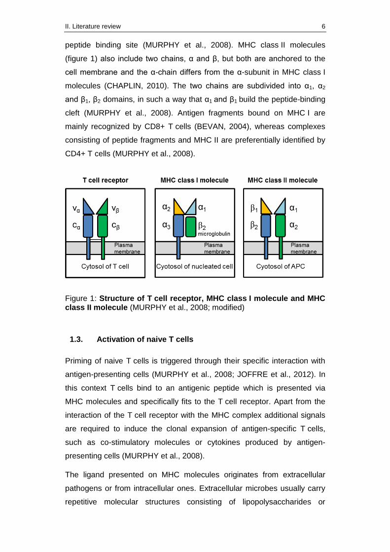
II. Literature review 6
peptide binding site (MURPHY et al., 2008). MHC class II molecules
(figure 1) also include two chains, α and β, but both are anchored to the
cell membrane and the α-chain differs from the α-subunit in MHC class I
molecules (CHAPLIN, 2010). The two chains are subdivided into α1, α2
and β1, β2 domains, in such a way that α1 and β1 build the peptide-binding
cleft (MURPHY et al., 2008). Antigen fragments bound on MHC I are
mainly recognized by CD8+ T cells (BEVAN, 2004), whereas complexes
consisting of peptide fragments and MHC II are preferentially identified by
CD4+ T cells (MURPHY et al., 2008).
Figure 1: Structure of T cell receptor, MHC class I molecule and MHC class II molecule (MURPHY et al., 2008; modified)
1.3. Activation of naive T cells
Priming of naive T cells is triggered through their specific interaction with
antigen-presenting cells (MURPHY et al., 2008; JOFFRE et al., 2012). In
this context T cells bind to an antigenic peptide which is presented via
MHC molecules and specifically fits to the T cell receptor. Apart from the
interaction of the T cell receptor with the MHC complex additional signals
are required to induce the clonal expansion of antigen-specific T cells,
such as co-stimulatory molecules or cytokines produced by antigen-
presenting cells (MURPHY et al., 2008).
The ligand presented on MHC molecules originates from extracellular
pathogens or from intracellular ones. Extracellular microbes usually carry
repetitive molecular structures consisting of lipopolysaccharides or
II. Literature review 7
carbohydrates on their bacterial cell wall, which are known as pathogen-
associated molecular patterns (PAMPs) (JANEWAY, 1989; IWASAKI &
MEDZHITOV, 2015). Macrophages, dendritic cells or other cells of the
innate immune system hold pattern recognition receptors (PRR) on their
cell surface which are able to recognize the PAMPs (JANEWAY, 1989;
IWASAKI & MEDZHITOV, 2015). The phagocytic cells furthermore
express receptors for the interaction with complement proteins. Apart from
direct binding, this additionally allows for recognition of opsonized
pathogens. After recognition and phagocytosis, antigen-presenting cells
process the pathogen intracellularly and present the newly formed
antigenic peptides via MHC class II on their cell surface to CD4+ T cells
(CHAPLIN, 2010). Antigen binding and processing furthermore also
induce enhanced cytokine production through the phagocytic cell. This
leads to the further activation of the innate immune response as well as
the adaptive one. Thus, antigen presenting cells, especially dendritic cells,
serve as a link between the innate and the adaptive immune system
(WARRINGTON et al., 2011). Intracellular pathogens like viruses or some
bacteria usually activate the immune system through another pathway.
Here, intracellularly produced antigenic proteins are degraded in the
proteasome and presented on MHC class I molecules. This leads to the
activation of CD8+ T cells (MURPHY et al., 2008).
Apart from the two major pathways described above there are also other
ways of antigen processing such as cross-presentation (CHAPLIN, 2010).
This pathway plays a key role for the activation of dendritic cells by viral
infections which do not directly affect the dendritic cells. Here, antigen-
presenting cells receive antigen delivered by other infected cells via
phagocytosis. This is mainly carried out by dendritic cells (CRESSWELL et
al., 2005). Afterwards the exogenous antigen can also be processed via
the proteasome and can finally be presented to cytotoxic T cells via MHC
class I molecules (MURPHY et al., 2008; JOFFRE et al., 2012). Generally
this strategy helps to control viruses which are able to avoid antigen
processing through the endogenous pathway (CHAPLIN, 2010).
After initial priming T lymphocytes start to proliferate and differentiate into
effector cells (AHMED & GRAY, 1996). In addition to the maturation of
II. Literature review 8
effector T cells, priming leads to the development of a small fraction of
memory T cells. These are long-lived T cells (AHMED & GRAY, 1996)
which accelerate the immune response in case of a subsequent infection
with the same pathogen responsible for priming. Furthermore memory
cells also exist despite the absence of their specific pathogenic antigens
(MURPHY et al., 2008). Generally, efficient vaccines should provide both,
T cell effector cells as well as memory T cells. This allows efficient control
of infectious pathogens including a higher magnitude of the secondary
response compared to primary responses (MURPHY et al., 2008;
SALLUSTO et al., 2010).
1.4. T cell response induced by vaccination
Up to date vaccines represent the most effective and cost-efficient
preventive method in modern medicine (SALLUSTO et al., 2010;
PULENDRAN & AHMED, 2011). The comprehensive use of vaccines is
able to protect individuals from infectious diseases and can at best even
result in the elimination of single pathogenic organisms (SALLUSTO et al.,
2010). For decades the development of vaccines has predominantly been
based on the induction of strong humoral immune responses. For this
reason it is quite common that most of the vaccines currently available are
based on the induction of antigen-specific antibodies (PLOTKIN, 2008).
However, antibody inducing vaccines are not effective enough against
numerous infectious pathogens which are more complex or even able to
chance their antigenic structure, such as human immunodeficiency virus
(HIV), influenza or Mycobacterium tuberculosis. The development of
vaccines against HIV for example has so far not been successful
(PULENDRAN & AHMED, 2011).
These findings clearly demonstrate the need for novel innovative
strategies in vaccine development. Moreover, data recently published also
highlight the importance of the T cell response and confirm that protection
against many threatening infectious diseases could be improved through
an increased induction of the cellular immune system (KREMER et al.,
2012a; ALTENBURG et al., 2014). The importance of the T cell response
is now generally accepted and therefore vaccine development is more and
II. Literature review 9
more focusing on the induction of T cells. The requirement of a strong
T cell response is for example clearly indicated in the control of
tuberculosis (HOFT, 2008) or HIV (PANTALEO & KOUP, 2004).
Additionally, influenza is also believed to require an efficient cellular
immune response. Here, the influenza-specific T cell response even
seems to correlate inversely with the risk of elderly people developing
sickness following vaccination (McELHANEY et al., 2006). Furthermore, it
is known that the protection against some infectious diseases such as
smallpox depends on T cell memory. In fact, antibodies can prevent
infection with smallpox and usually protect against severe disease, but the
infection will only be asymptomatic through the help of the cellular immune
response (PLOTKIN, 2008).
2. Modified Vaccinia virus Ankara (MVA) as vector
vaccine
2.1. MVA: a member of the family Poxviridae
Modified Vaccinia virus Ankara (MVA) is assigned to the genus
Orthopoxvirus which belongs to the family Poxviridae (MAYR et al., 1975).
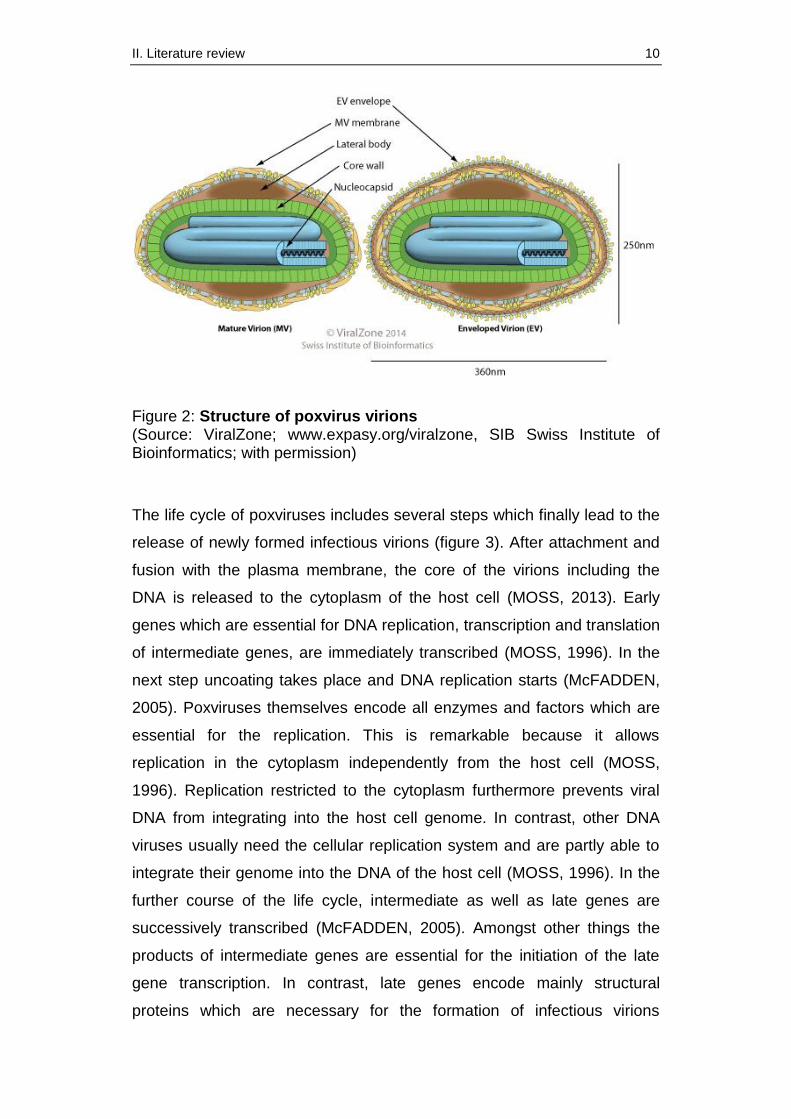
Poxviruses (figure 2) typically hold a single linear double-stranded DNA
genome of about 130 to 300 kilo base pairs which encodes a multiplicity of
viral proteins (MOSS, 1996). The virions of poxviruses are larger than
most other animal viruses and can even be seen in light microscopy (200
nm - 400 nm). Basically, the structure of the enveloped virions is relatively
complex. Additionally to the biconcave core, poxviruses form two lateral
bodies. The core includes the nucleocapsid with the s-shaped viral
genome and DNA associated proteins (MOSS, 2013).
II. Literature review 10
Figure 2: Structure of poxvirus virions (Source: ViralZone; www.expasy.org/viralzone, SIB Swiss Institute of Bioinformatics; with permission)
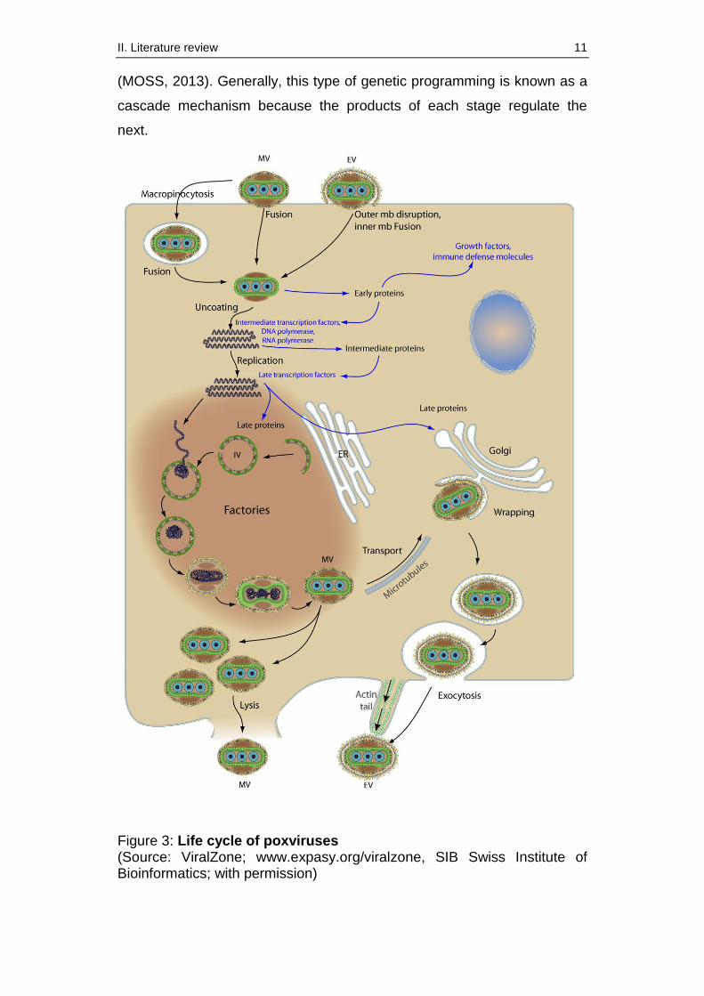
The life cycle of poxviruses includes several steps which finally lead to the
release of newly formed infectious virions (figure 3). After attachment and
fusion with the plasma membrane, the core of the virions including the
DNA is released to the cytoplasm of the host cell (MOSS, 2013). Early
genes which are essential for DNA replication, transcription and translation
of intermediate genes, are immediately transcribed (MOSS, 1996). In the
next step uncoating takes place and DNA replication starts (McFADDEN,
2005). Poxviruses themselves encode all enzymes and factors which are
essential for the replication. This is remarkable because it allows
replication in the cytoplasm independently from the host cell (MOSS,
1996). Replication restricted to the cytoplasm furthermore prevents viral
DNA from integrating into the host cell genome. In contrast, other DNA
viruses usually need the cellular replication system and are partly able to
integrate their genome into the DNA of the host cell (MOSS, 1996). In the
further course of the life cycle, intermediate as well as late genes are
successively transcribed (McFADDEN, 2005). Amongst other things the
products of intermediate genes are essential for the initiation of the late
gene transcription. In contrast, late genes encode mainly structural
proteins which are necessary for the formation of infectious virions
II. Literature review 11
(MOSS, 2013). Generally, this type of genetic programming is known as a
cascade mechanism because the products of each stage regulate the
next.
Figure 3: Life cycle of poxviruses (Source: ViralZone; www.expasy.org/viralzone, SIB Swiss Institute of Bioinformatics; with permission)
II. Literature review 12
After assembly and packaging of the newly replicated DNA, the viral
particles mature to infectious intracellular virions (mature virion, MV) and
get wrapped in the trans-Golgi (HILLER & WEBER, 1985; SCHMELZ et
al., 1994). The wrapped virions are directly transported to the plasma
membrane (STOKES, 1976). Finally, the virions are released to the
extracellular space as enveloped virions (EV) through fusion with the cell
membrane (MOSS, 2013).
2.2. Origin of MVA
The World Health Organization’s (WHO) smallpox eradication program
finally resulted in the elimination of smallpox in 1980. Vaccinia virus, the
virus used as vaccine during the program, was immunogenic, but it partly
induced severe side effects (KENNEDY et al., 2009; GILBERT, 2013).
Thus, it naturally was not the best vaccine possible, above all for people
with immunodeficiency. For this reason, the development of safe and
efficient smallpox vaccines was urgently needed. In this context, MVA was
developed at the LMU Institute for Microbiology and Infectious Diseases of
Animals in Munich (MAYR & MUNZ, 1964). It was derived from Vaccinia
virus strain Ankara after 516 serial passages on chicken embryo
fibroblasts (MAYR et al., 1975). This resulted among other things in six
major deletions of DNA, including several structural proteins, host range
genes (MEYER et al., 1991) and immune evasion genes (ANTOINE et al.,
1998). Apart from the six major deletions the genome was furthermore
affected by a couple of point mutations, shorter deletions and insertions
(ANTOINE et al., 1998; MEISINGER-HENSCHEL et al., 2007). Overall,
the loss of genomic original material amounts to approximately 15%
(about 30,000 base pairs) in comparison to the genome of the parenteral
Chorioallantois Vaccinia virus Ankara (CVA) (MEYER et al., 1991). Thus,
MVA lost the bigger part of its growth capacity and became replication
deficient in mammalian cells including human cells (MAYR et al., 1975;
CARROLL & MOSS, 1997; DREXLER et al., 1998). For this reason MVA
can even be used as a safe vaccine for immunocompromised people,
such as cancer patients receiving chemotherapy or people who have
contracted immunodeficiency virus. This finding basically resulted in the
II. Literature review 13
preferred use of MVA in vaccine development compared to the replication
competent Vaccinia virus. But despite the inability of MVA to replicate
early, intermediate as well as late gene expression still occurs in
mammalian cells (SUTTER & MOSS, 1992). In general, the high-level
attenuation of the virus allows handling of MVA in Germany under
biosafety level 1 conditions (ZKBS, 1997) and safe application in animal
experiments as well as in human clinical trials.
Originally, non-recombinant MVA was used for the vaccination of more
than 120,000 people in Germany (STICKL et al., 1974; MAYR et al.,
1978). During that period of time many field studies investigated and
confirmed the excellent safety profile of MVA (STICKL et al., 1974; MAYR
et al., 1975; MAYR et al., 1978). Today, MVA is approved in the European
Union and Canada as a smallpox vaccine for active immunization, even
for immunocompromised people (VOLLMAR et al., 2006; KENNEDY &
GREENBERG, 2009).
2.3. MVA: a vector for foreign genes
After the eradication of human smallpox, MVA has been used as a safe
viral vector for the generation of candidate vaccines for the prevention of
infectious diseases and cancer. In general, MVA has several useful
characteristics which make it suitable as a viral vector for vaccine
development (DREXLER et al., 2004; COTTINGHAM & CARROLL, 2013;
GILBERT, 2013; KREIJTZ et al., 2013; VOLZ & SUTTER, 2013;
ALTENBURG et al., 2014): i) The genome of MVA shows a high plasticity
and is relatively large which allows the insertion of foreign gene
sequences. ii) MVA is replication deficient in mammalian cells which
indicates the use even in immunocompromised people. iii) Several
studies showed an excellent safety record of MVA after use as a vaccine
in humans. iv) MVA is able to induce humoral as well as cellular immune
responses. v) Commonly available laboratory protocols allow easy and
efficient handling of MVA.
As a representative of poxviruses MVA shows a high genetic plasticity
which enables the insertion of heterologous gene sequences. The
II. Literature review 14
generation of recombinant MVA usually proceeds according to a well-
established and standardized laboratory system using a vector plasmid
(KREMER et al., 2012b). Thus, foreign genes can be inserted into the viral
genome via homologous recombination between flanking sequences in
the vector and the MVA genome (MOSS, 1996). Besides the gene of
interest, the vector plasmid also needs to include a promoter sequence.
The promoter has to be Vaccinia virus specific because poxviruses avoid
using the transcriptional system of the host cell through holding their
complete own transcription machinery (MOSS, 1996). Furthermore the
vector plasmid should also contain a marker gene which facilitates specific
selection of recombinant MVA. Screening for the expression of fluorescent
marker genes allows efficient isolation of recombinant viruses through
several plaque passages in the cell culture. Finally, recombinant MVAs are
able to synthesize high amounts of viral as well as recombinant protein in
the cytosol of infected cells. This provides the basis for the function of a
well-working expression vector (SUTTER & MOSS, 1992).
2.4. MVA as a platform for vaccine development
Preclinical studies already showed the suitability of MVA as a viral vector
more than 15 years ago (MOSS et al., 1996). So far, many different
recombinant MVAs delivering antigens of various infectious diseases have
been constructed and characterized. Previously published data for
example indicate the use of recombinant MVA for the prophylaxis of
malaria or human immunodeficiency virus (COSMA et al., 2003;
MOORTHY et al., 2003b; MOORTHY et al., 2003a). Several recombinant
MVAs have already been tested in phase I to phase IIb clinical trials
including vaccines against viral infections (table 1a) such as influenza,
hepatitis or human deficiency virus (HIV) and also against bacteria and
parasites (table 1b) (KREIJTZ et al., 2013). Vaccination with these
recombinant MVAs has generally been well-tolerated. Safety data from
these clinical trials have reported no severe or serious side effects
(MOORTHY et al., 2003b; GILBERT, 2013; GOMEZ et al., 2013).
Moreover, the MVA candidate vaccines were immunogenic as predicted in
previous preclinical studies, and they induced humoral as well as cellular
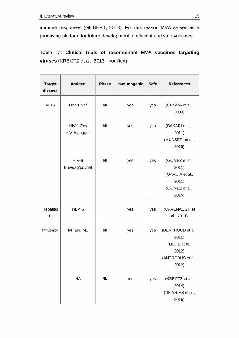
II. Literature review 15
immune responses (GILBERT, 2013). For this reason MVA serves as a
promising platform for future development of efficient and safe vaccines.
Table 1a: Clinical trials of recombinant MVA vaccines targeting
viruses (KREIJTZ et al., 2013; modified)
Target
disease
Antigen
Phase
Immunogenic
Safe
References
AIDS
HIV-1 Nef
HIV-1 Env
HIV-A gag/pol
HIV-B
Env/gag/pol/nef
I/II
I/II
I/II
yes
yes
yes
yes
yes
yes
(COSMA et al.,
2003)
(BAKARI et al.,
2011)
(MUNSERI et al.,
2015)
(GOMEZ et al.,
2011)
(GARCIA et al.,
2011)
(GOMEZ et al.,
2015)
Hepatitis
B
HBV S
I
yes
yes
(CAVENAUGH et
al., 2011)
Influenza
NP and M1
HA
I/II
I/IIa
yes
yes
yes
yes
(BERTHOUD et al.,
2011)
(LILLIE et al.,
2012)
(ANTROBUS et al.,
2012)
(KREIJTZ et al.,
2014)
(DE VRIES et al.,
2015)
II. Literature review 16
Table 1b: Clinical trials of recombinant MVA vaccines targeting
bacteria and parasites (KREIJTZ et al., 2013; modified)
Target
disease
Antigen
Phase
Immunogenic
Safe
References
Malaria
P. falcip.
AMA1
P.falcip. ME-
TRAP
P. falcip.
ME-TRAP
P. falcip.
ME-TRAP
I
IIb
IIb
I
yes
yes
yes
yes
yes
yes
yes
yes
(SHEEHY et al.,
2012)
(BEJON et al.,
2006)
(BEJON et al.,
2007)
(MOORTHY et al.,
2004)
(HODGSON et al.,
2015)
Tuberculosis
85A (MTB)
85A (MTB)
IIb
I
yes
yes
yes
yes
(TAMERIS et al.,
2013)
(SHEEHAN et al.,
2015)
Besides the advanced investigation of promising antigens in clinical trials,
preclinical studies consistently investigate new MVA vaccine candidates.
In this context, recently published data for example highlight the suitability
of MVA as a vector for more complex diseases such as Middle East
respiratory syndrome coronavirus (MERS-CoV) (SONG et al., 2013).
2.5. MVA’s antigen presentation pathway
Generally, thorough knowledge of MVA’s life cycle and its antigen
presentation pathway is essential for the development of new effective
vaccine candidates. In this context, the main mechanism of processing
intracellular viral antigens delivered by MVA, is the endogenous pathway
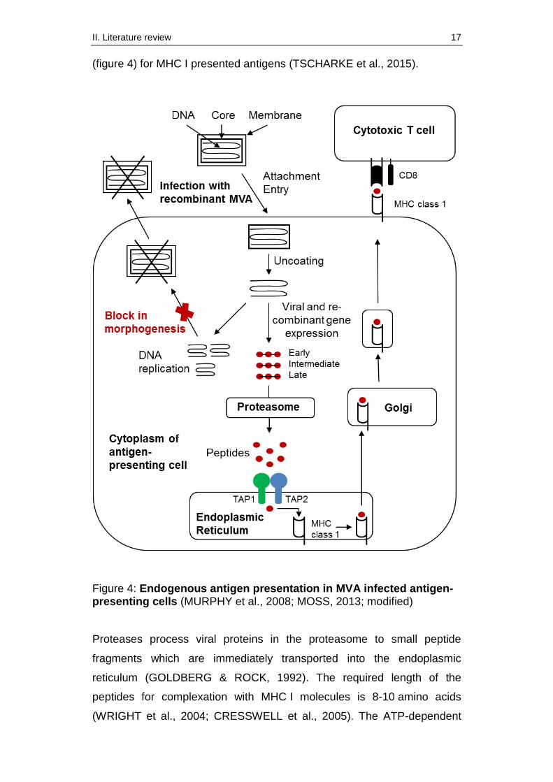
II. Literature review 17
(figure 4) for MHC I presented antigens (TSCHARKE et al., 2015).
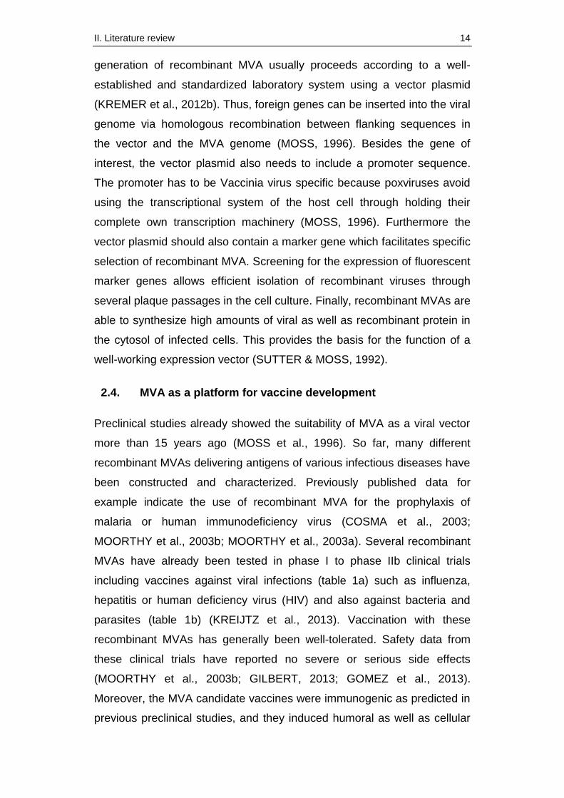
Figure 4: Endogenous antigen presentation in MVA infected antigen-presenting cells (MURPHY et al., 2008; MOSS, 2013; modified)
Proteases process viral proteins in the proteasome to small peptide
fragments which are immediately transported into the endoplasmic
reticulum (GOLDBERG & ROCK, 1992). The required length of the
peptides for complexation with MHC I molecules is 8-10 amino acids
(WRIGHT et al., 2004; CRESSWELL et al., 2005). The ATP-dependent
II. Literature review 18
transport of the peptides across the endoplasmic membrane is mediated
by two proteins called TAP 1, TAP 2 (transporters associated with antigen
processing) which build a heterodimer (ANDROLEWICZ et al., 1993;
CRESSWELL et al., 2005). This transport system belongs to the ATP-
binding cassette family (ABC) and consists of four domains, two
transmembrane and two ATP-binding domains (HIGGINS, 1992). The
encoding sequences of TAP 1 and TAP 2 are located within the MHC. It is
remarkable that the transcription and translation of these genes can be
enhanced by interferons which are produced by various cells of the
immune systems as a response to viral infections (MURPHY et al., 2008).
In the endoplasmic reticulum the viral peptide fragments are loaded on the
MHC class I molecules (CHAPLIN, 2010). This complex is subsequently
transported to the cell surface and presented to cytotoxic T cells
(MURPHY et al., 2008). Generally, this strategy allows the immune system
to detect transformed cells as well as infected cells (JOFFRE et al., 2012).
Apart from the endogenous pathway, MVA is also able to activate
cytotoxic T cells via cross-presentation by dendritic cells (TSCHARKE et
al., 2015).
3. Target antigens for MVA-induced CD8+ T cells
3.1. Influenza antigens
Influenza viruses cause infections in the respiratory system of humans as
well as of animals. They show a high genetic variability and an enormous
capacity for antigenic drift (HAMPSON & MACKENZIE, 2006; GAMBLIN &
SKEHEL, 2010) which can lead to the development of new viral subtypes.
This strategy allows the virus to evade the humoral immunity of the host
(WONG & PAMER, 2003). For this reason novel influenza subtypes
especially possess pandemic potential because they often cannot be
recognized and eliminated by antibodies induced by vaccination with the
seasonal influenza vaccine or by a previous infection (HILLAIRE et al.,
2011). Here, the virus-specific cellular immune response is considered to
play a key role in the control of the virus and could also strongly support
the induction of cross-protective immunity (HILLAIRE et al., 2011;
II. Literature review 19
ALTENBURG et al., 2014). Different studies have already shown the
relevance of CD8+ T cell responses in challenge experiments (FLYNN et
al., 1998; WONG & PAMER, 2003). Furthermore, it is known that memory
T cells are essential for rapid protection of mice in case of reinfection
(FLYNN et al., 1999; WONG & PAMER, 2003). Therefore the strong
activation of T cells is a vital target in today’s development of more broad-
protective vaccines. Successful development and licensing of such
vaccines is a major aim regarding the high morbidity and mortality of
epidemic influenza outbreaks which are especially threatening for elderly
or immunocompromised people.
In 1994 a study first described that vaccination with recombinant MVA
expressing hemagglutinin (HA) and nucleoprotein (NP) is able to protect
mice from lethal challenge with influenza virus (SUTTER et al., 1994).
Today several influenza antigens are known and investigated for their
potential to induce T cell responses. Recently published data evaluated
different MVA vaccines encoding influenza antigens for their potential to
induce antigen-specific CD8+ T cell responses. In this context,
hemagglutinin, nucleoprotein and matrixprotein 1 (M1) are promising
antigens used in the MVA vector system. Hemagglutinin is a glycoprotein
located on the surface of influenza virions (GAMBLIN & SKEHEL, 2010). It
mediates binding of the virus particle to the host cell and is also involved
into the fusion of the viral envelope with the endosome membrane
(GARTEN & KLENK, 1999). In contrast, nucleoprotein and matrixprotein
belong to the structural proteins of the virus. Nucleoprotein is known as an
RNA-binding protein which is responsible for the encapsidation of viral
RNA (PORTELA & DIGARD, 2002). Thus, it also interacts with
matrixprotein 1 and other viral molecules. It furthermore includes a nuclear
localization signal and mediates the translocation of viral RNA into the cell
nucleus. M1 is also a RNA-binding protein (YE et al., 1999), but it is able
to bind the plasma membrane as well. Simultaneous binding of RNA and
membrane through M1 is important for the encapsidation of the RNA into
the viral envelope. Table 2 shows a selection of recombinant MVA recently
tested expressing influenza proteins. It summarizes their potential to
induce cellular immune responses against homologous and heterologous
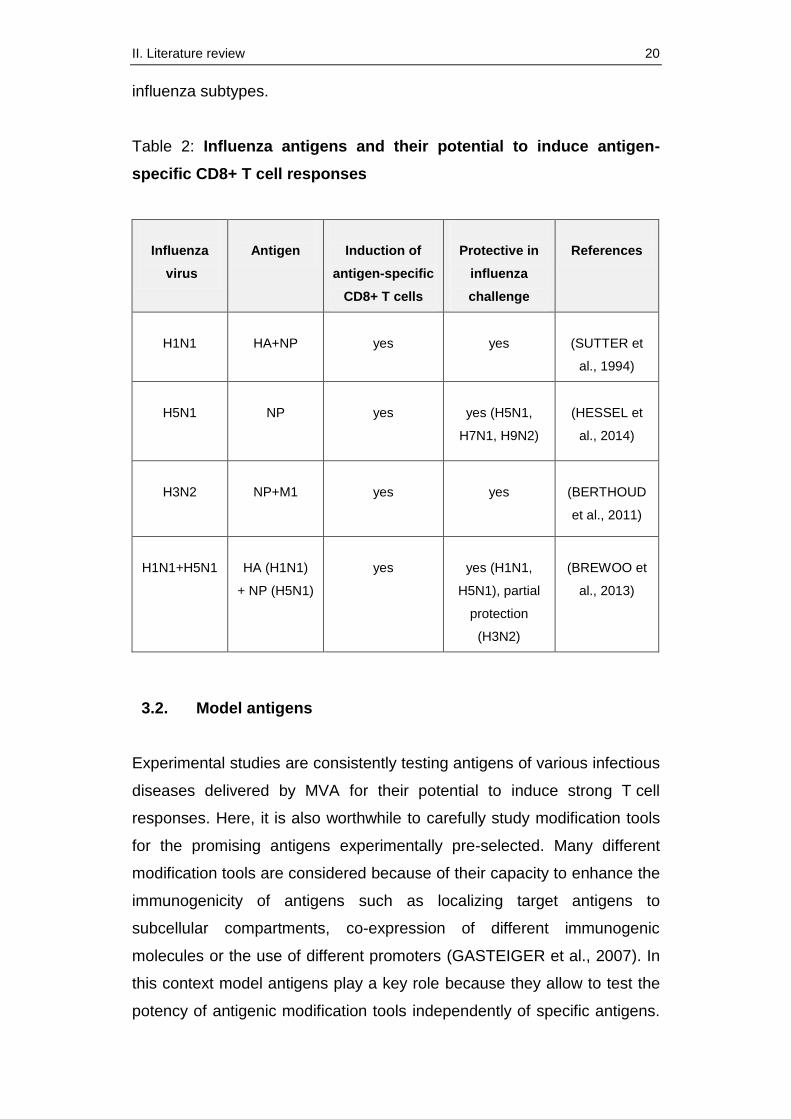
II. Literature review 20
influenza subtypes.
Table 2: Influenza antigens and their potential to induce antigen-
specific CD8+ T cell responses
Influenza
virus
Antigen
Induction of
antigen-specific
CD8+ T cells
Protective in
influenza
challenge
References
H1N1
HA+NP
yes
yes
(SUTTER et
al., 1994)
H5N1
NP
yes
yes (H5N1,
H7N1, H9N2)
(HESSEL et
al., 2014)
H3N2
NP+M1
yes
yes
(BERTHOUD
et al., 2011)
H1N1+H5N1
HA (H1N1)
+ NP (H5N1)
yes
yes (H1N1,
H5N1), partial
protection
(H3N2)
(BREWOO et
al., 2013)
3.2. Model antigens
Experimental studies are consistently testing antigens of various infectious
diseases delivered by MVA for their potential to induce strong T cell
responses. Here, it is also worthwhile to carefully study modification tools
for the promising antigens experimentally pre-selected. Many different
modification tools are considered because of their capacity to enhance the
immunogenicity of antigens such as localizing target antigens to
subcellular compartments, co-expression of different immunogenic
molecules or the use of different promoters (GASTEIGER et al., 2007). In
this context model antigens play a key role because they allow to test the
potency of antigenic modification tools independently of specific antigens.
II. Literature review 21
In the next step the combination of the most promising tools and pre-
selected antigens can be investigated for their immunogenicity. This
procedure clearly decreases the number of experiments and studies which
would usually be necessary to test all combinations of modifications and
antigens. Several model antigens with different characteristics are
established for the MVA vector system. Consequently the most commonly
used model systems are to be presented in the following chapter.
3.2.1. Ovalbumin
The chicken egg white protein ovalbumin (OVA) belongs to the serpin
superfamily (HUNT & DAYHOFF, 1980). The name of this superfamily is
based on the function of the main part of all associated proteins which
typically are serin proteinase inhibitors (GETTINS, 2002). The exact
function of the non-inhibitory OVA is still unknown, but it is supposed to be
a storage protein (GETTINS, 2002; BENARAFA & REMOLD-O'DONNELL,
2005). OVA’s X-ray structure was finally determined in 1990 (STEIN et al.,
1990). This well-conserved protein has so far been used as a model
antigen for recombinant MVA in many different experimental studies. Cells
infected with recombinant MVA expressing OVA (MVA-OVA) usually
secret ovalbumin constantly to the medium (BECKER et al., 2014). For
this reason OVA serves as an appropriate model antigen for the
comparison of modification tools which are supposed to have an influence
on the strength of the antigen expression such as different promoters
(BECKER et al., 2014). Furthermore, secretion to the medium allows
analysis of protein expressed without affecting the cells. OVA is also well-
established and frequently used as a model antigen for the analysis and
comparison of T cell responses in immunization experiments (BAUR et al.,
2010; BECKER et al., 2014). It is not only used as a model system, but
also as a promising antigen in the development of vaccines for the
prevention of allergic symptoms caused by food allergies. Thus, previously
published data clearly suggest MVA-OVA as a potential vaccine against
allergic symptoms induced by chicken egg white (ALBRECHT et al., 2008;
BOHNEN et al., 2013).
II. Literature review 22
3.2.2. β-Galactosidase
β-Galactosidase is a member of the enzyme family glycosyl hydrolases
which are typically able to hydrolase O-glycosyl linkages (HENRISSAT,
1991). For this reason these enzymes play a key role in the carbohydrate
metabolism of the human body. Enzyme deficiencies triggered through
mutations in the β-Galactosidase gene can result in GM1-gangliosidosis or
Morquio B syndrome. Both are rare autosomal recessive lysosomal
storage diseases (CACIOTTI et al., 2011). GM1-gangliosidosis results in a
pathological accumulation of acidic lipids in the lysosomes, especially in
cells of the central nerve system. In contrast, Morquio B syndrome is a
mucopolysaccharidosis which mainly affects the skeletal and the
cardiovascular system (CACIOTTI et al., 2011). The β-Galactosidase used
in molecular biology is originated from E. coli and is encoded by the lacZ
gene. This gene belongs to the inducible lac operon system (OSBOURN &
FIELD, 2009). It helps the bacteria to permanently adapt their enzyme
expression to the availability of alimentary substrate. In the presence of
lactose and in the simultaneous absence of glucose, the lac operon
system is activated and encodes - apart from other molecules - β-
Galactosidase which is essential for the lactose metabolism. β-
Galactosidase catalyzes the hydrolysis of the disaccharide lactose in
glucose and galactose (LEONARD et al., 2015). In molecular biology β-
Galactosidase serves on the one hand as a reporter for gene expression
because it is able to stain tissue cultures blue in the presence of enzyme
specific substrate (ARMIT, 2015). On the other hand it is also used as a
viral tumor associated antigen in cancer research. These antigens allow
the generation of recombinant anti-cancer vaccines on the basis of MVA.
Furthermore, β-Galactosidase delivered by MVA is able to specifically
induce T lymphocytes (CARROLL et al., 1997). For this reason research
especially makes use of recombinant MVA delivering β-Galactosidase in
studies about novel treatment options against cancer, in which cytotoxic
T cells play the key role (CARROLL et al., 1997). T cell leucemias induced
by T cell lymphotropic virus (DE THE et al., 1994) or Burkitt’s lymphoma
caused by Epstein-Barr virus (MOSS et al., 1994) for example belong to
this kind of cancer. More than 15 years ago MVA expressing β-
II. Literature review 23
Galactosidase has also successfully been used to protect and treat
pulmonary metastases which deliver β-Galactosidase (CARROLL et al.,
1997).
3.2.3. Green fluorescent protein (GFP)
In general, fluorescent proteins serve as an innovate tool in molecular
biology. They allow the visualization, observation and thus the better
understanding of processes within cells, bacteria or viruses. Today many
different fluorescent proteins are established for laboratory use such as
mCherry (red fluorescent protein), mOrange (orange fluorescent protein)
or Venus (yellow fluorescent protein) (KREMERS et al., 2011). But the
discovery of green fluorescent protein and its use in cell culture is still
considered as the milestone in the development of fluorescent proteins.
GFP was derived from the green fluorescent jellyfish Aequorea victoria
and firstly described in 1962 (SHIMOMURA et al., 1962). The crystal
structure of GFP was finally published in 1996 (YANG et al., 1996).
Generally, the field of fluorescent proteins has been studied very well over
years. In 2008 the Nobel Prize in chemistry was finally awarded to Martin
Chalfie, Osamu Shimomura and Roger Tsien for their outstanding
scientific studies about GFP (CHALFIE, 2009; REMINGTON, 2011). As
early as in 1994 Chalfie published the suitability of GFP as a marker for
gene expression in prokaryotic as well as in eukaryotic cells. It is
remarkable that the expression of GFP has no obvious negative influence
on cell growth or function which allows monitoring of gene expression and
protein localization in living cells (CHALFIE et al., 1994). Moreover, the
fluorescence is not affected by treatment with formaldehyde which is
useful for the analysis of fixed cell preparations (CHALFIE et al., 1994).
By now GFP has been established in many experimental settings. It is
very well characterized and probably the most commonly used fluorescent
protein in molecular biology. GFP is a protein of approximately 240 amino
acids which can spontaneously absorb blue light (maximum at 395 nm).
Furthermore it is able to very stably emit green fluorescence (peak at
309 nm) (CHALFIE et al., 1994). GFP can efficiently be targeted to the
major cell organelles as for example the cell nucleus, plasma membrane,
II. Literature review 24
endoplasmic reticulum or mitochondria (TSIEN, 1998). So far GFP has
also been used as a visualizable tool in many studies dealing with
recombinant MVA (for example: COSMA et al., 2004; PASCUTTI et al.,
2011; WONG et al., 2011) including the analysis of T cell responses (DI
PILATO et al., 2015).
In this study we used GFP as a model antigen to analyze the influence of
localization signals on the induction of antigen-specific cellular
immunogenicity. We preferred working with GFP as a model antigen
because it has several characteristics which are beneficial for the
experimental setting: i) GFP is - in difference to OVA - not secreted to the
medium which could influence and disturb the experimental testing
systems. ii) GFP allows direct visualization of the different subcellular
protein localization sites. iii) GFP as a model antigen allows to analyze the
effect triggered by localization signals independently of specific infectious
antigens. iv) GFP is very well characterized and established in many
experimental settings. v) GFP has already successfully been used in the
MVA vector system.
III. Objectives 25
III. OBJECTIVES
Due to the need of new efficient and safe vaccines for the control of
complex infectious diseases and due to the suitability of MVA as an
effective and safe viral vector this work describes:
(i) The generation of recombinant MVA expressing GFP in combination
with different localization signals
(ii) In vitro characterization of the recombinant MVA
Analysis on DNA level
Analysis on protein level
Fluorescence microscopy
(iii) In vivo characterization of the recombinant MVA
Vaccination experiment in mice
Analysis of the induced CD8+ T cell response
IV. Results 26
IV. RESULTS
The manuscript is presented in form accepted for publication (MARR et
al., 2016).
Myristoylation increases the CD8+ T cell response
to a green fluorescent protein prototype antigen
delivered by Modified Vaccinia virus Ankara
Lisa Marr1, Anna-Theresa Lülf1, Astrid Freudenstein1, Gerd Sutter1 and
Asisa Volz1
1German Centre for Infection Research (DZIF), Institute for Infectious
Diseases and Zoonoses, LMU University of Munich, Veterinaerstrasse 13,
D-80539 Munich, Germany
Journal of General Virology
February 2016
doi: 10.1099/jgv.0.000425
IV. Results 27
Activation of CD8+ T cells is an essential part of immune responses
elicited by recombinant Modified Vaccinia virus Ankara (MVA).
Strategies to enhance T cell responses to antigens may be
particularly necessary for broadly protective immunization against
influenza A virus infections or for candidate vaccines targeting
chronic infections and cancer. Here, we tested recombinant MVAs
that target a model antigen, green fluorescent protein (GFP), to
different localizations in infected cells. In vitro characterization
demonstrated that GFP accumulated in the nucleus (MVA-nls-GFP),
associated with cellular membranes (MVA-myr-GFP) or was equally
distributed throughout the cell (MVA-GFP). On vaccination, we found
significantly higher levels of GFP-specific CD8+ T cells in MVA-myr-
GFP vaccinated BALB/c mice than in those immunized with MVA-
GFP or MVA-nls-GFP. Thus, myristoyl modification may be a useful
strategy to enhance CD8+ T cell responses to MVA-delivered target
antigens.
Modified Vaccinia virus Ankara (MVA) is a replication-deficient and safety-
tested Vaccinia virus strain that can be engineered as a vector virus
encoding foreign antigens (SUTTER & MOSS, 1992; SUTTER et al.,
1994). Today, MVA vectors serve as an established platform technology
for developing vaccines against infectious diseases and cancer (KREIJTZ
et al., 2013; VOLZ & SUTTER, 2013; ALTENBURG et al., 2014;
SEBASTIAN & GILBERT, 2016). Various recombinant MVA have been
tested successfully in phase I to phase IIb clinical trials, and have been
found to be safe and immunogenic, inducing both target antigen-specific
antibodies and cellular immune responses (GILBERT, 2013; GOMEZ et
al., 2013). In a recent phase I study, immunizations with recombinant MVA
delivering the hemagglutinin (HA) antigen of Influenza A virus H5N1
(MVA-HA) induced high levels of H5-specific antibodies (KREIJTZ et al.,
2014).
In addition, the ability to activate strong cellular immune responses is an
important factor for the use of recombinant MVA in the search for influenza
vaccines with improved efficacy. Enhancing antigen specific T cell
responses might be a promising strategy for developing broadly protective
IV. Results 28
vaccines against influenza A virus. So far, two major parameters are
reported to influence efficient T cell responses: (i) optimal use of early
promoters for recombinant gene expression to support direct antigen
presentation and priming of T cells (BRONTE et al., 1997;
KASTENMULLER et al., 2006; KASTENMULLER et al., 2007) and (ii) the
synthesis and delivery of stable mature protein antigens as preferred
substrates for efficient priming of T cells by cross-presentation
(GASTEIGER et al., 2007; PASCUTTI et al., 2011).
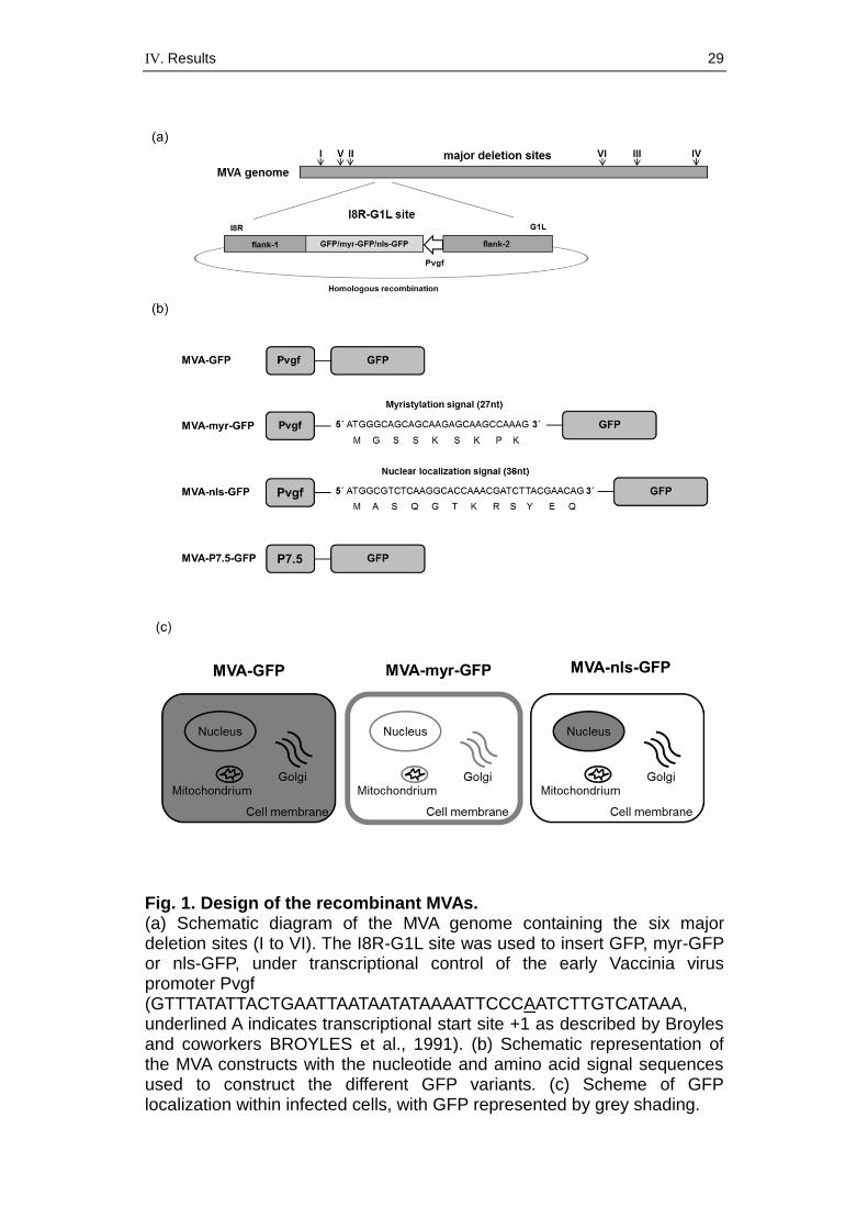
Localizing target antigens to subcellular compartments is also considered
as an innovative approach to enhance the cellular immune response
(GASTEIGER et al., 2007). To analyze the effect of different antigen
localizations on immunogenicity, we chose green fluorescent protein
(GFP) as a model antigen and assessed the induction of GFP epitope-
specific CD8+ T cells mediated by recombinant MVA producing either
unmodified GFP or GFPs containing nuclear localization (nls) or
myristoylation (myr) signals. The gene sequences encoding the target
antigens GFP, myr-GFP and nls-GFP were introduced at the site between
the Vaccinia virus (VACV) G1L and I8R genes (all gene nomenclatures as
established for VACV strain Copenhagen; GOEBEL et al., 1990) by
homologous recombination and placed under transcriptional control of
Pvgf promoter sequences (Fig. 1a). Pvgf is a natural Vaccinia virus
promoter controlling the abundant expression of VACV ORF C11R mRNA
(YANG et al., 2015).
Recombinant MVA expressing unmodified GFP served as a base-line
vaccine. To achieve increased transport of GFP to cellular membranes,
we added a myristoylation signal (MAURER-STROH et al., 2002; CHAN et
al., 2011), and for nuclear localization we tagged the GFP with a specific
nuclear localization signal (WANG et al., 1997) that translocates GFP to
the cell nucleus (Fig. 1b). As a further control, recombinant virus MVA-
P7.5-GFP was used, encoding GFP under the transcriptional control of the
natural VACV early-late promoter P7.5 (MACKETT et al., 1982), the first
and probably still most widely used promoter for constructing recombinant
Vaccinia viruses.
IV. Results 29
Fig. 1. Design of the recombinant MVAs. (a) Schematic diagram of the MVA genome containing the six major deletion sites (I to VI). The I8R-G1L site was used to insert GFP, myr-GFP or nls-GFP, under transcriptional control of the early Vaccinia virus promoter Pvgf (GTTTATATTACTGAATTAATAATATAAAATTCCCAATCTTGTCATAAA, underlined A indicates transcriptional start site +1 as described by Broyles and coworkers BROYLES et al., 1991). (b) Schematic representation of the MVA constructs with the nucleotide and amino acid signal sequences used to construct the different GFP variants. (c) Scheme of GFP localization within infected cells, with GFP represented by grey shading.
IV. Results 30
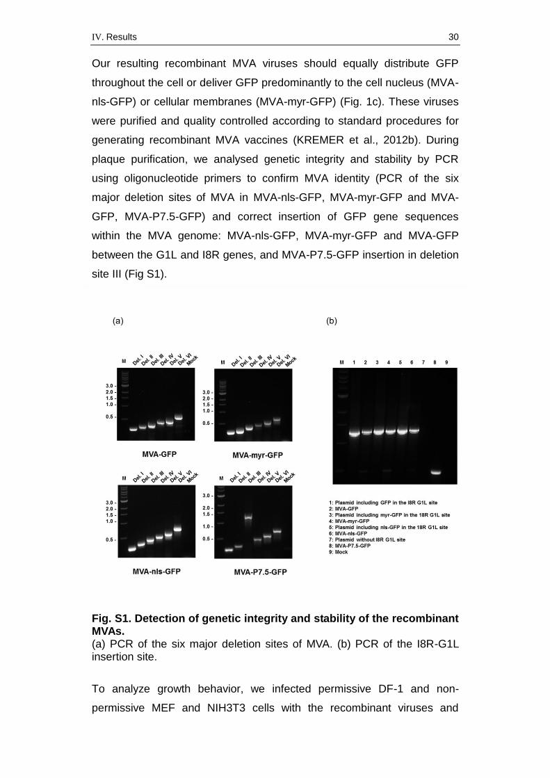
Our resulting recombinant MVA viruses should equally distribute GFP
throughout the cell or deliver GFP predominantly to the cell nucleus (MVA-
nls-GFP) or cellular membranes (MVA-myr-GFP) (Fig. 1c). These viruses
were purified and quality controlled according to standard procedures for
generating recombinant MVA vaccines (KREMER et al., 2012b). During
plaque purification, we analysed genetic integrity and stability by PCR
using oligonucleotide primers to confirm MVA identity (PCR of the six
major deletion sites of MVA in MVA-nls-GFP, MVA-myr-GFP and MVA-
GFP, MVA-P7.5-GFP) and correct insertion of GFP gene sequences
within the MVA genome: MVA-nls-GFP, MVA-myr-GFP and MVA-GFP
between the G1L and I8R genes, and MVA-P7.5-GFP insertion in deletion
site III (Fig S1).
Fig. S1. Detection of genetic integrity and stability of the recombinant MVAs. (a) PCR of the six major deletion sites of MVA. (b) PCR of the I8R-G1L insertion site.
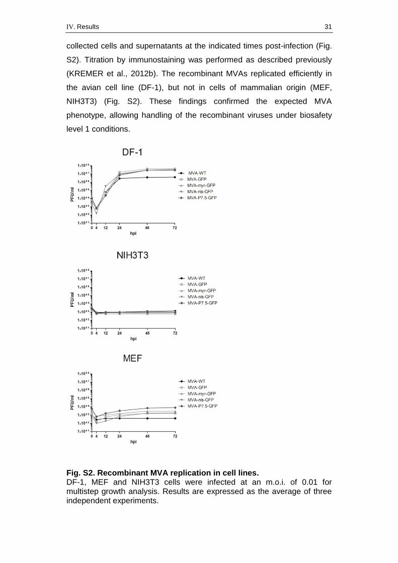
To analyze growth behavior, we infected permissive DF-1 and non-
permissive MEF and NIH3T3 cells with the recombinant viruses and
IV. Results 31
collected cells and supernatants at the indicated times post-infection (Fig.
S2). Titration by immunostaining was performed as described previously
(KREMER et al., 2012b). The recombinant MVAs replicated efficiently in
the avian cell line (DF-1), but not in cells of mammalian origin (MEF,
NIH3T3) (Fig. S2). These findings confirmed the expected MVA
phenotype, allowing handling of the recombinant viruses under biosafety
level 1 conditions.
Fig. S2. Recombinant MVA replication in cell lines. DF-1, MEF and NIH3T3 cells were infected at an m.o.i. of 0.01 for multistep growth analysis. Results are expressed as the average of three independent experiments.
IV. Results 32
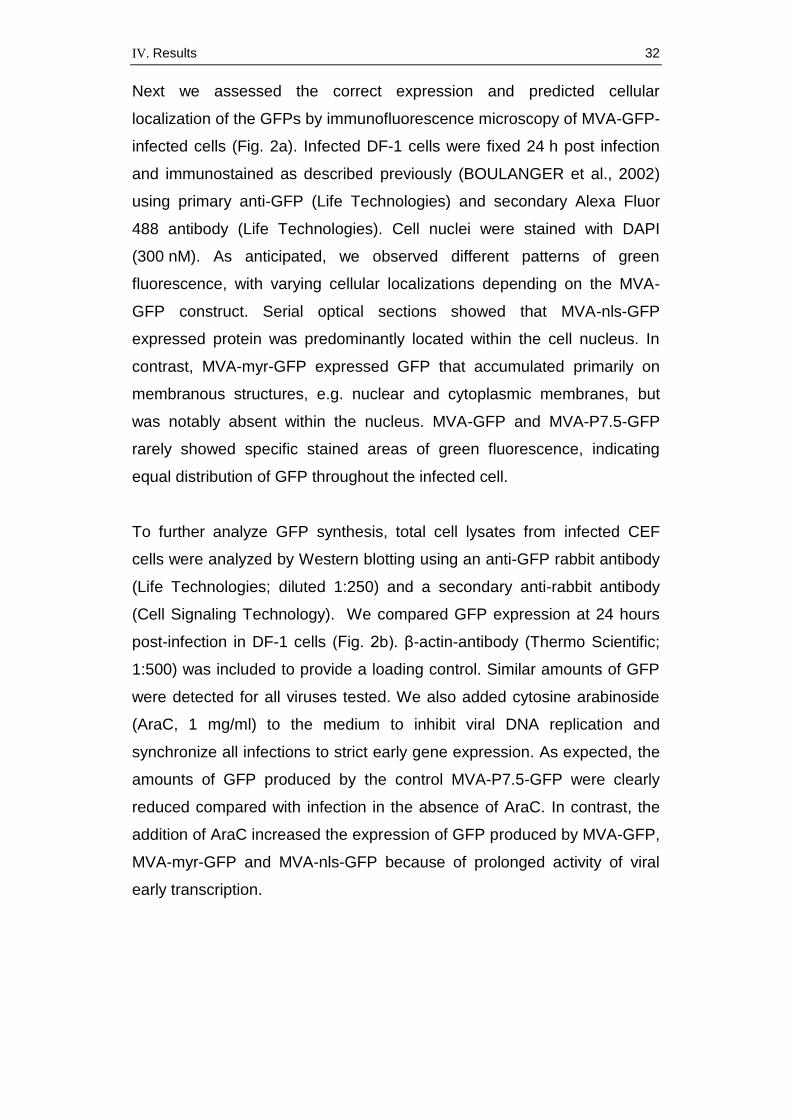
Next we assessed the correct expression and predicted cellular
localization of the GFPs by immunofluorescence microscopy of MVA-GFP-
infected cells (Fig. 2a). Infected DF-1 cells were fixed 24 h post infection
and immunostained as described previously (BOULANGER et al., 2002)
using primary anti-GFP (Life Technologies) and secondary Alexa Fluor
488 antibody (Life Technologies). Cell nuclei were stained with DAPI
(300 nM). As anticipated, we observed different patterns of green
fluorescence, with varying cellular localizations depending on the MVA-
GFP construct. Serial optical sections showed that MVA-nls-GFP
expressed protein was predominantly located within the cell nucleus. In
contrast, MVA-myr-GFP expressed GFP that accumulated primarily on
membranous structures, e.g. nuclear and cytoplasmic membranes, but
was notably absent within the nucleus. MVA-GFP and MVA-P7.5-GFP
rarely showed specific stained areas of green fluorescence, indicating
equal distribution of GFP throughout the infected cell.
To further analyze GFP synthesis, total cell lysates from infected CEF
cells were analyzed by Western blotting using an anti-GFP rabbit antibody
(Life Technologies; diluted 1:250) and a secondary anti-rabbit antibody
(Cell Signaling Technology). We compared GFP expression at 24 hours
post-infection in DF-1 cells (Fig. 2b). β-actin-antibody (Thermo Scientific;
1:500) was included to provide a loading control. Similar amounts of GFP
were detected for all viruses tested. We also added cytosine arabinoside
(AraC, 1 mg/ml) to the medium to inhibit viral DNA replication and
synchronize all infections to strict early gene expression. As expected, the
amounts of GFP produced by the control MVA-P7.5-GFP were clearly
reduced compared with infection in the absence of AraC. In contrast, the
addition of AraC increased the expression of GFP produced by MVA-GFP,
MVA-myr-GFP and MVA-nls-GFP because of prolonged activity of viral
early transcription.
IV. Results 33
Fig. 2. Characterization of recombinant GFP proteins. (a) Immunofluorescence staining of DF-1 cells infected with MVA viruses at an m.o.i. of 0.05 on cover slides and fixed 24 h post infection. Nuclei were stained with DAPI. Fluorescent images were captured with Keyence BZ-X710 at 100x magnification. (b) Western blot analysis of cell lysates prepared from DF-1 cells infected with MVA-GFP, MVA-nls-GFP, MVA-myr-GFP and MVA-P7.5-GFP at an m.o.i of 5, with or without the addition of cytosine arabinoside (AraC), 24 hours post infection.
IV. Results 34
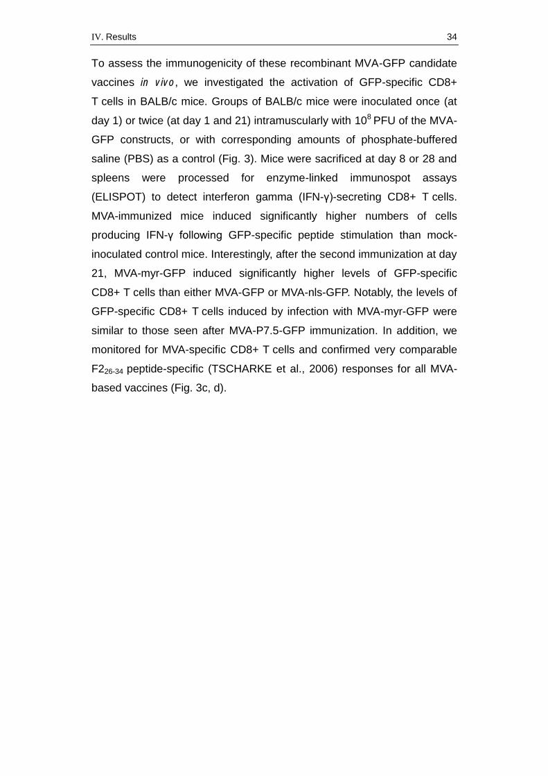
To assess the immunogenicity of these recombinant MVA-GFP candidate
vaccines in vivo, we investigated the activation of GFP-specific CD8+
T cells in BALB/c mice. Groups of BALB/c mice were inoculated once (at
day 1) or twice (at day 1 and 21) intramuscularly with 108 PFU of the MVA-
GFP constructs, or with corresponding amounts of phosphate-buffered
saline (PBS) as a control (Fig. 3). Mice were sacrificed at day 8 or 28 and
spleens were processed for enzyme-linked immunospot assays
(ELISPOT) to detect interferon gamma (IFN-γ)-secreting CD8+ T cells.
MVA-immunized mice induced significantly higher numbers of cells
producing IFN-γ following GFP-specific peptide stimulation than mock-
inoculated control mice. Interestingly, after the second immunization at day
21, MVA-myr-GFP induced significantly higher levels of GFP-specific
CD8+ T cells than either MVA-GFP or MVA-nls-GFP. Notably, the levels of
GFP-specific CD8+ T cells induced by infection with MVA-myr-GFP were
similar to those seen after MVA-P7.5-GFP immunization. In addition, we
monitored for MVA-specific CD8+ T cells and confirmed very comparable
F226-34 peptide-specific (TSCHARKE et al., 2006) responses for all MVA-
based vaccines (Fig. 3c, d).
IV. Results 35
Fig. 3. Immunogenicity of recombinant MVA-GFP candidate vaccines.
Groups of Balb/c mice (n=6) were immunized intramuscularly with 108
PFU MVA or mock vaccinated (PBS, n=4). (a, c) At day 8 post vaccination, or (b, d) at day 28, following a boost at day 21, GFP-specific (a, b) or MVA F226-34-specific (c, d) IFNγ-producing CD8+T cells were measured using an ELISPOT kit (Mabtech) for mouse IFN-γ following the manufacturer’s instructions. The cells were stimulated with GFP200-208 peptide (HYLSTQSAL, 2 ng/ml, Thermo Fisher Scientific) or MVA F226-34 peptide (SPGAAGYDL, 2 ng/ml, Thermo Fisher Scientific) automated ELISPOT plate reader software (A.EL.VIS Eli.Scan software) was used to count and analyze the spots and differences between the groups were compared by t-tests using GraphPad Prism for Windows (GraphPad Prism Software, USA). Data are representative of two independent experiments. Statistically significant differences between the groups are shown as follows: *, P < 0.05; **, P < 0.01, ns, not significant.
IV. Results 36
Our aim was to generate an MVA vector vaccine that optimizes the
induction of antigen-specific T cells. Recent data have increasingly
highlighted the importance of antigen-specific T cells for generating
protective immunity against infectious diseases, particularly in the context
of more complex viruses such as influenza virus and human
immunodeficiency virus, which characteristically mutate their antigenic
structure very rapidly (BRANDLER et al., 2010; KREMER et al., 2012a).
Moreover, long-lived memory CD8+ T cell immunity is considered
important for cross-protection to ensure broader efficacy against different
virus strains (BROWN & KELSO, 2009). Such vaccines are urgently
needed to control pathogens with pandemic potential such as influenza
(AHLERS & BELYAKOV, 2010).
Previous studies have proposed cross-priming as the most important
mechanism for antigen presentation upon primary immunization to induce
efficient T cell response (GASTEIGER et al., 2007). Here, antigen-
presenting cells such as dendritic cells package the antigen expressed by
donor cells using a major histocompatibility complex class I-molecule on
their cell surface.
We analysed whether localizing the antigen to different cellular sites could
affect the T cell immunogenicity of recombinant MVA. For this purpose we
constructed recombinant MVA expressing GFP or GFP linked to either a
nuclear localization signal or a cell membrane-locating myristoyl group.
Unmodified GFP delivered by MVA distributes equally throughout an
infected cell and is not secreted, providing a good base line to analyse the
influence of selected cell compartments. Our immunostaining results
confirmed the accumulation of GFP to different subcellular localizations.
Indeed, for this purpose GFP may be superior to other commonly used
model proteins, such as ovalbumin, which is glycosylated in the
endoplasmic reticulum/Golgi compartments and secreted from the cell
(NORDER et al., 2010; BECKER et al., 2014).
The choice of the early Pvgf promoter should not only drive early protein
synthesis but also avoid the potential hiding of antigens in viral factories
forming at late times of infection (KATSAFANAS & MOSS, 2007); both
IV. Results 37
parameters should facilitate efficient endogenous antigen presentation,
which is needed to appropriately compare the different localization signals.
Moreover, recent in vitro studies demonstrated the unique transcription
strength of Pvgf (YANG et al., 2015), which additionally recommends the
use of this promoter.
The localization signals of the recombinant MVA-GFP viruses did not
influence either their growth kinetics compared with non-recombinant
wildtype virus or the expression levels of the modified antigens. Direct
comparison of strict early expression induced by AraC treatment clearly
indicated the much more efficient early gene expression by the Pvgf
promoter compared with the well-established P7.5 promoter, as shown by
GFP protein amounts detected in Western blot analysis.
In vivo, we observed comparable activation levels of GFP epitope-specific
CD8+ T cells for all vaccines after single immunization. However, after
boost vaccination, expression of myristoylated GFP significantly enhanced
the induction of GFP CD8+ T cells compared with the nuclear localization
signal or unmodified GFP. These data are relevant because they confirm
that GFP accumulation on cellular membranes has beneficial effects for
activating antigen-specific CD8+ T cells. Moreover, these results support
the hypothesis of Gasteiger suggesting that subcellular localization of
target antigens could optimize the antigen characteristics to the
requirements of the MVA vector system (GASTEIGER et al., 2007).
Optimal interaction between the target antigen and the host cell system
could then also result in enhanced cross-presentation and thereby induce
an elevated immune response. Interestingly, early Pvgf in combination
with myristoylation produced an immunization efficacy comparable to
early-late P7.5-GFP. Pvgf is clearly the stronger early promoter, whereas
the P7.5 promoter can be assumed to allow for higher levels of
recombinant gene expression when combining early and late
transcriptional activities (YANG et al., 2015). However, based on previous
work (KASTENMULLER et al., 2007), we also expected a possible
disadvantage of the early-late P7.5 promoter in boosting the CD8+ T cell
response, which we did not observe. Thus, it could be that the late gene
expression provided by P7.5 does contribute to the in vivo amplification of
IV. Results 38
GFP-specific CD8+ T cells. Subsequent studies will be necessary to
investigate precisely the effect of myristoylation on antigen cross-
presentation and the activation of cytotoxic T cells in the context of MVA
early-late gene expression. In addition, while myristoylation can be
expected to enhance the immunogenicity of MVA-produced antigens
similar to GFP, it will be interesting to further test other target proteins
including glycosylated and membrane-anchored antigens.
Taken together, our data support the idea that myristoylation could be a
promising strategy for modifying antigens in the development of MVA-
based vaccines against threatening infectious diseases. The recombinant
MVA-GFP-myr prototype that we developed here merits further analysis in
the context of real antigens, for example the nucleoprotein antigen of
influenza A virus.
Acknowledgements
We thank Sylvia Jany for excellent support in ELISPOT analysis and
Ursula Klostermeier for expert help in animal work. This work was
supported by European Union grant FLUNIVAC (602604).
V. Discussion 39
V. DISCUSSION
In spite of ongoing progress in medicine, there are still several infectious
diseases especially threatening for public health. These diseases are
either hard to treat or completely untreatable (such as HIV), show high
pandemic potential (for example influenza) or can’t be prevented at all
because of the lack of efficient vaccines. Therefore the development of
novel vaccines including the investigation of innovative new vaccination
strategies has high priority in medical research. Regarding the excellent
safety profile and the results in preclinical as well as clinical trials, MVA
serves as a promising platform in the development of vector vaccines. The
objective in this study was to further enhance the cellular immunogenicity
of recombinant MVA. For this purpose recombinant MVAs were
constructed expressing green fluorescent protein as a model antigen
under the transcriptional control of the strong poxvirus specific promoter
Pvgf in combination with different subcellular localization signals. All
constructed recombinant MVAs showed genetic stability and replicated
efficiently in DF-1 cells. Moreover, the replication deficiency in mammalian
cells confirmed safe handling of the recombinant MVA under biosafety
level 1 conditions. The different MVA-GFP candidate vaccines efficiently
produced green fluorescent protein and induced GFP-specific CD8+
T cells in vaccinated mice. In addition, combining GFP to a myristoylation
signal statistically enhanced the cellular immunogenicity of GFP. Thus,
myristoylation could serve as a promising tool to modify recombinant MVA
expressing target proteins of various infectious diseases.
Why does development of vaccines still have high priority in medical
research?
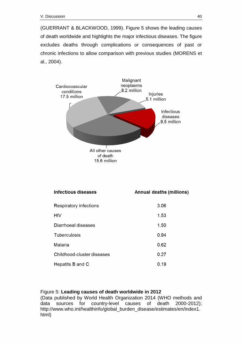
Today, infectious diseases yearly cause about 10 million deaths worldwide
despite an obvious decrease through medical progress compared to data
published about ten years ago (MORENS et al., 2004). Thus, infectious
diseases still amount to about 17% of the annual 56 million deaths in the
world in 2012 and represent one of the main problems in public health
(figure 5). Children in developing countries are mainly affected
V. Discussion 40
(GUERRANT & BLACKWOOD, 1999). Figure 5 shows the leading causes
of death worldwide and highlights the major infectious diseases. The figure
excludes deaths through complications or consequences of past or
chronic infections to allow comparison with previous studies (MORENS et
al., 2004).
Figure 5: Leading causes of death worldwide in 2012
(Data published by World Health Organization 2014 (WHO methods and data sources for country-level causes of death 2000-2012); http://www.who.int/healthinfo/global_burden_disease/estimates/en/index1.html)
V. Discussion 41
According to the data published in 2014 by the WHO respiratory infections
including influenza, HIV, tuberculosis and malaria are besides the
diarrhoeal diseases the most lethal infectious diseases. Unfortunately, the
development of an efficient and safe vaccine against HIV has so far not
been successful. Furthermore today no universal influenza vaccine is
available which would be needed to efficiently protect against all influenza
viruses including novel subtypes with pandemic potential. These more
complex diseases are known to require cellular immune responses for
protection (PANTALEO & KOUP, 2004; HOFT, 2008; ALTENBURG et al.,
2014). For this reason it is important to focus not only on the induction of
antibodies, but also on the strong activation of T cells in the development
of new vaccines. Generally, the development of vaccines efficient against
complex infectious diseases has high priority in medical research because
vaccination is regarded to be the most effective and cost-efficient
prevention method (SALLUSTO et al., 2010; PULENDRAN & AHMED,
2011).
Factors with potential influence on T cell immunity triggered by MVA
The activation of the cellular immune system does not only lead to the
generation of effector and memory T cells. It furthermore also supports the
B cell response (WEBER et al., 2009). Enhanced B cell response results
in antibody production and supports the efficacy of the vaccine as well.
Many different factors for the design of poxvirus vectors are considered to
have positive effects on the activation of the humoral immune system.
These factors can either modify the vector backbone or the target antigen.
The induction of T lymphocytes can for example be influenced through the
amount of target protein delivered. It is known that the amount of
heterologous protein clearly correlates with the immune response of
vaccinated mice (WYATT et al., 2008; GARCIA-ARRIAZA & ESTEBAN,
2014). These data suggest that recombinant MVA should produce the
highest amount of protein which is possible without affecting the stability
and growth behavior of the virus (WYATT et al., 2008). High production
levels of protein can mainly be achieved through the use of strong and
efficient promoters. For this reason numerous experimental studies
V. Discussion 42
consistently investigate and compare natural poxvirus-specific promoters
as well as new synthetic promoters for their efficacy and strength (for
example BAUR et al., 2010; BECKER et al., 2014; YANG et al., 2015). It is
remarkable that in poxvirus vector systems additionally to the expression
strength of the target antigen also the expression time during the life cycle
of the virus can be specifically chosen. The expression time is even
considered to be crucial for the type of the immune response (activation of
CD4+ or CD8+ T cells). CD4+ T cells mainly recognize late antigens,
whereas activation of cytotoxic T cells is predominantly associated with
early expression (MOUTAFTSI et al., 2007; YANG et al., 2011; GARCIA-
ARRIAZA & ESTEBAN, 2014). Today strong and well-characterized early,
early-late as well as late promoters for the MVA system are available.
These different promoters have specific advantages and disadvantages
depending on the experimental approach and setting. Early-late
expression allows antigen expression during the whole life cycle of MVA.
In contrast, the use of late promoters only leads to antigen expression
during the late cycle phase. Furthermore it can be associated with hiding
of antigen in viral factories which are formed at later times of MVA
infections (KATSAFANAS & MOSS, 2007). For this reason early
promoters are considered to be better suitable for efficient induction of the
endogenous pathway. Thus, early promoters are usually used in studies
which aim at investigating T cell responses (BAUR et al., 2010). Apart
from the promoter strength the spacer length also seems to play an
important role. 2015 a study firstly confirmed the positive effect of an
increased promoter spacer length on the activation of T lymphocytes (DI
PILATO et al., 2015).
Another strategy for the improvement of the cellular immune response
experienced over the last years consists in co-expression of different
molecules. In this context, it is a good approach to combine different
antigens of the same target virus. The combination of specific T cell
antigens with antigens which induce strong antibody production should
allow to efficiently activate both, the humoral immune system and the
cellular immune system. Furthermore it is also possible to combine
antigens of different viral subtypes for the induction of cross-protective
V. Discussion 43
immunity. This could especially be supportive in the development of
influenza vaccines. Besides the co-expression of different antigens,
another promising approach is to combine target antigen with co-
stimulatory molecules such as cytokines. Co-delivery of cytokines under
the control of poxvirus specific promoters or exogenous inoculation can
result in enhanced activation and recruitment of immune cells (GARCIA-
ARRIAZA & ESTEBAN, 2014). IL-2 (BERTLEY et al., 2004), IL-12
(ABAITUA et al., 2006), IFNγ (ABAITUA et al., 2006) or GM-CSF
(CHAVAN et al., 2006) have amongst other cytokines successfully been
used in the MVA vector system. In general, for the construction of
recombinant vectors co-expressing several proteins, the insertion of larger
DNA sequences is required. In this context MVA serves as an appropriate
vector because of its large genome and high genetic plasticity.
Next, the immune response could be improved through the combination of
different vaccines in prime-boost vaccination protocols (for review see
GARCIA-ARRIAZA & ESTEBAN, 2014). Today, poxvirus vectors
combined with other vaccines are commonly used in preclinical and
clinical trials. Here, the poxvirus vector can be used as the priming vaccine
as well as the booster component. Recombinant MVAs have so far
successfully been used in several experimental studies combined with
DNA vectors (GOMEZ et al., 2007), recombinant influenza viruses
(GONZALEZ-ASEGUINOLAZA et al., 2003), adenoviruses (REYES-
SANDOVAL et al., 2010; RATTO-KIM et al., 2012) or other poxvirus
vectors (SANTRA et al., 2007). Results from these data clearly
recommend further investigation and use of heterologous prime-boost
vaccination strategies.
Additionally to the improvement of the vector backbone described above
another approach is to optimize the target antigen. In this context the
choice and suitable design of the target antigen certainly plays the key
role. Optimal adjustment of the antigen to the specific requirements of the
delivering vector system is essential to efficiently activate the immune
system (GASTEIGER et al., 2007). Data published recently also highlight
the importance of long-lived antigen for the induction of the cellular
immune response. It is generally supposed that long-lived antigen could
V. Discussion 44
enhance the cross-presentation pathway and therefore induce a stronger
activation of cytotoxic T cells (GASTEIGER et al., 2007). Furthermore the
antigen chosen should preferably have full length instead of being
shortened. Full length antigen can support immune reaction not only
against a single, but potentially also against multiple T cell epitopes
(GASTEIGER et al., 2007). In this context we should keep in mind that
basically only 2% of all peptide fragments which are generated through
processing of foreign proteins within the cell have an amino acid structure
which can bind to MHC and is consequently able to activate T cells
(WEBER et al., 2009).
Data published recently including this study also consider effects of the
target protein’s localization on the immunogenicity of recombinant MVA
(GASTEIGER et al., 2007). Target protein can generally be expressed all
over an infected cell or it can be located in a specific subcellular
compartment. Distinct subcellular localizations can be achieved through
the use of cellular localization signals. But on the other hand it is also
possible to abrogate the natural subcellular distribution of a target antigen
through removing its own localization signal. The purpose of such
modifications is to improve the accessibility of target protein to the cellular
apparatus processing protein. Generally, enhanced protein depletion and
presentation of antigenic material obviously seem to support the activation
of T lymphocytes.
Another parameter which is discussed for further enhancement of
recombinant MVA’s immunogenicity is the use of adjuvants. Adjuvants are
commonly used in many vaccines currently available. The major task of
adjuvants is to enhance the efficacy of vaccines by triggering rapid, strong
and long-lasting immune responses (GUPTA & SIBER, 1995). Moreover
adjuvants are also able to selectively modulate immune responses to
MHC class I or MHC class II molecules. This could be helpful in the control
of diseases caused by intracellular pathogens (GUPTA & SIBER, 1995). In
addition, adjuvants containing synthetic T cell epitopes have already been
developed which can specifically enhance the cellular immunogenicity
(HADDEN, 1994). However, the use of adjuvants in the MVA vector
system is controversially discussed for good reasons. On the one hand
V. Discussion 45
adjuvants indeed enhance the immunogenicity of many vaccines available
and could therefore also be useful in the MVA vector system. But on the
other hand previous work clearly considers adjuvants as dispensable for
most MVA based vaccines because of the intrinsic immunogenicity of the
vector (ALTENBURG et al., 2014). Furthermore the use of adjuvants in
vaccines is generally discussed. Many adjuvants are known to have great
toxic potential and can induce adverse and severe side effects following
vaccination (GUPTA & SIBER, 1995). For this reason the dispensability of
adjuvants is considered to be one important factor which supports the
generation of safer vaccines. Therefore MVA renouncing the addition of
adjuvants serves as a promising vaccine platform.
Importance of T cell response for efficacy of MVA based vaccines
Today it is consensus that protective vaccines against more complex
diseases such as HIV, tuberculosis or malaria require both, induction of
strong T cell immunity and efficient activation of the humoral immune
system. Especially vaccines against HIV lack a coordinated B and T cell
response (GOMEZ et al., 2011; GOMEZ et al., 2012) because
T lymphocytes are crucial for the control of viremia during acute infections
(McELRATH & HAYNES, 2010). Additionally a large population-based
study generally provides evidence that Gag-specific T cell responses can
efficiently control chronic infections and are affiliated with lower viral loads
(KIEPIELA et al., 2007; McELRATH & HAYNES, 2010). Several
recombinant MVA vaccines expressing Gag have already been tested for
their potential to induce cellular immune responses (for review see
GOMEZ et al., 2012). The results of these studies clearly indicate MVA as
a promising Gag vector for combined induction of the cellular immune
system and the humoral immune system.
In case of malaria, T cell activation is also required for the protective
efficacy of vaccines because inducing protective immunity against
sporozoites only through humoral immune responses is obviously
challenging (HILL et al., 2010). Several animal studies and human clinical
trials have confirmed the key role of cytotoxic T cells in the elimination of
the parasites (DOOLAN & HOFFMAN, 2000; DE BARRA et al., 2014). In
V. Discussion 46
general, protection against malaria is clearly related to high levels of CD8+
T cells (SCHNEIDER et al., 1998; DE BARRA et al., 2014). Moreover,
cytotoxic T cell immunity even seems to be the vital factor for prevention of
parasitemia and clearance of all parasites (EWER et al., 2015). For this
reason malaria vaccine development is focused on the generation of
vaccines which can activate both, B cells and T lymphocytes.
The currently available tuberculosis vaccine, Bacille Calmette-Guérin
(BCG), is commonly used especially in developing countries (BROOKES
et al., 2008). However, a meta-analysis published in 1994 reports that
BCG generally only reduces the risk of tuberculosis by 50% (COLDITZ et
al., 1994). It efficiently protects children from tuberculous meningitis, but
the efficiency against pulmonary tuberculosis shows an inappropriately
high variability (COLDITZ et al., 1994; TRUNZ et al., 2006; BROOKES et
al., 2008). Therefore the development of new tuberculosis vaccines is still
ongoing. Experimental studies indicate the important role of cytotoxic
T cells for the induction of protective immunity in mice (FLYNN et al.,
1992). In this context, a MVA vaccine expressing 85A antigen has been
developed. This vaccine indeed induced 85A-specific T cells, when tested
on volunteers in Africa (BROOKES et al., 2008) and proved to be well
tolerated when tested in a large phase 2b trial in about 1,400 infants
(TAMERIS et al., 2013). These data showed that the 85A antigen alone
may not be sufficient to provide protective immunity, but the safety data
clearly recommend further development of tuberculosis vaccines on the
basis of MVA.
Even if it is often underlined that T cell based immunity through
vaccination with recombinant MVA is not able to completely prevent an
infection alone, it indeed can trigger rapid protection. In this context the
general rapid activation of T cells which can of course also be achieved in
the MVA vector system is especially vital. T cell responses usually only
need about four days to develop. In contrast antibody production takes
obviously longer with approximately eight to ten days (MURPHY et al.,
2008). So even if the T cell immunity is not able to prevent the infection
and the disease following, the rapid response can play the key role in the
prophylaxis of infectious diseases such as influenza. This point has been
V. Discussion 47
confirmed in recent studies which investigated MVA’s potential to induce
short term protection using ectromelia virus (ECTV) challenge infections in
mice (KREMER et al., 2012a; VOLZ et al., 2014). ECTV, the causative
agent of mousepox, is probably the best animal model available for human
smallpox infections. The results clearly show that the early protective
capacity of MVA depends on the induction of CD4+ and CD8+ T cells.
Here, MVA-specific T cells could already be detected at day two, whereas
the number of MVA-specific antibodies didn’t start to increase until day
eight. These studies furthermore show the adequacy of low-dose
immunization and consider the dispensability of the humoral immune
response for rapid protection.
Activation of CD8+ T cells has recently also been confirmed in
experimental studies with a recombinant MVA vaccine delivering spike
glycoprotein of Middle East respiratory syndrome coronavirus (MERS-
CoV) (SONG et al., 2013; VOLZ et al., 2015). MERS is a zoonotic
infectious disease which has its natural reservoir in camels. It has caused
severe human respiratory diseases in Middle Eastern countries since
2012. The MERS triggered respiratory syndrome is reported to result in
death of about 20% to 40% of those infected (MACKAY & ARDEN, 2015).
In 2015 the MVA-MERS-CoV vaccine was also successfully tested in a
challenge experiment using dromedary camels (HAAGMANS et al., 2016).
The success of this vaccination is hypothesized to be based on the
induction of MERS-CoV neutralizing antibodies. However, the potential
contribution of vaccine induced CD8+ and/or CD4+ T cells to protective
immunity is not known and warrants further investigation. Generally, the
development of camel-specific MERS-vaccines is very vital for the
protection of humans who work in close contact with the animals.
Furthermore the protective efficacy of recombinant MVA expressing
nucleoprotein of influenza also indicates the importance of T cell activation
in the MVA vector system. As previously mentioned, nucleoprotein is able
to efficiently induce T cells. Previous immunization experiments using
recombinant MVA delivering nucleoprotein highlight that protective
immunity against lethal challenge with influenza virus is affiliated with high
CD4+ and CD8+ T cell responses (HESSEL et al., 2014). These data also
V. Discussion 48
confirm that pre-existing CD4+ T cell responses obviously correlate with
protection even without influenza specific antibodies (WILKINSON et al.,
2012; HESSEL et al., 2014). The efficacy of a T cell-based MVA influenza
vaccine expressing nucleoprotein and matrixprotein 1 has also been
demonstrated in a phase 2a influenza challenge study published recently
(LILLIE et al., 2012).
The exact mechanism of short term protection triggered by T cells is still
subject of present research. However, it is supposed that rapid T cell
immunity could probably bypass the time until the infection leads to the
additional and long-lasting protective antibody response. This approach is
basically also regarded as progressively important for the prevention of
new threatening infections such as Ebola or MERS corona virus.
Role of T cells in MVA-based cancer immunotherapies
However, T cells are not only important for the protective capacity of
vaccines against infectious diseases. They are also considered to play a
key role in the development of efficient new cancer therapies. Here, T cells
could be determining factors for the efficacy of vaccines on the basis of
tumor-associated antigens. Such vaccines are especially promising in the
control of immunogenic tumors like melanomas. Spontaneous regression
of melanomas is obviously associated with infiltration of T cells (LOWES et
al., 1997). Therefore research investigates more and more tumor-
associated target antigens for their potential to induce tumor-specific T cell
responses.
In this context, tyrosinase serves as a promising tumor-associated target
antigen for cancer immunotherapies because it is regularly expressed in
melanomas (CHEN et al., 1995). Its natural function is being the key
enzyme of the melanin biosynthesis. Expressed by MVA, this enzyme is
able to efficiently trigger tyrosinase- and melanoma-specific cytotoxic
T cell responses in vitro and in vivo (DREXLER et al., 1999). In a phase I
clinical study melanoma patients were treated with dendritic cells which
had been infected with recombinant MVA delivering tyrosinase (DI
NICOLA et al., 2003). In general, this treatment was well tolerated and
V. Discussion 49
triggered tyrosinase-specific T cells in vivo (DI NICOLA et al., 2004). Apart
from tyrosinase, tyrosinase-related-protein-2 (TRP-2) also serves as an
appropriate target antigen for T cell based cancer therapy. It is remarkable
that the expression of TRP-2 even seems to correlate with the resistance
of melanoma cells against radio- and chemotherapy (PAK et al., 2004).
TRP-2 delivered by recombinant MVA can efficiently be recognized by
dendritic cells and therefore also activate T lymphocytes (PASCHEN et al.,
2005).
However, melanoma is not the only type of cancer which responds
positively on T cell based immunotherapies. In approximately 30% human
breast cancer is associated with the overexpression of human epidermal
growth factor receptor-2 (HER-2/neu) (SLAMON et al., 1989). This
overexpression has also been demonstrated in ovarian and renal cancer
(SLAMON et al., 1989). HER-2/neu is a proto-oncogenic glycoprotein
which belongs to the tyrosinase-kinase receptors family (COUSSENS et
al., 1985). Overexpression of HER-2/neu is related to a down-regulation of
MHC class I molecules and disturbance of CD8+ T cell mediated cell lysis
(HERRMANN et al., 2004). The down-regulation of MHC-class I molecules
can result in further tumor progression (HERRMANN et al., 2004). For this
reason it seems to be a promising approach to use HER-2/neu as a tumor-
associated antigen and recombinant MVA has been used for efficient
delivery of HER-2/neu antigen to human dendritic cells (KASTENMULLER
et al., 2006).
Future perspectives
Today safe and highly effective viral vectors on the basis of the well-
characterized MVA are commonly generated. Further preclinical as well as
clinical studies are essential to define the recombinant MVAs which are
able to fully protect against specific diseases. Thus, further enhancement
of the cellular immune response certainly plays an important role,
especially in the development of vaccines against complex infectious
diseases. In general, recombinant MVAs are also suitable candidates for
the development of cancer immunotherapies. In this context, the
combination of MVA with further innovative treatment tools such as the

V. Discussion 50
simultaneous use of checkpoint inhibitors seems to be a very promising
approach for future research.
VI. Summary 51
VI. SUMMARY
Improving the cellular immunogenicity of recombinant Modified
Vaccinia virus Ankara using green fluorescent protein as model
system
Infectious diseases are still threatening for public health and cause about
17% of the annual deaths worldwide despite great medical progress over
the last decades. In this context more complex diseases such as
influenza, HIV, tuberculosis or malaria are especially problematic. These
infectious diseases are characterized either by being untreatable or
unpreventable because of the lack of efficient vaccines.
The development of vaccines has for a long period of time predominantly
been based on the induction of humoral immune responses. But today the
cellular immune system is also considered to play an important role in the
prevention of more complex diseases. So the focus of vaccine
development is changing more and more. In this context, Modified
Vaccinia virus Ankara (MVA) serves as a promising platform for vaccine
development because it is able to induce both, humoral immune
responses and cellular immune responses. MVA is a safety tested,
replication deficient poxvirus vector which can stably express heterologous
proteins. Apart from being used as recombinant vector vaccine, MVA also
serves as a promising tool in the development of cancer immunotherapies
which are based on the induction of T cells.
In this study we investigated an innovative antigen modification tool for its
potential to further enhance the cellular immunogenicity of recombinant
MVA. For this purpose we introduced green fluorescent protein (GFP) in
combination with different localization signals to the MVA genome. GFP
was tacked either to myristoyl acid or to a nuclear localization signal and
compared with unmodified GFP. The genetic integrity and stability of the
recombinant MVAs was confirmed via polymerase chain reaction (PCR).
Moreover, growth kinetics of the candidate viruses in DF-1, MEF and
NIH3T3 cells showed the replication deficiency of the generated MVAs in
mammalian cells. The stable expression of the GFP variants was verified
VI. Summary 52
using immunoblot analysis. Furthermore, microscopical analysis confirmed
the expected specific subcellular localization sites of GFP triggered
through the different localization signals. All MVA-GFP candidate vaccines
induced GFP-specific CD8+ T cells after intramuscular inoculation of
BALB/c mice. However, after boost vaccination modification by
myristoylation signal resulted in an increased level of antigen-specific
CD8+ T cells statistically significant compared to unmodified GFP or to
nuclear localized GFP. Therefore the addition of myristoylation
successfully enhanced the cellular immunogenicity of the recombinant
MVA. Generally, this result supports the further use of myristoylation as
promising modification tool for the generation of MVA based vaccines
delivering specific antigens of infectious diseases.
VII. Zusammenfassung 53
VII. ZUSAMMENFASSUNG
Verbesserung der zellulären Immunogenität von rekombinantem
Modifizierten Vaccinia Virus Ankara unter Verwendung von Grün
fluoreszierendem Protein als Modellsystem
Trotz des großen medizinischen Fortschritts während der letzten
Jahrzehnte stellen Infektionskrankheiten eine nicht zu unterschätzende
Bedrohung für das öffentliche Gesundheitswesen dar.
Infektionskrankheiten zählen mit einem Anteil von 17% an den jährlich
weltweit registrierten Todesfällen immer noch zu den
Haupttodesursachen. Dabei gelten vor allem komplexere Infektionen wie
Influenza, HIV, Tuberkulose oder Malaria als besonders problematisch.
Diese Infektionskrankheiten weisen häufig einen schweren Verlauf auf
oder sind nicht heilbar. Aufgrund des Fehlens wirksamer und sicherer
Impfstoffe ist zudem die Prävention dieser Krankheiten nur eingeschränkt
oder überhaupt nicht möglich.
Die Entwicklung neuer Impfstoffe basierte lange Zeit hauptsächlich auf der
Induktion des humoralen Immunsystems. Inzwischen rückt aber auch die
Aktivierung des zellulären Immunsystems immer mehr in den Fokus, da
viele neuere Forschungsarbeiten in effizienten T-Zell-Antworten den
Schlüssel für die Prävention komplexerer Infektionskrankheiten sehen. In
diesem Zusammenhang dient Modifiziertes Vacciniavirus Ankara (MVA)
als eine vielversprechende Plattform für die Impfstoffentwicklung, denn
MVA kann sowohl die Aktivierung des humoralen als auch des zellulären
Immunsystems induzieren. Das Pockenvirus MVA kann stabil Fremdgene
exprimieren und dient somit als viraler Vektor in der Impfstoffentwicklung.
Da MVA in menschlichen Zellen und Säugetierzellen nicht replizieren
kann, gilt die Verwendung dieses Vektors als besonders sicher. Außer in
der Impfstoffentwicklung wird MVA auch in der Entwicklung von neuen
Krebstherapien, die auf der Induktion von T-Zellen basieren, eingesetzt.
Im Rahmen dieser Arbeit wurde eine neue Methode zur Modifizierung von
Proteinen auf ihre Immunogenität im MVA Vektorsystem getestet. Dazu
wurden Gensequenzen für Grün fluoreszierendes Protein (GFP) in
VII. Zusammenfassung 54
Kombination mit unterschiedlichen Lokalisierungssignalen in das MVA
Genom integriert. GFP wurde dabei entweder mit einer Myristoylierungs-
oder einer nukleären Lokalisierungssequenz versehen und mit
unmodifiziertem GFP verglichen. Die genetische Stabilität der
rekombinanten MVAs wurde mittels Polymerase-Ketten-Reaktion
nachgewiesen. Wachstumskurven auf DF-1, MEF und NIH3T3 Zellen
bestätigten sowohl die funktionierende Replikation in Zellen aviären
Ursprungs als auch die fehlende Vermehrungsfähigkeit der rekombinanten
Viren in Säugetierzellen. Zudem wurde die stabile Expression der
verschiedenen GFP-Varianten mithilfe von Immunfluoreszenz-Analysen
gezeigt. Die erhoffte subzelluläre Lokalisation von GFP in Abhängigkeit
von den eingesetzten Lokalisierungssequenzen wurde mittels
Fluoreszenzmikroskopie bestätigt. Im Tierversuch konnten alle
rekombinanten MVA nach intramuskulärer Injektion die Aktivität GFP-
spezifischer zytotoxischer T-Zellen induzieren. Nach der zweiten
Immunisierung war die Aktivität der CD8+ T-Zellen, induziert durch
myristoyliertes GFP, signifikant erhöht im Vergleich zu den weiteren GFP
Varianten. Die hier untersuchte Myristoylierung konnte somit erfolgreich
die zelluläre Immunogenität des rekombinanten MVA steigern.
Myristoylierung empfiehlt sich deshalb auch als zukunftsträchtige Methode
zur Modifizierung weiterer Antigene im MVA Vektorsystem.
VIII. References 55
VIII. REFERENCES
Abaitua F, Rodriguez JR, Garzon A, Rodriguez D, Esteban M. Improving
recombinant MVA immune responses: potentiation of the immune
responses to HIV-1 with MVA and DNA vectors expressing Env and the
cytokines IL-12 and IFN-gamma. Virus Res 2006; 116: 11-20.
Ahlers JD, Belyakov IM. Memories that last forever: strategies for
optimizing vaccine T-cell memory. Blood 2010; 115: 1678-89.
Ahmed R, Gray D. Immunological memory and protective immunity:
understanding their relation. Science 1996; 272: 54-60.
Albrecht M, Suezer Y, Staib C, Sutter G, Vieths S, Reese G. Vaccination
with a Modified Vaccinia Virus Ankara-based vaccine protects mice from
allergic sensitization. J Gene Med 2008; 10: 1324-33.
Altenburg AF, Kreijtz JH, de Vries RD, Song F, Fux R, Rimmelzwaan GF,
Sutter G, Volz A. Modified vaccinia virus ankara (MVA) as production
platform for vaccines against influenza and other viral respiratory
diseases. Viruses 2014; 6: 2735-61.
Androlewicz MJ, Anderson KS, Cresswell P. Evidence that transporters
associated with antigen processing translocate a major histocompatibility
complex class I-binding peptide into the endoplasmic reticulum in an ATP-
dependent manner. Proc Natl Acad Sci U S A 1993; 90: 9130-4.
Antoine G, Scheiflinger F, Dorner F, Falkner FG. The complete genomic
sequence of the modified vaccinia Ankara strain: comparison with other
orthopoxviruses. Virology 1998; 244: 365-96.
VIII. References 56
Antrobus RD, Lillie PJ, Berthoud TK, Spencer AJ, McLaren JE, Ladell K,
Lambe T, Milicic A, Price DA, Hill AV, Gilbert SC. A T cell-inducing
influenza vaccine for the elderly: safety and immunogenicity of MVA-
NP+M1 in adults aged over 50 years. PLoS One 2012; 7: e48322.
Armit C. Into the blue: the importance of murine lacZ gene expression
profiling in understanding and treating human disease. Dis Model Mech
2015; 8: 1341-3.
Bakari M, Aboud S, Nilsson C, Francis J, Buma D, Moshiro C, Aris EA,
Lyamuya EF, Janabi M, Godoy-Ramirez K, Joachim A, Polonis VR, Brave
A, Earl P, Robb M, Marovich M, Wahren B, Pallangyo K, Biberfeld G,
Mhalu F, Sandstrom E. Broad and potent immune responses to a low
dose intradermal HIV-1 DNA boosted with HIV-1 recombinant MVA among
healthy adults in Tanzania. Vaccine 2011; 29: 8417-28.
Baur K, Brinkmann K, Schweneker M, Patzold J, Meisinger-Henschel C,
Hermann J, Steigerwald R, Chaplin P, Suter M, Hausmann J. Immediate-
early expression of a recombinant antigen by modified vaccinia virus
ankara breaks the immunodominance of strong vector-specific B8R
antigen in acute and memory CD8 T-cell responses. J Virol 2010; 84:
8743-52.
Becker PD, Norder M, Weissmann S, Ljapoci R, Erfle V, Drexler I,
Guzman CA. Gene Expression Driven by a Strong Viral Promoter in MVA
Increases Vaccination Efficiency by Enhancing Antibody Responses and
Unmasking CD8(+) T Cell Epitopes. Vaccines (Basel) 2014; 2: 581-600.
Bejon P, Mwacharo J, Kai O, Mwangi T, Milligan P, Todryk S, Keating S,
Lang T, Lowe B, Gikonyo C, Molyneux C, Fegan G, Gilbert SC, Peshu N,
Marsh K, Hill AV. A phase 2b randomised trial of the candidate malaria
vaccines FP9 ME-TRAP and MVA ME-TRAP among children in Kenya.
PLoS Clin Trials 2006; 1: e29.
VIII. References 57
Bejon P, Ogada E, Mwangi T, Milligan P, Lang T, Fegan G, Gilbert SC,
Peshu N, Marsh K, Hill AV. Extended follow-up following a phase 2b
randomized trial of the candidate malaria vaccines FP9 ME-TRAP and
MVA ME-TRAP among children in Kenya. PLoS One 2007; 2: e707.
Benarafa C, Remold-O'Donnell E. The ovalbumin serpins revisited:
perspective from the chicken genome of clade B serpin evolution in
vertebrates. Proc Natl Acad Sci U S A 2005; 102: 11367-72.
Berthoud TK, Hamill M, Lillie PJ, Hwenda L, Collins KA, Ewer KJ, Milicic A,
Poyntz HC, Lambe T, Fletcher HA, Hill AV, Gilbert SC. Potent CD8+ T-cell
immunogenicity in humans of a novel heterosubtypic influenza A vaccine,
MVA-NP+M1. Clin Infect Dis 2011; 52: 1-7.
Bertley FM, Kozlowski PA, Wang SW, Chappelle J, Patel J, Sonuyi O,
Mazzara G, Montefiori D, Carville A, Mansfield KG, Aldovini A. Control of
simian/human immunodeficiency virus viremia and disease progression
after IL-2-augmented DNA-modified vaccinia virus Ankara nasal
vaccination in nonhuman primates. J Immunol 2004; 172: 3745-57.
Beutler B. Innate immunity: an overview. Mol Immunol 2004; 40: 845-59.
Bevan MJ. Helping the CD8(+) T-cell response. Nat Rev Immunol 2004; 4:
595-602.
Bohnen C, Wangorsch A, Schulke S, Nakajima-Adachi H, Hachimura S,
Burggraf M, Suzer Y, Schwantes A, Sutter G, Waibler Z, Reese G, Toda
M, Scheurer S, Vieths S. Vaccination with recombinant modified vaccinia
virus Ankara prevents the onset of intestinal allergy in mice. Allergy 2013;
68: 1021-8.
VIII. References 58
Boulanger D, Green P, Jones B, Henriquet G, Hunt LG, Laidlaw SM,
Monaghan P, Skinner MA. Identification and characterization of three
immunodominant structural proteins of fowlpox virus. J Virol 2002; 76:
9844-55.
Brandler S, Lepelley A, Desdouits M, Guivel-Benhassine F, Ceccaldi PE,
Levy Y, Schwartz O, Moris A. Preclinical studies of a modified vaccinia
virus Ankara-based HIV candidate vaccine: antigen presentation and
antiviral effect. J Virol 2010; 84: 5314-28.
Brewoo JN, Powell TD, Jones JC, Gundlach NA, Young GR, Chu H, Das
SC, Partidos CD, Stinchcomb DT, Osorio JE. Cross-protective immunity
against multiple influenza virus subtypes by a novel modified vaccinia
Ankara (MVA) vectored vaccine in mice. Vaccine 2013; 31: 1848-55.
Bronte V, Carroll MW, Goletz TJ, Wang M, Overwijk WW, Marincola F,
Rosenberg SA, Moss B, Restifo NP. Antigen expression by dendritic cells
correlates with the therapeutic effectiveness of a model recombinant
poxvirus tumor vaccine. Proc Natl Acad Sci U S A 1997; 94: 3183-8.
Brookes RH, Hill PC, Owiafe PK, Ibanga HB, Jeffries DJ, Donkor SA,
Fletcher HA, Hammond AS, Lienhardt C, Adegbola RA, McShane H, Hill
AV. Safety and immunogenicity of the candidate tuberculosis vaccine
MVA85A in West Africa. PLoS One 2008; 3: e2921.
Brown LE, Kelso A. Prospects for an influenza vaccine that induces cross-
protective cytotoxic T lymphocytes. Immunol Cell Biol 2009; 87: 300-8.
Broyles SS, Li J, Moss B. Promoter DNA contacts made by the vaccinia
virus early transcription factor. J Biol Chem 1991; 266: 15539-44.
VIII. References 59
Caciotti A, Garman SC, Rivera-Colon Y, Procopio E, Catarzi S, Ferri L,
Guido C, Martelli P, Parini R, Antuzzi D, Battini R, Sibilio M, Simonati A,
Fontana E, Salviati A, Akinci G, Cereda C, Dionisi-Vici C, Deodato F,
d'Amico A, d'Azzo A, Bertini E, Filocamo M, Scarpa M, di Rocco M, Tifft
CJ, Ciani F, Gasperini S, Pasquini E, Guerrini R, Donati MA, Morrone A.
GM1 gangliosidosis and Morquio B disease: an update on genetic
alterations and clinical findings. Biochim Biophys Acta 2011; 1812: 782-90.
Carroll MW, Overwijk WW, Chamberlain RS, Rosenberg SA, Moss B,
Restifo NP. Highly attenuated modified vaccinia virus Ankara (MVA) as an
effective recombinant vector: a murine tumor model. Vaccine 1997; 15:
387-94.
Carroll MW, Moss B. Host range and cytopathogenicity of the highly
attenuated MVA strain of vaccinia virus: propagation and generation of
recombinant viruses in a nonhuman mammalian cell line. Virology 1997;
238: 198-211.
Cavenaugh JS, Awi D, Mendy M, Hill AV, Whittle H, McConkey SJ.
Partially randomized, non-blinded trial of DNA and MVA therapeutic
vaccines based on hepatitis B virus surface protein for chronic HBV
infection. PLoS One 2011; 6: e14626.
Chalfie M, Tu Y, Euskirchen G, Ward WW, Prasher DC. Green fluorescent
protein as a marker for gene expression. Science 1994; 263: 802-5.
Chalfie M. GFP: lighting up life (Nobel Lecture). Angew Chem Int Ed Engl
2009; 48: 5603-11.
Chan TO, Zhang J, Rodeck U, Pascal JM, Armen RS, Spring M, Dumitru
CD, Myers V, Li X, Cheung JY, Feldman AM. Resistance of Akt kinases to
dephosphorylation through ATP-dependent conformational plasticity. Proc
Natl Acad Sci U S A 2011; 108: E1120-7.
VIII. References 60
Chaplin DD. Overview of the immune response. J Allergy Clin Immunol
2010; 125: S3-23.
Chavan R, Marfatia KA, An IC, Garber DA, Feinberg MB. Expression of
CCL20 and granulocyte-macrophage colony-stimulating factor, but not
Flt3-L, from modified vaccinia virus ankara enhances antiviral cellular and
humoral immune responses. J Virol 2006; 80: 7676-87.
Chen YT, Stockert E, Tsang S, Coplan KA, Old LJ. Immunophenotyping of
melanomas for tyrosinase: implications for vaccine development. Proc Natl
Acad Sci U S A 1995; 92: 8125-9.
Colditz GA, Brewer TF, Berkey CS, Wilson ME, Burdick E, Fineberg HV,
Mosteller F. Efficacy of BCG vaccine in the prevention of tuberculosis.
Meta-analysis of the published literature. JAMA 1994; 271: 698-702.
Cosma A, Nagaraj R, Buhler S, Hinkula J, Busch DH, Sutter G, Goebel
FD, Erfle V. Therapeutic vaccination with MVA-HIV-1 nef elicits Nef-
specific T-helper cell responses in chronically HIV-1 infected individuals.
Vaccine 2003; 22: 21-9.
Cosma A, Buhler S, Nagaraj R, Staib C, Hammarin AL, Wahren B, Goebel
FD, Erfle V, Sutter G. Neutralization assay using a modified vaccinia virus
Ankara vector expressing the green fluorescent protein is a high-
throughput method to monitor the humoral immune response against
vaccinia virus. Clin Diagn Lab Immunol 2004; 11: 406-10.
Cottingham MG, Carroll MW. Recombinant MVA vaccines: dispelling the
myths. Vaccine 2013; 31: 4247-51.
Coussens L, Yang-Feng TL, Liao YC, Chen E, Gray A, McGrath J,
Seeburg PH, Libermann TA, Schlessinger J, Francke U, et al. Tyrosine
kinase receptor with extensive homology to EGF receptor shares
chromosomal location with neu oncogene. Science 1985; 230: 1132-9.
VIII. References 61
Cresswell P, Ackerman AL, Giodini A, Peaper DR, Wearsch PA.
Mechanisms of MHC class I-restricted antigen processing and cross-
presentation. Immunol Rev 2005; 207: 145-57.
de Barra E, Hodgson SH, Ewer KJ, Bliss CM, Hennigan K, Collins A,
Berrie E, Lawrie AM, Gilbert SC, Nicosia A, McConkey SJ, Hill AV. A
phase Ia study to assess the safety and immunogenicity of new malaria
vaccine candidates ChAd63 CS administered alone and with MVA CS.
PLoS One 2014; 9: e115161.
de The G, Bomford R, Kazanji M, Ibrahim F. Human T cell lymphotropic
virus: necessity for and feasibility of a vaccine. Ciba Found Symp 1994;
187: 47-55; discussion -60.
de Vries RD, De Gruyter HL, Bestebroer TM, Pronk M, Fouchier RA,
Osterhaus AD, Sutter G, Kreijtz JH, Rimmelzwaan GF. Induction of
influenza (H5N8) antibodies by modified vaccinia virus Ankara H5N1
vaccine. Emerg Infect Dis 2015; 21: 1086-8.
Di Nicola M, Carlo-Stella C, Anichini A, Mortarini R, Guidetti A, Tragni G,
Gallino F, Del Vecchio M, Ravagnani F, Morelli D, Chaplin P, Arndtz N,
Sutter G, Drexler I, Parmiani G, Cascinelli N, Gianni AM. Clinical protocol.
Immunization of patients with malignant melanoma with autologous
CD34(+) cell-derived dendritic cells transduced ex vivo with a recombinant
replication-deficient vaccinia vector encoding the human tyrosinase gene:
a phase I trial. Hum Gene Ther 2003; 14: 1347-60.
Di Nicola M, Carlo-Stella C, Mortarini R, Baldassari P, Guidetti A, Gallino
GF, Del Vecchio M, Ravagnani F, Magni M, Chaplin P, Cascinelli N,
Parmiani G, Gianni AM, Anichini A. Boosting T cell-mediated immunity to
tyrosinase by vaccinia virus-transduced, CD34(+)-derived dendritic cell
vaccination: a phase I trial in metastatic melanoma. Clin Cancer Res 2004;
10: 5381-90.
VIII. References 62
Di Pilato M, Sanchez-Sampedro L, Mejias-Perez E, Sorzano CO, Esteban
M. Modification of promoter spacer length in vaccinia virus as a strategy to
control the antigen expression. J Gen Virol 2015; 96: 2360-71.
Doolan DL, Hoffman SL. The complexity of protective immunity against
liver-stage malaria. J Immunol 2000; 165: 1453-62.
Drexler I, Heller K, Wahren B, Erfle V, Sutter G. Highly attenuated
modified vaccinia virus Ankara replicates in baby hamster kidney cells, a
potential host for virus propagation, but not in various human transformed
and primary cells. J Gen Virol 1998; 79 ( Pt 2): 347-52.
Drexler I, Antunes E, Schmitz M, Wolfel T, Huber C, Erfle V, Rieber P,
Theobald M, Sutter G. Modified vaccinia virus Ankara for delivery of
human tyrosinase as melanoma-associated antigen: induction of
tyrosinase- and melanoma-specific human leukocyte antigen A*0201-
restricted cytotoxic T cells in vitro and in vivo. Cancer Res 1999; 59: 4955-
63.
Drexler I, Staib C, Sutter G. Modified vaccinia virus Ankara as antigen
delivery system: how can we best use its potential? Curr Opin Biotechnol
2004; 15: 506-12.
Ewer KJ, Sierra-Davidson K, Salman AM, Illingworth JJ, Draper SJ,
Biswas S, Hill AV. Progress with viral vectored malaria vaccines: A multi-
stage approach involving "unnatural immunity". Vaccine 2015; 33: 7444-
51.
Flynn JL, Goldstein MM, Triebold KJ, Koller B, Bloom BR. Major
histocompatibility complex class I-restricted T cells are required for
resistance to Mycobacterium tuberculosis infection. Proc Natl Acad Sci U
S A 1992; 89: 12013-7.
VIII. References 63
Flynn KJ, Belz GT, Altman JD, Ahmed R, Woodland DL, Doherty PC.
Virus-specific CD8+ T cells in primary and secondary influenza
pneumonia. Immunity 1998; 8: 683-91.
Flynn KJ, Riberdy JM, Christensen JP, Altman JD, Doherty PC. In vivo
proliferation of naive and memory influenza-specific CD8(+) T cells. Proc
Natl Acad Sci U S A 1999; 96: 8597-602.
Gamblin SJ, Skehel JJ. Influenza hemagglutinin and neuraminidase
membrane glycoproteins. J Biol Chem 2010; 285: 28403-9.
Garcia-Arriaza J, Esteban M. Enhancing poxvirus vectors vaccine
immunogenicity. Hum Vaccin Immunother 2014; 10: 2235-44.
Garcia F, Bernaldo de Quiros JC, Gomez CE, Perdiguero B, Najera JL,
Jimenez V, Garcia-Arriaza J, Guardo AC, Perez I, Diaz-Brito V, Conde
MS, Gonzalez N, Alvarez A, Alcami J, Jimenez JL, Pich J, Arnaiz JA,
Maleno MJ, Leon A, Munoz-Fernandez MA, Liljestrom P, Weber J,
Pantaleo G, Gatell JM, Plana M, Esteban M. Safety and immunogenicity of
a modified pox vector-based HIV/AIDS vaccine candidate expressing Env,
Gag, Pol and Nef proteins of HIV-1 subtype B (MVA-B) in healthy HIV-1-
uninfected volunteers: A phase I clinical trial (RISVAC02). Vaccine 2011;
29: 8309-16.
Garten W, Klenk HD. Understanding influenza virus pathogenicity. Trends
Microbiol 1999; 7: 99-100.
Gasteiger G, Kastenmuller W, Ljapoci R, Sutter G, Drexler I. Cross-
priming of cytotoxic T cells dictates antigen requisites for modified vaccinia
virus Ankara vector vaccines. J Virol 2007; 81: 11925-36.
Germain RN. T-cell development and the CD4-CD8 lineage decision. Nat
Rev Immunol 2002; 2: 309-22.
VIII. References 64
Gettins PG. Serpin structure, mechanism, and function. Chem Rev 2002;
102: 4751-804.
Gilbert SC. Clinical development of Modified Vaccinia virus Ankara
vaccines. Vaccine 2013; 31: 4241-6.
Goebel SJ, Johnson GP, Perkus ME, Davis SW, Winslow JP, Paoletti E.
The complete DNA sequence of vaccinia virus. Virology 1990; 179: 247-
66, 517-63.
Goldberg AL, Rock KL. Proteolysis, proteasomes and antigen
presentation. Nature 1992; 357: 375-9.
Gomez CE, Najera JL, Jimenez EP, Jimenez V, Wagner R, Graf M,
Frachette MJ, Liljestrom P, Pantaleo G, Esteban M. Head-to-head
comparison on the immunogenicity of two HIV/AIDS vaccine candidates
based on the attenuated poxvirus strains MVA and NYVAC co-expressing
in a single locus the HIV-1BX08 gp120 and HIV-1(IIIB) Gag-Pol-Nef
proteins of clade B. Vaccine 2007; 25: 2863-85.
Gomez CE, Najera JL, Perdiguero B, Garcia-Arriaza J, Sorzano CO,
Jimenez V, Gonzalez-Sanz R, Jimenez JL, Munoz-Fernandez MA, Lopez
Bernaldo de Quiros JC, Guardo AC, Garcia F, Gatell JM, Plana M,
Esteban M. The HIV/AIDS vaccine candidate MVA-B administered as a
single immunogen in humans triggers robust, polyfunctional, and selective
effector memory T cell responses to HIV-1 antigens. J Virol 2011; 85:
11468-78.
Gomez CE, Perdiguero B, Garcia-Arriaza J, Esteban M. Poxvirus vectors
as HIV/AIDS vaccines in humans. Hum Vaccin Immunother 2012; 8: 1192-
207.
VIII. References 65
Gomez CE, Perdiguero B, Garcia-Arriaza J, Esteban M. Clinical
applications of attenuated MVA poxvirus strain. Expert Rev Vaccines
2013; 12: 1395-416.
Gomez CE, Perdiguero B, Garcia-Arriaza J, Cepeda V, Sanchez-Sorzano
CO, Mothe B, Jimenez JL, Munoz-Fernandez MA, Gatell JM, Lopez
Bernaldo de Quiros JC, Brander C, Garcia F, Esteban M. A Phase I
Randomized Therapeutic MVA-B Vaccination Improves the Magnitude and
Quality of the T Cell Immune Responses in HIV-1-Infected Subjects on
HAART. PLoS One 2015; 10: e0141456.
Gonzalez-Aseguinolaza G, Nakaya Y, Molano A, Dy E, Esteban M,
Rodriguez D, Rodriguez JR, Palese P, Garcia-Sastre A, Nussenzweig RS.
Induction of protective immunity against malaria by priming-boosting
immunization with recombinant cold-adapted influenza and modified
vaccinia Ankara viruses expressing a CD8+-T-cell epitope derived from
the circumsporozoite protein of Plasmodium yoelii. J Virol 2003; 77:
11859-66.
Gowans JL. The lymphocyte-a disgraceful gap in medical knowledge.
Immunol Today 1996; 17: 288-91.
Grawunder U, West RB, Lieber MR. Antigen receptor gene
rearrangement. Curr Opin Immunol 1998; 10: 172-80.
Guerrant RL, Blackwood BL. Threats to global health and survival: the
growing crises of tropical infectious diseases-our "unfinished agenda". Clin
Infect Dis 1999; 28: 966-86.
Gupta RK, Siber GR. Adjuvants for human vaccines-current status,
problems and future prospects. Vaccine 1995; 13: 1263-76.
VIII. References 66
Haagmans BL, van den Brand JM, Raj VS, Volz A, Wohlsein P, Smits SL,
Schipper D, Bestebroer TM, Okba N, Fux R, Bensaid A, Solanes Foz D,
Kuiken T, Baumgartner W, Segales J, Sutter G, Osterhaus AD. An
orthopoxvirus-based vaccine reduces virus excretion after MERS-CoV
infection in dromedary camels. Science 2016; 351: 77-81.
Hadden JW. T-cell adjuvants. Int J Immunopharmacol 1994; 16: 703-10.
Hampson AW, Mackenzie JS. The influenza viruses. Med J Aust 2006;
185: S39-43.
Henrissat B. A classification of glycosyl hydrolases based on amino acid
sequence similarities. Biochem J 1991; 280 (Pt 2): 309-16.
Herrmann F, Lehr HA, Drexler I, Sutter G, Hengstler J, Wollscheid U,
Seliger B. HER-2/neu-mediated regulation of components of the MHC
class I antigen-processing pathway. Cancer Res 2004; 64: 215-20.
Hessel A, Savidis-Dacho H, Coulibaly S, Portsmouth D, Kreil TR, Crowe
BA, Schwendinger MG, Pilz A, Barrett PN, Falkner FG, Schafer B. MVA
vectors expressing conserved influenza proteins protect mice against
lethal challenge with H5N1, H9N2 and H7N1 viruses. PLoS One 2014; 9:
e88340.
Higgins CF. ABC transporters: from microorganisms to man. Annu Rev
Cell Biol 1992; 8: 67-113.
Hill AV, Reyes-Sandoval A, O'Hara G, Ewer K, Lawrie A, Goodman A,
Nicosia A, Folgori A, Colloca S, Cortese R, Gilbert SC, Draper SJ. Prime-
boost vectored malaria vaccines: progress and prospects. Hum Vaccin
2010; 6: 78-83.
VIII. References 67
Hillaire ML, Osterhaus AD, Rimmelzwaan GF. Induction of virus-specific
cytotoxic T lymphocytes as a basis for the development of broadly
protective influenza vaccines. J Biomed Biotechnol 2011; 2011: 939860.
Hiller G, Weber K. Golgi-derived membranes that contain an acylated viral
polypeptide are used for vaccinia virus envelopment. J Virol 1985; 55:
651-9.
Hodgson SH, Ewer KJ, Bliss CM, Edwards NJ, Rampling T, Anagnostou
NA, de Barra E, Havelock T, Bowyer G, Poulton ID, de Cassan S, Longley
R, Illingworth JJ, Douglas AD, Mange PB, Collins KA, Roberts R, Gerry S,
Berrie E, Moyle S, Colloca S, Cortese R, Sinden RE, Gilbert SC, Bejon P,
Lawrie AM, Nicosia A, Faust SN, Hill AV. Evaluation of the efficacy of
ChAd63-MVA vectored vaccines expressing circumsporozoite protein and
ME-TRAP against controlled human malaria infection in malaria-naive
individuals. J Infect Dis 2015; 211: 1076-86.
Hoft DF. Tuberculosis vaccine development: goals, immunological design,
and evaluation. Lancet 2008; 372: 164-75.
Hunt LT, Dayhoff MO. A surprising new protein superfamily containing
ovalbumin, antithrombin-III, and alpha 1-proteinase inhibitor. Biochem
Biophys Res Commun 1980; 95: 864-71.
Iwasaki A, Medzhitov R. Control of adaptive immunity by the innate
immune system. Nat Immunol 2015; 16: 343-53.
Janeway CA, Jr. Approaching the asymptote? Evolution and revolution in
immunology. Cold Spring Harb Symp Quant Biol 1989; 54 Pt 1: 1-13.
Janeway CA, Jr. Pillars article: approaching the asymptote? Evolution and
revolution in immunology. Cold spring harb symp quant biol. 1989. 54: 1-
13. J Immunol 2013; 191: 4475-87.
VIII. References 68
Joffre OP, Segura E, Savina A, Amigorena S. Cross-presentation by
dendritic cells. Nat Rev Immunol 2012; 12: 557-69.
Kastenmuller W, Drexler I, Ludwig H, Erfle V, Peschel C, Bernhard H,
Sutter G. Infection of human dendritic cells with recombinant vaccinia virus
MVA reveals general persistence of viral early transcription but distinct
maturation-dependent cytopathogenicity. Virology 2006; 350: 276-88.
Kastenmuller W, Gasteiger G, Gronau JH, Baier R, Ljapoci R, Busch DH,
Drexler I. Cross-competition of CD8+ T cells shapes the
immunodominance hierarchy during boost vaccination. J Exp Med 2007;
204: 2187-98.
Katsafanas GC, Moss B. Colocalization of transcription and translation
within cytoplasmic poxvirus factories coordinates viral expression and
subjugates host functions. Cell Host Microbe 2007; 2: 221-8.
Kennedy JS, Greenberg RN. IMVAMUNE: modified vaccinia Ankara strain
as an attenuated smallpox vaccine. Expert Rev Vaccines 2009; 8: 13-24.
Kennedy RB, Ovsyannikova I, Poland GA. Smallpox vaccines for
biodefense. Vaccine 2009; 27 Suppl 4: D73-9.
Kiepiela P, Ngumbela K, Thobakgale C, Ramduth D, Honeyborne I,
Moodley E, Reddy S, de Pierres C, Mncube Z, Mkhwanazi N, Bishop K,
van der Stok M, Nair K, Khan N, Crawford H, Payne R, Leslie A, Prado J,
Prendergast A, Frater J, McCarthy N, Brander C, Learn GH, Nickle D,
Rousseau C, Coovadia H, Mullins JI, Heckerman D, Walker BD, Goulder
P. CD8+ T-cell responses to different HIV proteins have discordant
associations with viral load. Nat Med 2007; 13: 46-53.
Klein L, Kyewski B, Allen PM, Hogquist KA. Positive and negative
selection of the T cell repertoire: what thymocytes see (and don't see). Nat
Rev Immunol 2014; 14: 377-91.
VIII. References 69
Kreijtz JH, Gilbert SC, Sutter G. Poxvirus vectors. Vaccine 2013; 31: 4217-
9.
Kreijtz JH, Goeijenbier M, Moesker FM, van den Dries L, Goeijenbier S,
De Gruyter HL, Lehmann MH, Mutsert G, van de Vijver DA, Volz A,
Fouchier RA, van Gorp EC, Rimmelzwaan GF, Sutter G, Osterhaus AD.
Safety and immunogenicity of a modified-vaccinia-virus-Ankara-based
influenza A H5N1 vaccine: a randomised, double-blind phase 1/2a clinical
trial. Lancet Infect Dis 2014; 14: 1196-207.
Kremer M, Suezer Y, Volz A, Frenz T, Majzoub M, Hanschmann KM,
Lehmann MH, Kalinke U, Sutter G. Critical role of perforin-dependent
CD8+ T cell immunity for rapid protective vaccination in a murine model for
human smallpox. PLoS Pathog 2012a; 8: e1002557.
Kremer M, Volz A, Kreijtz JH, Fux R, Lehmann MH, Sutter G. Easy and
efficient protocols for working with recombinant vaccinia virus MVA.
Methods Mol Biol 2012b; 890: 59-92.
Kremers GJ, Gilbert SG, Cranfill PJ, Davidson MW, Piston DW.
Fluorescent proteins at a glance. J Cell Sci 2011; 124: 157-60.
Leonard SR, Lacher DW, Lampel KA. Acquisition of the lac operon by
Salmonella enterica. BMC Microbiol 2015; 15: 173.
Lillie PJ, Berthoud TK, Powell TJ, Lambe T, Mullarkey C, Spencer AJ,
Hamill M, Peng Y, Blais ME, Duncan CJ, Sheehy SH, Havelock T, Faust
SN, Williams RL, Gilbert A, Oxford J, Dong T, Hill AV, Gilbert SC.
Preliminary assessment of the efficacy of a T-cell-based influenza vaccine,
MVA-NP+M1, in humans. Clin Infect Dis 2012; 55: 19-25.
Lowes MA, Bishop GA, Crotty K, Barnetson RS, Halliday GM. T helper 1
cytokine mRNA is increased in spontaneously regressing primary
melanomas. J Invest Dermatol 1997; 108: 914-9.
VIII. References 70
Luckheeram RV, Zhou R, Verma AD, Xia B. CD4(+)T cells: differentiation
and functions. Clin Dev Immunol 2012; 2012: 925135.
Mackay IM, Arden KE. MERS coronavirus: diagnostics, epidemiology and
transmission. Virol J 2015; 12: 222.
Mackett M, Smith GL, Moss B. Vaccinia virus: a selectable eukaryotic
cloning and expression vector. Proc Natl Acad Sci U S A 1982; 79: 7415-
9.
Marr L, Lulf AT, Freudenstein A, Sutter G, Volz A. Myristoylation increases
the CD8+ T cell response to a green fluorescent protein prototype antigen
delivered by Modified Vaccinia virus Ankara. J Gen Virol 2016; doi:
10.1099/jgv.0.000425.
Maurer-Stroh S, Eisenhaber B, Eisenhaber F. N-terminal N-myristoylation
of proteins: refinement of the sequence motif and its taxon-specific
differences. J Mol Biol 2002; 317: 523-40.
Mayr A, Munz E. [Changes in the vaccinia virus through continuing
passages in chick embryo fibroblast cultures]. Zentralbl Bakteriol Orig
1964; 195: 24-35.
Mayr A, Hochstein-Mintzel V, Stickl H. Abstammung, Eigenschaften und
Verwendung des attenuierten Vaccinia-Stammes MVA. Infection 1975; 3:
6-14.
Mayr A, Stickl H, Muller HK, Danner K, Singer H. [The smallpox
vaccination strain MVA: marker, genetic structure, experience gained with
the parenteral vaccination and behavior in organisms with a debilitated
defence mechanism (author's transl)]. Zentralbl Bakteriol B 1978; 167:
375-90.
VIII. References 71
McElhaney JE, Xie D, Hager WD, Barry MB, Wang Y, Kleppinger A, Ewen
C, Kane KP, Bleackley RC. T cell responses are better correlates of
vaccine protection in the elderly. J Immunol 2006; 176: 6333-9.
McElrath MJ, Haynes BF. Induction of immunity to human
immunodeficiency virus type-1 by vaccination. Immunity 2010; 33: 542-54.
McFadden G. Poxvirus tropism. Nat Rev Microbiol 2005; 3: 201-13.
Meisinger-Henschel C, Schmidt M, Lukassen S, Linke B, Krause L,
Konietzny S, Goesmann A, Howley P, Chaplin P, Suter M, Hausmann J.
Genomic sequence of chorioallantois vaccinia virus Ankara, the ancestor
of modified vaccinia virus Ankara. J Gen Virol 2007; 88: 3249-59.
Meyer H, Sutter G, Mayr A. Mapping of deletions in the genome of the
highly attenuated vaccinia virus MVA and their influence on virulence. J
Gen Virol 1991; 72 (Pt 5): 1031-8.
Moorthy VS, Pinder M, Reece WH, Watkins K, Atabani S, Hannan C,
Bojang K, McAdam KP, Schneider J, Gilbert S, Hill AV. Safety and
immunogenicity of DNA/modified vaccinia virus ankara malaria vaccination
in African adults. J Infect Dis 2003a; 188: 1239-44.
Moorthy VS, McConkey S, Roberts M, Gothard P, Arulanantham N,
Degano P, Schneider J, Hannan C, Roy M, Gilbert SC, Peto TE, Hill AV.
Safety of DNA and modified vaccinia virus Ankara vaccines against liver-
stage P. falciparum malaria in non-immune volunteers. Vaccine 2003b; 21:
1995-2002.
Moorthy VS, Imoukhuede EB, Milligan P, Bojang K, Keating S, Kaye P,
Pinder M, Gilbert SC, Walraven G, Greenwood BM, Hill AS. A
randomised, double-blind, controlled vaccine efficacy trial of DNA/MVA
ME-TRAP against malaria infection in Gambian adults. PLoS Med 2004; 1:
e33.
VIII. References 72
Morens DM, Folkers GK, Fauci AS. The challenge of emerging and re-
emerging infectious diseases. Nature 2004; 430: 242-9.
Moss B. Genetically engineered poxviruses for recombinant gene
expression, vaccination, and safety. Proc Natl Acad Sci U S A 1996; 93:
11341-8.
Moss B, Carroll MW, Wyatt LS, Bennink JR, Hirsch VM, Goldstein S,
Elkins WR, Fuerst TR, Lifson JD, Piatak M, Restifo NP, Overwijk W,
Chamberlain R, Rosenberg SA, Sutter G. Host range restricted, non-
replicating vaccinia virus vectors as vaccine candidates. Adv Exp Med Biol
1996; 397: 7-13.
Moss B. Poxviridae: the viruses and their replication. In: Field's Virology
6th edition. Knipe DM, Howley P, eds. United States: Lippincott Williams &
Wilkins 2013.
Moss DJ, Burrows SR, Suhrbier A, Khanna R. Potential antigenic targets
on Epstein-Barr virus-associated tumours and the host response. Ciba
Found Symp 1994; 187: 4-13; discussion -20.
Moutaftsi M, Bui HH, Peters B, Sidney J, Salek-Ardakani S, Oseroff C,
Pasquetto V, Crotty S, Croft M, Lefkowitz EJ, Grey H, Sette A. Vaccinia
virus-specific CD4+ T cell responses target a set of antigens largely
distinct from those targeted by CD8+ T cell responses. J Immunol 2007;
178: 6814-20.
VIII. References 73
Munseri PJ, Kroidl A, Nilsson C, Joachim A, Geldmacher C, Mann P,
Moshiro C, Aboud S, Lyamuya E, Maboko L, Missanga M, Kaluwa B,
Mfinanga S, Podola L, Bauer A, Godoy-Ramirez K, Marovich M, Moss B,
Hoelscher M, Gotch F, Stohr W, Stout R, McCormack S, Wahren B, Mhalu
F, Robb ML, Biberfeld G, Sandstrom E, Bakari M. Priming with a simplified
intradermal HIV-1 DNA vaccine regimen followed by boosting with
recombinant HIV-1 MVA vaccine is safe and immunogenic: a phase IIa
randomized clinical trial. PLoS One 2015; 10: e0119629.
Murphy K, Travers P, Walport M. Janeway's immunobiology 7th edition.
New York, London: Garland Science 2008.
Norder M, Becker PD, Drexler I, Link C, Erfle V, Guzman CA. Modified
vaccinia virus Ankara exerts potent immune modulatory activities in a
murine model. PLoS One 2010; 5: e11400.
Osbourn AE, Field B. Operons. Cell Mol Life Sci 2009; 66: 3755-75.
Pak BJ, Lee J, Thai BL, Fuchs SY, Shaked Y, Ronai Z, Kerbel RS, Ben-
David Y. Radiation resistance of human melanoma analysed by retroviral
insertional mutagenesis reveals a possible role for dopachrome
tautomerase. Oncogene 2004; 23: 30-8.
Pantaleo G, Koup RA. Correlates of immune protection in HIV-1 infection:
what we know, what we don't know, what we should know. Nat Med 2004;
10: 806-10.
Paschen A, Jing W, Drexler I, Klemm M, Song M, Muller-Berghaus J,
Nguyen XD, Osen W, Stevanovic S, Sutter G, Schadendorf D. Melanoma
patients respond to a new HLA-A*01-presented antigenic ligand derived
from a multi-epitope region of melanoma antigen TRP-2. Int J Cancer
2005; 116: 944-8.
VIII. References 74
Pascutti MF, Rodriguez AM, Falivene J, Giavedoni L, Drexler I, Gherardi
MM. Interplay between modified vaccinia virus Ankara and dendritic cells:
phenotypic and functional maturation of bystander dendritic cells. J Virol
2011; 85: 5532-45.
Plotkin SA. Vaccines: correlates of vaccine-induced immunity. Clin Infect
Dis 2008; 47: 401-9.
Portela A, Digard P. The influenza virus nucleoprotein: a multifunctional
RNA-binding protein pivotal to virus replication. J Gen Virol 2002; 83: 723-
34.
Pulendran B, Ahmed R. Immunological mechanisms of vaccination. Nat
Immunol 2011; 12: 509-17.
Ratto-Kim S, Currier JR, Cox JH, Excler JL, Valencia-Micolta A, Thelian D,
Lo V, Sayeed E, Polonis VR, Earl PL, Moss B, Robb ML, Michael NL, Kim
JH, Marovich MA. Heterologous prime-boost regimens using rAd35 and
rMVA vectors elicit stronger cellular immune responses to HIV proteins
than homologous regimens. PLoS One 2012; 7: e45840.
Remington SJ. Green fluorescent protein: a perspective. Protein Sci 2011;
20: 1509-19.
Reyes-Sandoval A, Berthoud T, Alder N, Siani L, Gilbert SC, Nicosia A,
Colloca S, Cortese R, Hill AV. Prime-boost immunization with adenoviral
and modified vaccinia virus Ankara vectors enhances the durability and
polyfunctionality of protective malaria CD8+ T-cell responses. Infect
Immun 2010; 78: 145-53.
Sallusto F, Lanzavecchia A, Araki K, Ahmed R. From vaccines to memory
and back. Immunity 2010; 33: 451-63.
VIII. References 75
Santra S, Sun Y, Parvani JG, Philippon V, Wyand MS, Manson K, Gomez-
Yafal A, Mazzara G, Panicali D, Markham PD, Montefiori DC, Letvin NL.
Heterologous prime/boost immunization of rhesus monkeys by using
diverse poxvirus vectors. J Virol 2007; 81: 8563-70.
Schmelz M, Sodeik B, Ericsson M, Wolffe EJ, Shida H, Hiller G, Griffiths
G. Assembly of vaccinia virus: the second wrapping cisterna is derived
from the trans Golgi network. J Virol 1994; 68: 130-47.
Schneider J, Gilbert SC, Blanchard TJ, Hanke T, Robson KJ, Hannan CM,
Becker M, Sinden R, Smith GL, Hill AV. Enhanced immunogenicity for
CD8+ T cell induction and complete protective efficacy of malaria DNA
vaccination by boosting with modified vaccinia virus Ankara. Nat Med
1998; 4: 397-402.
Sebastian S, Gilbert SC. Recombinant modified vaccinia virus Ankara-
based malaria vaccines. Expert Rev Vaccines 2016; 15: 91-103.
Sheehan S, Harris SA, Satti I, Hokey DA, Dheenadhayalan V, Stockdale
L, Manjaly Thomas ZR, Minhinnick A, Wilkie M, Vermaak S, Meyer J,
O'Shea MK, Pau MG, Versteege I, Douoguih M, Hendriks J, Sadoff J,
Landry B, Moss P, McShane H. A Phase I, Open-Label Trial, Evaluating
the Safety and Immunogenicity of Candidate Tuberculosis Vaccines
AERAS-402 and MVA85A, Administered by Prime-Boost Regime in BCG-
Vaccinated Healthy Adults. PLoS One 2015; 10: e0141687.
Sheehy SH, Duncan CJ, Elias SC, Biswas S, Collins KA, O'Hara GA,
Halstead FD, Ewer KJ, Mahungu T, Spencer AJ, Miura K, Poulton ID,
Dicks MD, Edwards NJ, Berrie E, Moyle S, Colloca S, Cortese R, Gantlett
K, Long CA, Lawrie AM, Gilbert SC, Doherty T, Nicosia A, Hill AV, Draper
SJ. Phase Ia clinical evaluation of the safety and immunogenicity of the
Plasmodium falciparum blood-stage antigen AMA1 in ChAd63 and MVA
vaccine vectors. PLoS One 2012; 7: e31208.
VIII. References 76
Shimomura O, Johnson FH, Saiga Y. Extraction, purification and
properties of aequorin, a bioluminescent protein from the luminous
hydromedusan, Aequorea. J Cell Comp Physiol 1962; 59: 223-39.
Slamon DJ, Godolphin W, Jones LA, Holt JA, Wong SG, Keith DE, Levin
WJ, Stuart SG, Udove J, Ullrich A, et al. Studies of the HER-2/neu proto-
oncogene in human breast and ovarian cancer. Science 1989; 244: 707-
12.
Song F, Fux R, Provacia LB, Volz A, Eickmann M, Becker S, Osterhaus
AD, Haagmans BL, Sutter G. Middle East respiratory syndrome
coronavirus spike protein delivered by modified vaccinia virus Ankara
efficiently induces virus-neutralizing antibodies. J Virol 2013; 87: 11950-4.
Stein PE, Leslie AG, Finch JT, Turnell WG, McLaughlin PJ, Carrell RW.
Crystal structure of ovalbumin as a model for the reactive centre of
serpins. Nature 1990; 347: 99-102.
Stickl H, Hochstein-Mintzel V, Mayr A, Huber HC, Schafer H, Holzner A.
[MVA vaccination against smallpox: clinical tests with an attenuated live
vaccinia virus strain (MVA) (author's transl)]. Dtsch Med Wochenschr
1974; 99: 2386-92.
Stokes GV. High-voltage electron microscope study of the release of
vaccinia virus from whole cells. J Virol 1976; 18: 636-43.
Sutter G, Moss B. Nonreplicating vaccinia vector efficiently expresses
recombinant genes. Proc Natl Acad Sci U S A 1992; 89: 10847-51.
Sutter G, Wyatt LS, Foley PL, Bennink JR, Moss B. A recombinant vector
derived from the host range-restricted and highly attenuated MVA strain of
vaccinia virus stimulates protective immunity in mice to influenza virus.
Vaccine 1994; 12: 1032-40.
VIII. References 77
Tameris MD, Hatherill M, Landry BS, Scriba TJ, Snowden MA, Lockhart S,
Shea JE, McClain JB, Hussey GD, Hanekom WA, Mahomed H, McShane
H. Safety and efficacy of MVA85A, a new tuberculosis vaccine, in infants
previously vaccinated with BCG: a randomised, placebo-controlled phase
2b trial. Lancet 2013; 381: 1021-8.
Topfer E, Boraschi D, Italiani P. Innate Immune Memory: The Latest
Frontier of Adjuvanticity. J Immunol Res 2015; 2015: 478408.
Trunz BB, Fine P, Dye C. Effect of BCG vaccination on childhood
tuberculous meningitis and miliary tuberculosis worldwide: a meta-analysis
and assessment of cost-effectiveness. Lancet 2006; 367: 1173-80.
Tscharke DC, Woo WP, Sakala IG, Sidney J, Sette A, Moss DJ, Bennink
JR, Karupiah G, Yewdell JW. Poxvirus CD8+ T-cell determinants and
cross-reactivity in BALB/c mice. J Virol 2006; 80: 6318-23.
Tscharke DC, Croft NP, Doherty PC, La Gruta NL. Sizing up the key
determinants of the CD8(+) T cell response. Nat Rev Immunol 2015; 15:
705-16.
Tsien RY. The green fluorescent protein. Annu Rev Biochem 1998; 67:
509-44.
van der Merwe PA, Dushek O. Mechanisms for T cell receptor triggering.
Nat Rev Immunol 2011; 11: 47-55.
Vollmar J, Arndtz N, Eckl KM, Thomsen T, Petzold B, Mateo L, Schlereth
B, Handley A, King L, Hulsemann V, Tzatzaris M, Merkl K, Wulff N,
Chaplin P. Safety and immunogenicity of IMVAMUNE, a promising
candidate as a third generation smallpox vaccine. Vaccine 2006; 24: 2065-
70.
VIII. References 78
Volz A, Sutter G. Protective efficacy of Modified Vaccinia virus Ankara in
preclinical studies. Vaccine 2013; 31: 4235-40.
Volz A, Langenmayer M, Jany S, Kalinke U, Sutter G. Rapid expansion of
CD8+ T cells in wild-type and type I interferon receptor-deficient mice
correlates with protection after low-dose emergency immunization with
modified vaccinia virus Ankara. J Virol 2014; 88: 10946-57.
Volz A, Kupke A, Song F, Jany S, Fux R, Shams-Eldin H, Schmidt J,
Becker C, Eickmann M, Becker S, Sutter G. Protective Efficacy of
Recombinant Modified Vaccinia Virus Ankara Delivering Middle East
Respiratory Syndrome Coronavirus Spike Glycoprotein. J Virol 2015; 89:
8651-6.
Wang P, Palese P, O'Neill RE. The NPI-1/NPI-3 (karyopherin alpha)
binding site on the influenza a virus nucleoprotein NP is a nonconventional
nuclear localization signal. J Virol 1997; 71: 1850-6.
Warrington R, Watson W, Kim HL, Antonetti FR. An introduction to
immunology and immunopathology. Allergy Asthma Clin Immunol 2011; 7
Suppl 1: S1.
Weber CA, Mehta PJ, Ardito M, Moise L, Martin B, De Groot AS. T cell
epitope: friend or foe? Immunogenicity of biologics in context. Adv Drug
Deliv Rev 2009; 61: 965-76.
Wilkinson TM, Li CK, Chui CS, Huang AK, Perkins M, Liebner JC,
Lambkin-Williams R, Gilbert A, Oxford J, Nicholas B, Staples KJ, Dong T,
Douek DC, McMichael AJ, Xu XN. Preexisting influenza-specific CD4+ T
cells correlate with disease protection against influenza challenge in
humans. Nat Med 2012; 18: 274-80.
Wong P, Pamer EG. CD8 T cell responses to infectious pathogens. Annu
Rev Immunol 2003; 21: 29-70.
VIII. References 79
Wong YC, Lin LC, Melo-Silva CR, Smith SA, Tscharke DC. Engineering
recombinant poxviruses using a compact GFP-blasticidin resistance fusion
gene for selection. J Virol Methods 2011; 171: 295-8.
Wright CA, Kozik P, Zacharias M, Springer S. Tapasin and other
chaperones: models of the MHC class I loading complex. Biol Chem 2004;
385: 763-78.
Wyatt LS, Earl PL, Vogt J, Eller LA, Chandran D, Liu J, Robinson HL,
Moss B. Correlation of immunogenicities and in vitro expression levels of
recombinant modified vaccinia virus Ankara HIV vaccines. Vaccine 2008;
26: 486-93.
Yang F, Moss LG, Phillips GN, Jr. The molecular structure of green
fluorescent protein. Nat Biotechnol 1996; 14: 1246-51.
Yang Z, Reynolds SE, Martens CA, Bruno DP, Porcella SF, Moss B.
Expression profiling of the intermediate and late stages of poxvirus
replication. J Virol 2011; 85: 9899-908.
Yang Z, Cao S, Martens CA, Porcella SF, Xie Z, Ma M, Shen B, Moss B.
Deciphering poxvirus gene expression by RNA sequencing and ribosome
profiling. J Virol 2015; 89: 6874-86.
Ye Z, Liu T, Offringa DP, McInnis J, Levandowski RA. Association of
influenza virus matrix protein with ribonucleoproteins. J Virol 1999; 73:
7467-73.
Zinkernagel RM. On immunological memory. Philos Trans R Soc Lond B
Biol Sci 2000; 355: 369-71.

VIII. References 80
ZKBS. Position statement on handling recombinant vaccinia viruses. In:
Ref. No 6790-10-14. Bundesamt für Verbraucherschutz und
Lebensmittelsicherheit. Berlin, Germany: Zentrale Kommission für die
biologische Sicherheit 1997.
IX. Abbreviations 81
IX. ABBREVIATIONS
ABC ATB-binding cassette family
AIDS Acquired immunodeficiency syndrome
AMA1 Apical membrane antigen 1
APC Antigen-presenting cell
ATP Adenosine triphosphate
BALB/c Albino, laboratory bred strain of house mouse
BCG Bacille Calmette-Guérin
CD4 Cluster of differentiation 4
CD8 Cluster of differentiation 8
CEF Chicken embryo fibroblasts
CVA Chorioallantois Vaccinia virus Ankara
DAPI 4’,6-Diamidin-2-phenylindol
DF-1 Chicken embryo fibroblast cell line prepared by Doug Foster (University of Minnesota)
DNA Desoxyribonucleic acid
ECTV Ectromelia virus
ELISPOT Enzyme-linked immuno spot assay
ENF HIV gene encoding for envelope proteins
EV Enveloped virion
GAG HIV gene encoding for proteins of matrix and capside
GFP Green fluorescent protein
GM1 Monosialotetrahexosylganglioside 1
GM-CSF Granulocyte-macrophage colony-stimulating factor
HA Hemagglutinin
HBV-S Hepatits B virus surface protein
HER-2/neu Human epidermal growth factor receptor 2
HIV Human immunodeficiency virus
IX. Abbreviations 82
hpi Hours post infection
IAV Influenza A virus
IL Interleukin
INF Interferon
Kb Kilobase
kDa Kilodalton
LMU Ludwig-Maximilians-Universität München
M1 Matrixprotein1
MEF Mouse embryonic fibroblasts
MERS-CoV Middle East respiratory syndrome coronavirus
ME-TRAP Thrombospondin related adhesive protein fused to a multi-epitope
mg Milligram
MHC Major histocompatibility complex
ml Milliliter
MOI Multiplicity of infection
mRNA Messenger ribonucleic acid
MTB Mycobacterium tuberculosis bacteria
MV Mature virion
MVA Modified Vaccinia virus Ankara
NA Neuraminidase
NEF Negative factor
ng Nanogram
NIH3T3 Mouse embryonic fibroblast cell line derived from albino mice from the National Institutes of Health, Bethesda
nM Nanomolar
NP Nucleoprotein
ORF Origin of replication
OVA Ovalbumin
PAMP Pathogen associated molecular pattern
IX. Abbreviations 83
PBS Phosphate buffered saline
PCR Polymerase chain reaction
P. falcip Plasmodium falciparum
POL HIV gene encoding viral enzymes
PRR Pattern recognition receptor
RNA Ribonucleic acid
TAP Transporters associated with antigen processing
TRP Tyrosinase-related-protein
WHO World Health Organization
WT Wildtype
VACV Vaccinia virus
X. Danksagung 84
X. DANKSAGUNG
Mein besonderer Dank gilt Herrn Prof. Dr. Sutter, der meine ersten
Schritte in der Wissenschaft begleitet hat es und mir ermöglicht hat meine
Arbeit auf Kongressen in Belgien und München zu präsentieren und als
Publikation zu veröffentlichen. Besonders bedanken möchte ich mich auch
für die Unterstützung meines parallel zur Doktorarbeit laufenden
Medizinstudiums.
Meiner Betreuerin Frau Dr. Asisa Volz danke ich ganz herzlich für ihre
Hilfe und die schnelle und fundierte Korrektur meiner Publikation und
Doktorarbeit. Ich habe bei der Fertigstellung meiner Arbeit sehr von ihren
hilfreichen Kommentaren und umfassenden Literaturkenntnissen profitiert.
Ganz besonders möchte ich mich bei Anna-Theresa Lülf für ihre
Unterstützung bedanken. Anna war für mich die beste Kollegin, die ich mir
nur hätte vorstellen können und ist für mich auch eine wunderbare
Freundin geworden.
Auch allen anderen (ehemaligen) Doktoranden der Virologie - Carina
Herzog, Ellen Link, Juan Jose Rojas, Katharina Müller, Lino Torres,
Martina Resch und Monique Richards - möchte ich für ihre Unterstützung
und die schöne gemeinsame Zeit danken. Ich hoffe, wir gehen noch oft
miteinander Karaoke singen!
Astrid Freudenstein und Sylvia Jany danke ich ganz herzlich für die
Einführung ins Labor, die gute Arbeitsatmosphäre und ihre Hilfe bei allen
experimentellen Fragen.
Bei Herrn Dr. Martin Langenmeyer möchte ich mich für die Unterstützung
bei der Bildbearbeitung bedanken.
Allen weiteren Mitarbeitern der Virologie danke ich ganz besonders für
ihre Hilfsbereitschaft und die schöne gemeinsame Zeit am Institut.
X. Danksagung 85
Der größte Dank gilt aber meiner Familie, besonders meinen Eltern, die
mich zu jeder Zeit uneingeschränkt unterstützt haben und mir meinen
ungewöhnlichen Weg durch zwei medizinische Studiengänge und diese
Doktorarbeit ermöglicht haben. Meinen Großeltern danke ich ganz herzlich
für ihre Unterstützung und ihren uneingeschränkten Glauben an mich. Bei
meinen Schwestern Theresa und Vera möchte ich mich für ihre Hilfe
bedanken und dafür, dass sie immer für mich da sind. Sabine und
Susanne danke ich besonders für alle lieben, unterstützenden Briefe und
Päckchen, die ich erhalten habe. Gisela und Harald danke ich ganz
herzlich für die Unterstützung meines Studiums und meiner Arbeit.
Johannes, you are simply the best!
Ich bin sehr froh, euch alle auch auf dem weiteren Weg durch das
Medizinstudium an meiner Seite zu haben und freue mich schon auf die
nächste Doktorarbeit mit euch!