Embed Size (px)
Citation preview
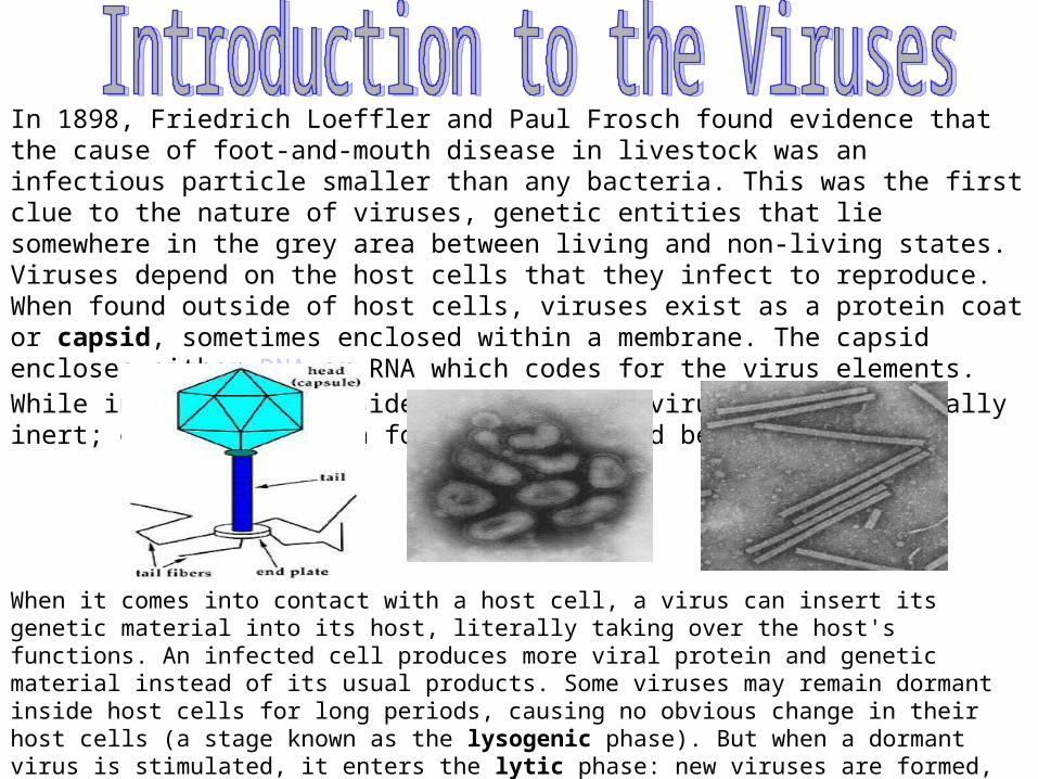
In 1898, Friedrich Loeffler and Paul Frosch found evidence that the cause of foot-and-mouth disease in livestock was an infectious particle smaller than any bacteria. This was the first clue to the nature of viruses, genetic entities that lie somewhere in the grey area between living and non-living states. Viruses depend on the host cells that they infect to reproduce. When found outside of host cells, viruses exist as a protein coat or capsid, sometimes enclosed within a membrane. The capsid encloses either DNA or RNA which codes for the virus elements. While in this form outside the cell, the virus is metabollically inert; examples of such forms are pictured below.
When it comes into contact with a host cell, a virus can insert its genetic material into its host, literally taking over the host's functions. An infected cell produces more viral protein and genetic material instead of its usual products. Some viruses may remain dormant inside host cells for long periods, causing no obvious change in their host cells (a stage known as the lysogenic phase). But when a dormant virus is stimulated, it enters the lytic phase: new viruses are formed, self-assemble, and burst out of the host cell, killing the cell and going on to infect other cells. The diagram below at right shows a virus that attacks bacteria, known as the lambda bacteriophage, which measures roughly 200 nanometers.
The origin of viruses is not known. Two theories of viral origin:1- Viruses may be derived from DNA or RNA or from both nucleic acid components of host cells that became able to replicate autonomously and evolve independently.2- Viruses may be degenerate forms of intracellular parasites.
Viruses are separated into major groupings called families on the basis of morphology, genome structure, and replication. Virus families have the suffix –viridae-. Within each family, subdivisions, called genera, are usually based on physicochemical or serologic properties. Genus names carry the suffix –virus-. Subfamilies virinae.
In 1995, the international committee on taxonomy of viruses had organized more than 4000 animal and plant viruses into 71 families, 11 subfamilies, and 164 genera, with hundreds of viruses still unassigned. Currently 24 families contain viruses that infect humans and animals.
According to the type of nucleic acid viruses are classified into DNA and RNA viruses.
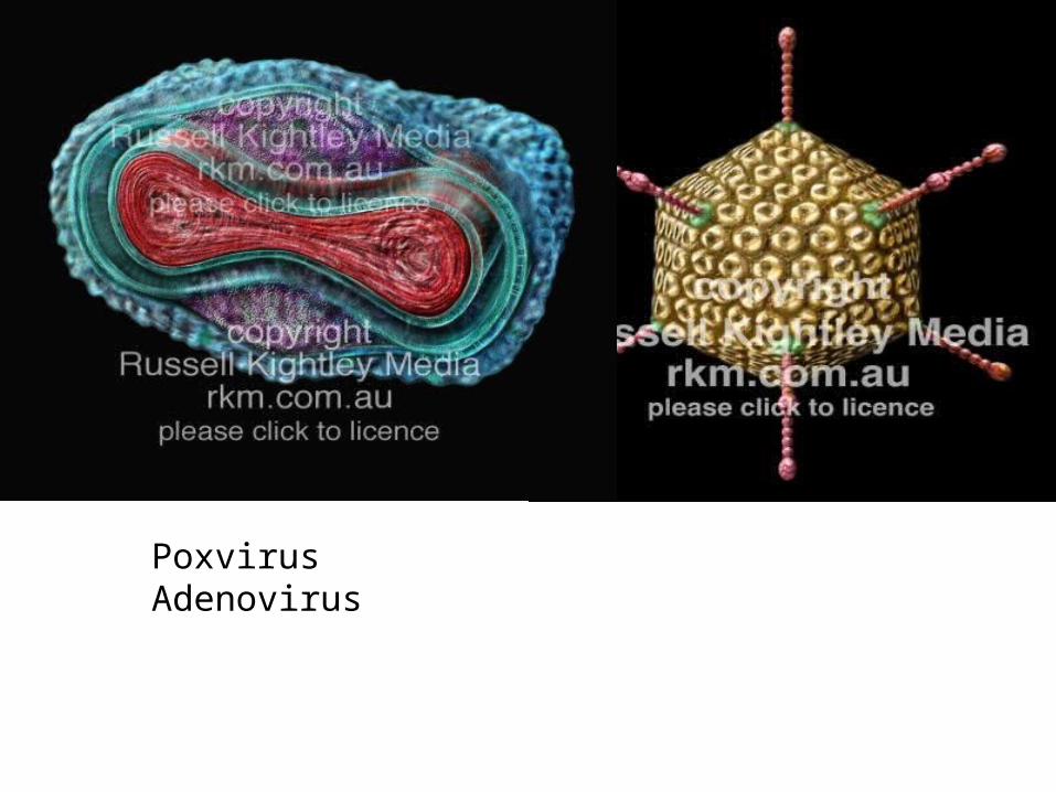
Poxvirus Adenovirus
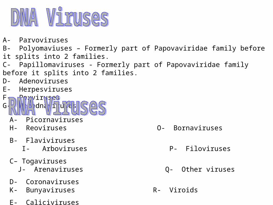
A- ParvovirusesB- Polyomaviuses – Formerly part of Papovaviridae family before it splits into 2 families.C- Papillomaviruses - Formerly part of Papovaviridae family before it splits into 2 families.D- AdenovirusesE- HerpesvirusesF- PoxvirusesG- Hepadnaviruses
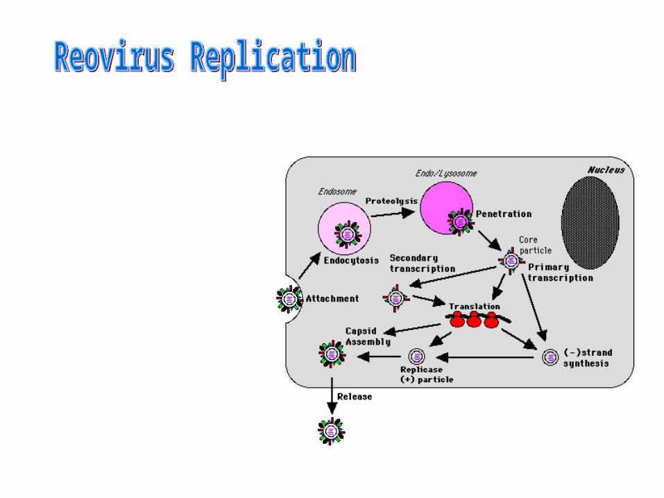
A- Picornaviruses H- Reoviruses O- Bornaviruses
B- Flaviviruses I- Arboviruses P- Filoviruses
C– Togaviruses J- Arenaviruses Q- Other viruses
D- Coronaviruses K- Bunyaviruses R- Viroids
E- Caliciviruses L- Orthomyxoviruses S- Prions
F- Retroviruses M- Paramyxoviruses
G- Astrovirses N- Rhabdoviruses
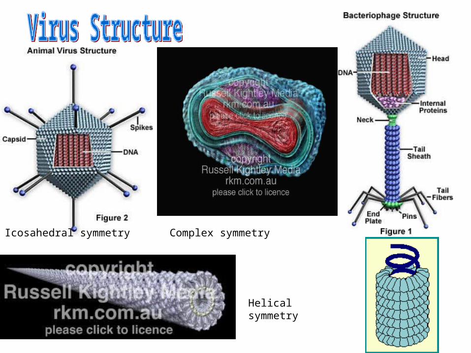
Helical symmetry
Icosahedral symmetry Complex symmetry
Capsid: protein shell that encloses the nucleic acid. It is built of structure units.
• STRUCTURE UNITS are the smallest functional equivalent building units of the capsid.
• CAPSOMERS are morphological units seen on the surface of particles and represent clusters of structure units.
• The capsid together with its enclosed nucleic acid is called the NUCLEOCAPSID.
• The nucleocapsid may be invested in an ENVELOPE which may contain material of host cell as well as viral origin.
• The VIRION is the complete infective virus particle
1- Icosahedral ( Cubic ) Symmetry
2- Helical Symmetry
3- Complex Structure
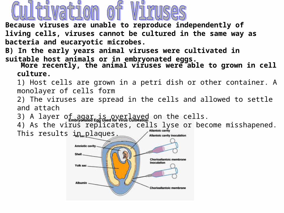
Because viruses are unable to reproduce independently of living cells, viruses cannot be cultured in the same way as bacteria and eucaryotic microbes. B) In the early years animal viruses were cultivated in suitable host animals or in embryonated eggs.
More recently, the animal viruses were able to grown in cell culture.1) Host cells are grown in a petri dish or other container. A monolayer of cells form2) The viruses are spread in the cells and allowed to settle and attach3) A layer of agar is overlayed on the cells.4) As the virus replicates, cells lyse or become misshapened. This results in plaques.

1- Heat and Cold.
2- Stabilization of Viruses by Salts.
3- pH.
4- Radiation.
5- Photodynamic Inactivation.
6- Ether Susceptibility.
7- Detergents.
8- Formaldehyde.
9- Antibiotics and Other Antibacterial Agents.
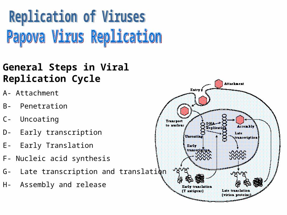
General Steps in Viral Replication Cycle
A- Attachment
B- Penetration
C- Uncoating
D- Early transcription
E- Early Translation
F- Nucleic acid synthesis
G- Late transcription and translation
H- Assembly and release
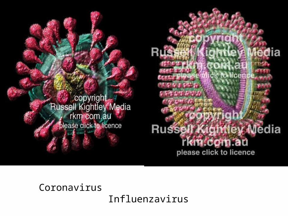
Coronavirus Influenzavirus
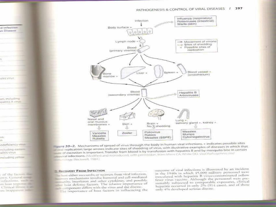
More than 300 viruses are known to infect humans and to cause as many as 50 different syndromes.
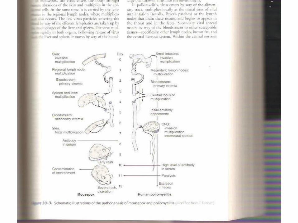
Steps in Viral Pathogenesis
A- Viral entry and primary replication.
B- Viral Spread and Cell Tropism.
C- Cell Injury and Clinical Illness.
D- Recovery From Infection.
E- Virus Shedding.
Host Immune Response
1- Both humeral and cellular immunity are involved in control of viral infection.
2- Mononuclear cells and lymphocytes are involved in viral infection.
3- The capsid serves as the targetfor the immune response.
4- Cytotoxic T lymphocytes lyse virus infected cells.
5- Secretory IgA antibody is important against viral infections of the respiratory or gastrointestinal tract.
6- Among the nonimmune responses is the induction of interferone.
1- Some viruses infect and damage cells of the immune system(AIDS).
2- Development of pathologic changes and clinical illness.
3- Immunopathologic disorder due to vaccine immunization.
4- Development of autoantibodies.
Viruses have a variety of ways that serve to suppress or evade the host immune response and thus avoid eradication:
1- Oftentimes the viral proteins involved in modulating the host response are not essential for the growth of the virus.
2- Some viruses infect cells of the immune system and abrogate their function(AIDS).
3- They may infect neurons that express little or no class 1 MHC(herpesviruses).
4- Form proteins that inhibit MHC function(adenoviruses).
5- Viruses may mutate and change the antigenic sites on virion proteins(influenza virus).
6- Regulate the level of viral surface proteins(herpesvirus).
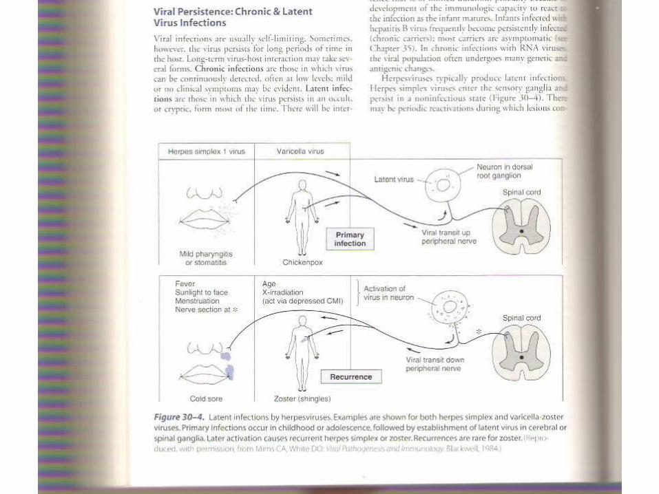
Viral infections are usually self-limiting. Sometimes, however, the virus persists for long periods of time in the host. Long-term virus-host interaction may take several forms:
1- Chronic infections.
2- Latent infections.
3- Inapparent or subclinical infections.
Acute Viral Respiratory Infections.
Viral Infections of the Gastrointestinal Tract.
Viral skin Infections
Viral Infections of the CNS
Congenital Viral Infections.
Rubella, CMV, Herpes simplex, Varicella-zoster, HBV, Enterovirus, HIV, Parvovirus B19
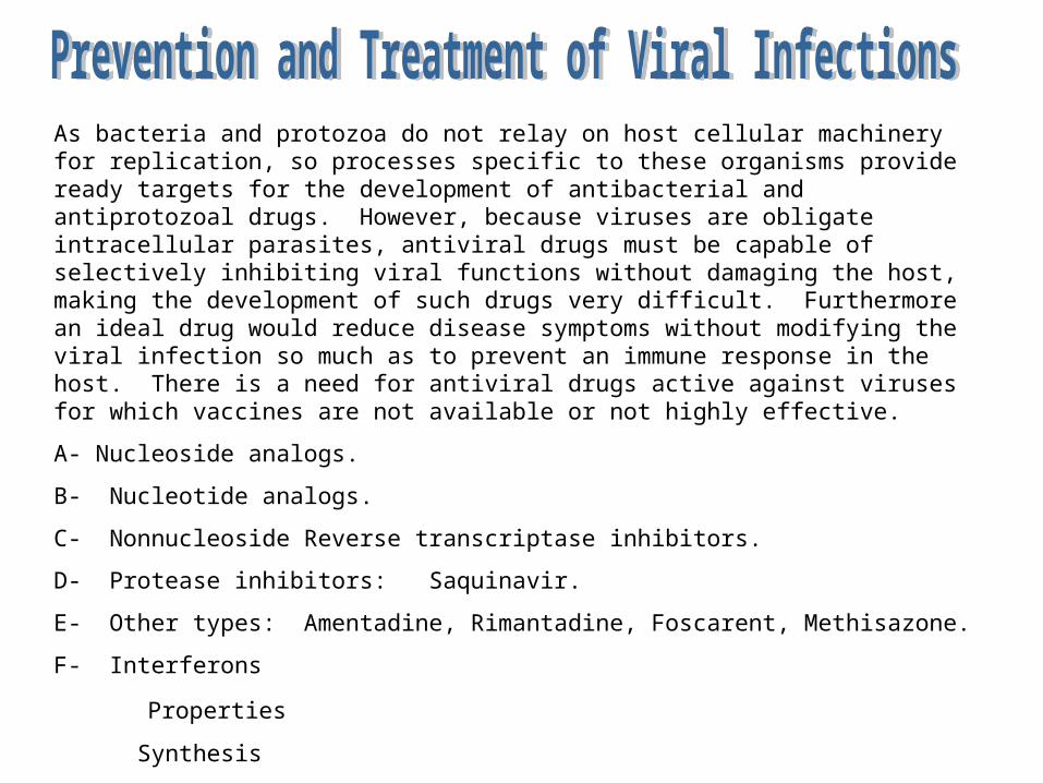
As bacteria and protozoa do not relay on host cellular machinery for replication, so processes specific to these organisms provide ready targets for the development of antibacterial and antiprotozoal drugs. However, because viruses are obligate intracellular parasites, antiviral drugs must be capable of selectively inhibiting viral functions without damaging the host, making the development of such drugs very difficult. Furthermore an ideal drug would reduce disease symptoms without modifying the viral infection so much as to prevent an immune response in the host. There is a need for antiviral drugs active against viruses for which vaccines are not available or not highly effective.
A- Nucleoside analogs.
B- Nucleotide analogs.
C- Nonnucleoside Reverse transcriptase inhibitors.
D- Protease inhibitors: Saquinavir.
E- Other types: Amentadine, Rimantadine, Foscarent, Methisazone.
F- Interferons
Properties
Synthesis
Antiviral activity and other biologic effects.
Clinical studies.
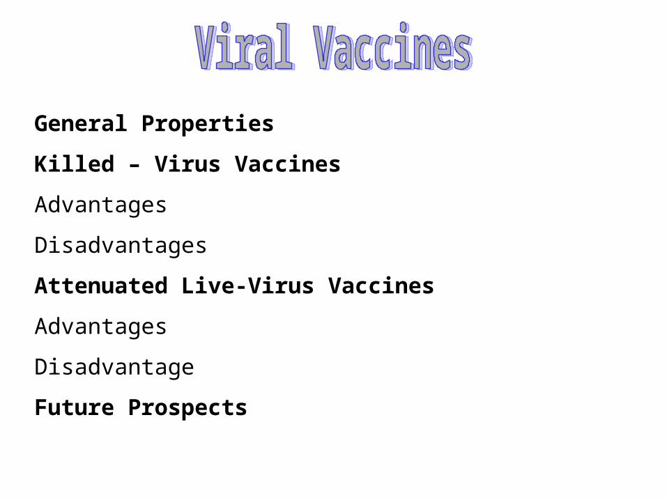
General Properties
Killed – Virus Vaccines
Advantages
Disadvantages
Attenuated Live-Virus Vaccines
Advantages
Disadvantage
Future Prospects

Simplified diagram of the Bacteriophage p22 virus. Original measures
995 pixels across
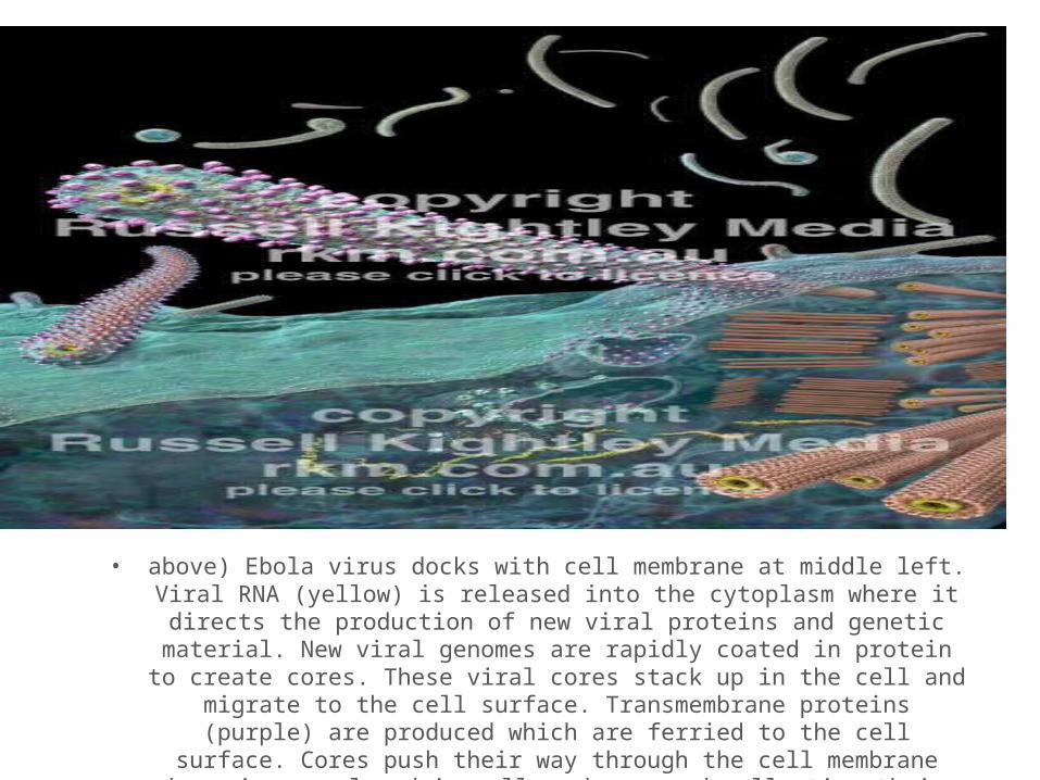
• above) Ebola virus docks with cell membrane at middle left. Viral RNA (yellow) is released into the cytoplasm where it directs the production of new viral proteins and
genetic material. New viral genomes are rapidly coated in protein to create cores. These viral cores stack up in the cell and migrate to the cell surface. Transmembrane proteins
(purple) are produced which are ferried to the cell surface. Cores push their way through the cell membrane becoming enveloped in cell membrane and collecting their
transmembrane proteins (spikes) as they do so. Examples of coiled virions are shown in the background.
•
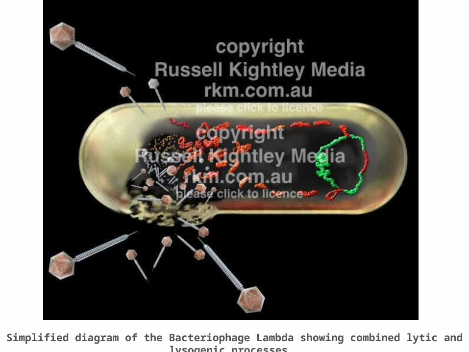
Simplified diagram of the Bacteriophage Lambda showing combined lytic and lysogenic processes.
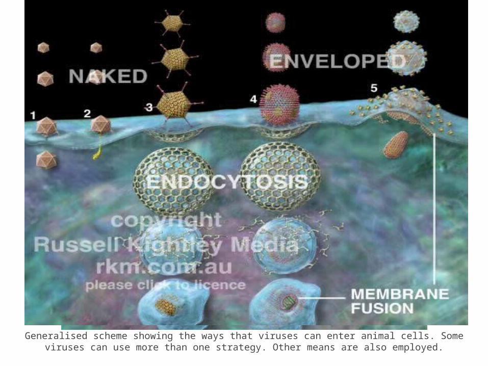
Generalised scheme showing the ways that viruses can enter animal cells. Some viruses can use more than one strategy.
Other means are also employed.
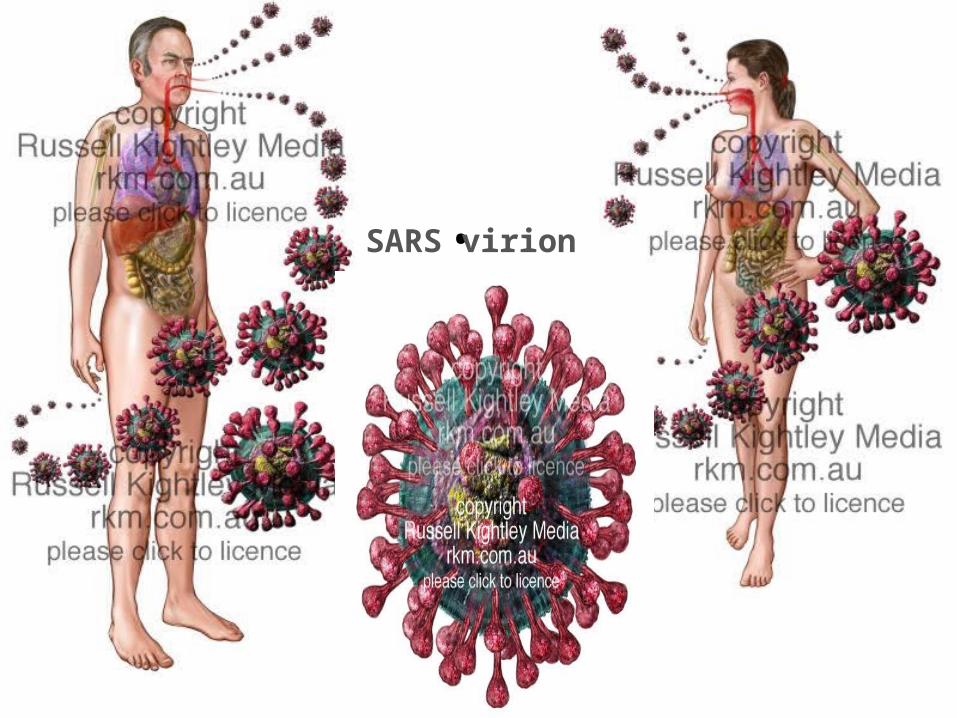
• SARS virion
•
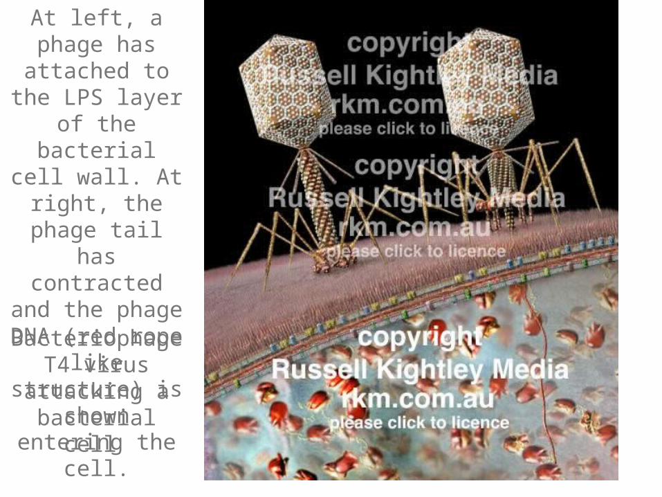
Bacteriophage T4 virus attacking a
bacterial cell
At left, a phage has attached to the LPS
layer of the bacterial cell wall. At right, the phage tail has contracted
and the phage DNA (red rope like
structure) is shown entering the cell.
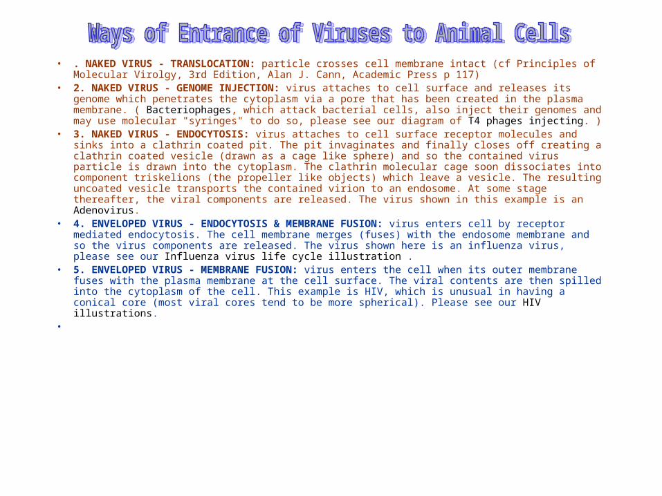
• . NAKED VIRUS - TRANSLOCATION: particle crosses cell membrane intact (cf Principles of Molecular Virolgy, 3rd Edition, Alan J. Cann, Academic Press p 117)
• 2. NAKED VIRUS - GENOME INJECTION: virus attaches to cell surface and releases its genome which penetrates the cytoplasm via a pore that has been created in the plasma membrane. ( Bacteriophages, which attack bacterial cells, also inject their genomes and may use molecular "syringes" to do so, please see our diagram of T4 phages injecting. )
• 3. NAKED VIRUS - ENDOCYTOSIS: virus attaches to cell surface receptor molecules and sinks into a clathrin coated pit. The pit invaginates and finally closes off creating a clathrin coated vesicle (drawn as a cage like sphere) and so the contained virus particle is drawn into the cytoplasm. The clathrin molecular cage soon dissociates into component triskelions (the propeller like objects) which leave a vesicle. The resulting uncoated vesicle transports the contained virion to an endosome. At some stage thereafter, the viral components are released. The virus shown in this example is an Adenovirus.
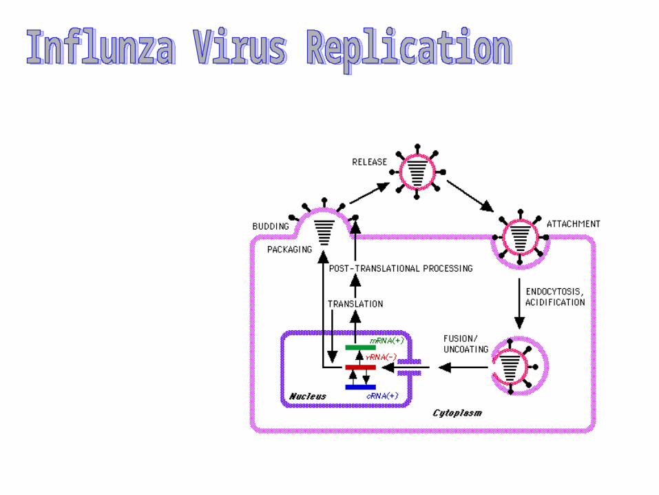
• 4. ENVELOPED VIRUS - ENDOCYTOSIS & MEMBRANE FUSION: virus enters cell by receptor mediated endocytosis. The cell membrane merges (fuses) with the endosome membrane and so the virus components are released. The virus shown here is an influenza virus, please see our Influenza virus life cycle illustration .
• 5. ENVELOPED VIRUS - MEMBRANE FUSION: virus enters the cell when its outer membrane fuses with the plasma membrane at the cell surface. The viral contents are then spilled into the cytoplasm of the cell. This example is HIV, which is unusual in having a conical core (most viral cores tend to be more spherical). Please see our HIV illustrations.
•
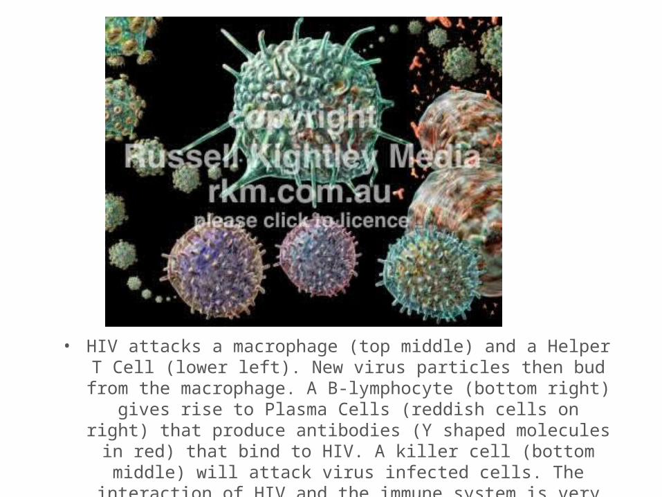
• HIV attacks a macrophage (top middle) and a Helper T Cell (lower left). New virus particles then bud from the macrophage. A B-
lymphocyte (bottom right) gives rise to Plasma Cells (reddish cells on right) that produce antibodies (Y shaped molecules in red) that bind to HIV. A killer cell (bottom middle) will attack virus infected cells. The interaction of HIV and the immune system is very complex and varies
over time. This image is available for licensing worldwide. The
•


![virovétéchap01-origines.pps [Mode de compatibilité]...Paul Frosch (Greifswald, île de Riems) Paul Frosch (1860-1928) Friedrich Loeffler (1852–1915) , Robert Koch (1843-1910)](https://img.pdfslide.net/doc/110x75/5ec26423837cd533aa0bde81/virovtchap01-mode-de-compatibilit-paul-frosch-greifswald-le-de.jpg)