Embed Size (px)
Citation preview

HEAD & FACE MEDICINE
Knösel et al. Head & Face Medicine (2015) 11:11 DOI 10.1186/s13005-015-0069-6
RESEARCH Open Access
In-vivo durability of a fluoride-releasing sealant(OpalSeal) for protection against white-spot lesionformation in orthodontic patientsMichael Knösel1*, David Ellenberger2, Yvonne Göldner3, Paulo Sandoval4 and Dirk Wiechmann5,6
Abstract
Background: Sealant application during fixed appliances orthodontic treatment for enamel protection is common,however, reliable data on its durability in vivo are rare.
Objective: This study aims at assessing the durability of a sealant (OpalSeal, Ultradent) for protection againstwhite-spot lesion formation in orthodontic patients over 26 weeks in vivo, taking into account the provision orabsence of an adequate oral hygiene. We tested the null hypothesis of (1) no significant abatement of the sealantafter 26 weeks in fixed orthodontic treatment compared to baseline, and (2) no significant influence of the factor ofbrushing and oral hygiene (as screened by approximal plaque index, API) on the abatement of the sealant.
Methods: Integrity and abatement of OpalSeal applicated directly following bracketing was assessed in thirty-sixconsecutive patients (nteeth = 796) undergoing orthodontic treatment with fixed appliances (male/female12/24;mean age/SD 14.4/1.33 Y). Assessment of the fluorescing sealant preservation was by a black-light lamp, using aclassification that was concepted in analogy to the ARI index: (3, sealant completely preserved; 2= > 50% preserved;1 = <50%; 0 = no sealant observable) immediately following application (Baseline, T0), after 2 (T1), 8 (T2), 14 (T3), 20(T4) and 26 weeks (T5). API was assessed at T0 and T1. Statistical analysis was by non-parametric repeated measuresANOVA (α = 5%, power >80%).
Results: At baseline, 43.4% of teeth had a positive API. Oral hygiene deteriorated after bracketing (T1, 53%)significantly. Null hypothesis (1) was rejected, while (2) was accepted: Mean values of both the well brushed andnon-brushed anterior teeth undercut the score “1” at T3 (week 14). Despite a slightly better preservation of thesealer before and after T3 in not-sufficiently brushed (API-positive) teeth, this finding was statistically not significant.
Conclusion: One single application of OpalSeal is unlikely to last throughout the entire fixed appliance treatmentstage. On average, re-application of the sealant can be expected to be necessary after 3.5 months (week 14) intreatment.
Keywords: Orthodontic sealant, Durability, OpalSeal, White-spot lesions, In-vivo
IntroductionPrevention of white-spot lesions (WSL) during fixed appli-ances orthodontic treatment is still a challenge in today’sorthodontic treatment: There is evidence that neglectingoral hygiene during orthodontic treatment with fixed ap-pliances can cause WSL formation within weeks [1-4].Other than mechanical plaque removal by tooth brushing,
* Correspondence: [email protected] of Orthodontics, University Medical Center Göttingen (UMG),37099 Göttingen, GermanyFull list of author information is available at the end of the article
© 2015 Knösel et al.; licensee BioMed Central.Commons Attribution License (http://creativecreproduction in any medium, provided the orDedication waiver (http://creativecommons.orunless otherwise stated.
local fluoridation by dentifrices and mouth rinses, or theuse of fluoride-releasing bonding materials, major prevent-ive strategies for a prevention of enamel demineralizationduring fixed orthodontic treatment focus on the applica-tion of fluoride-releasing sealants [5,6].Sealant application for enamel protection is common
in fixed appliances orthodontic treatment patients, how-ever, reliable data on its durability in vivo are rare [7].Tüfekçi et al. investigated the preservation of a sealanton extracted premolars 67 ± 28 days following bracketbonding and sealant application in vivo, and found that
This is an Open Access article distributed under the terms of the Creativeommons.org/licenses/by/4.0), which permits unrestricted use, distribution, andiginal work is properly credited. The Creative Commons Public Domaing/publicdomain/zero/1.0/) applies to the data made available in this article,

Knösel et al. Head & Face Medicine (2015) 11:11 Page 2 of 8
layers of OpalSeal (Ultradent) remained on an average of50% at the time of assessment, and found no correlationbetween sealant residues and the variation of time theteeth were in the mouth [7]. However, it is conceivablethat the factors of oral hygiene and abrasion caused bymechanical tooth brushing, as well as acidic or mechan-ical assaults during consumption of food and beveragesmay have an impact on the sealant condition and dur-ability in vivo: Varnish layers may be reduced in thick-ness and extension by daily mechanical wear. However,whilst there have been studies on reduction of WSL oc-currence following fluoride-releasing sealant application,there is a lack of studies concerning an vivo-screening ofthe integrity or abatement of sealants, in interferencewith oral hygiene habits and observation time.
Study aimsThis study aims at assessing the durability of a sealant(OpalSeal, Ultradent Products, South Jordan, Utah) forprotection against white-spot lesion formation in ortho-dontic patients over more than six months (26 weeks)in vivo, taking into account the provision or absence ofan adequate oral hygiene.We tested the null hypotheses of (1) no significant
abatement of the sealant (as screened by a score from0–3) after 26 weeks in fixed orthodontic treatment com-pared to baseline, and (2) no significant influence of thefactor of oral hygiene (as screened by approximal plaqueindex, API [8]) on the abatement of the sealant.
Subjects and MethodsThirty-six consecutive patients undergoing orthodontictreatment with fixed appliances (male/female 12/24; age12–17 years; mean age 14.44 Y; SD 1.33) were consecu-tively recruited at an orthodontic practice in Hannover,Germany, between Nov 1st, 2011 and April 30, 2012.Subjects were included upon meeting the following in-clusion criteria:
� upcoming indirect Damon-3 (Ormco, Orange, CA,USA) bracket placement of least of sixteen teeth,
� application of a sealant (OpalSeal, UltradentProducts, South Jordan, Utah) on that sameappointment, and
� having given consent for participation and acceptingfollow-up assessments during recall visits.
Subject were excluded upon refusal of sealant applica-tion, or less than sixteen teeth bracketed, or if they dis-agreed to participate. Other than exclusion of subjects,single teeth of included trial subjects were not assessedby this study in case they were not bracketed on the sameappointment, or in case they were subject to upcoming
extraction. Of 864 potentially eligible teeth, a number of796 trial teeth was included (drop-out: n = 68 teeth).Standardized indirect bracket placement using a dry-
field system for isolation was performed prior to sealantapplication, in order to allow for a removal of excessiveadhesives without setting damages to sealant layers. Fol-lowing cleaning of tooth surfaces with fluoride-free pum-ice, adhesive and sealant application routine was carriedout following manufacturer’s instructions and included a15 s interval of etching with 37 % phosphoric acid of thecomplete labial enamel surface, followed by indirect bond-ing using chemically-cured Monolok2 composite adhesivesystem (Rocky Mountain Orthodontics, Denver, Colo,USA). Adhesive residues have been removed prior tosealant application. According to the manufacturer’s in-structions, OpalSeal was gently air-dried following applica-tion, prior to light-curing for 20s per tooth (Bluephase C8,800 mW/cm2, IvoclarVivadent, Schaan, Liechtenstein).
Ethical approvalThe study was performed in extension of an earlier posi-tively voted study protocol (# 4/8/09). All procedures usedin this prospective observational study had been presentedto the ethics committee of the University of Göttingen,Germany, earlier. There were no objections against publi-cation. The patients and their guardians gave informedconsent for taking part in the study.
Parameter 1: Screening of oral hygieneThe approximal plaque index (API) has been introducedin dentistry for a quick assessment of oral hygiene status[8]. Although being based on more or less subjective de-cisions that are made chair-side, API assessments havebeen established as a basic clinical methodology used inresearch on the subject of cariology and periodontology[9]. Oral hygiene status was screened using the API foreach bracketed tooth, as a yes/no decision (results givenin % of teeth with plaque) prior to bracket placementand sealant application at T0, and after 14 days in treat-ment (T1). All patients received identical, standardizedinstructions on both tooth- and inter-bracket brushingduring orthodontic treatment with fixed appliances, andwere advised to do so three times daily, using typicalcommercially available 1,400-1,450 ppm fluoridated den-tifrices. They were provided with the same type of toothbrushes with medium filaments, and interdental brushes(TePe, Malmö, Sweden).
Parameter 2: Scoring of sealant layer integrityIntegrity and condition of the OpalSeal-layer was assessedusing a black-light UV lamp provided by the manufacturerfor screening purposes of the fluorescing sealant. Similarto previous trials [4], assessments were done chair-side bya clinician who was blinded to the patient’s trial time

Knösel et al. Head & Face Medicine (2015) 11:11 Page 3 of 8
frame, while notes were made by a study nurse: Immedi-ately after bonding and sealant application (Baseline, T0),after 14 days (T1), 8 weeks (T2), 14 weeks (T3), 20 weeks(T4) and 26 weeks (T5). Abatement of the varnish wasparameterized using a classification from 0 to 3 that wasconcepted in analogy to the adhesive remnant index(ARI, [10]): (3 = sealant undamaged/completely preserved,2 > =50% preserved, 1 < =50%, 0 = no sealant observable tothe naked eye), assessed for every bracketed tooth (max.24 per patient, Figure 1a, b and c).
Statistical analysisThe factor of ‘oral hygiene’ as assessed by API scores atBaseline (T0) and two weeks following bracketing (T1)was tested for potential changes (increases in API score =deterioration of oral hygiene) using a t-test for dependentsamples. The status of the durability of the OpalSeal-layeras well as potential impacts and interactions of the initialAPI (T0), trial time elapse (T1-T5), tooth type (#1-#6; 1,central -; 2, lateral incisor; 3, canine; 4, first-; 5, secondpremolar; 6, first molar) and jaw (maxilla, mandible) weretested by non-parametric, repeated measures ANOVA,with the OpalSeal-Score as dependent variable. Correlatedmeasurements within one patient as well as over time foreach tooth were modeled by a random factor ‘subject’along with a random factor ‘tooth’ yielding a nested com-pound symmetry structure. In the case of significant
Figure 1 a Assessment of sealant integrity was done by black-light illuminexamples of sealant scores 3 (sealant undamaged/completely preserved), a
interactions between the experimental factors, the datawere split and further analyzed in subgroups. The sig-nificance level was set to 5%. Sample size calculation ac-cording to O’Brien-Castelloe yielded a power in excessof 80% for an inclusion of 36 subjects/796 teeth. Allanalyses were performed using SAS 9.3 (SAS Institute,Cary, NC, USA) and Statistica 10 (StatSoft (Europe)GmbH, Hamburg, Germany).
ResultsAt baseline, 43.4% of teeth had a positive API (SD:20.5%). Oral hygiene deteriorated after bracketing (T1,53%, SD: 22.0%) significantly: The T-test for dependentsamples API (complete, %) T0 vs T1 yielded p = 0.01.
Effect of oral hygiene (API) on sealant abatementAt T1, we found that teeth with positive API scoresshowed no significant differences in terms of sealantlayer preservation, in contrast to teeth with negative APIscores (Table 1). Generally spoken, there was an increasein the abatement of the sealant from front teeth 1–4 toposterior teeth #5 or #6, which is globally significant(Table 2) and was found to be more rapid in wellbrushed lower posterior teeth (teeth #5 and #6 withnegative API). (Table 1, Figures 2a and b). Mean valuesof both the well brushed and non-brushed anteriorteeth undercut the score “1” (<50% sealer left) at T3
ation, using the fluorescent properties of the OpalSeal. b and c givend 1 (<=50% of sealant left).

Table 1 As anterior teeth #1-#4 were found to behomogeneous in terms of abatement of the sealantscore, pair-wise comparisons between this group of teethwith teeth #5 and #6 were implemented
Time API (T0) Toothgroupscompared
Opalsealscoredifference
Standarderror
p-value
1 positive 1-4 5 0.24 0.16 0.20
1 positive 1-4 6 0.26 0.12 0.07
1 positive 5 6 0.03 0.19 0.94
1 negative 1-4 5 0.21 0.08 0.02
1 negative 1-4 6 0.69 0.09 <.0001
1 negative 5 6 0.49 0.10 <.0001
2 positive 1-4 5 0.26 0.16 0.08
2 positive 1-4 6 0.41 0.12 0.001
2 positive 5 6 0.15 0.19 0.57
2 negative 1-4 5 0.39 0.08 <.0001
2 negative 1-4 6 0.69 0.08 <.0001
2 negative 5 6 0.31 0.10 0.005
3 positive 1-4 5 0.20 0.16 0.15
3 positive 1-4 6 0.45 0.12 0.0002
3 positive 5 6 0.25 0.19 0.25
3 negative 1-4 5 0.33 0.08 <.0001
3 negative 1-4 6 0.54 0.09 <.0001
3 negative 5 6 0.21 0.10 0.07
4 positive 1-4 5 0.22 0.16 0.17
4 positive 1-4 6 0.61 0.12 <.0001
4 positive 5 6 0.39 0.19 0.03
4 negative 1-4 5 0.21 0.08 0.003
4 negative 1-4 6 0.46 0.09 <.0001
4 negative 5 6 0.26 0.10 0.02
5 positive 1-4 5 0.30 0.16 0.04
5 positive 1-4 6 0.58 0.12 <.0001
5 positive 5 6 0.27 0.19 0.13
5 negative 1-4 5 0.00 0.08 0.79
5 negative 1-4 6 0.32 0.09 <.0001
5 negative 5 6 0.32 0.10 0.001
Especially during the first weeks in treatment, sealant preservation was betterin API-negative teeth, although this finding was globally not significant whenconsidering all time points (see also Table 3).
Table 2 Factors and interactions that have a potentialimpact on sealant durability scores
Effect ANOVA
p-Value
Jaw (Maxilla, Mandible) <.0001
Tooth type (#1,#2,#3,#4,#5,#6) <.0001
Jaw * Tooth type 0.01
Time (T 1,2,3,4,5) <.0001
Jaw * Time 0.45
Tooth type * Time 0.69
Jaw * Tooth type * Time 0.83
Oral hygiene by initial API (0) 0.54
Jaw * Oral hygiene 0.24
Tooth type * Oral hygiene 0.73
Jaw * Tooth type * Oral hygiene 0.26
Time * Oral hygiene 0.10
Jaw * Time * Oral hygiene 0.08
Tooth type * Time * Oral hygiene 0.0002
Jaw * Tooth type * Time * Oral hygiene 0.83
The explained variance by within-subject measurements was found to becrucial with R2 = 0.34 (p < .0001) for the random factor ‘subject’ and R2 = 0.27(p < .0001) for the random factor ‘tooth’.
Knösel et al. Head & Face Medicine (2015) 11:11 Page 4 of 8
(week 14) (Figure 2a and b). Percentages of teeth with ascore higher than 0 are depicted by Table 3 and Figure 2cand d. Despite the overall slightly better preservation ofthe sealant before and after T3 in not-sufficiently brushed(API positive) teeth compared to API-negative teeth, thisfinding was statistically not significant (Table 2). That is,considering the total trial time, the factor oral hygieneitself has no global significant effect on the abatement
of the sealant. See (Figure 2a and b) for a visualisationof this effect.
Maxilla vs. MandiblePairwise comparisons indicate a more pronounced abate-ment of the sealant in the mandible than in the maxilla atT1, and it was significantly increased in mandibularteeth #1, #2, and #6 when compared to the maxillaryequivalent (Figure 3).
DiscussionAn inhibition of enamel demineralization during ortho-dontic fixed treatment can be achieved by the applica-tion of fluoride-releasing sealants [11,12], however, theefficacy of those sealants also depends on their integrityor durability [7]. It is a popular fallacy to assume thatone sealant application at the start of fixed appliancesorthodontic treatment will suffice for enamel protectionthroughout the entire fixed treatment stage, without arenewal [13,14]. In-vivo research yielded evidence thatsealants offer some protection and are suitable for redu-cing frequencies of new WSL [12], but do not offer out-right protection from WSL formation for the full durationof treatment [7]. Diligence during application and fre-quencies of re-application may be relevant in terms ofsealant durability, as may be the presence of differentlevels of oral hygiene and intensities of tooth- and inter-bracket brushing as a factor that is potentially causingsealant abrasion. In-vivo data on the durability of those
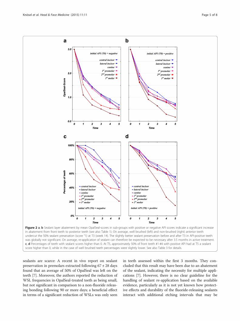
Figure 2 a, b Sealant layer abatement by mean OpalSeal-scores in sub-groups with positive or negative API scores indicate a significant increasein abatement from front teeth to posterior teeth (see also Table 1). On average, well brushed (left) and non-brushed (right) anterior teethundercut the 50% sealant presevation (score “1) at T3 (week 14). The slightly better sealant preservation before and after T3 in API-positive teethwas globally not significant. On average, re-application of sealant can therefore be expected to be necessary after 3.5 months in active treatment.c, d Percentages of teeth with sealant scores higher than 0. At T5, approximately 50% of front teeth #1-#4 with positive API had at T5 a sealantscore higher than 0, while in the case of well brushed teeth percentages were slightly lower. See also Table 3 for details.
Knösel et al. Head & Face Medicine (2015) 11:11 Page 5 of 8
sealants are scarce: A recent in vivo report on sealantpreservation in premolars extracted following 67 ± 28 daysfound that an average of 50% of OpalSeal was left on theteeth [7]. Moreover, the authors reported the reduction ofWSL frequencies in OpalSeal-treated teeth as being small,but not significant in comparison to a non-fluoride releas-ing bonding following 90 or more days; a beneficial effectin terms of a significant reduction of WSLs was only seen
in teeth assessed within the first 3 months. They con-cluded that this result may have been due to an abatementof the sealant, indicating the necessity for multiple appli-cations [7]. However, there is no clear guideline for thehandling of sealant re-application based on the availableevidence, particularly as it is not yet known how protect-ive effects and durability of the fluoride-releasing sealantsinteract with additional etching intervals that may be

Table 3 Sealant abatement: Percentages of teeth withsealant scores higher than 0 in sub-groups with adequateor inadequate oral hygiene (negative or positive APIscores)
Tooth type # Time Teeth with sealantscore 1 or higher (%)
Negative API Positive API
1 0 100 100
1 1 92.59 93.02
1 2 83.33 77.91
1 3 75.93 70.93
1 4 66.67 59.30
1 5 53.70 54.65
2 0 100 100
2 1 100 90.11
2 2 93.62 71.43
2 3 78.72 67.03
2 4 63.83 59.34
2 5 40.43 56.04
3 0 100 100
3 1 90.91 91.84
3 2 80.00 77.55
3 3 70.91 65.31
3 4 56.36 61.23
3 5 40.00 48.98
4 0 100 100
4 1 95.56 92.59
4 2 83.33 79.01
4 3 70.00 72.84
4 4 58.89 66.67
4 5 46.67 51.85
5 0 100 100
5 1 84.09 88.46
5 2 61.36 73.08
5 3 46.59 65.38
5 4 39.77 61.54
5 5 37.50 34.62
6 0 100 100
6 1 66.67 85.29
6 2 51.39 67.65
6 3 44.44 50.00
6 4 30.56 26.47
6 5 20.83 14.71
Figure 3 Pairwise comparisons of time-averaged mean OpalSeal-scores(with 95% confidence intervals) stratified by tooth type (#1-6) andjaw (maxilla, mandible). Abatement of sealant layer was significantlyincreased in the mandibular teeth #1, #2, #6 (p < 0.001; 0.03; <.0001)when compared to the maxillary equivalent.
Knösel et al. Head & Face Medicine (2015) 11:11 Page 6 of 8
necessary to achieve sealant retention after months intreatment: It is a fact that additional, surplus etching ofenamel surfaces not covered by bracket bases may triggerWSL formation itself [15]. The data presented here add
some evidence to the unanswered questions of recom-mended time intervals for a renewal of the sealant, and,second, the unclear role of individual intensities of oralhygiene and mechanical tooth brushing abrasion on seal-ant durability: The null-hypothesis (1) of no significantglobal abatement of the sealant after 26 weeks in treat-ment was rejected, while the null hypothesis (2) was ac-cepted, as we did not find a significant global effect of thefactor of oral hygiene on the abatement of the sealant.Nonetheless, there was an increase in the abatement ofthe sealant from front teeth 1–4 to posterior teeth #5 or#6, which is significant (Table 1) and was found to bemore rapid in well brushed lower posterior teeth (teeth #5and #6 with negative API; Figure 2a). One possible inter-pretation might be that sealant layers may be literallywiped off by tooth brushing, and it may be concluded thatsealants offer a slightly better protection of those teeththat are not sufficiently brushed. Another possible explan-ation would be that this abatement in mandibular poster-ior teeth may also be attributed to enhanced attrition bychewing activities. This would explain reductions of var-nish layers in the upper enamel third of lower first molarsand premolars, as those areas are more exposed to dailymechanical wear during consumption of food.Mean sealant scores indicate that in both the well
brushed and non-brushed teeth anterior teeth undercutthe score “1” (less than 50% sealer left) at T3 (week 14),while there seems overall a slightly better preservationof the sealer before and after T3 in positive API-teeth,especially in the case of second premolars (Figure 3c).

Knösel et al. Head & Face Medicine (2015) 11:11 Page 7 of 8
However, globally, these slightly better results are notsignificantly improved compared to the non-brushedteeth (Table 2). Re-application can therefore be expectedto be necessary from week 14 in the average case treatedwith orthodontic fixed appliances. This result is in agree-ment with Tüfekçi et al. who reported a preservation of50% of the same sealant following ninety days [7].
Sealant re-application: Time intervals and modeAccording to the manufacturer, re-application of OpalSealimplies prior etching for 15-30s, and subsequent light-curing for 10 s. Previous research hints at a potential iat-rogenic triggering of WSL especially in those cases withsurplus orthodontic etching prior to bracket bonding, es-pecially when those areas are subsequently neither cov-ered by bracket bases nor by bonding material [15]: Thishas been shown to have a significant deteriorating effecton WSL formation. Subsequent covering of those etchedareas by a sealant prevents WSL formation, however,the results of the current study show that the sealantabates on average in about 3.5 months, and it is unclearwhether there is an increased susceptibility to WSL afterdisappearance of the sealant. According to previous re-ports, teeth are especially susceptible to decalcificationduring the first six months in fixed appliances ortho-dontic treatment [4]. If this was due to some type ofintra-oral customization to the fixed orthodontic appli-ances, simple re-application of sealants without repeat-ing the step of etching may also be viable.
Advantages and drawbacks of the study designIntegrity and condition of the OpalSeal layers was assessedchair-side by a clinician who was blinded to the patient’strial time schedule, while notes were made by a studynurse, similar to previous studies on the topic of WSLformation in orthodontic patients [4]. The assessment ofphotos would not have been feasible here, as every singletooth would have to be photographed while being illumi-nated by the black-light lamp. However, as an advantage,the trial sample size and numbers of performed assess-ments are remarkable.To the best of our knowledge, this is the first in-vivo
trial with a screening of the integrity and durability of asealant at different time points with a consideration of oralhygiene as a co-factor. While in-vivo research offers amore realistic picture of the study subject, standardizationis more difficult to achieve in comparison to using an in-vitro setup, where standardization is easy, but applicabilityand generalisability of results are rather poor. This studyhas a limitation in that there was no standardization ofthe brushing regimes other than providing standardizedbrushing instructions and handing out identical toothbrushes. There was no left-/right handed distinction. Also,oral hygiene was screened before placement of brackets,
and then again 14 days following incorporation ofbrackets. That is, subjects were allocated to the groupsof adequate or inadequate oral hygiene based on thesetwo assessments, not considering potential improve-ment or deterioration afterwards.The results of this study provide some evidence on the
abatement characteristics of the longevity of a typicalsealant used in orthodontics, and they indicate that a re-newal of the OpalSeal layer after an average elapse of3.5 months may be beneficial or necessary for an enduringsuppression of WSL formation and frequencies. Previousstudies on WSL formation following sealant applicationmay be revisited in terms of fixed appliances orthodontictreatment duration versus presence or absence of sealantre-application. However, in this trial on the effects of timeelapse and oral hygiene measures on sealant abatement,there was no assessment of WSL formation, but of sealantpreservation only. That is, no conclusion can be drawnon the basis of our data that renewal of sealants after3.5 months does indeed result in a further reduction ofWSL numbers: It may also be conceivable that sealingof the enamel surfaces during the first months in fixedappliances orthodontic treatment may suffice, as it hasbeen reported that teeth are especially susceptible towhite-spot formation during this time period. Also, theinteraction of sealant renewal with potentially necessaryadditional etching intervals needs to be considered.
Future researchFurther research is required on the subject of the modeof OpalSeal re-application (with or without additionaletching) in terms of (1) reduction of frequencies of WSL,and (2) durability and abatement of sealants re-applicatedthe one way, or the other: As it is known that excessivephosphoric-acid etching of enamel as required prior tosealant application may trigger WSL formation itself [15],it is e.g. not clear whether enamel surfaces should beetched again prior to re-application, or if OpalSeal appli-cation renewal should be performed without a secondetching interval, or not at all.
ConclusionsThe following conclusions can be drawn from the studypresented here:
� Diligent screening of sealant preservation in patientstreated with fixed orthodontic appliances isa necessity.
� One single sealant application is unlikely to lastthroughout the entire stage of orthodontictreatment with fixed appliances.
� On average, re-application of OpalSeal can beexpected to be necessary after 3.5 months (week 14)in treatment. Further clinical trials should address

Knösel et al. Head & Face Medicine (2015) 11:11 Page 8 of 8
the question if a re-application of sealants would bebeneficial in terms of reducing frequencies of WSL.
� As etching of enamel surfaces is known topotentially trigger WSL formation, future researchshould also clarify if re-application of sealantsshould include an additional etching interval for animprovement of sealant durability, or not.
Competing interestsAll authors declare that they have no competing interests.
Authors’ contributionsAll authors contributed extensively to the work presented in this paper. MKprovided the idea for the project, was responsible for the study design,treated the patients orthodontically, carried out the measurements with YG,interpreted the data together with DE, implemented the literature search,and wrote the manuscript. YG collected the data, and compiled all medicalrecords. DE carried out the statistical analyses. PS and DW reviewed thepaper, and contributed to the writing. All authors approved the manuscriptprior to submission.
AcknowledgementWe thank Dr. J. Raiman (Hannover, Germany) for the permission to conductthis study in his orthodontic practice and to release these data, and also C.Jerchel for her help in organizing the data.
Author details1Department of Orthodontics, University Medical Center Göttingen (UMG),37099 Göttingen, Germany. 2Department of Medical Statistics, UniversityMedical Center Göttingen (UMG), 37099 Göttingen, Germany. 3PrivatePractice, Hannover, Germany. 4Department of Orthodontics, Universidad dela Frontera (UFRO), Temuco, Chile. 5Orthodontic Practice, Lindenstrasse 44,49152 Bad, Essen, Germany. 6Department of Orthodontics, Hannover MedicalSchool (MHH), 30625 Hannover, Germany.
Received: 19 January 2015 Accepted: 2 April 2015
References1. Øgaard B, Rolla G, Arends J. Orthodontic appliances and enamel
demineralization. Part 1. Lesion development. Am J Orthod DentofacialOrthop. 1988;94:68–73.
2. Melrose CA, Appleton J, Lovius BB. A scanning electron microscopic studyof early enamel caries formed in vivo beneath orthodontic bands. Br JOrthod. 1996;23:43–7.
3. Richter AE, Arruda AO, Peters MC, Sohn W. Incidence of caries lesionsamong patients treated with comprehensive orthodontics. Am J OrthodDentofacial Orthop. 2011;139:657–64.
4. Tüfekçi E, Dixon JS, Gunsolley JC, Lindauer SJ. Prevalence of white spotlesions during orthodontic treatment with fixed appliances. Angle Orthod.2011;81:206–10.
5. Geiger AM, Gorelick L, Gwinnett AJ, Benson BJ. Reducing white spot lesionsin orthodontic populations with fluoride rinsing. Am J Orthod DentofacialOrthop. 1992;101:403–7.
6. Benson PE, Parkin N, Dyer F, Millett DT, Furness S, Germain P. Fluorides forthe prevention of early tooth decay (demineralised white lesions) duringfixed brace treatment. Cochrane Database Syst Rev. 2013;12, CD003809.
7. Tüfekçi E, Pennella DR, Mitchell JC, Best AM, Lindauer SJ. Efficacy of afluoride-releasing orthodontic primer in reducing demineralization aroundbrackets: an in-vivo study. Am J Orthod Dentofacial Orthop. 2014;146:207–14.
8. Lange DE, Plagmann HC, Eenboom A, Promesberger A. KlinischeBewertungsverfahren zur Objektivierung der Mundhygiene [Clinicalmethods for the objective evaluation of oral hygiene]. Dtsch Zahnarztl Z.1977;32:44–7.
9. Splieth CH, Treuner A, Gedrange T, Berndt C. Caries-preventive andremineralizing effect of fluoride gel in orthodontic patients after 2 years.Clin Oral Investig. 2012;16:1395–9.
10. Årtun J, Bergland S. Clinical trials with crystal growth conditioning as analternative to acid-etch enamel pretreatment. Am J Orthod. 1984;85:333–40.
11. Hu W, Featherstone JDB. Prevention of enamel demineralization: an in-vitrostudy using light-cured filled sealant. Am J Orthod Dentofacial Orthop.2005;128:592–600.
12. O'Reilly MT, De Jesús VJ, Hatch JP. Effectiveness of a sealant compared withno sealant in preventing enamel demineralization in patients with fixedorthodontic appliances: a prospective clinical trial. Am J Orthod DentofacialOrthop. 2013;143:837–44.
13. Todd MA, Staley RN, Kanellis MJ, Donly KJ, Wefel JS. Effect of fluoridevarnish on demineralization adjacent to orthodontic brackets. Am J OrthodDentofacial Orthop. 1999;116:159–67.
14. Farhadian N, Miresmaeili A, Eslami B, Mehrabi S. Effect of fluoride varnish onenamel demineralization around brackets: an in-vivo study. Am J OrthodDentofacial Orthop. 2008;133(4 Suppl):S95–8.
15. Knösel M, Bojes M, Jung K, Ziebolz D. Increased susceptibility for white spotlesions by surplus orthodontic etching exceeding bracket base area. Am JOrthod Dentofacial Orthop. 2012;141:574–82.
Submit your next manuscript to BioMed Centraland take full advantage of:
• Convenient online submission
• Thorough peer review
• No space constraints or color figure charges
• Immediate publication on acceptance
• Inclusion in PubMed, CAS, Scopus and Google Scholar
• Research which is freely available for redistribution
Submit your manuscript at www.biomedcentral.com/submit


![F]FDG...discuss the production of [18 F]fluoride ion. Next, I’ll talk about trapping and releasing the [18 F]fluoride ion, followed by it’s preparation for the subsequent processing](https://img.pdfslide.net/doc/110x75/5fccaab1fe42b040e11f0866/ffdg-discuss-the-production-of-18-ffluoride-ion-next-iall-talk-about.jpg)