Embed Size (px)
Citation preview

Increase or Decrease Hydrogen Sulfide Exert OppositeLipolysis, but Reduce Global Insulin Resistance in HighFatty Diet Induced Obese MiceBin Geng1*, Bo Cai1,2, Feng Liao1, Yang Zheng1, Qiang Zeng3, Xiaofang Fan4, Yongsheng Gong4,
Jichun Yang1, Qing hua Cui1, Chaoshu Tang1,4, Guo heng Xu1*
1Department of Physiology and Pathophysiology, School of Basic Medical Science, Peking University, Bei Jing, P.R. China, 2Department of Physiology, Guangdong
Medical Collage, Zhan Jiang, Guang dong Province, P.R. China, 3 International Medical Center, Chinese PLA General Hospital, Bei Jing, P.R. China, 4 Institute of Hypoxia
Medicine, Wenzhou Medical University, Wen Zhou, Zhe jiang Province, P.R. China
Abstract
Objective: Adipose tissue expressed endogenous cystathionine gamma lyase (CSE)/hydrogen sulfide (H2S) system. H2Sprecursor inhibited catecholamine stimulated lipolysis. Thus, we hypothesized that CSE/H2S system regulates lipolysis whichcontributed to the pathogenesis of insulin resistance.
Methods: We treated rat adipocyte with DL-propargylglycine (PAG, a CSE inhibitor), L-cysteine (an H2S precursor) pluspyridoxial phosphate (co-enzyme) or the H2S chronic release donor GYY4137, then the glycerol level was assayed forassessing the lipolysis. Then, the effects of PAG and GYY4137 on insulin resistance in high fatty diet (HFD) induced obesemice were investigated.
Results: Here, we found that PAG time-dependently increased basal or isoproterenol stimulated lipolysis. However, L-cysteine plus pyridoxial phosphate or GYY4137 significantly reduced it. PAG increased phosphorylated protein kinase Asubstrate, perilipin 1 and hormone sensitive lipase, but L-cysteine and GYY4137 decreased the parameters. In HFD inducedobese mice, PAG increased adipose basal lipolysis, thus blunted fat mass increase, resulting in lowering insulin resistanceevidenced by reduction of fasting glucose, insulin level, HOMA index, oral glucose tolerance test (OGTT) curve area andelevating the insulin tolerance test (ITT) response. GYY4137 inhibited lipolysis in vivo without increasing fat mass, but alsoameliorated the insulin resistance in HFD mice.
Conclusion: These results implicated that inhibition endogenous CSE/H2S system in adipocytes increased lipolysis by aprotein kinase A-perilipin/hormone-sensitive lipase pathway, thus blunted fat mass increase and reduced insulin resistancein obese mice; giving H2S donor decreased lipolysis, also reduced insulin resistance induced by HFD. Our data showed thatincrease or decrease H2S induced opposite lipolysis, but had the same effect on insulin resistance. The paradoxicalregulation may be resulted from different action of H2S on metabolic and endocrine function in adipocyte.
Citation: Geng B, Cai B, Liao F, Zheng Y, Zeng Q, et al. (2013) Increase or Decrease Hydrogen Sulfide Exert Opposite Lipolysis, but Reduce Global InsulinResistance in High Fatty Diet Induced Obese Mice. PLoS ONE 8(9): e73892. doi:10.1371/journal.pone.0073892
Editor: Victor Sanchez-Margalet, Virgen Macarena University Hospital, School of Medicine, Spain
Received February 20, 2013; Accepted July 26, 2013; Published September 13, 2013
Copyright: � 2013 Geng et al. This is an open-access article distributed under the terms of the Creative Commons Attribution License, which permitsunrestricted use, distribution, and reproduction in any medium, provided the original author and source are credited.
Funding: This work was supported by the Major State Basic Research Development Program of the People’s Republic of China (nos. 2012CB517806 and2009CB941603) and the National Natural Science Foundation of the People’s Republic China (nos. 81170235 and 81070114). The funders had no role in studydesign, data collection and analysis, decision to publish, or preparation of the manuscript.
Competing Interests: The authors have declared that no competing interests exist.
* E-mail: [email protected] (BG); [email protected] (GHX)
Introduction
Obesity is popular diseases in developed and developing
countries and major characteristic is fat mass increase. In obesity
individual, un-balance of over energy uptake and lowered energy
expenditure is the major reason of obesity. All mammals store
excess amounts of energy in the form of intracellular triglycerides,
mainly in lipid droplets. During food deprivation or stress,
triglyceride lipolysis provides the primary source of energy [1].
In obese adipocyte, starvation or stress-stimulated lipolysis
reduced, but basal triglyceride lipolysis elevated then released
more free fatty acids (FFAs) into the bloodstream. The excess FFAs
from obese adipocyte induced target tissues local inflammatory
response, oxidative stress, endoplasmic reticulum stress and
metabolic disorder etc. which seem to be metabolic risk factors
contributing to the pathogenesis of diabetes and insulin resistance
[2]. Three major lipases control lipolysis: adipose triglyceride
lipase (ATGL), hormone-sensitive lipase (HSL) and monoglyceride
lipase [3]. ATGL exhibits high substrate for triacylglycerol [4] and
mediates basal lipolysis [5]. HSL is a well-known rate-limiting
enzyme of lipolysis under starvation and stress [1]; PKA
phosphorylated HSL at Ser659, and Ser660 site increased [6],
and AMP-activated protein kinase (AMPK) phosphorylated HSL
at Ser565 [7] inhibited HSL activity. Perilipin 1 (perilipin A) is a
major lipid droplet scaffold protein and blocked the access of
cytosolic lipases to lipid droplet. Phosphorylation perilipin by PKA
PLOS ONE | www.plosone.org 1 September 2013 | Volume 8 | Issue 9 | e73892

results in perilipin conformational changes that expose lipid
droplet stores and facilitates translocation of phosphorylated HSL,
thereby elevating the fat mobilization [8].
Hydrogen sulfide (H2S) is a gasotransmitter and plays important
regulatory roles in cardiovascular, gastrointestinal and neurolog-
ical diseases [9,10]. Cystathionine b synthase (CBS), cystathionine
c lyase (CSE) or 3-mercaptopyruvate sulfurtransferase are key
enzymes generating H2S as L-cysteine as a substrate [11]. Our
recent work found that visceral white adipose, subcutaneous
adipose and perivascular adipose tissues expressed CSE protein
and endogenously generated H2S [12,13]. Interestingly, in normal
culture condition, an H2S donor inhibited basal or insulin-
stimulated glucose uptake in mature adipocytes,whereas blocked
endogenous H2S production by DL-propargylglycine (PAG)
increased glucose uptake activity [12]. However, in 3T3-L1
differentiated adipocytes exposed to high glucose (25 mM), H2S or
its precursor L-cysteine increased glucose utilization [14]. These
works suggested H2S might play different roles in glucose
utilization in physiological and diabetic condition, which also
means that H2S might regulate balance of energy storage (lipid
accumulation) and consumption (lipolysis) while adipocyte is in
different energy statues. H2S precursor-cysteine dose-dependently
inhibited catecholamine-stimulated lipolysis or inhibited HSL
activity with TNF-a stimulation [15,16] in rat adipocytes. So we
hypothesized that adipocyte endogenous CSE/H2S pathway
regulated lipolysis, which contributed to insulin resistance induced
by obesity.
To test our hypothesis, we used PAG inhibition of CSE activity
and GYY4137 as H2S donor, to investigate the possible role of
endogenous CSE/H2S system in adipose lipolysis. To confirm the
effect in vivo, we also evaluated the effects of PAG and GYY4137
on lipolysis and insulin resistance in obesity mice induced by HFD.
Materials and Methods
MaterialMale Sprague-Dawley rats (180–200 g) and C57BL/6J mice
(13–15 g) were provided by the Animal Department, Health
Science Center of Peking University. All animal care and
experimental protocols complied with the Animal Management
Rules of the Ministry of Health of the People’s Republic of China
and the guide for the Care and Use of the Laboratory Animals of
the Peking University. L-cysteine, pyridoxal phosphate, isoproter-
enol, bovine insulin and phenol red-free DMEM were from Sigma
(St. Louis, MO). Polyclonal antibody against rat perilipin was a gift
from C. Londos (US National Institutes of Health). Antibodies
recognizing HSL, phosphorylated HSL (phospho-HSL), phospho-
PKA substrate (RRXS*/T*), AMPK and IRS-1 were from Cell
Signaling Technologies (Boston, MA). Defatted bovine serum
albumin (BSA) and enhanced chemiluminescence (ECL) reagent
were from Applygen Technologies (Beijing).
Primary Adipocyte Isolation and CulturePrimary adipocytes were isolated from epididymal fat pads of
male Sprague-Dawley rats (160,180 g) according to our labora-
tory method [17]. The minced fat pads (2 g) were suspended in
Krebs-Ringer solution (5 mL) containing 0.75 mg/ml type I
collagenase, 200 nM adenosine, 25 mM Hepes, pH 7.4, and 1%
free fatty acid free-BSA, then digested at 37uC by horizontal
shaking (110 rpm) for 30 min. Primary adipocytes were collected,
washed and counted, then pre-incubated in phenol red-free and
serum-free DMEM in an atmosphere of 5% CO2 at 37uC for 1-h
before treatments.
Evaluation of Lipolysis by Glycerol AssayAdipocyte lipolysis leads to the hydrolysis of triglyceride release
of three free fatty acids and a glycerol backbone. Thus, we
measured glycerol content in medium to assess lipolytic reaction.
Adipocytes were incubated in serum-free and phenol red-free
DMEM. After treatment with different reagents for different times,
the culture medium was collected and heated at 70uC for 10 min
to inactivate residue lipase activity. Glycerol was determined by
the enzyme-coupled colorimetric assay (GPO Trinder reaction)
[18]. For evaluation adipose tissues lipolysis, 20 mg adipose tissue
from normal chow mice or high fatty diet mice was incubated in
serum-free and phenol red-free DMEM for 1-h, the release
glycerol was measured.
Western Blot AssayAdipose tissues or adipocytes were collected and lysed in buffer
containing 62.5 mM Tris-HCl, pH 6.8, 2% SDS, 0.1 mM sodium
orthovanadate, and 50 mM sodium fluoride. The lysates were
centrifuged by 12000 g at 4uC, then solid lipid was removed, and
protein content was determined by the BCA assay. Equal amounts
of protein were denatured, then loaded and separated by SDS-
PAGE. For detecting phospho-perilipin with anti-perilipin anti-
body, we used a low-Bis–concentration polyacrylamide gel
prepared with 10% acrylamide and 0.07% N, N9-methylene-bis-
acrylamide (at 142:1 ratio versus 39:1 for traditional gel), to provide
maximal resolution of proteins in the 60- to 70-kDa range [17,19–
21]. Proteins transferred on membranes were recognized with use
of primary antibodies and horseradish peroxidase-conjugated
secondary antibodies. The bands were developed by use of ECL
reagents. If required, membranes were stripped by a commercial
stripping buffer (2% SDS, 62.5 mM Tris-HCl pH 6.8, 0.8% b-mercaptoethanol), and blots were reprobed with other antibodies.
High Fatty Diet Induced Insulin Resistance MiceMale C57BL/6J mice were housed in standard cages in a
temperature- and mumidity-controlled environment, on a 12-h
light/dark cycle, and with free access to water. From the age of 7-
wk, animals were fed a normal diet (equivalent of 10% energy
from fat) or a high-fat diet (HFD: equivalent of 45% energy from
fat, both purchased from Beijing HFK Bioscience CO. LTD).
After 12-wk of feeding, an oral glucose tolerance test (OGTT) and
insulin tolerance test (ITT) were performed on non-anesthetized
mice. Mice were fasted for 16-h before OGTT and for 4-h before
ITT. For OGTT, blood (tail vein) glucose levels were measured at
baseline and 15, 30, 60, 90, and 120 min after gavage (gastric
tube, outer diameter 1.2 mm) glucose (150 mg) by using an Accu
CheK Active glucometer (Roche Diagnostics). For ITT, the blood
glucose levels were measured at baseline and 15, 30, 45, 60 and
90 min after injection bovine insulin (0.25 U/kg body weight). For
HFD feeding 13-wk, mice were fasted 6-h and blood was collected
by eyeball. Fasting blood glucose was determined by glucose
oxidase method. Fasting serum insulin was determined radio-
immunochemically using a rabbit anti-mouse insulin antibody,125I-labeled bovine insulin as tracer, and mouse insulin as
standard. HOMA index was counted by (fasting glucose 6fasting
insulin)/22.5.
Statistical AnalysisData are means 6 SD. Differences among groups were
analyzed by one-way ANOVA, then Student-Newman-Keuls test
or nonparametric t test. A P,0.05 was considered statistically
significant.
Same Effect in IR by Opposed Manipulation of H2S
PLOS ONE | www.plosone.org 2 September 2013 | Volume 8 | Issue 9 | e73892
Results
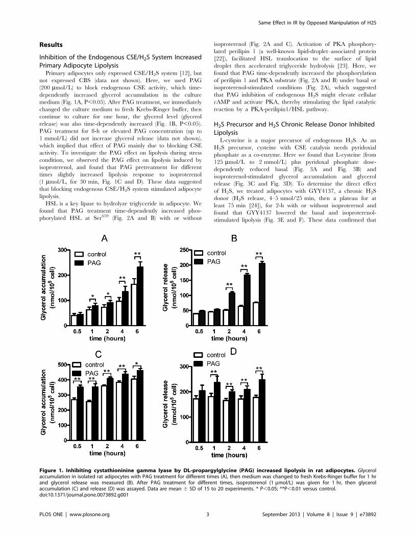
Inhibition of the Endogenous CSE/H2S System IncreasedPrimary Adipocyte LipolysisPrimary adipocytes only expressed CSE/H2S system [12], but
not expressed CBS (data not shown). Here, we used PAG
(200 mmol/L) to block endogenous CSE activity, which time-
dependently increased glycerol accumulation in the culture
medium (Fig. 1A, P,0.05). After PAG treatment, we immediately
changed the culture medium to fresh Krebs-Ringer buffer, then
continue to culture for one hour, the glycerol level (glycerol
release) was also time-dependently increased (Fig. 1B, P,0.05).
PAG treatment for 8-h or elevated PAG concentration (up to
1 mmol/L) did not increase glycerol release (data not shown),
which implied that effect of PAG mainly due to blocking CSE
activity. To investigate the PAG effect on lipolysis during stress
condition, we observed the PAG effect on lipolysis induced by
isoproterenol, and found that PAG pretreatment for different
times slightly increased lipolysis response to isoproterenol
(1 mmol/L, for 30 min, Fig. 1C and D). These data suggested
that blocking endogenous CSE/H2S system stimulated adipocyte
lipolysis.
HSL is a key lipase to hydrolyze triglyceride in adipocyte. We
found that PAG treatment time-dependently increased phos-
phorylated HSL at Ser659 (Fig. 2A and B) with or without
isoproterenol (Fig. 2A and C). Activation of PKA phosphory-
lated perilipin 1 (a well-known lipid-droplet–associated protein
[22]), facilitated HSL translocation to the surface of lipid
droplet then accelerated triglyceride hydrolysis [23]. Here, we
found that PAG time-dependently increased the phosphorylation
of perilipin 1 and PKA substrate (Fig. 2A and B) under basal or
isoproterenol-stimulated conditions (Fig. 2A), which suggested
that PAG inhibition of endogenous H2S might elevate cellular
cAMP and activate PKA, thereby stimulating the lipid catalytic
reaction by a PKA-perilipin1/HSL pathway.
H2S Precursor and H2S Chronic Release Donor InhibitedLipolysisL-cysteine is a major precursor of endogenous H2S. As an
H2S precursor, cysteine with CSE catalysis needs pyridoxial
phosphate as a co-enzyme. Here we found that L-cysteine (from
125 mmol/L to 2 mmol/L) plus pyridoxal phosphate dose-
dependently reduced basal (Fig. 3A and Fig. 3B) and
isoproterenol-stimulated glycerol accumulation and glycerol
release (Fig. 3C and Fig. 3D). To determine the direct effect
of H2S, we treated adipocytes with GYY4137, a chronic H2S
donor (H2S release, 4–5 nmol/25 min, then a plateau for at
least 75 min [24]), for 2-h with or without isoproterenol and
found that GYY4137 lowered the basal and isoproterenol-
stimulated lipolysis (Fig. 3E and F). These data confirmed that
Figure 1. Inhibiting cystathioninine gamma lyase by DL-propargylglycine (PAG) increased lipolysis in rat adipocytes. Glycerolaccumulation in isolated rat adipocytes with PAG treatment for different times (A), then medium was changed to fresh Krebs-Ringer buffer for 1 hrand glycerol release was measured (B). After PAG treatment for different times, isoproterenol (1 mmol/L) was given for 1 hr, then glycerolaccumulation (C) and release (D) was assayed. Data are mean 6 SD of 15 to 20 experiments. * P,0.05; **P,0.01 versus control.doi:10.1371/journal.pone.0073892.g001
Same Effect in IR by Opposed Manipulation of H2S
PLOS ONE | www.plosone.org 3 September 2013 | Volume 8 | Issue 9 | e73892

L-cysteine endogenous release of H2S via CSE inhibited lipolysis
in rat adipocytes.
H2S reduced isoproterenol-induced cAMP elevation and
forskolin-stimulated adenylyl cyclase activity [25], then dose-
dependently lowered cAMP-dependent PKA activation, as
evidenced by decreased phosphorylation of the PKA substrate
(Fig. 4A) under basal (Fig. 4B) and isoproterenol-stimulated
conditions (Fig. 4C). H2S also inhibited phosphorylation of
perilipin and HSL at Ser659 site (Fig. 4A, D) with or without
isoproterenol stimulation, thus blocking HSL translocation to lipid
droplet [8] for decreased lipolysis activity. These data suggest that
the cAMP-PKA-perilipin/HSL pathway is involved in regulating
the lipolysis by endogenous H2S in adipocytes.
PAG and GYY4137 Regulated the Adipose TissueLipolysis In VivoDysfunction of adipose lipolysis contributed to pathogenesis of
insulin resistance in obesity. To investigate the role of adipose
endogenous CSE/H2S in triglyceride lipolysis in vivo, we fed mice
by HFD (45% energy from fat) for 13 weeks to induce obesity and
normal diet (10% energy from fat) as control. As Fig. 5A–D
shown, HFD significantly increased C57BL/6J body weight
(Fig. 5A, P,0.01), visceral fat weight (including epididymal fat
pad, perinephric fat and retroperitoneal fat, Fig. 5B, P,0.01) and
subcutaneous fat weight (Fig. 5C, P,0.01), resulting in increasing
ratio of fat weight/body weight (Fig. 5D, P,0.01). Association
with fat mass increased by HFD, CSE protein expression and
endogenous H2S production decreased in adipose tissues (Figure
S1 in File S1). PAG inhibited CSE expression and H2S production
(Figure S1 in File S1), lowered the basal and HFD induced body
weight growth (Fig. 5A, P,0.01) and blunted the fat mass increase
Figure 2. PAG increased phosphorylated PKA substrate, perilipin and hormone sensitive lipase (HSL) in rat adipocytes. (A) Lysates ofadipocytes treated with 200 mmol/L PAG were separated by SDS-PAGE on low-Bis concentration gels and underwent immunoblot analysis with ananti-perilipin antibody. The band shift from 65 kDa (native) to 67 kDa (phosphorylated) perilipin 1 indicates hyperphosphorylation of full-lengthperilipin 1. The 46-kDa band is perilipin 2. Phosphorylated HSL at Ser659 and phosphorylated PKA (p-PKA) substrate was determined. Relativeexpression of p-HSL to total HSL was analyzed by band density under basal (B) or isoproterenol (1 mmol/L)-stimulated conditions (C). Data are mean6 SD. ** P,0.01 vs. untreated with PAG.doi:10.1371/journal.pone.0073892.g002
Same Effect in IR by Opposed Manipulation of H2S
PLOS ONE | www.plosone.org 4 September 2013 | Volume 8 | Issue 9 | e73892

(evidenced by visceral, subcutaneous fat weight and ratio of fat
weight/body weight, Fig. 5B–D, all P,0.01). Leptin is a marker of
obesity by HFD. Here we found that PAG antagonized the high
plasma leptin level induced by HFD (Figure S2 in File S1), which
was according to reduction of fat mass. H2S donor (GYY4137) did
not affect these basal physiological characteristics. Triglyceride
Figure 3. H2S precursor and donor inhibited lipolysis in rat adipocytes. Adipocytes were supplemented with L-cysteine plus pyridoxialphosphate (PLP) for 1 hr to increase endogenous H2S, then glycerol accumulation (A) or release in medium (B) was measured. The isoproterenol-stimulated glycerol accumulation (C) and release (D) was assayed with L-cysteine and pyridoxial phosphate treatment. After treatment with GYY-4137(H2S release donor, 4–5 nmol/25 min then plateaued at least 75 min or more) for 2 hr, glycerol accumulation (E) or released (F) was measured underbasal or isoproterenol-stimulated conditions. Data are mean 6 SD from 10 experiments. * P,0.05; **P,0.01.doi:10.1371/journal.pone.0073892.g003
Same Effect in IR by Opposed Manipulation of H2S
PLOS ONE | www.plosone.org 5 September 2013 | Volume 8 | Issue 9 | e73892

lipolysis release 1 glycerol and 3 free fatty acid. Circulatory free
fatty acid was quickly lowered by uptake, oxidation or reesterfica-
tion in tissue. So we used circulatory glycerol to assess the adipose
lipolysis in vivo. As Fig. 5E shown, PAG treatment increased
fasting blood glycerol in normal chow mice (P,0.01), slightly (but
not statistical significant) increased it in HFD mice. GYY4137
lowered fasting blood glycerol level in HFD mice (Fig. 5E,
P,0.01) which suggested that H2S lowered basal lipolysis in obese
fat. To confirm the effects of PAG and GYY4137, we measured
the direct lipolysis in isolated adipose tissues and found that PAG-
treatment increased glycerol release from adipose tissues in both
control and HFD mice; GYY-treatment reduced lipolysis in HFD
adipose tissues (Fig. 5F, P,0.01). Food uptake is an important
factor of obesity, so we measured the food consumption and found
that PAG and GYY4137 treatment did not affect the food
consumption (FigureS3 in File S1). These data implied that PAG
continuously elevated lipolysis blunted HFD induced obesity; H2S
donor seemly lowered the lipolysis in obese adipose, but did not
accelerate fat mass deposition.
Both Increase and Decrease CSE/H2S Reduced GlobalInsulin Resistance In VivoTo investigate the role of endogenous CSE/H2S in insulin
resistance, we treated mice with PAG or GYY4137 for 13 week,
then we assessed the insulin sensitivity. Here, we found that PAG
per se did not affect fasting blood glucose, fasting blood insulin and
HOMA index (Fig. 6A–C); slightly increased insulin sensitivity
evidenced by decrease OGTT curve area (Fig. 6D and F) and
Figure 4. Endogenous H2S or chronic H2S donor reduced phosphorylated PKA substrate, perilipin 1 and HSL activity. (A) Aftertreatment with L-cysteine plus pyridoxial phosphate or GYY4137, adipocyte lysates were separated by SDS-PAGE, and phosphorylated PKA substrate,perilipin 1 and HSL activity were assayed. The relative phosphorylated level of HSL to total HSL was compared after treatment with L-cysteine (B),isoproterenol (C) or GYY4137 (D).doi:10.1371/journal.pone.0073892.g004
Same Effect in IR by Opposed Manipulation of H2S
PLOS ONE | www.plosone.org 6 September 2013 | Volume 8 | Issue 9 | e73892

increase glucose response to insulin (ITT assay, Fig. 6G and I) in
normal chow mice. Interestingly, PAG significantly lowered
insulin resistance in HFD obese mice evidenced by decrease
fasting blood glucose and insulin, HOMA index (Fig. 6A–C,
P,0.01), OGTT curve area (Fig. 6E and F, P,0.01) and ITT
curve area (Fig. 6H and I, P,0.01).
Figure 5. The basal characteristics changes and in vivo lipolysis after treatment by PAG and GYY4137 in HFD obesity mice. Obesitymice were induced by high fatty diet (45% energy from fat), normal diet (10% energy from fat) as control. PAG (30 mg/kg/day in saline) and GYY4137(200 mmol/kg/day in saline) were administrated by subcutaneous injection and saline injection as control. After 13 weeks, body weight (A), visceral fatweight (B), subcutaneous fat weight (C) were measured then the fat weight/body weight (D) were counted. Fasting blood glucose (E) was assayed byglucometer. 20 mg epidymal adipose tissue from each mouse was incubated in serum-free and phenol red-free DMEM for 1-h, the release glycerol forevaluation lipolysis of mice (F) was assayed. All data are means 6 SD. * P,0.05, ** P,0.01 versus control mice; # P,0.05 versus HFD saline injectionmice.doi:10.1371/journal.pone.0073892.g005
Same Effect in IR by Opposed Manipulation of H2S
PLOS ONE | www.plosone.org 7 September 2013 | Volume 8 | Issue 9 | e73892

In normal chow mice, H2S donor-GYY4137 slightly increased
blood glucose and insulin levels for fasting 6-h (Fig. 6A and B,
P,0.05), resulting in elevation of HOMA index (Fig. 6C, P,0.05)
comparison to saline injection mice. However, GYY4137 lowered
these parameters in HFD mice (Fig. 6A–C, P,0.05). In normal
chow mice, GYY4137 delayed the blood glucose peak in OGTT
curve (Fig. 6C), and slightly decreased the glucose response to
insulin (Fig. 6F). In HFD obese mice, GYY4137 lowered the
OGTT curve area (Fig. 6E and F) and increased the effects of
lowering blood glucose by insulin (Fig. 6H and I). These data
suggested that blocked endogenous CSE enzyme activity lowered
fat mass growth association with reduction insulin resistance in
HFD obese mice. More interestingly, H2S donor also reduced
insulin resistance in HFD mice, but slightly decreased insulin
sensitivity in normal chow mice. The bilateral regulation of CSE/
H2S in insulin resistance also implied that different pathway or
signals were involved in H2S regulation in physiological and/or
pathological condition.
AMPK is an energy sensor and play an essential role in insulin
signal. AMPK directly phosphorylated IRS-1 then increased
insulin sensitivity. Here, we found that PAG up-regulated AMPK
protein (Fig. 7A and B, P,0.05) in adipose tissues of control mice
and HFD mice, but GYY4137 did not. PAG also increased IRS-1
protein expression in HFD mice. These findings suggested that
AMPK-IRS-1 pathway may be involved in the regulation of PAG
to antagonize adipose tissues insulin resistance. Giving GYY4137
treatment, lowered adipose IRS-1 protein expression in control
mice but up-regulated it in HFD mice (Fig7.A and C). In present
study, PAG and GYY4137 were systemic administration, so we
also measured the AMPK and IRS-1 expression in skeletal muscle.
As supplemental data Fig. S4 (Figure S4 in File S1)shown, both
PAG and GYY4137 up-regulated AMPK and IRS-1protein
expression in HFD mice; PAG also increased IRS-1 protein
Figure 6. The effects of PAG and GYY4137 on insulin sensitivity in HFD mice. For assessing insulin sensitivity, fasting blood glucose (A),fasting serum insulin level (B) and HOMA index (C) was counted. After fasting 16-hrs, oral glucose tolerance test (OGTT) in normal chow mice (D) andHFD mice (E), area under curve for OGTT (F); insulin tolerance test (ITT) in control mice (G) and HFD mice (H) and area under curve for ITT (I) wereassayed. All data are means 6 SD. * P,0.05, ** P,0.01 versus control mice; # P,0.05 versus HFD saline injection mice.doi:10.1371/journal.pone.0073892.g006
Same Effect in IR by Opposed Manipulation of H2S
PLOS ONE | www.plosone.org 8 September 2013 | Volume 8 | Issue 9 | e73892

expression in normal chow mice. These results might explain the
bilateral regulation of H2S in insulin sensitivity in vivo.
Discussion
Dysfunction of lipolysis contributed to pathogenesis of insulin
resistance. In present study, we found that inhibition of adipocyte
endogenous CSE/H2S with PAG increased basal and isoproter-
enol stimulated lipolysis, oppositely H2S donor (GYY4137)
inhibited them and PKA-HSL/perilipin pathway involved in the
regulation. PAG increased blood glycerol and adipose lipolysis but
not lowered food uptake, which thus blunted HFD induced
obesity, and reduced insulin resistance from HFD mice; GYY4137
did not change HFD induced fat mass increase, but ameliorated
the insulin resistance in obese mice.
H2S is a metabolic production source from cysteine dependent
on CSE in adipose tissues. Several clinical studies have reported a
positive association of total cysteine level (including cysteine,
reduced cysteine, cystine, and mixed disulphides) with body mass
index (BMI) [26–30]; with fat mass (as measured by dual energy
X-ray absorptiometry) contributing the most to the BMI [31].
CSE-knockout mice showed lower plasma cysteine and H2S levels,
and lower body weight, of which white adipose tissue mass (34% of
wild-type) was the most contribution to the body weight lost [32].
These studies strongly suggest that cysteine/CSE contributes to
functional regulation of adipocytes. Here we found that CSE
inhibitor-PAG induced robust basal and isoproterenol stimulated
lipolysis; H2S precursor-L-Cysteine or donor-GYY4137 (a water
soluble, stable, chronic releasing H2S donor [24]) inhibited them.
In normal chow and HFD mice, PAG also increased lipolysis
evidenced by elevated serum glycerol and lipolysis reaction in
isolated adipose tissues, but did not affect food consumption, then,
blunting fat mass deposition and body weight increase. H2S donor
lowered lipolysis in vivo in HFD mice but not in normal chow
mice. GYY4137 just offer about 4–5 nmol per 25 min [24], and
accumulated in liver, kidney and other organ during 2 hours after
bonus injection [33]. Thus systemic administration GYY4137 just
partly inhibited lipolysis in short times, which partly explained that
GYY4137 treatment did not increase fat mass and body weight.
These findings supported that adipose endogenous CSE/H2S
system contributed to lipolysis reaction, which might be a reason
of high serum blood cysteine positive regression with BMI and
body fatty mass [31].
H2S attenuated catecholamine-induced cellular cAMP elevation
[25], inhibited PKA activation which reduced phosphorylated
HSL and perilipin 1, thus blocked HSL translocation to lipid
droplet [1] resulting in lowered triglyceride lipolysis. The
hypothesis is evidenced by the phosphorylation of PKA substrate,
HSL at ser659 site, perilipin 1, and cysteine activated by PAG or
inhibited by GYY4137 (Fig. 4). These findings suggested that
PKA-HSL/perilipin 1 pathway is involved in the lipolytic
regulation by H2S. Plasma cysteine releases H2O2 via Cu2+-
dependent auto-oxidation [34,35]. H2O2 also inhibits hormone-
sensitive lipase activity by forming an intersubunit disulfide bond
within cAMP-dependent PKA [36], which might be a molecular
mechanism of cysteine action in the lipolysis response in vivo. In
isolated adipocytes, Cu2+ absence limited this response model.
Antioxidants such as N-acetyl-L-cysteine or diphenyleneiodonium
lowered the adenylyl cyclase activity by increasing Gi protein
expression, then decreasing intracellular cAMP level [37]. H2S is a
strong antioxidant [38]; whether H2S upregulates receptor-
dependent or -independent Gi protein expression or function
causing inhibition adenylyl cyclase activity needs further investi-
gation. Global CSE-knockout mice showed lower fat weight [32],
which might be caused by increasing adipose lipolysis because of
CSE deficient. Unfortunately, authors did not measure adipocyte
lipolysis activity in CSE-knockout mice [32].
Obesity is an independent risk factor of diabetes. In adipocyte of
HFD obese mice, basal lipolysis is increased but catecholamine-
stimulated lipolysis is blunted [39]. Saturated fat in the high fatty
diet increased adipose TNF-a expression and macrophage
infiltration [40], these local inflammatory cytokine impaired
insulin effects on glucose uptake activity and lipolysis [41]. PAG
inhibited endogenous CSE/H2S system and dose-dependently
recovered the glucose consumption and uptake which impaired by
TNF-a in adipocyte [42]. PAG also up-regulated AMPKa (an
important kinase involved in insulin sensitivity) and IRS-1 protein
(insulin signal transduction) in adipose and skeletal muscle, which
means blocking CSE/H2S stimulated adipose energy output
similar to energy deprivation. These data suggested PAG
ameliorated adipose insulin resistance partly by reduction adipose
inflammation and activated AMPK pathway.
H2S donor inhibited the basal lipolysis in obese adipose tissues,
increased insulin resistance in normal chow mice, but decreased
that in HFD mice. H2S per se inhibited rat mature adipocyte basal
glucose uptake and insulin stimulated glucose uptake [12], which
may contribute to increase insulin resistance in normal chow.
Whereas, H2S donor antagonized high glucose lowered phos-
phorylated Akt, phospho-IRS-1 and type four glucose transporter
(GLUT4) protein expression by inhibition PTEN and NF-kBactivation [43]. Another work also found that CSE involved in the
Figure 7. Alterations of AMPK and IRS-1 protein expression inepidymal adipose tissues. Relative protein expression of AMPK andIRS-1 in adipose were measured by western blot (A). Gray analysis wasperformed for quantization of AMPK (B) and IRS-1 (C). Six independentexperiments were performed. All data are means6 SD. * P,0.05 versuscontrol mice; # P,0.05 versus HFD saline injection mice.doi:10.1371/journal.pone.0073892.g007
Same Effect in IR by Opposed Manipulation of H2S
PLOS ONE | www.plosone.org 9 September 2013 | Volume 8 | Issue 9 | e73892

protective effects of vitamin D on insulin resistance by high glucose
[14]. The present study also found that H2S donor increase IRS-1
protein expression in obese adipose and skeletal muscle. These
data suggested that CSE/H2S system might regulate gene
transcription of glucose metabolic enzyme or transporter protein
by nuclear receptor such as vitamin D receptor. The bilateral
regulation of CSE/H2S system in glucose metabolism suggested
that CSE/H2S might act as an energy balancer. In physiological
condition, CSE/H2S system is apt to reduce energy consumption
thus slightly decrease glucose utilization; while under stress or
inflammation, CSE/H2S antagonize injury and increase glucose
utilization resulting in increasing insulin sensitivity.
In conclusion, the endogenous CSE/H2S system in adipocytes
regulated lipolysis by PKA-perilin/HSL pathway. Inhibition of
CSE/H2S induced robust lipolysis thus blunted adipose increase
and lowered insulin resistance by AMPK pathway. Although H2S
donor lowered lipolysis, it ameliorated insulin resistance by up-
regulating IRS-1 protein. The paradoxical regulation of CSE/H2S
system in insulin sensitivity implied that CSE and H2S might have
independent regulation mechanism and differential signal trans-
duction. Clearance the regulation of CSE/H2S in energy
metabolism may be helpful to understand the complicated
interactive linkage of glucose, fat and sulfur containing amino
acid in physiology and diseases.
Supporting Information
File S1 File containing all supporting informationfigures. Figure S1: Changes of endogenous CSE/H2S system
in epididymal adipose tissues. (A): CSE protein expression was
measured by western blot. (B): Relative quantitative of CSE
protein expression was analyzed by gray density of CSE and b-actin band. (C): Endogenous H2S production in adipose tissue was
assayed by the methylene blue method. All data are means 6 SD.
* P,0.05, ** P,0.01 versus normal chow mice; # P,0.05 versus
HFD mice. Figure S2: Alterations of plasma leptin measured by
ELISA assay (ELISA kit from R&D Minneapolis, MN). All data
are means 6 SD. ** P,0.01 versus normal chow mice; # P,0.05
versus HFD mice. Figure S3: Food consumption was measured
every 3 days. Figure S4: Alterations of AMPK and IRS-1 protein
expression in skeletal muscle tissues. Relative protein expression of
AMPK and IRS-1 in skeletal muscle were measured by western
blot (A). Gray analysis was performed for quantization of AMPK
(B) and IRS-1 (C). Six independent experiments were performed.
All data are means 6 SD. * P,0.05 versus normal chow mice; #P,0.05 versus HFD mice.
(DOC)
Author Contributions
Conceived and designed the experiments: BG GHX. Performed the
experiments: BG BC FL YZ. Analyzed the data: BG QZ QHC CT.
Contributed reagents/materials/analysis tools: XF YG CT. Wrote the
paper: BG GHX QHC JY.
References
1. Londos C, Brasaemle DL, Schultz CJ, Adler-Wailes DC, Levin DM, et al. (1999)On the control of lipolysis in adipocytes. Ann N Y Acad Sci 892: 155–168.
2. Samuel VT, Shulman GI (2012) Mechanisms for insulin resistance: commonthreads and missing links. Cell 148: 852–871.
3. Lass A, Zimmermann R, Oberer M, Zechner R (2011) Lipolysis - a highlyregulated multi-enzyme complex mediates the catabolism of cellular fat stores.
Prog Lipid Res 50: 14–27.
4. Zimmermann R, Strauss JG, Haemmerle G, Schoiswohl G, Birner-Gruenberger
R, et al. (2004) Fat mobilization in adipose tissue is promoted by adiposetriglyceride lipase. Science 306: 1383–1386.
5. Schweiger M, Schreiber R, Haemmerle G, Lass A, Fledelius C, et al. (2006)Adipose triglyceride lipase and hormone-sensitive lipase are the major enzymes
in adipose tissue triacylglycerol catabolism. J Biol Chem 281: 40236–40241.
6. Anthonsen MW, Ronnstrand L, Wernstedt C, Degerman E, Holm C (1998)
Identification of novel phosphorylation sites in hormone-sensitive lipase that arephosphorylated in response to isoproterenol and govern activation properties
in vitro. J Biol Chem 273: 215–221.
7. Garton AJ, Campbell DG, Carling D, Hardie DG, Colbran RJ, et al. (1989)
Phosphorylation of bovine hormone-sensitive lipase by the AMP-activatedprotein kinase. A possible antilipolytic mechanism. Eur J Biochem 179: 249–254.
8. Londos C, Gruia-Gray J, Brasaemle DL, Rondinone CM, Takeda T, et al.(1996) Perilipin: possible roles in structure and metabolism of intracellular
neutral lipids in adipocytes and steroidogenic cells. Int J Obes Relat Metab
Disord 20 Suppl 3: S97–101.
9. Kimura H (2011) Hydrogen sulfide: its production, release and functions. AminoAcids 41: 113–121.
10. Hu LF, Lu M, Hon Wong PT, Bian JS (2011) Hydrogen sulfide:neurophysiology and neuropathology. Antioxid Redox Signal 15: 405–419.
11. Kabil O, Banerjee R (2010) Redox biochemistry of hydrogen sulfide. J BiolChem 285: 21903–21907.
12. Feng X, Chen Y, Zhao J, Tang C, Jiang Z, et al. (2009) Hydrogen sulfide fromadipose tissue is a novel insulin resistance regulator. Biochem Biophys Res
Commun 380: 153–159.
13. Fang L, Zhao J, Chen Y, Ma T, Xu G, et al. (2009) Hydrogen sulfide derived
from periadventitial adipose tissue is a vasodilator. J Hypertens 27: 2174–2185.
14. Manna P, Jain SK (2012) Vitamin D Up-regulates Glucose Transporter 4
(GLUT4) Translocation and Glucose Utilization Mediated by Cystathionine-gamma-lyase (CSE) Activation and H2S Formation in 3T3L1 Adipocytes. J Biol
Chem 287: 42324–42332.
15. Olefsky JM (1979) Comparison of the effects of insulin and insulin-like agents on
different aspects of adipocyte metabolism. Horm Metab Res 11: 209–213.
16. Kawasaki M, Miura Y, Yagasaki K (2010) Effects of sulfur amino acids, L: -
methionine, L: -cystine and L: -cysteine on lipoprotein lipase and hormone-
sensitive lipase in differentiated mouse 3T3-L1 adipocytes. Cytotechnology 62:
225–233.
17. Zu L, He J, Jiang H, Xu C, Pu S, et al. (2009) Bacterial endotoxin stimulates
adipose lipolysis via toll-like receptor 4 and extracellular signal-regulated kinase
pathway. J Biol Chem 284: 5915–5926.
18. Xu C, He J, Jiang H, Zu L, Zhai W, et al. (2009) Direct effect of glucocorticoids
on lipolysis in adipocytes. Mol Endocrinol 23: 1161–1170.
19. Egan JJ, Greenberg AS, Chang MK, Londos C (1990) Control of endogenous
phosphorylation of the major cAMP- dependent protein kinase substrate in
adipocytes by insulin and beta-adrenergic stimulation. J Biol Chem 265: 18769–
18775.
20. Greenberg AS, Egan JJ, Wek SA, Garty NB, Blanchette-Mackie EJ, et al. (1991)
Perilipin, a major hormonally regulated adipocyte-specific phosphoprotein
associated with the periphery of lipid storage droplets. J Biol Chem 266: 11341–
11346.
21. He J, Jiang H, Tansey JT, Tang C, Pu S, et al. (2006) Calyculin and okadaic acid
promote perilipin phosphorylation and increase lipolysis in primary rat
adipocytes. Biochim Biophys Acta 1761: 247–255.
22. Greenberg AS, Egan JJ, Wek SA, Garty NB, Blanchette-Mackie EJ, et al. (1991)
Perilipin, a major hormonally regulated adipocyte-specific phosphoprotein
associated with the periphery of lipid storage droplets. J Biol Chem 266: 11341–
11346.
23. Sztalryd C, Xu G, Dorward H, Tansey JT, Contreras JA, et al. (2003) Perilipin
A is essential for the translocation of hormone-sensitive lipase during lipolytic
activation. J Cell Biol 161: 1093–1103.
24. Li L, Whiteman M, Guan YY, Neo KL, Cheng Y, et al. (2008) Characterization
of a novel, water-soluble hydrogen sulfide-releasing molecule (GYY4137): new
insights into the biology of hydrogen sulfide. Circulation 117: 2351–2360.
25. Yong QC, Pan TT, Hu LF, Bian JS (2008) Negative regulation of beta-
adrenergic function by hydrogen sulphide in the rat hearts. J Mol Cell Cardiol
44: 701–710.
26. Dhawan SS, Eshtehardi P, McDaniel MC, Fike LV, Jones DP, et al. (2011) The
role of plasma aminothiols in the prediction of coronary microvascular
dysfunction and plaque vulnerability. Atherosclerosis 219: 266–272.
27. Elshorbagy AK, Refsum H, Smith AD, Graham IM (2009) The association of
plasma cysteine and gamma-glutamyltransferase with BMI and obesity. Obesity
(Silver Spring) 17: 1435–1440.
28. Giral P, Jacob N, Dourmap C, Hansel B, Carrie A, et al. (2008) Elevated
gamma-glutamyltransferase activity and perturbed thiol profile are associated
with features of metabolic syndrome. Arterioscler Thromb Vasc Biol 28: 587–
593.
Same Effect in IR by Opposed Manipulation of H2S
PLOS ONE | www.plosone.org 10 September 2013 | Volume 8 | Issue 9 | e73892

29. van den Brandhof WE, Haks K, Schouten EG, Verhoef P (2001) The relation
between plasma cysteine, plasma homocysteine and coronary atherosclerosis.Atherosclerosis 157: 403–409.
30. Elshorbagy AK, Valdivia-Garcia M, Graham IM, Palma Reis R, Sales Luis A, et
al. (2011) The association of fasting plasma sulfur-containing compounds withBMI, serum lipids and apolipoproteins. Nutr Metab Cardiovasc Dis.
31. Elshorbagy AK, Nurk E, Gjesdal CG, Tell GS, Ueland PM, et al. (2008)Homocysteine, cysteine, and body composition in the Hordaland Homocysteine
Study: does cysteine link amino acid and lipid metabolism? Am J Clin Nutr 88:
738–746.32. Mani S, Yang G, Wang R (2011) A critical life-supporting role for cystathionine
gamma-lyase in the absence of dietary cysteine supply. Free Radic Biol Med 50:1280–1287.
33. Yu F, Zhao J, Tang CS, Geng B (2010) [Effect of synthesized GYY4137, a slowlyreleasing hydrogen sulfide donor, on cell viability and distribution of hydrogen
sulfide in mice]. Beijing Da Xue Xue Bao 42: 493–497.
34. Czech MP, Fain JN (1972) Cu++-dependent thiol stimulation of glucosemetabolism in white fat cells. J Biol Chem 247: 6218–6223.
35. Czech MP, Lawrence JC, Jr., Lynn WS (1974) Evidence for electron transferreactions involved in the Cu2+ -dependent thiol activation of fat cell glucose
utilization. J Biol Chem 249: 1001–1006.
36. de Pina MZ, Vazquez-Meza H, Pardo JP, Rendon JL, Villalobos-Molina R, etal. (2008) Signaling the signal, cyclic AMP-dependent protein kinase inhibition
by insulin-formed H2O2 and reactivation by thioredoxin. J Biol Chem 283:12373–12386.
37. Lappas G, Daou GB, Anand-Srivastava MB (2005) Oxidative stress contributes
to the enhanced expression of Gialpha proteins and adenylyl cyclase signaling in
vascular smooth muscle cells from spontaneously hypertensive rats. J Hypertens
23: 2251–2261.
38. Chang L, Geng B, Yu F, Zhao J, Jiang H, et al. (2008) Hydrogen sulfide inhibits
myocardial injury induced by homocysteine in rats. Amino Acids 34: 573–585.
39. Commerford SR, Pagliassotti MJ, Melby CL, Wei Y, Gayles EC, et al. (2000)
Fat oxidation, lipolysis, and free fatty acid cycling in obesity-prone and obesity-
resistant rats. Am J Physiol Endocrinol Metab 279: E875–885.
40. Enos RT, Davis JM, Velazquez KT, McClellan JL, Day SD, et al. (2013)
Influence of dietary saturated fat content on adiposity, macrophage behavior,
inflammation, and metabolism: composition matters. J Lipid Res 54: 152–163.
41. Wueest S, Rapold RA, Rytka JM, Schoenle EJ, Konrad D (2009) Basal lipolysis,
not the degree of insulin resistance, differentiates large from small isolated
adipocytes in high-fat fed mice. Diabetologia 52: 541–546.
42. Huang CY, Yao WF, Wu WG, Lu YL, Wan H, et al. (2012) Endogenous CSE/
H(2) S system mediates TNF-alpha-induced insulin resistance in 3T3-L1
adipocytes. Cell Biochem Funct.
43. Manna P, Jain SK (2011) Hydrogen sulfide and L-cysteine increase
phosphatidylinositol 3,4,5-trisphosphate (PIP3) and glucose utilization by
inhibiting phosphatase and tensin homolog (PTEN) protein and activating
phosphoinositide 3-kinase (PI3K)/serine/threonine protein kinase (AKT)/
protein kinase Czeta/lambda (PKCzeta/lambda) in 3T3l1 adipocytes. J Biol
Chem 286: 39848–39859.
Same Effect in IR by Opposed Manipulation of H2S
PLOS ONE | www.plosone.org 11 September 2013 | Volume 8 | Issue 9 | e73892