Embed Size (px)
Citation preview

(CANCER RESEARCH 48, 4222-4226, August 1, 19881
Increased Ornithine Decarboxylase Activity in Cultured Cells Exposed to LowEnergy Modulated Microwave Fields and Phorbol Ester Tumor Promoters1
Craig V. Byus, K. Kartun, S. Pieper, and W. Ross AdeyDivision of BiomédicalSciences and Dépannent of Biochemistry, University of California, Riverside, California 92521 [C. V. B., K. K., S. P.]; and VétéransAdministration Medical Center, Loma Linda, California 92357 [W. R. A.]
ABSTRACT
Ornithine decarboxylase (OIK) is present in all nucleated cells and isthe rate-limiting enzyme for synthesis of polyamines. In turn, the poly-amines are required for DNA synthesis and cell growth. In Reuber H35hepatoma cells, we show that ODC activity is increased by about 50%during exposure to a 1-h "athermal" (<0.1°Ctemperature rise) (450MHz, 1.0 mW/ciir peak-envelope-power) microwave field sinusoidali)'amplitude-modulated at 16 Hz. The increased activity of ODC persistedfor several hours following the 1-h exposure to the field. A similar fieldamplitude-modulated at 60 and 100 Hz did not alter the hepatoma cellODC activity. The stimulated ODC activity in the cultured cells thatfollowed treatment with a phorhoi ester tumor promoter (12-O-tetru-decanoylphorbol-13-acetate) was further potentiated by prior exposureto the same low energy electromagnetic field. This field did not altereither basal or 12-O-tetradecanoylphorbol-13-acetate-stimulated DNAsynthesis.
We observed a similar increase in the basal ODC activity of culturesof two additional cell lines (Chinese hamster ovary, and 294T melanoma)exposed for l h to the amplitude-modulated field. Chinese hamster ovarycells exposed to the radio frequency field for l h also responded tosubsequent treatment with 12-0-tetradecanoylphorbol-13-acetate by exhibiting a further increase in ODC activity.
We have observed previously that the activity of this enzyme isincreased in cultured cells following a transient exposure to a 60-Hzelectric field (30). Altered ODC activity may serve as a sensitive andspecific molecular marker of the transductive coupling of weak pericel-lular electromagnetic fields to biological systems.
INTRODUCTION
In previous studies, we have shown that weak-imposed lowfrequency EM3 fields modulate the activity of adenylate cyclase
in cyclic AMP production by bone cells (1). These studies havealso shown that this modulating action occurs at the cell membrane (2). Lymphocyte protein kinases also respond to weak-imposed EM fields (3). The EM fields in these studies inducedelectric gradients in pericellular fluid in the range 10~3-10~' V/
cm. They were therefore six to eight orders of magnitude lessthan the membrane potential gradient of 10s V/cm, and their
actions were not attributable to thermal effects. The importanceof understanding the molecular mechanism(s) whereby theseweak fields interact with biological organisms cannot be overestimated due to the pervasive presence of such fields in theenvironment. A series of epidemiological studies over the past5 years have suggested an association between industrial anddomestic exposures to low-level environmental electromagneticfields and an increased risk of cancer (4-14).
Studies in the Denver area first drew attention to an increased
Received 7/14/86; revised 8/6/87, 12/30/87; accepted 4/18/88.The costs of publication of this article were defrayed in part by the payment
of page charges. This article must therefore be hereby marked advertisement inaccordance with 18 U.S.C. Section 1734 solely to indicate this fact.
1Supported by U. S. Department of Energy, U. S. Veterans Administration,
U.S.F.D.A. Bureau of Radiological Health, the Southern California EdisonCompany, and General Motors Medical Research Institute.
2To whom requests for reprints should be addressed, at the University of
California, Division of Biomedicai Sciences, Department of Biochemistry, Riverside, CA 92521-0121.
3The abbreviations used are: EM, electromagnetic, TP A, 12-O-tetradecanoyl-phorbol-13-acetate; ODC, ornithine decarboxylase; CHO, Chinese hamster ovary.
incidence of malignant diseases in children living in closeproximity to 60-Hz high voltage power lines (11). These epidemiology studies have subsequently been confirmed and expanded in a recent report (12). In addition, the incidence ofadult cancer in general (6, 13), leukemia (4, 6, 7, 15), eyemelanoma (9), neuroblastomas (8), and primary brain tumors(16) have all been correlated with exposure to EM fields. EMfield effects of a related nature on the growth and developmentof the human organism have been reported in preliminarystudies of problem pregnancies, including spontaneous abortionin women sleeping under electric blankets (14). Some controversy, however, exists as to whether these epidemiological relationships to cancer could be attributed to the weak EM fieldsthemselves, or to associated factors such as hypothetical releaseof lead in water pipes (11), or to job-related polychlorinatedbiphenyls, solvents, fluxes, metal fumes, synthetic waxes, andepoxy resins which are known to occur in electrical occupations,or to other as yet undefined factors (15, 16).
This study of the enzyme ornithine decarboxylase was undertaken because its activity is regulated by a wide variety of growthfactors and hormones active at the cell surface (17, 18). Inaddition, ornithine decarboxylase is unique among enzymemarkers in that its activity can change markedly and rapidly inresponse to extracellular signals. The transductive coupling oflow frequency EM fields and of radio frequency and microwavefields amplitude-modulated at low frequencies has been shownto occur primarily at cell membranes (19-21). Cyclic AMP andthe phorbol ester tumor promoters, in addition to other membrane-active compounds, have been shown to markedly andrapidly modulate ornithine decarboxylase activity in a widevariety of cells and tissues (17, 18, 22-25). The tumor-promoting phorbol esters, including TPA used in this study, have aspecific membrane receptor, the calcium phospholipid kinase,protein kinase C (26, 27). Since latent cell transformationmanifested in the presence of phorbol esters has been reportedfollowing low-level microwave exposure (28,29), we anticipatedthat ornithine decarboxylase might be a convenient and sensitive molecular marker of field-related actions in cell membranetransductive coupling directly related to the process of tumorpromotion. In this regard, we have reported recently that a 1-hexposure of cultured mammalian cells to low energy 60-Hzelectric fields led to an increase in ODC activity (30).
MATERIALS AND METHODS
Cell Culture Procedures. Low passage number Reuber H35 hepatomacells were seeded at low density onto 120-mm plastic culture dishes inDulbecco's modified Eagle's medium containing 10% fetal calf serum
as described previously (25, 31). These monolayer cultures were grownfor 3 days at 37°Cin 5% COi in a conventional incubator and achieveda cell density of 5 x 10' cells/dish. Cultures grown in this manner havebeen previously determined to be in log-phase of growth and to possessrelatively high basal ODC activity in comparison to more dense confluent cultures (31). CHO cells and human 294T melanoma cells weregrown in a similar manner in minimal essential medium containing10% fetal calf serum as described previously (24).
4222
on March 10, 2020. © 1988 American Association for Cancer Research.cancerres.aacrjournals.org Downloaded from

FIELD-INDUCED ORNITHINE DECARBOXYLASE ACTIVITY
The optimal culture conditions for investigating the regulation of [3H]thymidine (0.3 MM;specific activity, 28 Ci/mmol, NEN) for 2 h.
ODC activity by TPA have been determined previously (25, 31). Essentially, the Reuber H35 hepatoma cells were grown to confluency (adensity of 7 x IO6 cells/dish, 6 days in culture) and the mediumcontaining serum replaced with the same Dulbecco's modified Eagle's
medium free of serum. The cells were grown for an additional 2 daysunder the serum-free conditions prior to addition of TPA (21, 25).Cultures treated in this manner are 100% viable as determined bynumerous parameters and are highly responsive to a number of agentsincluding TPA, insulin, glucagon, and corticosteroids (25, 31). Culturedishes were then placed in a Crawford cell and exposed to the 450-MHz amplitude-modulated field as described in detail below. Followingthe 1-h field exposure, TPA was added at a final concentration of 1.6MMto exposed and unexposed cultures and the activity of ODC determined. The cultures of CHO cells were grown in medium containing2% serum for 16 h prior to the addition of TPA.
Field Exposure Parameters. A Crawford cell exposure system (Instruments for Industry, model 3000) was used. This cell operates as acoaxial transmission line with a characteristic impedance of 50 ohmsover the spectrum from DC to 520 MHz. The long axis of the Crawfordcell was oriented vertically inside a large incubator maintained at 37 ±0.5°C.These were the largest perturbations observed and related to
prolonged opening of the chamber door. Temperature changes afterequilibration and during field exposure were typically around 0.1 "(.'.
Cell cultures in circular Petri dishes (75 cm2) in the Crawford cellwere placed in a gas-tight lucite box. The volume of each culture was10 ml and they were placed near the center of the long axis of the cellwith a separation of 1 cm between adjoining cultures. Control cultureswere placed in the same incubator in a similar box outside the Crawfordcell. Air containing 5% CO; at 100% humidity was passed continuouslyinto the lucite culture chambers. Test cultures were placed in theCrawford cell 16 h before exposure, and controls were also placed inthe incubator concurrently.
All cultures were exposed to a 450-MHz field, sinusoidally amplitudemodulated at 16 Hz. The field-generating system used a phase-locked-loop-controlled signal generator (Wavetek, model 3000), amplitudemodulated through a PIN diode modulator by a low frequency waveform generator (Wavetek, model 159). The 450-MHz signal drove abroadband linear power amplifier (Ailtech, model 35512). Modulationdepth was maintained at 75-85%, and was monitored with an oscilloscope and with an in-line modulation meter/forward-reflected powermeter (Bird, model 5483). An input of 1.7 W peak envelope power tothe cell produced a peak field intensity of 1.0 mW/cm2. Peak envelope
power levels were therefore adjusted to this level at a modulation depthof 75-85% in all experiments.
With the cultures placed in the Crawford cell and a 50-ohm nonin-ductive termination on the cell, the standing wave ratio did not exceed1.15:1 at the operating frequency of 450 MHz. Measurements with afield intensity probe (N'arda, model 8300) and with an experimental
tripole probe reported elsewhere (32, 33) showed reasonable isotropyof the electric field distribution, except near the side walls of theCrawford cell, where the fields were lower than in the central zonewhere the cultures were placed.
All cultures were exposed for 1 h. Their viability was not altered byfield exposure. No changes in temperature of the culture mediumbetween the beginning and the end of the exposure were detected usinga thermistor probe with a temperature resolution of 0.05'C (3).
Ornithine Decarboxylase Assay. The cells were homogenized inbuffer containing 50 mM Na-phosphate, pH 7.4, 1 mM dithiothreitol,0.1 mM EDTA, 0.05 M pyridoxal phosphate, and the amount of ODCin the supernatants (13,000 x g) was measured as the amount of 14CO2evolved from 0.25 MCi[MC]L-ornithine (Amersham, specific activity 60mCi/mmol) during a 60-min incubation at 37°Cat a total L-ornithine
concentration of 0.25 mM (see References 22, 24, and 25 for details).Determination of DNA Synthesis. H35 cells were plated at a density
of 10s cells/well (3.5 cm in diameter) into six-well dishes (Flow Lab.).Cultures were allowed to grow to approximately 60-70% confluencybefore being exposed to a real or sham field for 1 h. TPA was added tothe medium at zero time following the 1-h exposure. Fourteen h afterTPA treatment and/or field exposure, cells were pulsed with 1 //Ci/ml
DNA synthesis has been determined previously to be maximal at thistime period (34). At the end of the labeling period, cells were rinsedextensively with cold Hanks' balanced salt solution containing 0.1%
unlabeled thym idinc. precipitated two times with ice-cold 10% trichlo-roacetic acid, and the DNA in the resultant pellets was hydrolyzed in0.5 N HC1O4 at 90°Cfor 15 min. Aliquots of each sample hydrolysatewere examined for 3H-radioactivity by liquid scintillation counting.Results were expressed as pmol thymidine incorporated/2 h/10' cells.
Previous autoradiographic analysis of cells treated in this mannershowed a linear relationship between [3H]thymidine incorporation intoacid-insoluble material and 3H-label in the nucleus of these cells (34).
RESULTS
Ornithine decarboxylase is known to be a cell cycle-regulatedenzyme and is higher in activity in cells in (., of the cell cycle(18). Cells that are growing rapidly or are stimulated to growby a variety of hormones have high levels of ODC activityrelative to their quiescent counterparts (18, 25). Initially, wewished to determine whether exposure of a population of log-phase or growing cells to a low energy electromagnetic fieldwould lead to an alteration in the basal activity of ODC. Forthese studies, we chose field parameters previously determinedto alter a number of biochemical parameters in several celltypes, including protein kinase activity (3), Ca2+ binding at the
cell surface (35-37), the cytotoxicity of allogeneic T-lympho-cytes (38), and of catecholamines (39) and insulin (40). The450-MHz field caused no detectable (i.e., <0.1*C) change in
temperature of the culture medium throughout a 1-h exposure(9) (see "Materials and Methods").
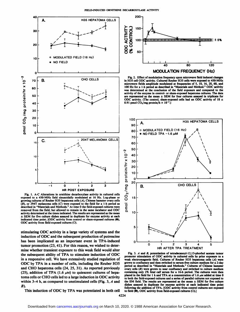
Initially we investigated the ability of a brief exposure to thisfield to alter the basal or unstimulated activity of ODC in threetypes of continuous monolayer cultured cells; Reuber H35hepatoma, Chinese hamster ovary, and 294T human melanomacells. Culture dishes of the various cell types in log-phase ofgrowth were arranged for exposure in the Crawford cell in apattern that minimized mutual coupling between the dishes andthe associated distortion of the imposed field due to antennaeffects. Cultures were exposed to the field for 1 h and theactivity of ODC was determined as described upon removalfrom the field (time 0) and throughout an additional 3-h period(Fig. \,A-C).
In all three cell types, a 1-h exposure produced a significant(up to 50%) increase in ODC activity in comparison to theunexposed cultures (Fig. 1, A-C, time 0). Following removalfrom the field, ODC activity remained elevated in the field-exposed cells for more than a 3-h period in the Reuber H35and CHO cell cultures (Fig. 1, A and B). The field-inducedincrease in ODC activity in the 294T melanoma cells persistedfor only 1 h after removal of the cultures from the field (Fig.1C).
Frequencies of amplitude modulation of the 450-MHz fieldin the range of 5-100 Hz were tested for their ability to alterODC activity in the cultured Reuber H35 hepatoma cells (Fig.2). Modulation frequencies of 60 and 100 Hz failed to causeany alteration in ODC activity following a 1-h exposure to thefield, in contrast to the 50% increase in enzyme activity broughtabout by the 16-Hz modulated field (Fig. 2). However, modulation frequencies of 10 and 20 Hz also resulted in an increasein ODC activity.
Many hormones and agents with cell membrane receptorselicit a rapid and marked increase in ODC activity in appropriate target cells or tissues. Phorbol ester tumor promoters, ofwhich TPA is the most potent, are particularly effective in
4223
on March 10, 2020. © 1988 American Association for Cancer Research.cancerres.aacrjournals.org Downloaded from
FIELD-INDUCED ORNITHINE DECARBOXYLASE ACTIVITY
40
30
20
10
A H35 HEPATOMA CELLS
o MODULATED FIELD (16 Hz)
•NO FIELD
200-1
150-
100
50-
±5%
120
MODULATION FREQUENCY (Hz)01'o
70i—*
60.c.£
50«2
40Q.0>
30Ec?
20ü_
10o
I og
CHOCELLSTÎ*-—
T~~~-<>^T~~
~^ ~- "*"+T^^ ^)"^^^l~~"~~--^
^^^^Is/^^^^^^y_-lili!rig.
1. Lllect ol modulation frequency upon microwave Held induced changesin H35 cell ODC activity. Cultured Reuber H35 cells were exposed to450-MHzind100
Hz for a 1-h period as described in "Materials and Method." ODCactivitywas
determined at the conclusion of the field exposure and comparedtotheactivityof the enzyme in control- or sham-exposed hepatoma cultures. Thedataare
represented as the mean ±SEM for four cultures assayed intriplicateforODCactivity. (The control, sham-exposed cells had an ODC activity of 18±0.91
pmol CO2/mg protein/h xIO"2.)1009080O.
u1510SQ
T 204T MELANOMACELLS"* ¿>•
^ " \Â¥\\\\r~
^""~"~--ij.^*"^\-^^^iiiii/
\J60CM
50I0
40*
30^
20c'Q)100^
H35 HEPATOMACELLS-_
o MODULATED FIELD (16 Hz)^•NO FIELD TPA -1.6 /uM ^ /\/TV
\/if \\r
\\/\\i \\li
]L\//^\5r
\^^i>^^^sf^^'ii0^--**^^,, ,,i-
O —Q.on-10123
0)HR
POST EXPOSURE-IFig.1. A-C Alterations in ornithine decarboxylase activity in cultured cellsJ^exposedto a 450-MHz field sinusoidally modulated at 16 Hz. Log-phase or ~-15growingcultures of Reuber H35 hepatoma cells (A), Chinese hamster ovarycells(B),
or 294T melanoma cells (C) were exposed to the field for a 1-h period as"^describedin "Materials and Methods." At time 0 the field-exposed cultures were£removed
from the field, but allowed to remain in the same incubator and ODC Q_activity determined at the times indicated. The results are represented as the mean '0±
SEM for five culture dishes assayed in duplicate for enzyme activity ateachindicatedtime point. [ODC activity from control or sham-exposed cultures(•),ODC
activity from field-exposed cultures(O).5stimulating
ODC activity in a large variety of systems andtheinductionof ODC and the subsequent production ofputrescinehas
been implicated as an important event in TPA-induced ng
CHO CELLSA/T\/
l\/\\YNA
i\/~×~~~~—TV//^\!/
\i>-^//\i^5"^^ /\^^W^\iiiiii
tumor promotion (23, 41). For this reason, we wished to determine whether transient exposure to this weak field would alterthe subsequent ability of TPA to stimulate induction of ODCin a responsive cell. We have extensively studied regulation ofODC by TPA in a number of cells, including the Reuber H35and CHO hepatoma cells (24, 25, 31). As reported previously(25), addition of TPA (1.6 ¿¿M)to quiescent cultures of hepatoma cells or CHO cells led to a large induction in ODC activitywithin 3-4 h, as compared to unstimulated cells (Fig. 3, A and
B).This induction of ODC by TPA was potentiated in both cell
1234HR AFTER TPA TREATMENT
Fig. 3. A and B, piitem lution of tetradecanoyl-12,13-phorbol acetate tumorpromoter stimulation of ODC activity in cultured cells by prior exposure to aweak electromagnetic field. Cultures of Reuber H35 hepatoma cells (A) weregrown to confluency and then switched to serum-free culture medium for a 2-dayperiod as described in "Materials and Methods." Cultures of Chinese hamster
ovary cells (li) were grown to near confluency and switched to culture mediumcontaining only 2% fetal calf serum for a 16-h period. The cultures were thenplaced in the field for l h and TPA at a concentration of 1.6 pM added at time 0to both the field-exposed cultures and a series of parallel cultures not exposed tothe EM field. The results are represented as the mean ±SEM for five culturedishes assayed in duplicate for enzyme activity at each indicated time pointfollowing the addition of TPA. [ODC activity from control cultures not exposedto field (•),ODC activity from field-exposed cultures (O).]
4224
on March 10, 2020. © 1988 American Association for Cancer Research.cancerres.aacrjournals.org Downloaded from

FIELD-INDUCED ORNITHINE DECARBOXYLASE ACTIVITY
types by prior exposure of the cells for l h in the low energyfield. ODC activity was further stimulated by TPA in the field-
exposed cells at 4 and 5 h following the addition of the phorbolester in the Reuber H35 hepatoma cells (Fig. 3A). In the CHOcell cultures a 1-h field exposure resulted in a significant increase in ODC activity in comparison to the nonexposed cellat 3, 4, and 5 h following TPA (Fig. 3fi).
Upon removal from the 1-h exposure to the field, prior tothe addition of TPA (i.e., time 0 in Fig. 3, A and B), the levelof ODC activity was similar in both exposed and unexposedcultures. This is in contrast to the data represented in Fig. 1, Aand li, where a 1-h field exposure led to a significant stimulationof ODC activity at time 0. The lack of a field-induced increasein ODC activity at time 0 in Fig. 3, A and B, was a result ofcultures for the TPA experiments being placed in serum-freemedium or low serum for 16 h to 2 days prior to field exposure(see "Materials and Methods" and Fig. 3, A and B, legend).
The ability of a 1-h field exposure to alter DNA synthesis inunstimulated or TPA-treated cultures was also determined (Table 1). Reuber H35 hepatoma cell cultures were exposed tosham fields or 450-MHz fields amplitude modulated at 16 Hzfor l h as described previously. TPA (1.6 UM)was then addedwhere indicated at time 0 and the incorporation of [3H]thymi-dine into acid-insoluble material during a 2-h pulse was determined 14 h later (see "Materials and Methods" and Reference
25). In both unexposed and field-exposed cultures, TPA treatment resulted in a stimulation of [3H]thymidine incorporation
in comparison to the controls (Table 1). However, in contrastto its effect upon ODC activity, a 1-h field exposure did notalter either control or TPA-stimulated DNA synthesis (i.e.,[3H]thymidine incorporation) (Table 1).
DISCUSSION
There has been a progressive refinement in ionic and molecular markers of the bioeffects of weak-imposed EM fields.
Initial observations of highly cooperative field effects on calcium binding in cerebral tissue (35-37) led to models of thefirst steps in transductive coupling of these EM fields at cellmembrane surface sites. In addition, studies by Lu hen (1, 2) onthe activation of adenylate cyclase by parathyroid hormone inbone cells further indicated the cell membrane as the prime siteof low frequency EM field interaction and suggested that thefield were potentiating the ability of the receptor to couple tothe cyclase. In this regard, the alterations in ornithine decar-boxylase activity reported here may provide a sensitive andeasily assayable molecular marker of the sensitivity of a givencell or tissue to a specific EM field. It does not appear that theability of the modulated microwave field to lead to an increasein ODC activity is a secondary effect of a stimulation of the
Table I Effect of exposure to a 450 MHz field sinusoidal/y modulated at 16Hzupon DNA synthesis in unstimulated and TPA-treated H35 hepatoma cells
Reuber H35 hepatoma cell cultures were exposed to sham field conditions orto 450-MHz fields amplitude modulated at 16 Hz for 1 h as described in thelegend to Fig. 1. TPA (0.6 JIM)was then added where indicated at time 0 and theincorporation of ['H|th\ mietine into acid-insoluble material during a 2-h pulsewas determined 13 h later (see "Materials and Methods"). The data are repre
sented as the mean ±SEM for five cultures assayed in duplicate. The data shownis a representative experiment which was performed three times.
[3H]Thymidine incorporationField (1 h) TPA (0.6 KM) (pmol/10'cells)
Sham-exposed (control)
4SO MHz, amplitudemodulated at 16 Hz
0.563 ±0.0301.068 ±0.1040.582 ±0.024
1.139 ±0.013
cell to divide, since field exposure did not alter control or TPA-stimulated DNA synthesis (Table 1). While an increase in ODCactivity can be caused by many different stimuli in given cellsand tissue (17, 18), the data presented here support the suggestion that an alteration in the activity of this enzyme may be aspecific response of a cell to certain low energy EM fields.
Maximum sensitivity to the microwave field in terms of ODCinduction occurred in the range of 10-20 Hz (Fig. 2), coincidingwith the frequency-dependent responses in brain tissue Ca2+
efflux both for low frequency fields and for radio frequencyfields modulated at low frequencies (35, 36). The transientmicrowave field when modulated at 16 Hz also brought aboutthe largest alteration in histone kinase activity in culturedhuman lymphocytes (3). In contrast, allogeneic T-lymphocytesshowed maximum sensitivity in cytolytic activity when exposedto fields similar to those used here modulated at 60 Hz (38).This highly differential frequency sensitivity in the low frequency range has been modeled in terms of quasiparticle behavior in linear macromolecules, such as transmembrane lipopro-teins (42).
Exposure to the field resulted in an increase in the basal orunstimulated activity of ODC in three different cultured cellsin the absence of TPA (Fig. 1, A-C). However, field exposuredid not lead to a measurable increase in ODC activity when thecells were deprived of serum factors (Fig. 3, A and B, time 0).ODC activity in cultured growing cells is maintained or stimulated by the complex mixture of serum-growth factors andhormones found in calf serum. When the serum was removedfrom the culture medium, ODC activity decreased markedlyand the cells ceased growing (i.e., compare control ODC activityat time 0 in Figs. 1, A and H. and 3, A and B). The lack of field-induced ODC activity in the serum-free cultures suggests thatthe field might potentiate normal growth factor receptor-me
diated mechanisms at the cell membrane involved in the regulation of ODC activity.
The exact mechanism by which the field leads to an increasein ODC activity remains to be elucidated. However, severalinferences can be drawn from this and other studies. Briefexposure of the cells to the EM field altered their responsivenessto TPA (Fig. 3, A and B). This compound has been shown tohave a specific receptor in the membranes of all cells. Thisphorbol ester receptor is believed to be a calcium phospholipid-dependent protein kinase or "protein kinase C." Protein kinase
C has been directly implicated in the regulation of a variety ofcellular events, including modulation of receptor functions formost of the major classes of hormones ( 1, 43-46), including
adenylate cyclase (43) as well as the induction of ornithinedecarboxylase (23, 24, 35). Our data and those of others areconsistent with the concept that protein kinase C in the membrane may be a target for low energy EM fields, leading sequentially to a variety of altered intracellular events. In addition,Mustelin (47) has recently reported that ODC activity in T-lymphocytes can be stimulated several-fold without new proteinsynthesis by events apparently involving membrane boundGTP-binding regulatory protein. We are currently investigatingthe role of these proteins in the regulation of ODC activity.
Recently it has been reported that prior exposure to low levelmicrowave fields (2.45 GHz, 130 pulses/s) led to an enhancedeffect of benzpyrene- or X-ray-induced transformation frequencies, provided the cells were also treated with TPA (28). Theseauthors (28) suggest also that their data is consistent with thehypothesis that the primary cellular effects of the low levelmicrowave fields and of TPA is at the level of the cell membrane
4225
on March 10, 2020. © 1988 American Association for Cancer Research.cancerres.aacrjournals.org Downloaded from

FIELD-INDUCED ORNITHINE DECARBOXYLASE ACTIVITY
and may involve as yet undefined hormone/growth factor receptor interactions.
The current evidence in the literature clearly indicate that,unlike ionizing radiation, low energy EM fields do not damageDNA and hence do not cause mutations or serve as initiatorsin the cancer process (28). Since the data reported here showthat EM field exposure is capable of increasing ODC activity,an enzyme involved in tumor promotion in a number of animalmodels (23), it is conceivable that exposure to low energy fieldsmay promote the formation of tumors in a manner analogousto the phorbol ester compounds. However, this suggestion isentirely speculative and awaits experimental evidence in anestablished animal or cell culture model of the tumor promotionprocess. In this regard, the increase in ODC activity in a cell,in and of itself, does not mean necessarily that the cell hasbecome transformed or is in the process of becoming a tumor.
REFERENCES
1. Luben, R. A., Cain, C. D., Chen, M. C, Rosen, D. M., and Adey, W. R.Effects of electromagnetic stimuli on bone and bone cells in vitro. Inhibitionof responses to parathyroid hormone by low energy fields. Proc. Nati. Acad.Sci. USA, 79:4180-4184, 1982.
2. Luben, R. A., and Cain, C. D. Use of bone cell hormone response systems toinvestigate bioelectromagnetic effects on membranes in vitro. In: W. R. Adeyand A. F. Lawrence (eds.), Nonlinear Electrodynamics in Biological Systems,pp. 23-34. New York: Plenum Press, 1984.
3. Byus, C. V., Lundak, R. L., Fletcher, R., and Adey, W. R. Alterations inprotein kinase activity following exposure of cultured human lymphocytes tomodulated microwave fields. Bioelectromagnetics, 5: 341-352, 1984.
4. Gilman, P. A., Ames, R. G., and McCawley, M. A. Leukemia risks amongU. S. while male coal workers. J. Occup. Med., 27:669-671, 1985.
5. Lin, R. S., Dischinger, P. C., Conde, J., and Farrell, K. P. Occupationalexposure to electromagnetic fields and the occurrence of brain tumors. J.Occup. Med., 27:413-419, 1985.
6. Milham, S. Mortality in workers exposed to electromagnetic fields. Environ.Health Perspect., 62: 297-300, 1985.
7. Pearson, N. E., Sheppard, R. A., Howard, J. K., Fräser,K., and Lilley, B.M. Leukaemia in electrical workers in New Zealand. Lancet, 7: 811-812,1985.
8. Spitz, R. M., and Cole-Johnson, C. Neuroblastoma and paternal occupation:a case control analysis. Am. J. Epidemici., ¡21:924-929, 1985.
9. Swerdlow, A. J. Epidemiology of eye cancer in adults in England and Wales.Am. J. Epidemici., 118: 292-300, 1983.
10. Tomenius, L. 50-Hz electromagnetic environment and the incidence ofchildhood tumors in Stockholm county. Bioelectromagnetics, 7: 191-207,1986.
11. Wertheimer, N., and Leeper, E. Electrical wiring configurations and childhood cancer. Am. J. Epidemici., 109: 273-284, 1979.
12. Savtiz, D. A., and Kalle, E. E. Leukemia and occupational exposure toelectromagnetic fields: Review of epidemologic survey. J. Occup. Med., 29:47-51, 1987.
13. Wertheimer, N., and Leeper, E. Adult cancer related to electrical wires nearthe home. Int. J. Epidemici., 11: 345-355, 1982.
14. Wertheimer, N., and Leeper, E. Possible effects of electric blankets andheated waterbeds on fetal development. Bioelectromagnetics, 7:13-22,1986.
15. Wright, W. E., Peters, J. M., and Mack, T. M. Leukaemia in workersexposed to electrical and magnetic lie Ids. Lancet, 2:1160-1161, 1982.
16. Thomas, T. L., Stolley, P. D., Stemhagen, A., Fontham, E. T. H., Bleecker,M. L., Stewart, P. A., and Hoover, R. N. Brain tumor mortality risk amongmen with electrical and electronic jobs: a case-controlled study. J. Nati.Cancer Inst., 79:233-237, 1987.
17. Pegg, A. E., and Williams-Ashman, H. G. In: D. R. Morris and L. J. Marlon(eds.), Polyamines in Biology and Medicine, pp. 3-42. New York: MarcelDecker, 1981.
18. Russell, D. H., Byus, C. V., and Manen, C. A. Proposed model of majorsequential biochemical events of a trophic response. Life Sci., 19: 1297-1306, 1976.
19. Adey, W. R. Tissue interactions with nonionizing electromagnetic fields.Physiol. Rev., 66: 435-514, 1981.
20. Adey, W. R. Ionic nonequilibrium phenomena in tissue interactions withelectromagnetic fields. /;;: K. H. Illinger (ed.), American Chemical SocietySymposium, No. 157, 1981.
21. Adey, W. R., and Lawrence, A. F. (eds.) Nonlinear Electrodynamics inBiological Systems. New York: Plenum Press, 1984.
22. Byus, C. V., and Weiner, R. A. Tumor promoting phorbol ester derivativesincrease ornithine decarboxylase activity and polyamine biosynthesis in theliver of the rat and mouse. Carcinogenesis (Lond.), J: 751 -755, 1982.
23. O'Brien, T. G., Simsiman, R. C., and Boutwell, R. K. Induction of polyamine-biosynthetic enzymes in mouse epidermis by tumor-promoting agents. CancerRes., 35: 1662-1670, 1975.
24. Trevillyan, J. M., and Byus, C. V. Cyclic AMP and tumor promoters causedifferential induction of ornithine decarboxylase and accumulation of putres-cine in CHO cells deficient in cyclic AMP-dependent protein kinase.Biochim. Biophys. Acta, 762: 187-197, 1983.
25. Wu, V., Donato, N. M., and Byus, C. V. Growth-state dependent alterationsin the ability of 12-O-tetradecanoyl-13-acetate to increase ornithine decarboxylase activity in Reuber H35 hepatoma cells. Cancer Res., 41: 3384-3391, 1981.
26. Kikkawa, U., Takai, Y., Tanaka, Y., Miyake, R., and Nishizuka, Y. Calcium-activated, phospholipid-dependent protein kinase from rat brain. J. Biol.Chem., 25«:11442-11445, 1983.
27. Niedel, J. E., Kühn,L. J., and Vandenbark, G. R. Phorbol diester receptorcopurifies with protein kinase C. Proc. Nati. Acad. Sci. USA, 80: 36-40,1983.
28. Balcer-Kubiczek, E. K., and Harrison, G. H. Evidence for microwave carci-nogenesis in vitro. Carcinogenesis (Lond.), 6: 859-864, 1985.
29. Szmigielski, S., Szudzinski, A., Pietraszek, A., Bielec, M., Janiak, M., andWrembel, J. K. Accelerated development of spontaneous and benzopyrene-induced skin cancer in mice exposed to 2450 MHz microwave radiation.Bioelectromagnetics, 3: 179-191, 1982.
30. Byus, C. V., Pieper, S. E., and Adey, W. R. The effects of low-energy 60-Hzenvironmental fields upon the growth-related enzyme omithine decarboxylase. Carcinogenesis (Lond.), 8: 1385-1389, 1987.
31. Wu, V. S., and Byus, C. V. The role of omithine in the regulation of putrescineaccumulation and omithine decarboxylase activity in Reuber H35 hepatomacells. Biochim. Biophys. Acta, 804: 89-99, 1984.
32. Adey, W. R., Bawin, S. M., Barge, R. F., Bässen,H. L, and Franke, K. E.Electric fields in cat brain exposed to 450 MHz semi-far fields, (Abstract).Proceedings of the Bioelectromagnetics Society, 34th Annual Meeting,Washington, DC, p. 35, 1981.
33. Adey, W. R., Bawin, S. M., and Lawrence, A. F. Effects of weak amplitude-modulated microwave fields on calcium efflux from awake cat cerebral cortex.Bioelectromagnetics, 3: 295-308, 1982.
34. Wu, V. S. Involvement of polyamines in the proliferation of rat hepatomacells stimulated with tumor promoter. PhD Thesis, University of California,Riverside, CA, 1982.
35. Bawin, S. M., and Adey, W. R. Sensitivity of calcium binding in cerebraltissue to weak environmental electric fields oscillating at low frequency. Proc.Nati. Acad. Sci. USA, 73: 1999-2003, 1976.
36. Bawin, S. M., Kaczmarek, L. K., and Adey, W. R. Effects of modulated VHPfields on the central nervous system. Ann. NY Acad. Sci., 247:74-81,1975.
37. Lin-Liu, S., and Adey, W. R. Low-frequency amplitude-modulated microwave fields change calcium efflux rates from synaptosomes. Bioelectromagnetics, 3:309-322, 1982.
38. Lyle, D. B., Schechter, P., Adey, W. R., and Lundak, R. L. Suppression ofT lymphocyte cytotoxicity following exposure to sinusoidally amplitude-modulated fields. Bioelectromagnetics, 4: 281-292, 1983.
39. Dixey, R., and Rein, G. 'I I Noradrenaline release potentiated in a clonalnerve cell line by low-intensity pulsed magnetic fields. Nature (Lond.), 296:253-256, 1982.
40. Jolley, W. B., Hinshaw, D. B., Knierim, K., and Hinshay, D. B. Magneticfield effects on calcium efflux and insulin secretion in isolated rabbit islets ofLangerhans. Bioelectromagnetics, 4: 103-106, 1983.
41. Lichti, U., Yuspa, S. H., and Hennings, H. Omithine and 5-adenosyl methionine decarboxylase in mouse epidermal cell cultures treated with tumorpromoters. In: T. J. Slaga, A. Sivak, and R. K. Boutwell (eds.), Carcinogenesis, Vol. 2, pp. 221-232. New York: Raven Press, 1978.
42. Lawrence, A. F., and Adey, W. R. Nonlinear wave mechanisms in interactionsbetween excitable tissue and electromagnetic fields. Neurol. Res., 4: 115-153, 1982.
43. Jacobs, S. S., Saltiel, N. E., and Cuatracasas, P. Phorbol ester stimulate thephosphorylation of receptors for insulin and somatomedin C. Proc. Nati.Acad. Sci. USA, 80:6211-6213,1983.
44. Nambi, P., Peters, J. R., Sibley, D. R., and Lefkowitz, R. J. Desensitizationof the turkey erythrocyte (J-adrenergic receptor in a cell-free system. J. Biol.Chem., 260: 2165-2171, 1985.
45. Shoyab, M., DeLarco, J., and 'Ioduro, G. Biologically active phorbol esters
specifically alter affinity of epidermal growth factor membrane receptors.Nature (Lond.), 279: 387-391, 1979.
46. /wilier, J., Revel, Marie-Odile, and Malviva. A. N. Protein kinase C catalyzesphosphorylation of guanylate cyclase in vitro. J. Biol. Chem., 260: 1350-1353, 1985.
47. Mustelin, T. GTP dependence of the transduction of mitogenic signalsthrough the T3 complex in T lymphocytes indicates the involvement of a G-protein. FEBS Lett., 2/3: 193-203, 1987.
4226
on March 10, 2020. © 1988 American Association for Cancer Research.cancerres.aacrjournals.org Downloaded from

1988;48:4222-4226. Cancer Res Craig V. Byus, K. Kartun, S. Pieper, et al. Phorbol Ester Tumor PromotersExposed to Low Energy Modulated Microwave Fields and Increased Ornithine Decarboxylase Activity in Cultured Cells
Updated version
http://cancerres.aacrjournals.org/content/48/15/4222
Access the most recent version of this article at:
E-mail alerts related to this article or journal.Sign up to receive free email-alerts
Subscriptions
Reprints and
To order reprints of this article or to subscribe to the journal, contact the AACR Publications
Permissions
Rightslink site. Click on "Request Permissions" which will take you to the Copyright Clearance Center's (CCC)
.http://cancerres.aacrjournals.org/content/48/15/4222To request permission to re-use all or part of this article, use this link
on March 10, 2020. © 1988 American Association for Cancer Research.cancerres.aacrjournals.org Downloaded from


![t e c h n ol gy Journal of Biotechnology & Biomaterials · argF proB kgd) for L-ornithine production, which could produce 4.62 g/L of L-ornithine [13]. The level of L-ornithine production](https://img.pdfslide.net/doc/110x75/5e22e2c1220ab9163b5a39e7/t-e-c-h-n-ol-gy-journal-of-biotechnology-biomaterials-argf-prob-kgd-for-l-ornithine.jpg)