Embed Size (px)
Citation preview
4443RESEARCH ARTICLE
INTRODUCTIONThe vast majority of axons in the vertebrate central nervous systemare eventually myelinated by oligodendrocytes (Hildebrand et al.,1993), which facilitates rapid saltatory conduction along theirlength (Bakiri et al., 2011; Waxman and Swadlow, 1977), ensurestheir long-term viability (Nave, 2010) and contributes to functionalregulation of nervous system plasticity (Fields, 2010). Although weknow much about the early development of oligodendrocytes (Liet al., 2009; Richardson et al., 2006) and that oligodendrocytes candifferentiate and express myelin proteins in the absence of axonsin vitro (Barres et al., 1994; Knapp et al., 1987; Nawaz et al., 2009;Temple and Raff, 1986), the key question of whether the timingand extent of myelination in vivo represents intrinsic properties ofoligodendrocytes or is regulated by the axons remains unanswered.Although it has been known for nearly 100 years thatoligodendrocyte morphology correlates with the caliber of axonsthat they myelinate, whereby oligodendrocytes usually associatewith either a small number of large caliber axons or a largernumber of smaller axons (Bunge, 1968; Butt and Berry, 2000; DelRio-Hortega, 1921; Del Rio-Hortega, 1928; Remahl andHildebrand, 1990), a causal role for axons in establishing theserelationships, although predicted (Ueda et al., 1999), remains to beformally demonstrated.
Here, we use zebrafish, which have recently become establishedas a powerful model for the study of glial cells and myelinated axons(Brosamle and Halpern, 2002; Buckley et al., 2010; Kazakova et al.,2006; Kirby et al., 2006; Li et al., 2007; Lyons et al., 2009; Monk et
al., 2009; Pogoda et al., 2006; Takada and Appel, 2010), to study therole that axons play in regulating CNS myelination in vivo. As is thecase in mammals, we show that single oligodendrocytes in zebrafishmyelinate either a small number of large caliber axons or a largernumber of smaller axons. We identify the very first axon myelinatedin the zebrafish CNS as the very large caliber Mauthner axon, andshow that increasing the number of Mauthner axons in vivodramatically regulates myelination by single oligodendrocytes. In thepresence of supernumerary Mauthner axons we observeoligodendrocytes that usually myelinate just one Mauthner axonmyelinating many Mauthner axons. In addition, we observedoligodendrocytes that usually myelinate only smaller caliber axonsreadily myelinating both large caliber Mauthner axons as well asaxons of much smaller caliber, which happens very rarely in wildtypes. These results show that oligodendrocytes can myelinate axonsof very different size and that individual axons can regulate themyelinating potential and, thus, the morphology of singleoligodendrocytes in vivo.
MATERIALS AND METHODSZebrafishWe used the following standard zebrafish strains and lines: AB, Golden andTg(sox10(7.2):mRFP) (Kirby et al., 2006). For this study, we alsogenerated Tg(mbp:EGFP-CAAX) and Tg(mbp:EGFP). All animals weremaintained in accordance with UK Home Office guidelines.
Plasmid constructionA 2 kb fragment of mbp regulatory sequence (Jung et al., 2010) wasamplified from genomic DNA of the AB strain using the following primersthat contain att recombination sites (underlined): attB4_mbpF, GGGGA-CAACTTTGTATAGAAAAGTTGCAGATGCTGAGATGTGACTACT-GCAAATGA; attB1R_mbpR, GGGGACTGCTTTTTTGTACAAA -CTTGGTTGATCTGTTCAGTGGTCTACAGTCTGGA.
Purified PCR products were recombined with pDONRP4-P1R(Invitrogen) using BP clonase (Invitrogen) to generate the 5� element clonep5E_mbp. Tol2 transgenesis constructs were generated by recombinationof the abovementioned and other donor clones, all of which arecomponents of the Tol2kit (Kwan et al., 2007), with pDEST_Tol2_CG2using LR clonase II Plus (Invitrogen).
Development 138, 4443-4450 (2011) doi:10.1242/dev.071001© 2011. Published by The Company of Biologists Ltd
1Centre for Neuroregeneration, University of Edinburgh, 49 Little France Crescent,Edinburgh EH16 4SB, UK. 2Centre for Multiple Sclerosis Research, University ofEdinburgh, 47-49 Little France Crescent, Edinburgh EH16 4SB, UK. 3MRC Centre forRegenerative Medicine, University of Edinburgh, 47-49 Little France Crescent,Edinburgh EH16 4SB, UK.
*These authors contributed equally to this work†Author for correspondence ([email protected])
Accepted 29 July 2011
SUMMARYThe majority of axons in the central nervous system (CNS) are eventually myelinated by oligodendrocytes, but whether the timingand extent of myelination in vivo reflect intrinsic properties of oligodendrocytes, or are regulated by axons, remainsundetermined. Here, we use zebrafish to study CNS myelination at single-cell resolution in vivo. We show that the large caliberMauthner axon is the first to be myelinated (shortly before axons of smaller caliber) and that the presence of supernumerarylarge caliber Mauthner axons can profoundly affect myelination by single oligodendrocytes. Oligodendrocytes that typicallymyelinate just one Mauthner axon in wild type can myelinate multiple supernumerary Mauthner axons. Furthermore,oligodendrocytes that exclusively myelinate numerous smaller caliber axons in wild type can readily myelinate small caliber axonsin addition to the much larger caliber supernumerary Mauthner axons. These data indicate that single oligodendrocytes canmyelinate diverse axons and that their myelinating potential is actively regulated by individual axons.
KEY WORDS: CNS Myelination, In vivo imaging, Oligodendrocyte, Zebrafish
Individual axons regulate the myelinating potential of singleoligodendrocytes in vivoRafael G. Almeida1,2,*, Tim Czopka1,2,3,*, Charles ffrench-Constant2,3 and David A. Lyons1,2,†
DEVELO
PMENT
4444
DNA injections and generation of transgenic zebrafishFertilised eggs were co-injected with 1 nl of a solution containing 10 ng/mlplasmid DNA and 25 ng/ml tol2 transposase mRNA. Injected fish wereanalysed as mosaics or grown to adulthood to raise stable transgenic lines.
notch1a morpholino injectionWe injected zebrafish embryos with 500 pg of a morpholino directedagainst a notch1a-specific splice junction (Ma and Jiang, 2007) totemporarily abrogate embryonic notch1a expression. We carried out RT-PCR to assay notch1a mRNA expression levels using the primers 5�-CTTCTGCACTTTCTGGAGATTTAAAGAAG-3� (Ma and Jiang, 2007)and 5�-CACACGTCTGACCTGTGAAGC-3�.
hoxb1 mRNA injectionWe injected zebrafish embryos with 50 pg of hoxb1 mRNA. The majorityof hoxb1 mRNA-injected animals were morphologically normal andtypically had two Mauthner axons on one side of the CNS and one on thecontralateral side. A subset of hoxb1 mRNA-injected animals hadmorphological abnormalities as have been previously described (Hale etal., 2004), and were therefore excluded from further study.
ImmunohistochemistryWhole-mount antibody labelling was carried out using standard protocols.Anti-3A10 (Developmental Studies Hybridoma Bank) was used at aconcentration of 1:200.
Confocal imagingEmbryonic and larval zebrafish were embedded in 1.5% low melting pointagarose (Sigma) in embryo medium with tricaine. Imaging was performedat a Zeiss LSM710 confocal microscope. Quantification of cell and myelinsheath number was carried out on confocal z-stacks using unbiasedstereological methods.
Transmission electron microscopyPreparation of tissue was as carried out as described previously (Lyons etal., 2008). Microwave stimulation during tissue processing was carried outusing a Panasonic microwave with ‘Inverter’ technology. Images weretaken with a Phillips CM120 Biotwin. Data analyses were carried out usingAdobe Photoshop and ImageJ.
RESULTSCNS myelination proceeds according tostereotyped gradients in vivoTo define CNS myelination in vivo at single cell resolution, wegenerated transgenic constructs and stable transgenic lines to drivegene expression in myelinating glial cells of the zebrafish (Fig. 1A;see Fig. S1 in the supplementary material). Time-course analysesof fluorescent reporter lines generated using previously identified
regulatory sequences of the myelin basic protein (mbp) gene (Junget al., 2010) showed that the very first axon to be myelinated in theCNS is that of the large Mauthner neuron (Fig. 1B). The Mauthnercell is an individually identifiable reticulospinal neuron; each fishhas two Mauthner neurons, one on each side of the midline ofrhombomere 4 (Kimmel et al., 1982) and each Mauthner neuronprojects a very large caliber axon along a stereotyped path in theventral spinal cord (Jontes et al., 2000; Kimmel et al., 1982).mbp:EGFP-CAAX (which drives a membrane-tethered variant ofEGFP) was first detected along proximal (anterior) parts of theMauthner axon from about 60 hours post-fertilisation (hpf) andproceeded progressively towards more distal (posterior) parts of theaxon over time (Fig. 1B,C). Myelination of axons in more dorsalregions of the spinal cord occurred soon after the appearance ofmyelination of the ventral Mauthner axon (Fig. 1C). Myelinationof these more dorsally located axons also occurred in an anterior toposterior gradient (data not shown). These observations show thatmyelination occurs according to stereotyped anterior-posterior andventral-dorsal gradients at the level of axonal tracts and individualaxons.
Individual myelinating oligodendrocytes havediverse morphologies in vivoIn order to visualise individual myelinating oligodendrocytes invivo we injected wild-type embryos with plasmid DNA encodingmbp:EGFP or mbp:EGFP-CAAX and imaged them between 3 and9 days post-fertilisation (dpf). We observed that theoligodendrocytes that associate with the very large Mauthner axontypically associate only with this axon (Fig. 2A). The vast majorityof such cells (103/133) associated with just one Mauthner axon,whereas the remaining cells associated with the Mauthner axon onboth sides of the embryonic midline (Fig. 2A). We also found thata very small proportion of wild-type oligodendrocytes (5%, 11/204)were capable of myelinating the Mauthner axon as well as axonsof much smaller caliber (Fig. 2B). The position of oligodendrocytesthat associate with the Mauthner axons was very stereotyped: thecell bodies always resided in the ventral spinal cord, almost alwaysventral to the Mauthner axon itself (Fig. 1B,C; Fig. 2A,B).
As expected, the vast majority of oligodendrocytes in the spinalcord extended multiple myelinating processes that associated withnumerous axons of relatively small caliber compared with theMauthner axons. These cells exhibited striking morphologicaldiversity with respect to the number and length of their individual
RESEARCH ARTICLE Development 138 (20)
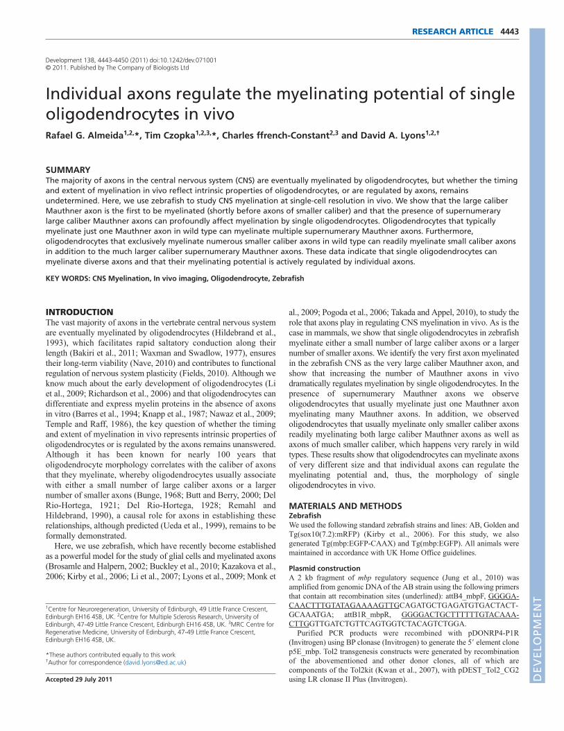
Fig. 1. Transgenic reporters reveal first axon myelinated in vivo in zebrafish CNS. (A)Lateral view of a stable Tg(mbp:EGFP-CAAX) zebrafishat 8 dpf. Myelinating glia of the CNS and PNS are labelled, as is the heart, which serves as a marker of transgenesis. (A�)Lateral view of the spinalcord (area indicated by box in A). Prominent myelinated tracts myelinated in the dorsal spinal cord and ventral spinal cord are apparent. (B)Lateralviews of a stable Tg(mbp:EGFP-CAAX) zebrafish at 60 hpf show that the very first axon to be myelinated is the large Mauthner axon in the ventralspinal cord, which is first myelinated in the anterior spinal cord. (C)Lateral views of a stable Tg(mbp:EGFP-CAAX) zebrafish at 70 hpf. Myelination ofthe Mauthner axon has now commenced in the more posterior part of the spinal cord. At this stage, oligodendrocytes have started to myelinateaxons in the dorsal spinal cord. Dorsal is up and anterior is to the left in all images. Scale bars: 500mm in A; 20mm in C. D
EVELO
PMENT
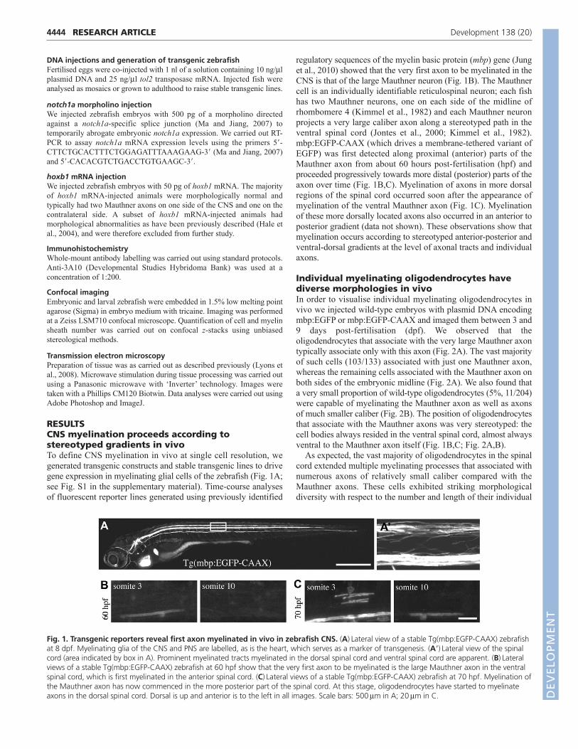
myelin sheaths (Fig. 2C-E). At 4 dpf, we found that the majorityof such cells (14 of 22) had 11-14 myelinating processes, althoughprocess number varied between 4 and 22 (Fig. 2D). Also, thelength of individual nascent myelin sheaths per cell was highlyvariable and ranged from 6 mm to 49 mm (Fig. 2E). The number ofmyelin sheaths per oligodendrocyte was relatively stable from 3-7dpf, but average myelin sheath length per cell increased almosttwofold over the same time period (Fig. 2F-G).
Supernumerary Mauthner axons are robustlymyelinated in vivoIn order to test how individual large caliber axons might regulateCNS myelination, we generated animals with supernumeraryMauthner neurons using two independent genetic manipulations.Previous studies have shown that disruption of notch1a causes amild neurogenic phenotype in zebrafish, characterised in part bythe appearance of multiple Mauthner neurons (Gray et al., 2001).Such extra Mauthner neurons are born between 9 and 12 hpf, as inwild type, and extend axons to the posterior spinal cord, as in wildtype (Liu et al., 2003). It is important to point out that the birth ofthese neurons and their axonal growth occurs long before theappearance of oligodendrocytes. Because Notch1 has beenassociated with oligodendrocyte development and myelination(Genoud et al., 2002; Hu et al., 2003; Park and Appel, 2003; Parket al., 2005; Wang et al., 1998), we sought to only temporarilyreduce Notch1a function, and found that injection of 500 pg of apreviously published morpholino (Ma and Jiang, 2007) wassufficient to generate animals with supernumerary Mauthnerneurons and axons (Fig. 3A,B). The level of notch1a mRNA insuch morphants is reduced relative to control at 10 hpf (whenMauthner neurons are born) but is indistinguishable from controls
by 3 dpf (when extensive myelination commences) (Fig. 3D). Inorder to have an independent method to generate extra Mauthneraxons we injected embryos with hoxb1 mRNAs (Fig. 3C), whichhas previously been shown to be capable of generating animalswith ectopic Mauthner neurons by duplicating rhombomere 4identity in the hindbrain (Hale et al., 2004).
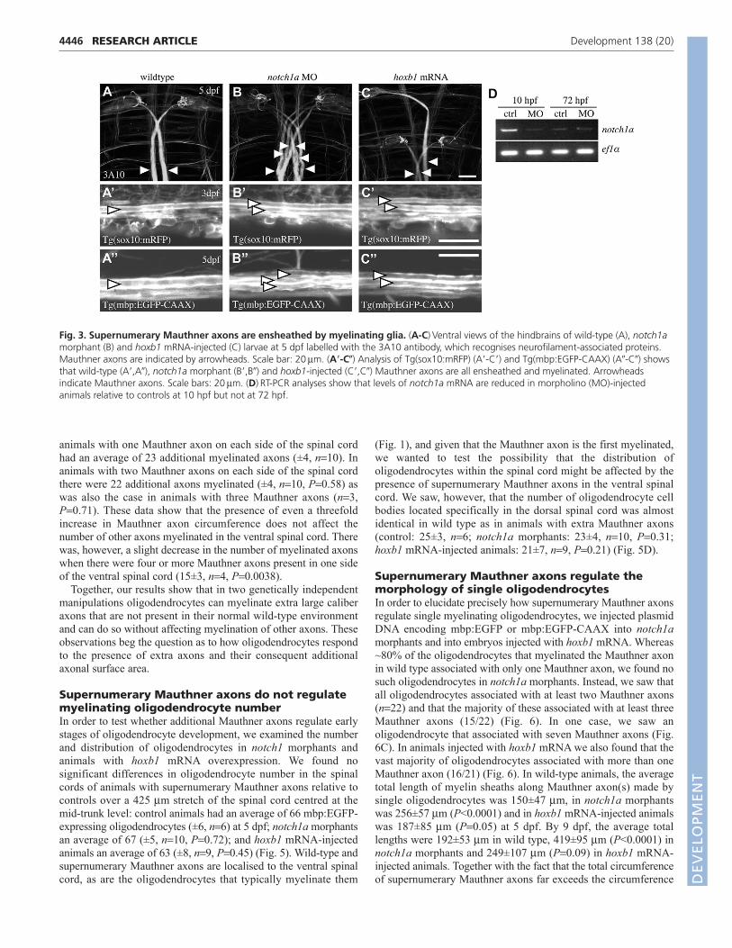
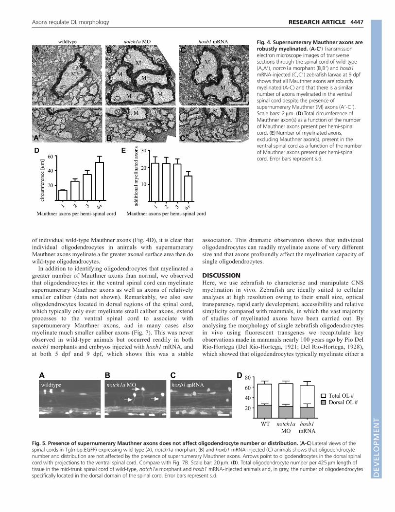
Examination of notch1a morphants and embryos injected withhoxb1 mRNA between 3 and 9 dpf using transgenic reporters(sox10:mRFP and mbp:EGFP-CAAX) and transmission electronmicroscopy showed that normal and supernumerary Mauthneraxons were covered by glial membrane and robustly myelinated(Fig. 3; Fig. 4; data not shown). The Mauthner axon, like manyaxons in situ, is not circular in cross-sectional profile (Fig. 4). Wetherefore quantified the circumference(s) of Mauthner axon(s) inour analyses, because this variable (in combination with axonlength) reflects the axonal surface available for potentialinteractions with oligodendrocytes. We saw that the summedcircumference(s) of all Mauthner axon(s) per hemi-spinal cordincreased almost linearly with the number of Mauthner axonspresent (Fig. 4D). In control animals that have one Mauthner axonon each side of the spinal cord, the average circumference per axonwas 13.5±0.8 mm (mean±s.d., n12) at 9 dpf. In animals with twoMauthner axons on one side of the spinal cord, the combinedcircumference was approximately doubled (26±3.5 mm, n11,P<0.0001) and those with three on each side nearly tripled (35±5mm, n3, P<0.0001). In animals with even more Mauthner axons,their total circumference increased even further.
In order to test whether myelination of extra Mauthner axonsoccurred at the expense of other axons, we counted the totalnumber of myelinated axons (excluding the Mauthner axons) incontrol and experimental ventral spinal cords (Fig. 4E). Control
4445RESEARCH ARTICLEAxons regulate OL morphology
Fig. 2. Single-cell analysis reveals morphological diversity of individual CNS oligodendrocytes. (A)Lateral views of single mbp:EGFP-expressing oligodendrocytes associating with one Mauthner axon (top) and both Mauthner axons (bottom). (B)Lateral view of a single mbp:EGFP-expressing oligodendrocyte associating with the large Mauthner axon (left) and an axon of much smaller caliber (right). (C)Lateral views of singleoligodendrocytes associating with multiple axons in the dorsal spinal cord (top) and ventral spinal cord (bottom). (D)Myelin sheath number peroligodendrocyte (excluding those that myelinate the Mauthner axons) at 4 dpf. (E)Myelin sheath length in four sample oligodendrocytes (that donot associate with the Mauthner axon) at 4 dpf. (F)Average myelin sheath number per cell over time. This does not include oligodendrocytes thatmyelinate the Mauthner axon. (G)Average myelin sheath length per cell over time. This does not include oligodendrocytes that myelinate theMauthner axon. Error bars represent s.d. Scale bars: 10mm.
DEVELO
PMENT
4446
animals with one Mauthner axon on each side of the spinal cordhad an average of 23 additional myelinated axons (±4, n10). Inanimals with two Mauthner axons on each side of the spinal cordthere were 22 additional axons myelinated (±4, n10, P0.58) aswas also the case in animals with three Mauthner axons (n3,P0.71). These data show that the presence of even a threefoldincrease in Mauthner axon circumference does not affect thenumber of other axons myelinated in the ventral spinal cord. Therewas, however, a slight decrease in the number of myelinated axonswhen there were four or more Mauthner axons present in one sideof the ventral spinal cord (15±3, n4, P0.0038).
Together, our results show that in two genetically independentmanipulations oligodendrocytes can myelinate extra large caliberaxons that are not present in their normal wild-type environmentand can do so without affecting myelination of other axons. Theseobservations beg the question as to how oligodendrocytes respondto the presence of extra axons and their consequent additionalaxonal surface area.
Supernumerary Mauthner axons do not regulatemyelinating oligodendrocyte numberIn order to test whether additional Mauthner axons regulate earlystages of oligodendrocyte development, we examined the numberand distribution of oligodendrocytes in notch1 morphants andanimals with hoxb1 mRNA overexpression. We found nosignificant differences in oligodendrocyte number in the spinalcords of animals with supernumerary Mauthner axons relative tocontrols over a 425 mm stretch of the spinal cord centred at themid-trunk level: control animals had an average of 66 mbp:EGFP-expressing oligodendrocytes (±6, n6) at 5 dpf; notch1a morphantsan average of 67 (±5, n10, P0.72); and hoxb1 mRNA-injectedanimals an average of 63 (±8, n9, P0.45) (Fig. 5). Wild-type andsupernumerary Mauthner axons are localised to the ventral spinalcord, as are the oligodendrocytes that typically myelinate them
(Fig. 1), and given that the Mauthner axon is the first myelinated,we wanted to test the possibility that the distribution ofoligodendrocytes within the spinal cord might be affected by thepresence of supernumerary Mauthner axons in the ventral spinalcord. We saw, however, that the number of oligodendrocyte cellbodies located specifically in the dorsal spinal cord was almostidentical in wild type as in animals with extra Mauthner axons(control: 25±3, n6; notch1a morphants: 23±4, n10, P0.31;hoxb1 mRNA-injected animals: 21±7, n9, P0.21) (Fig. 5D).
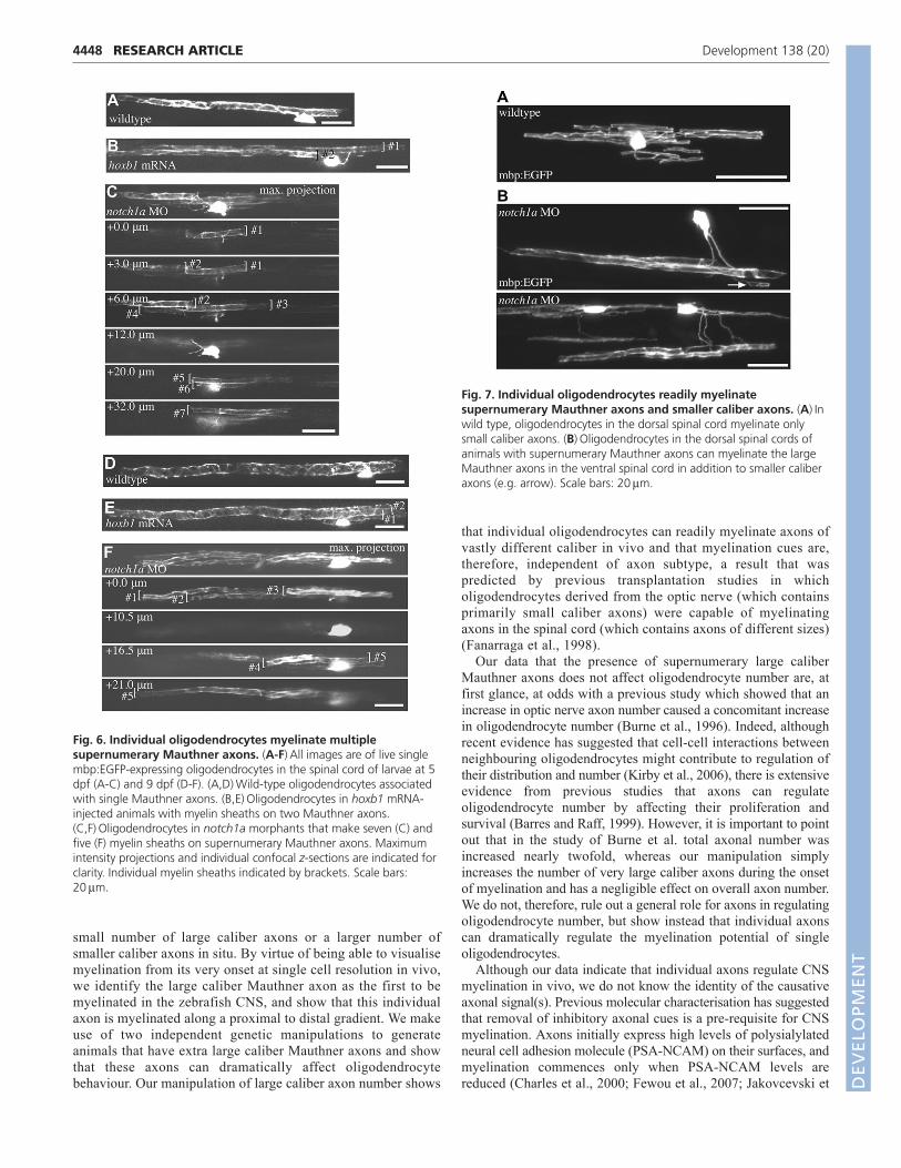
Supernumerary Mauthner axons regulate themorphology of single oligodendrocytesIn order to elucidate precisely how supernumerary Mauthner axonsregulate single myelinating oligodendrocytes, we injected plasmidDNA encoding mbp:EGFP or mbp:EGFP-CAAX into notch1amorphants and into embryos injected with hoxb1 mRNA. Whereas~80% of the oligodendrocytes that myelinated the Mauthner axonin wild type associated with only one Mauthner axon, we found nosuch oligodendrocytes in notch1a morphants. Instead, we saw thatall oligodendrocytes associated with at least two Mauthner axons(n22) and that the majority of these associated with at least threeMauthner axons (15/22) (Fig. 6). In one case, we saw anoligodendrocyte that associated with seven Mauthner axons (Fig.6C). In animals injected with hoxb1 mRNA we also found that thevast majority of oligodendrocytes associated with more than oneMauthner axon (16/21) (Fig. 6). In wild-type animals, the averagetotal length of myelin sheaths along Mauthner axon(s) made bysingle oligodendrocytes was 150±47 mm, in notch1a morphantswas 256±57 mm (P<0.0001) and in hoxb1 mRNA-injected animalswas 187±85 mm (P0.05) at 5 dpf. By 9 dpf, the average totallengths were 192±53 mm in wild type, 419±95 mm (P<0.0001) innotch1a morphants and 249±107 mm (P0.09) in hoxb1 mRNA-injected animals. Together with the fact that the total circumferenceof supernumerary Mauthner axons far exceeds the circumference
RESEARCH ARTICLE Development 138 (20)
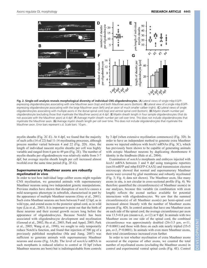
Fig. 3. Supernumerary Mauthner axons are ensheathed by myelinating glia. (A-C)Ventral views of the hindbrains of wild-type (A), notch1amorphant (B) and hoxb1 mRNA-injected (C) larvae at 5 dpf labelled with the 3A10 antibody, which recognises neurofilament-associated proteins.Mauthner axons are indicated by arrowheads. Scale bar: 20mm. (A�-C�) Analysis of Tg(sox10:mRFP) (A�-C�) and Tg(mbp:EGFP-CAAX) (A�-C�) showsthat wild-type (A�,A�), notch1a morphant (B�,B�) and hoxb1-injected (C�,C�) Mauthner axons are all ensheathed and myelinated. Arrowheadsindicate Mauthner axons. Scale bars: 20mm. (D)RT-PCR analyses show that levels of notch1a mRNA are reduced in morpholino (MO)-injectedanimals relative to controls at 10 hpf but not at 72 hpf.
DEVELO
PMENT
of individual wild-type Mauthner axons (Fig. 4D), it is clear thatindividual oligodendrocytes in animals with supernumeraryMauthner axons myelinate a far greater axonal surface area than dowild-type oligodendrocytes.
In addition to identifying oligodendrocytes that myelinated agreater number of Mauthner axons than normal, we observedthat oligodendrocytes in the ventral spinal cord can myelinatesupernumerary Mauthner axons as well as axons of relativelysmaller caliber (data not shown). Remarkably, we also sawoligodendrocytes located in dorsal regions of the spinal cord,which typically only ever myelinate small caliber axons, extendprocesses to the ventral spinal cord to associate withsupernumerary Mauthner axons, and in many cases alsomyelinate much smaller caliber axons (Fig. 7). This was neverobserved in wild-type animals but occurred readily in bothnotch1 morphants and embryos injected with hoxb1 mRNA, andat both 5 dpf and 9 dpf, which shows this was a stable
association. This dramatic observation shows that individualoligodendrocytes can readily myelinate axons of very differentsize and that axons profoundly affect the myelination capacity ofsingle oligodendrocytes.
DISCUSSIONHere, we use zebrafish to characterise and manipulate CNSmyelination in vivo. Zebrafish are ideally suited to cellularanalyses at high resolution owing to their small size, opticaltransparency, rapid early development, accessibility and relativesimplicity compared with mammals, in which the vast majorityof studies of myelinated axons have been carried out. Byanalysing the morphology of single zebrafish oligodendrocytesin vivo using fluorescent transgenes we recapitulate keyobservations made in mammals nearly 100 years ago by Pio DelRio-Hortega (Del Rio-Hortega, 1921; Del Rio-Hortega, 1928),which showed that oligodendrocytes typically myelinate either a
4447RESEARCH ARTICLEAxons regulate OL morphology
Fig. 4. Supernumerary Mauthner axons arerobustly myelinated. (A-C�) Transmissionelectron microscope images of transversesections through the spinal cord of wild-type(A,A�), notch1a morphant (B,B�) and hoxb1mRNA-injected (C,C�) zebrafish larvae at 9 dpfshows that all Mauthner axons are robustlymyelinated (A-C) and that there is a similarnumber of axons myelinated in the ventralspinal cord despite the presence ofsupernumerary Mauthner (M) axons (A�-C�).Scale bars: 2mm. (D)Total circumference ofMauthner axon(s) as a function of the numberof Mauthner axons present per hemi-spinalcord. (E)Number of myelinated axons,excluding Mauthner axon(s), present in theventral spinal cord as a function of the numberof Mauthner axons present per hemi-spinalcord. Error bars represent s.d.
Fig. 5. Presence of supernumerary Mauthner axons does not affect oligodendrocyte number or distribution. (A-C)Lateral views of thespinal cords in Tg(mbp:EGFP)-expressing wild-type (A), notch1a morphant (B) and hoxb1 mRNA-injected (C) animals shows that oligodendrocytenumber and distribution are not affected by the presence of supernumerary Mauthner axons. Arrows point to oligodendrocytes in the dorsal spinalcord with projections to the ventral spinal cord. Compare with Fig. 7B. Scale bar: 20mm. (D). Total oligodendrocyte number per 425mm length oftissue in the mid-trunk spinal cord of wild-type, notch1a morphant and hoxb1 mRNA-injected animals and, in grey, the number of oligodendrocytesspecifically located in the dorsal domain of the spinal cord. Error bars represent s.d. D
EVELO
PMENT
4448
small number of large caliber axons or a larger number ofsmaller caliber axons in situ. By virtue of being able to visualisemyelination from its very onset at single cell resolution in vivo,we identify the large caliber Mauthner axon as the first to bemyelinated in the zebrafish CNS, and show that this individualaxon is myelinated along a proximal to distal gradient. We makeuse of two independent genetic manipulations to generateanimals that have extra large caliber Mauthner axons and showthat these axons can dramatically affect oligodendrocytebehaviour. Our manipulation of large caliber axon number shows
that individual oligodendrocytes can readily myelinate axons ofvastly different caliber in vivo and that myelination cues are,therefore, independent of axon subtype, a result that waspredicted by previous transplantation studies in whicholigodendrocytes derived from the optic nerve (which containsprimarily small caliber axons) were capable of myelinatingaxons in the spinal cord (which contains axons of different sizes)(Fanarraga et al., 1998).
Our data that the presence of supernumerary large caliberMauthner axons does not affect oligodendrocyte number are, atfirst glance, at odds with a previous study which showed that anincrease in optic nerve axon number caused a concomitant increasein oligodendrocyte number (Burne et al., 1996). Indeed, althoughrecent evidence has suggested that cell-cell interactions betweenneighbouring oligodendrocytes might contribute to regulation oftheir distribution and number (Kirby et al., 2006), there is extensiveevidence from previous studies that axons can regulateoligodendrocyte number by affecting their proliferation andsurvival (Barres and Raff, 1999). However, it is important to pointout that in the study of Burne et al. total axonal number wasincreased nearly twofold, whereas our manipulation simplyincreases the number of very large caliber axons during the onsetof myelination and has a negligible effect on overall axon number.We do not, therefore, rule out a general role for axons in regulatingoligodendrocyte number, but show instead that individual axonscan dramatically regulate the myelination potential of singleoligodendrocytes.
Although our data indicate that individual axons regulate CNSmyelination in vivo, we do not know the identity of the causativeaxonal signal(s). Previous molecular characterisation has suggestedthat removal of inhibitory axonal cues is a pre-requisite for CNSmyelination. Axons initially express high levels of polysialylatedneural cell adhesion molecule (PSA-NCAM) on their surfaces, andmyelination commences only when PSA-NCAM levels arereduced (Charles et al., 2000; Fewou et al., 2007; Jakovcevski et
RESEARCH ARTICLE Development 138 (20)
Fig. 6. Individual oligodendrocytes myelinate multiplesupernumerary Mauthner axons. (A-F)All images are of live singlembp:EGFP-expressing oligodendrocytes in the spinal cord of larvae at 5dpf (A-C) and 9 dpf (D-F). (A,D)Wild-type oligodendrocytes associatedwith single Mauthner axons. (B,E)Oligodendrocytes in hoxb1 mRNA-injected animals with myelin sheaths on two Mauthner axons.(C,F)Oligodendrocytes in notch1a morphants that make seven (C) andfive (F) myelin sheaths on supernumerary Mauthner axons. Maximumintensity projections and individual confocal z-sections are indicated forclarity. Individual myelin sheaths indicated by brackets. Scale bars:20mm.
Fig. 7. Individual oligodendrocytes readily myelinatesupernumerary Mauthner axons and smaller caliber axons. (A)Inwild type, oligodendrocytes in the dorsal spinal cord myelinate onlysmall caliber axons. (B)Oligodendrocytes in the dorsal spinal cords ofanimals with supernumerary Mauthner axons can myelinate the largeMauthner axons in the ventral spinal cord in addition to smaller caliberaxons (e.g. arrow). Scale bars: 20mm.
DEVELO
PMENT
al., 2007; Keirstead et al., 1999). Analyses of the transmembraneprotein Lingo1 (Lee et al., 2007; Mi et al., 2005) also suggest thatit too might function on axons to inhibit myelination. In theperipheral nervous system (PNS) there is strong evidence thataxonal Neuregulin 1 type III serves as an instructive cue formyelination (Michailov et al., 2004; Taveggia et al., 2005).Although Neuregulin 1 type III is not required for CNSmyelination in vivo, it can stimulate hypermyelination of CNSaxons in vivo (Brinkmann et al., 2008), which indicates thatoligodendrocytes can respond to instructive axonal cues. It will beinteresting to see whether future studies will identify instructiveaxonal signals required for CNS myelination.
Our data that single oligodendrocytes faced with supernumerarylarge caliber Mauthner axons can myelinate a larger axonal surfacearea than they normally do during development prompts thequestion of whether there is a maximum amount of axonal surfacethat any single oligodendrocyte can myelinate. If singleoligodendrocytes myelinate close to their maximum capacity, e.g.by adulthood, then unless every oligodendrocyte is replacedfollowing demyelination after injury or disease, incomplete repairmight be inevitable. Our demonstration that axon size can regulateoligodendrocyte morphology is also relevant to the remyelinationof axons. Given the significant cross-sectional growth ofmyelinated axons that takes place after the onset of myelination, itis likely that the caliber of any axon demyelinated long afterdevelopment will be significantly larger than when it was firstmyelinated. This discrepancy in target caliber might mean that themorphology of remyelinating oligodendrocytes will be differentthan during development. It is possible that during remyelinationmore individual oligodendrocytes make fewer myelin sheaths onsuch larger caliber axons (Blakemore, 1974). If this is indeed thecase, remyelination might necessitate the employment of a greaternumber of oligodendrocytes than are required to myelinate thesame complement of axons during development. It is, however,also possible that oligodendrocytes faced with such relatively largecaliber axons during remyelination do not regulate theirmorphology dramatically, but rather extend less myelin aroundindividual axons. This possibility could explain the fact thatremyelinated axon profiles are often surrounded by thin myelin(Blakemore, 1974). Future studies of demyelination andremyelination may benefit from high-resolution analyses that canassess precisely how much axonal surface is myelinated or howmuch myelin is made by individual oligodendrocytes.
In summary, our study shows that individual axons canprofoundly regulate the myelinating potential of singleoligodendrocytes, which has implications for the formation andregulation of myelinated axons throughout life and for mechanismsthat may need to be considered during their repair.
AcknowledgementsWe thank Carl Tucker, John Mullins, Patricia Smart and Sebastien Rider forfishroom support; Bruce Appel, Koichi Kawakami, Jiang Yun Jin, Hae-Chul Parkand Victoria Prince for sharing reagents; Crerar hotels for free confocal access;and Stephen Mitchell for TEM assistance. Very special thanks to Chi-Bin Chienand the Chien laboratory for the Tol2kit. We are grateful to Thomas Becker,Peter Brophy, Ben Emery, Kelly Monk, Alya Raphael, William Talbot, AnnaWilliams and members of the Lyons, ffrench-Constant and Brophy laboratoriesfor critical reading of the manuscript.
FundingThis work was supported by a David Phillips Fellowship from the BBSRC, anInnovative grant from the UK MS Society and an International ReintegrationGrant to D.A.L., an FCT doctoral studentship to R.G.A. and a Wellcome TrustProgramme Grant and UK MS Society Centre award to C.ff.-C. Deposited inPMC for release after 6 months.
Competing interests statementThe authors declare no competing financial interests.
Supplementary materialSupplementary material for this article is available athttp://dev.biologists.org/lookup/suppl/doi:10.1242/dev.071001/-/DC1
ReferencesBakiri, Y., Karadottir, R., Cossell, L. and Attwell, D. (2011). Morphological and
electrical properties of oligodendrocytes in the white matter of the corpuscallosum and cerebellum. J. Physiol. 589, 559-573.
Barres, B. A. and Raff, M. C. (1999). Axonal control of oligodendrocytedevelopment. J. Cell Biol. 147, 1123-1128.
Barres, B. A., Lazar, M. A. and Raff, M. C. (1994). A novel role for thyroidhormone, glucocorticoids and retinoic acid in timing oligodendrocytedevelopment. Development 120, 1097-1108.
Blakemore, W. F. (1974). Pattern of remyelination in the CNS. Nature 249, 577-578.
Brinkmann, B. G., Agarwal, A., Sereda, M. W., Garratt, A. N., Muller, T.,Wende, H., Stassart, R. M., Nawaz, S., Humml, C., Velanac, V. et al. (2008).Neuregulin-1/ErbB signaling serves distinct functions in myelination of theperipheral and central nervous system. Neuron 59, 581-595.
Brosamle, C. and Halpern, M. E. (2002). Characterization of myelination in thedeveloping zebrafish. Glia 39, 47-57.
Buckley, C. E., Marguerie, A., Roach, A. G., Goldsmith, P., Fleming, A.,Alderton, W. K. and Franklin, R. J. (2010). Drug reprofiling using zebrafishidentifies novel compounds with potential pro-myelination effects.Neuropharmacology 59, 149-159.
Bunge, R. P. (1968). Glial cells and the central myelin sheath. Physiol. Rev. 48, 197-251.
Burne, J. F., Staple, J. K. and Raff, M. C. (1996). Glial cells are increasedproportionally in transgenic optic nerves with increased numbers of axons. J.Neurosci. 16, 2064-2073.
Butt, A. M. and Berry, M. (2000). Oligodendrocytes and the control ofmyelination in vivo: new insights from the rat anterior medullary velum. J.Neurosci. Res. 59, 477-488.
Charles, P., Hernandez, M. P., Stankoff, B., Aigrot, M. S., Colin, C., Rougon,G., Zalc, B. and Lubetzki, C. (2000). Negative regulation of central nervoussystem myelination by polysialylated-neural cell adhesion molecule. Proc. Natl.Acad. Sci. USA 97, 7585-7590.
Del Rio-Hortega, P. (1921). Estudios sobre la neuroglia. La glia de escasasradiaciones oligodendroglia. Bol. Real Soc. Esp. Hist. Nat. 21, 63-92.
Del Rio-Hortega, P. (1928). Tercera aportacion al conocimiento morfologico einterpretacion funcional de la oligodendroglia. Mem. Real Soc. Esp. Hist. Nat.14, 40-122.
Fanarraga, M. L., Griffiths, I. R., Zhao, M. and Duncan, I. D. (1998).Oligodendrocytes are not inherently programmed to myelinate a specific size ofaxon. J. Comp. Neurol. 399, 94-100.
Fewou, S. N., Ramakrishnan, H., Bussow, H., Gieselmann, V. and Eckhardt,M. (2007). Down-regulation of polysialic acid is required for efficient myelinformation. J. Biol. Chem. 282, 16700-16711.
Fields, R. D. (2010). Neuroscience. Change in the brain’s white matter. Science330, 768-769.
Genoud, S., Lappe-Siefke, C., Goebbels, S., Radtke, F., Aguet, M., Scherer, S.S., Suter, U., Nave, K. A. and Mantei, N. (2002). Notch1 control ofoligodendrocyte differentiation in the spinal cord. J. Cell Biol. 158, 709-718.
Gray, M., Moens, C. B., Amacher, S. L., Eisen, J. S. and Beattie, C. E. (2001).Zebrafish deadly seven functions in neurogenesis. Dev. Biol. 237, 306-323.
Hale, M. E., Kheirbek, M. A., Schriefer, J. E. and Prince, V. E. (2004). Hox genemisexpression and cell-specific lesions reveal functionality of homeoticallytransformed neurons. J. Neurosci. 24, 3070-3076.
Hildebrand, C., Remahl, S., Persson, H. and Bjartmar, C. (1993). Myelinatednerve fibres in the CNS. Prog. Neurobiol. 40, 319-384.
Hu, Q. D., Ang, B. T., Karsak, M., Hu, W. P., Cui, X. Y., Duka, T., Takeda, Y.,Chia, W., Sankar, N., Ng, Y. K. et al. (2003). F3/contactin acts as a functionalligand for Notch during oligodendrocyte maturation. Cell 115, 163-175.
Jakovcevski, I., Mo, Z. and Zecevic, N. (2007). Down-regulation of the axonalpolysialic acid-neural cell adhesion molecule expression coincides with the onsetof myelination in the human fetal forebrain. Neuroscience 149, 328-337.
Jontes, J. D., Buchanan, J. and Smith, S. J. (2000). Growth cone and dendritedynamics in zebrafish embryos: early events in synaptogenesis imaged in vivo.Nat. Neurosci. 3, 231-237.
Jung, S. H., Kim, S., Chung, A. Y., Kim, H. T., So, J. H., Ryu, J., Park, H. C. andKim, C. H. (2010). Visualization of myelination in GFP-transgenic zebrafish. Dev.Dyn. 239, 592-597.
Kazakova, N., Li, H., Mora, A., Jessen, K. R., Mirsky, R., Richardson, W. D.and Smith, H. K. (2006). A screen for mutations in zebrafish that affect myelingene expression in Schwann cells and oligodendrocytes. Dev. Biol. 297, 1-13.
4449RESEARCH ARTICLEAxons regulate OL morphology
DEVELO
PMENT
4450
Keirstead, H. S., Ben-Hur, T., Rogister, B., O’Leary, M. T., Dubois-Dalcq, M.and Blakemore, W. F. (1999). Polysialylated neural cell adhesion molecule-positive CNS precursors generate both oligodendrocytes and Schwann cells toremyelinate the CNS after transplantation. J. Neurosci. 19, 7529-7536.
Kimmel, C. B., Powell, S. L. and Metcalfe, W. K. (1982). Brain neurons whichproject to the spinal cord in young larvae of the zebrafish. J. Comp. Neurol. 205,112-127.
Kirby, B. B., Takada, N., Latimer, A. J., Shin, J., Carney, T. J., Kelsh, R. N. andAppel, B. (2006). In vivo time-lapse imaging shows dynamic oligodendrocyteprogenitor behavior during zebrafish development. Nat. Neurosci. 9, 1506-1511.
Knapp, P. E., Bartlett, W. P. and Skoff, R. P. (1987). Cultured oligodendrocytesmimic in vivo phenotypic characteristics: cell shape, expression of myelin-specificantigens, and membrane production. Dev. Biol. 120, 356-365.
Kwan, K. M., Fujimoto, E., Grabher, C., Mangum, B. D., Hardy, M. E.,Campbell, D. S., Parant, J. M., Yost, H. J., Kanki, J. P. and Chien, C. B.(2007). The Tol2kit: a multisite gateway-based construction kit for Tol2transposon transgenesis constructs. Dev. Dyn. 236, 3088-3099.
Lee, X., Yang, Z., Shao, Z., Rosenberg, S. S., Levesque, M., Pepinsky, R. B.,Qiu, M., Miller, R. H., Chan, J. R. and Mi, S. (2007). NGF regulates theexpression of axonal LINGO-1 to inhibit oligodendrocyte differentiation andmyelination. J. Neurosci. 27, 220-225.
Li, H., Lu, Y., Smith, H. K. and Richardson, W. D. (2007). Olig1 and Sox10interact synergistically to drive myelin basic protein transcription inoligodendrocytes. J. Neurosci. 27, 14375-14382.
Li, H., He, Y., Richardson, W. D. and Casaccia, P. (2009). Two-tier transcriptionalcontrol of oligodendrocyte differentiation. Curr. Opin. Neurobiol. 19, 479-485.
Liu, K. S., Gray, M., Otto, S. J., Fetcho, J. R. and Beattie, C. E. (2003).Mutations in deadly seven/notch1a reveal developmental plasticity in the escaperesponse circuit. J. Neurosci. 23, 8159-8166.
Lyons, D. A., Naylor, S. G., Mercurio, S., Dominguez, C. and Talbot, W. S.(2008). KBP is essential for axonal structure, outgrowth and maintenance inzebrafish, providing insight into the cellular basis of Goldberg-Shprintzensyndrome. Development 135, 599-608.
Lyons, D. A., Naylor, S. G., Scholze, A. and Talbot, W. S. (2009). Kif1b isessential for mRNA localization in oligodendrocytes and development ofmyelinated axons. Nat. Genet. 41, 854-858.
Ma, M. and Jiang, Y. J. (2007). Jagged2a-notch signaling mediates cell fatechoice in the zebrafish pronephric duct. PLoS Genet. 3, e18.
Mi, S., Miller, R. H., Lee, X., Scott, M. L., Shulag-Morskaya, S., Shao, Z.,Chang, J., Thill, G., Levesque, M., Zhang, M. et al. (2005). LINGO-1negatively regulates myelination by oligodendrocytes. Nat. Neurosci. 8, 745-751.
Michailov, G. V., Sereda, M. W., Brinkmann, B. G., Fischer, T. M., Haug, B.,Birchmeier, C., Role, L., Lai, C., Schwab, M. H. and Nave, K. A. (2004).Axonal neuregulin-1 regulates myelin sheath thickness. Science 304, 700-703.
Monk, K. R., Naylor, S. G., Glenn, T. D., Mercurio, S., Perlin, J. R.,Dominguez, C., Moens, C. B. and Talbot, W. S. (2009). A G protein-coupledreceptor is essential for Schwann cells to initiate myelination. Science 325, 1402-1405.
Nave, K. A. (2010). Myelination and support of axonal integrity by glia. Nature468, 244-252.
Nawaz, S., Kippert, A., Saab, A. S., Werner, H. B., Lang, T., Nave, K. A. andSimons, M. (2009). Phosphatidylinositol 4,5-bisphosphate-dependentinteraction of myelin basic protein with the plasma membrane inoligodendroglial cells and its rapid perturbation by elevated calcium. J. Neurosci.29, 4794-4807.
Park, H. C. and Appel, B. (2003). Delta-Notch signaling regulates oligodendrocytespecification. Development 130, 3747-3755.
Park, H. C., Boyce, J., Shin, J. and Appel, B. (2005). Oligodendrocytespecification in zebrafish requires notch-regulated cyclin-dependent kinaseinhibitor function. J. Neurosci. 25, 6836-6844.
Pogoda, H. M., Sternheim, N., Lyons, D. A., Diamond, B., Hawkins, T. A.,Woods, I. G., Bhatt, D. H., Franzini-Armstrong, C., Dominguez, C., Arana,N. et al. (2006). A genetic screen identifies genes essential for development ofmyelinated axons in zebrafish. Dev. Biol. 298, 118-131.
Remahl, S. and Hildebrand, C. (1990). Relation between axons andoligodendroglial cells during initial myelination I. The glial unit. J. Neurocytol. 19,313-328.
Richardson, W. D., Kessaris, N. and Pringle, N. (2006). Oligodendrocyte wars.Nat. Rev. Neurosci. 7, 11-18.
Takada, N. and Appel, B. (2010). Identification of genes expressed by zebrafisholigodendrocytes using a differential microarray screen. Dev. Dyn. 239, 2041-2047.
Taveggia, C., Zanazzi, G., Petrylak, A., Yano, H., Rosenbluth, J., Einheber, S.,Xu, X., Esper, R. M., Loeb, J. A., Shrager, P. et al. (2005). Neuregulin-1 type IIIdetermines the ensheathment fate of axons. Neuron 47, 681-694.
Temple, S. and Raff, M. C. (1986). Clonal analysis of oligodendrocytedevelopment in culture: evidence for a developmental clock that counts celldivisions. Cell 44, 773-779.
Ueda, H., Levine, J. M., Miller, R. H. and Trapp, B. D. (1999). Rat optic nerveoligodendrocytes develop in the absence of viable retinal ganglion cell axons. J.Cell Biol. 146, 1365-1374.
Wang, S., Sdrulla, A. D., diSibio, G., Bush, G., Nofziger, D., Hicks, C.,Weinmaster, G. and Barres, B. A. (1998). Notch receptor activation inhibitsoligodendrocyte differentiation. Neuron 21, 63-75.
Waxman, S. G. and Swadlow, H. A. (1977). The conduction properties of axonsin central white matter. Prog. Neurobiol. 8, 297-324.
RESEARCH ARTICLE Development 138 (20)
DEVELO
PMENT