Embed Size (px)
Citation preview
BIOCHEMICAL AND BIOPHYSICAL RESEARCH COMMUNICATIONS 239, 823–829 (1997)ARTICLE NO. RC977561
Induction of Ubiquitin Conjugating Enzyme Activity forDegradation of Topoisomerase IIa during AdenovirusE1A-Induced Apoptosis
Takuma Nakajima,*,1 Michiko Kimura,* Kazufumi Kuroda,† Masato Tanaka,† Akihiko Kikuchi,‡Hiroaki Seino,§ Fumiaki Yamao,§ and Kinichiro Oda**Department of Biological Science and Technology, Science University of Tokyo, 2641 Yamazaki, Noda 278, Japan;†Mitsubisi Kasei Institute of Life Sciences, 11, Minamiohya, Machida 194, Japan; ‡Laboratory of Medical Mycology,Research Institute of Disease Mechanism and Control, Nagoya University, 65 Tsurumae-cho, Showa-ku, Nagoya 466,Japan; and §National Institute of Genetics, Mishima 411, Japan
Received September 29, 1997
virus long terminal repeat (MMTV-LTR), expressesTopoisomerase (topo) IIa is degraded via polyubi- E1A12S in response to dexamethasone (dex) and elicits
quitination during adenovirus E1A-induced apoptosis apoptosis after stabilization of wt p53 (5). Topoisomer-in MA1 cells, a derivative of the human epidermoid ase (topo) IIa seems to be a target of E1A-inducedcarcinoma cell line KB. Topo IIa ubiquitination activ-
apoptosis and begin to be degraded before the onset ofity in MA1 cells increased nearly 10-fold after induc-DNA fragmentation (5). The degradation of topo IIation of E1A in response to dexamethasone. To identifyoccurs in a ATP and ubiquitin dependent manner bya topo IIa ubiquitination factor(s), the S100 fractionsinduction of a component(s) involved in the polyubiqui-prepared from apoptosis-induced (42 h) and unin-tination of topo IIa. Ubiquitination activity for topo IIaduced (0 h) MA1 cells were first fractionated by ubiqui-increased several fold upon induction of apoptosis intin–Sepharose columns. The ubiquitination activityMA1 cells (6).induced by E1A was predominantly eluted with 20 mM
AMP. Further fractionation of the AMP eluates on Re- The expression of topo IIa is cell cycle-regulatedsource-Q columns and the thiolester formation of the peaking in G2/M phase and declining to a minimal levelproteins resolved by electrophoresis with biotinylated at the end of M phase (7-11). This pattern of expressionubiquitin revealed that a species of E2 isozyme recov- is similar to that of cyclin B which has been shown toered in the QFT2 fraction increased markedly in MA1 be degraded by induction of a specific ubiquitin ligasecells after E1A expression. These results indicate that activity E3-C. E3-C cooperates with a specific ubiquitina ubiquitination factor(s) specific to topo IIa is in- conjugating enzyme E2-C for degradation of cyclin Bduced during E1A-induced apoptosis in MA1 cells. q 1997 at metaphase-anaphase transition (12-15). However,Academic Press nothing is known about enzymes involved in the polyu-
biquitination of topo IIa. Conjugation of ubiquitin totarget proteins occurs, in general, in three steps. Ubi-
The adenovirus E1A gene products E1A have multi- quitin is first activated by the formation of thiolesterple functions to induce cell growth and death and to intermediate with a ubiquitin-activating enzyme E1inhibit cell differenciation. The induction of apoptosis and then transferred to a ubiquitin-conjugating en-by E1A depends on the expression of wild type (wt) p53 zyme E2. Ubiquitin is transferred to target proteins byin cells (1, 2) and the E1A domain required for induc- E2, often in combination with a ubiquitin-ligase E3tion of apoptosis has been mapped in the amino-termi- (Reviewed by Ciechanover (16)). Polyubiquitinated pro-nal and CR (conserved region) 1 regions (3, 4). The teins are then degraded by the 26S proteasome (17,cell line MA1, established from a human epithermoid 18). Both E2 and E3 enzymes constitute the family andcarcinoma cell line KB by introducing the adenovirus consist of many members.E1A 12S cDNA linked to the mouse mammary tumor In the present study, an enzyme(s) involved in the
polyubiquitination of topo IIa in the apoptosis-inducedand uninduced MA1 cells were fractionated by affinity1 To whom correspondence should be addressed. Fax: /81-471-25-
1841, E-mail: [email protected]. chromatography on ubiquitin-Sepharose (19) and by
0006-291X/97 $25.00Copyright q 1997 by Academic PressAll rights of reproduction in any form reserved.
823
AID BBRC 7561 / 693c$$$681 10-21-97 10:13:59 bbrcg AP: BBRC
Vol. 239, No. 3, 1997 BIOCHEMICAL AND BIOPHYSICAL RESEARCH COMMUNICATIONS
fractions (UFT) were collected the columns were washed with 35 mlanion-exchange chromatography on Resource-Q. Theof buffer-A containing 2 mM ATP, and adsorbed components wereE2 enzyme activity involved in the ubiquitination ofeluted with 14 ml of 1 M KCl in 50 mM Tris-HCl, pH7.2 (KCl frac-topo IIa increased nearly 10-fold in MA1 cells after tion), 14 ml of 2 mM AMP in 50 mM Tris-HCl, pH7.2 containing 40
induction of apoptosis by E1A. Ubiquitin-conjugation mM Na-pyrophosphate (AMP fraction), 14 ml of 10 mM DTT in 50mM Tris-HCl, pH7.2 (DTT fraction) and 14 ml of 2 mM DTT in 50assays revealed a species of E2 isozyme with molecularmM Tris-HCl, pH9.0 (pH9 fraction). Fractions of 2 ml were collectedweight of about 20 kDa, which increased markedlyat a flow rate of 0.2 ml/min into tubes containing 2 ml of 50 mM p-after expression of E1A.ABSF. Fractions eluted by the same buffer were combined. All thefractions were concentrated by ultrafiltration with Macrosep-10k.The salts in the eluates were removed by another ultrafiltration toMATERIALS AND METHODSa final volume of 0.5 ml, after 1:100 dilution with QB0.5.
Further fractionation of the AMP eluates was performed by Re-Cell culture and viability assay. The cell line MA1 was estab-sorce-Q column chromatography. Aliquots of 40 mg of protein werelished from the human epidermoid carcinoma cell line KB by intro-diluted with 10 volumes of QB1 buffer and applied to 1-ml columnsducing the adenovirus E1A 12S cDNA linked to the MMTV-LTR (5).equilibrated with QB1 buffer. The columns were washed with 9 mlThe cells were cultivated at 37 7C in Dulbecco’s modified Eagle’sof QB1 buffer and adsorbed proteins were eluted with 9 ml each ofmedium supplemented with 10% fetal calf serum at 377C. For theQB buffer containing 0.25 M KCl and 0.4 M KCl. The eluates wereviability assay, both floating and adherent cells were assesed bydesalted and concentrated as described above, and further concen-trypan blue exclusion.trated by using Millisep-1k (Paul-Filtron) to a final volume of 100 ml.
Preparation of cell extracts. Highspeed supernatant of cell extractAssays for topoIIa-ubiquitination activity. TopoIIa-ubiquitina-(S100) was prepared as described previously (6) with minor modifi-
tion activity was assayed as described previously (6). Briefly, 2 ml ofcation. Subconfluent cultures of MA1 cells, untreated or treated with20% suspension of the protein A-Sepharose beads conjugated with1 mM dexamethasone (dex) for 42 h, were washed twice in ice coldimmunoprecipitated topoIIa were incubated in 50 ml of QB0.5 bufferphosphate buffered saline, once in ice-cold hypotonic buffer (20 mMcontaining 20 mg of bacterially expressed GST-ubiquitin fusion pro-TrisrHCl, pH7.4, 5 mM MgCl2, 8 mM KCl and 1 mM dithiothreitoltein, 2 mM ATP and ATP regeneration system (20 mM phosphocre-[DTT]), and resuspended in 10 volumes of the hypotonic buffer. Afteratine and 0.1 units/ml creatine phosphokinase) for 15 min at 307C.incubation on ice for 15 min, the swollen cells were disrupted byThe beads were washed 3 times in 50 mM HEPES-KOH, pH7.0 con-homogenization with a tight pestle for 40 times on ice. The homoge-taining 1 mM DTT, 0.4 M NaCl and 0.1% Nonidet P-40, and boilednate was centrifuged at 105,000 x g for 1 h to remove cell debris,after addition of 30 ml of 2 1 Laemmli sample buffer (21). The sam-and the supernatant was centrifuged again at 105,000 1 g for 5 hples were subjected to SDS-PAGE and Western blotting. Polyubiqui-to prepare the S100 extract, which lacks most of the proteasometinated and unprocessed forms of topoIIa were detected on X-rayactivity. Protein concentration was determined by a dye-bindingfilm by enhanced chemiluminescence (ECL, Amersham) using anti-assay (20).GST polyclonal antibody (Z-5, SantaCruz) and anti-human topoIIa
Fractionation of topoIIa-ubiquitination factors. For fractionation monoclonal antibody 8D2, respectively. The specific activity of ubi-of S100 extracts by anion exchange chromatography on Resource-Q, quitination was quantitated by the densitometer scanning of X-ray60 mg protein of S100-0 and S100-42 (S100 prepared from MA1 cells film.untreated or treated with dex for 42 h) were diluted with 10 volumes
Ubiquitin thiolester formation. The ubiquitin-thiolester forma-of QB buffer (40 mM Tris-HCl, pH7.7 / 5 mM MgCl2 / 2 mM DTT)tion of E1 and E2s was assayed with biotinylated ubiquitin. Twenty-containing 0.1 M KCl (referred as QB1 buffer) and centrifuged atfive milligrams of bovine erythrocyte ubiquitin (Sigma) were dis-15,0001 g for 15 min. The supernatant was applied to 6-ml Resource-solved in 1 ml of 50 mM Na-bicarbonate buffer, pH8.3 and reactedQ columns (Pharmacia) equilibrated with QB1 buffer. The columnswith 1 mg of Sulfo-NHS-LC-Biotin (Pierce) for 2 h on ice. The reactionwere washed with 30 ml of QB1 buffer, and adsorbed componentswas stopped by adding 100 ml of 2 M Tris-HCl, pH7.2, and the solu-were eluted with 30 ml each of QB buffer containing 0.25 M KCl andtion was applied to a 15-ml Sephadex G-25 column equilibrated with0.4 M KCl. Fractions of 1.5 ml were collected at a flow rate of 0.5QB0.5 buffer to remove unreacted biotinylation reagent. Biotinylatedml/min into tubes containing 1.5 ml of 50 mM 4-(2-aminoethyl)-ben-ubiquitin was concentrated with Millisep-1k to a final volume of 2.5zenesulfonyl fluoriderHCl (p-ABSF). Fractions eluted by the sameml. The reaction of the ubiquitin-thiolester formation was carriedKCl concentration were combined to make QFT (the flowthough),out in 10 ml of QB0.5 buffer containing 4 mg of biotinylated ubiquitin,QA (0.25 M KCl eluate) and QB (0.4 M KCl eluate) fractions. Frac-2 mM ATP, ATP reganeration system and aliquots of the fractionatedtions were concentrated by centrifugal ultrafiltration with Macrosep-ubiquitination factors for 30 min at 187C. The reaction was stopped10k (Paul-Filtron) or Centriprep-10 (Amicon) concentrators and ex-on ice and the mixture was boiled with 10 ml of 21 stop buffer (100cess salt was removed by another ultrafiltration to a final volume ofmM Tris-HCl, pH6.8 / 4% SDS / 8 M Urea / 20% glycerol / 0.01% BPB)1 ml, after dilution with QB buffer containing 50 mM KCl (referredfor 3 min. Ubiquitin-thiolester was detected by Western blotting.as QB0.5).The filters were treated with ECL-detection system using peroxidaseFor covalent affinity separation of S100, ubiquitin-Sepharoseconjugated streptavisin.beads (É25 mg of ubiquitin per ml of swollen gel) were prepared
as described by Ciechanover et al. (19). Four milliliter of swollenubiquitin-Sepharose beads were stuffed in 16/10 column (Pharmacia) RESULTSand equilibrated with 15 ml of buffer-A (50 mM Tris-HCl, pH7.2 /10 mM MgCl2 / 0.2 mM DTT) containing 2 mM ATP. Fifteen mg
Activation of the Ubiquitination Activity of topo IIaprotein of S100-0 and S100-42 were incubated after addition of 10mM DTT on ice for 30 min to dissociate ubiquitin conjugates of E1 during Adenovirus E1A-Induced Apoptosisand E2s, and endogenous ubiquitin was removed by ultrafiltrationwith Macrosep-10k after 1:40 dilution with buffer-A. The samples Topoisomerase (topo) IIa is one of the targets of ade-were concentrated to a final volume of 2 ml. The samples were mixed novirus E1A-induced apoptosis and degraded via thewith equal volume of buffer-A containing 4 mM ATP, 20 mM phos- ubiquitin-proteasome pathway before the onset of DNAphocreatine and 10 units/ml inorganic pyrophosphatase, and applied
fragmentation (5, 6). To determine an optimal time pe-to the ubiquitin-Sepharose columns after removal of insoluble mate-rials by centrifugation at 15,000 1 g for 15 min. After unadsorbed riod to analyze an ubiquitination factor(s), degradation
824
AID BBRC 7561 / 693c$$$681 10-21-97 10:13:59 bbrcg AP: BBRC

Vol. 239, No. 3, 1997 BIOCHEMICAL AND BIOPHYSICAL RESEARCH COMMUNICATIONS
These results together with our previous result indi-cate that ubiquitination of topo IIa in MA1 cells wasactivated after about 36 h of dex treatment maximizingduring 40 to 44 h.
Topo IIa -Ubiquitination Factors Were Induced inMA1 Cells after Expression of E1A
To characterize a factor(s) responsible for ubiquitina-tion of topo IIa, during E1A-induced apoptosis, theS100 fractions were prepared from both the 0 h (un-treated) and 42 h (after treatment with dex for 42 h)MA1 cell extracts by high speed centrifugation, andaliquots of 60 mg of protein were applied to Resource-Q columns. After collection of the flowthrough fraction
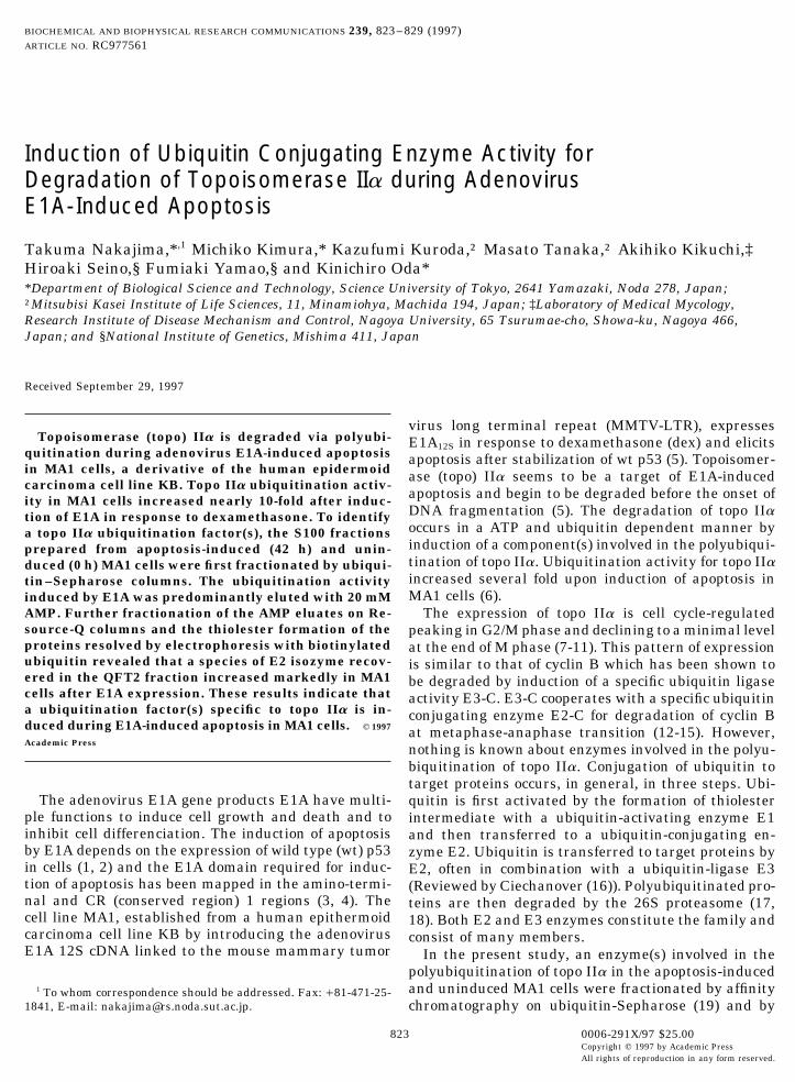
FIG. 1. Degradation of topo IIa during E1A-induced apoptosis.(QFT), the adsorbed proteins were eluted with QB(A) The levels of E1A, p53 and topo IIa in MA1 cells after inductionbuffer containing 0.25 M KCl (QA) and 0.4 M KCl (QB)of E1A. Subconfluent cultures of MA1 cells were treated with 1 mM
dex and cell lysates were prepared by lysing the cells in 21 Laemmli as shown in Fig. 2A. After centrifugal concentration ofsample buffer (20). Aliquots of 50 mg of protein per lane were electro- these fractions using Macrosep 10k, topo IIa ubiquiti-phoresed on 7.5% polyacrylamide gels and the amounts of E1A, p53 nation activity was assayed by Western blotting withand topo IIa were analysed by Western blotting. Monoclonal antibod-
immunoprecipitated topo IIa, GST-ubiquitin and 50 mgies, M73, Bp53–12 and 8D2 were used as primary antibodies, respec-protein of each fraction. The S100-0 extract (S100 pre-tively, and HRP-conjugated anti-mouse IgG was used as the second-
ary antibody. The filters were treated with ECL-detection system pared from the 0 h extract, 100 mg of protein) was addedand exposed to X-ray film. Viable cell numbers were determined by to all the reaction mixture as a source of ubiquitinationtrypan blue exclusion. (B) Ubiquitination of topo IIa. The MA1 cell enzymes. After the reaction at 307C for 15 min, thelysates were similarly prepared at the times indicated. Z-LLnV (100
mixture was subjected to SDS-PAGE. Polyubiquiti-mM) was added to the medium 30 min prior to the cell harvest.nated form of topo IIa was detected by using anti-GSTAliquots of extract containing 5 mg (0, 36 h) or 100 mg (40, 44, 48 h)
of proteins were electrophoresed. Topo IIa was detected by Western polyclonal antibody Z-5 and unprocessed topo IIa byblotting as stated above. using monoclonal antibody 8D2. The amounts of polyu-
biquitinated topo IIa were quantitated by densitometerscanning. The specific activity was shown by theamount of ubiquitinated topo IIa formed. The amountand ubiquitination of topo IIa in MA1 cells were fol-formed with 50 mg protein in S100-0 was taken as 1lowed after induction of E1A by the treatment withand the activities in other fractions are shown as rela-dexamethasone (dex) (Fig. 1). Following expression oftive values in Fig. 2C. The ubiquitination activity ofE1A and subsequent stabilization of p53, the level ofS100-42 was eight times higher than that of S100-0.topo IIa began to decrease steeply after about 30 h,Most of the activity in S100-0 was eluted in QA andpreceding the loss of cell viability (Fig. 1A) as pre-QB, while the activity in S100-42 was recovered in allviously shown (5). Ubiquitination of topo IIa was as-the fractions. The activity recovered in QB was highersayed by Western blotting with monoclonal antibodythan those recovered in QFT and QA. The activities in8D2 specific to topo IIa (Fig. 1B). The cell lysates wereQFT-42 and QB-42 were 10 and 5 fold higher thanprepared at the times indicated immediately after incu-those in QFT-0 and QB-0, respectively, suggesting thebation of the cells with 100 mM of proteasome inhibitor,induction of more than one ubiquitination factors aftercarbobenzoxy-L-leucyl-L-leucyl-L-norvalynal (Z-LLnV,expression of E1A.also known as MG115) for 30 min and boiled in 21
Laemmli sample buffer. To normalize the amounts of Separation of topo IIa Ubiquitination Activities bytopo IIa, 20-fold more amounts of proteins were electro-Affinity Chromatographyphoresed, when the cell lysates were prepared after 40
h of dex treatment. As shown in Fig. 1B, the thick To identify factors responsible for ubiquitination ofbands of topo IIa with molecular weight of 170 kDa topo IIa, both S100-0 and S100-42 were applied to ubi-was predominantly seen in the 0 h and 36 h lysates, quitin-Sepharose columns in the presence of ATP. Thewhile the ladder of slow migrating bands became visi- adsorbed proteins were sequentially eluted with highble above the unprocessed topo IIa in the 40 h lysate. salt (1 M KCl), 20 mM AMP, 10 mM DTT and pH9The ladder was also visible in the 44 h and 48 h lysates, solution as shown in Fig. 3A. Most of ubiquitin-activat-but the amounts decreased steeply along with the pro- ing enzyme E1 is recovered in the AMP eluate, becausegression of the apoptotic process. These slow migrating E1 binds to ubiquitin only in the presence of ATP (22).bands could not be detected, when the cells were not DTT dissociates E1 and ubiquitin-conjugating en-
zymes E2s that bound to ubiquitin through thiolestertreated with Z-LLnV before preparation of lysate.
825
AID BBRC 7561 / 693c$$$681 10-21-97 10:13:59 bbrcg AP: BBRC

Vol. 239, No. 3, 1997 BIOCHEMICAL AND BIOPHYSICAL RESEARCH COMMUNICATIONS
FIG. 2. Fractionation of topo IIa ubiquitination activity by Resource-Q column. (A) Protein elution profile of Resource-Q column chroma-tography. The S100 extracts were prepared from MA1 cells (S100–0) and MA1 cells treated with 1 mM dex for 42 h (S100-42). Aliquots of60 mg of protein were applied to the columns and fractions of 2 ml were collected. QFT, flowthrough fraction; QA, QB, fractions eluted withQB buffer containing 0.25 M and 0.4 M KCl, respectively. (B) Topo IIa ubiquitination activity. Aliquots of immunoprecipitated topo IIawere incubated in S100-0, S100-42, QFT, QA and QB fractions (each 50 mg of protein) in the presence of 10 mg of GST-ubiquitin and 100mg protein of S100-0 as a source of ubiquitination enzymes at 307C for 15 min. Ubiquitinated and unprocessed topo IIas were detected byWestern blotting with rabbit anti-GST antibody Z-5 and anti-topo IIa monoclonal antibody 8D2. (C) Ubiquitinated topo IIa was quantitatedby densitometer scanning. The specific activity was defined as 1 with the amount of polyubiquitinated topo IIa formed with 50 mg of proteinin S100-0 and the activities in other fractions are shown as relative values.
bond. Some E2 isozymes are also eluted in the AMP Identification of an E2 Isozyme Induced in MA1 Cellsand pH9 eluates (22). Under the conditions, most of after Expression of E1Aubiquitin-ligases E3s were recovered in the flow-
To confirm the induction of an E2 isozyme(s) in MA1through fraction (22).cells after expression of E1A, both AMP eluates recov-Each fraction was concentrated by centrifugal ultra-ered from S100-0 and S100-42 were further fraction-filtration, and an aliquot of 1/500 volume was assayedated on Resource-Q column and QFT-2, QA2 and QB2for the ubiquitination activity with immunoprecipi-fractions were collected. After concentration, aliquotstated topo IIa and GST-ubiuqitin. A fraction of S100-of 1/20 volume of these fractions were subjected to SDS-0 (100 mg of protein) was added as a source of ubiquiti-PAGE and the proteins resolved were silver-stainednation enzymes. As shown in Fig. 3B, the majority of(Fig. 4A). The slowest migrating thick band in QA2the activity was not adsorbed, however, a significantfractions of S100-0 and S100-42 seemed to be E1 judg-fraction of the activity was eluted in the AMP, DTTing from its molecular weight of about 120 kDa (lanesand pH9 fractions with a great increase in the specific2 and 5). The multiple bands migrating within theactivity (Fig. 3C). Although the activity recovered inrange of molecular weights 15 to 60 kDa correspondthese fractions was somewhat variable, most of the ac-to heterogenous species of E2 isozymes. Most of thesetivities of S100-0 and S100-42 were eluted in the AMPbands were recovered in QFT2 and QA2 fractionsand DTT fractions. The activity eluted in the AMP frac-(lanes 1, 2, 4 and 5) and very little in QB2. The presencetion of S100-42 was nearly 10-fold higher than thatof E1 and E2s in these bands was detected by ubiquitin-eluted in the AMP fraction of S100-0. Since E3s haveconjugation assay, since these enzymes form thiolesterbeen shown not to be eluted in these fractions (22),bond with ubiquitin in the presence of ATP. The reac-the result suggests that a species of E2 responsibletion was performed with 1/20 volume of each eluatefor ubiquitination of topo IIa is induced during E1A-
induced apoptosis. in the presence of ATP and biotinylated ubiquitin. A
826
AID BBRC 7561 / 693c$$$681 10-21-97 10:13:59 bbrcg AP: BBRC

Vol. 239, No. 3, 1997 BIOCHEMICAL AND BIOPHYSICAL RESEARCH COMMUNICATIONS
FIG. 3. Fractionation of topo IIa ubiquitination activity by affinity column. (A) Ubiquitin–Sepharose chromatography of S100-0 andS100-42. Aliquots of 15 mg of protein were applied to the columns. The flowthorugh (UFT) and KCl, AMP, DTT and pH9 eluates werecollected. Elution profile of S100-42 proteins is shown. (B) A 1/500 volume of flowthrough and each eluate was mixed with 100 mg proteinof S100-0 as a source of ubiquitination enzymes and topo IIa ubiquitination activity was assayed as described in Fig. 2B. (C) The specificactivity of each fraction was determined as described in Fig. 2C.
fraction of 1/200 volume of QA2 from S100-0 was added matin in the nuclei. The expression of topo IIa is cellto QFT2 and QB2 fractions to supply E1, because the cycle-regulated, peaking in G2/M phase and decliningformation of ubiquitin-E2 conjugates requires E1. As to a minimal level at the end of M phase. Althoughshown in Fig. 4B, thiolester formation was predomi- polyubiquitination of topo IIa occurred only slightlynantly observed with the slowest migrating band of E1 with the uninduced MA1 cell extract, there presents ain QA2 and the heterogenous bands of E2s in all the significant activity in the extract, suggesting that thefractions. Among them, the band of about 31 kDa in level of topo IIa in the cell cycle may be regulated byQFT2 of the 42 h AMP eluate (lane 4) was prominent the ubiquitin-proteasome system. The ubiquitinationas indicated by the arrow. The same band was scarcely activity of topo IIa in MA1 cells increased nearly 10-detected in QFT2 of the 0 h AMP eluate (lane 1). This fold after induction of E1A and subsequent stabiliza-species of E2 may correspond to either of the two bands tion of wt p53. Topo IIa was then degraded steeplymigrated in moleculer mass range of 20 to 18 kDa (Fig. before the onset of DNA fragmentation characteristics4A, indicated by the arrow), since moleculer mass of of apoptosis.the biotinylated ubiquitin moiety of the 31 kDa protein A new component(s) responsible for ubiquitination ofis about 12 kDa. These bands too were prominent in topo IIa seems to be induced in MA1 cells after expres-QFT2 of the 42 h AMP eluate (lane 4) as compared sion of E1A, since the elution patterns of the ubiquiti-with those in QFT2 of the 0 h AMP eluate (lane 1). nation activity from ubiquitin-Sepharose columns wereAlthough the involvement of this E2 isozyme in polyu- consistently different between S100-0 and S100-42 pre-biquitination of topo IIa is presently unknown, the re- pared from MA1 cells untreated or treated with dex forsults presented here clearly show the induction of topo 42 h. The activity in the AMP eluate of S100-42 wasIIa ubiquitination factors including a species of E2 dur- nearly 10-fold higher than that in the AMP eluate ofing E1A-induced apoptosis. S100-0 (Fig. 3B, C). As previously shown (22), most of
E1 and a significant fraction of E2 isozymes are elutedDISCUSSION in the AMP fraction, while most of E3 isozymes were
not adsorbed to ubiquitin-Sepharose, E2 isozymes recov-ered in the AMP eluates were further fractionated onTopo IIa is a component of nuclear matrix (23, 24)
which determines the topological organization of chro- Resource-Q columns. The presence of E1 and E2s in
827
AID BBRC 7561 / 693c$$$681 10-21-97 10:13:59 bbrcg AP: BBRC

Vol. 239, No. 3, 1997 BIOCHEMICAL AND BIOPHYSICAL RESEARCH COMMUNICATIONS
FIG. 4. Resource-Q chromatography of ubiquitination factors in AMP eluates. (A) Silver staining of proteins. The AMP eluates of S100-0 and S100-42 from ubiquitin-Sepharose columns (each 40 mg of protein) were applied to 1-ml Resource-Q columns. Aliquots of 1/20 volumeof the flowthrough (QFT2), and the 0.25 M and 0.4 M KCl eluates (QA2 and QB2) were electrophoresed on 8%/14% stepwise gradientacrylamide gels and the proteins resolved were stained with 2D-silver staining reagent (Daiichi). (B) Thiolester formation of the proteinswith biotinylated ubiquitin. Aliquots of 1/20 volume of each eluate was incubated in 10 ml of QB0.5 buffer with 4 mg of biotinylated ubiquitin,2 mM ATP and 1/200 volume of QA2 to supply E1 at 187C for 30 min. Ubiquitin conjugated proteins were electrophoresed, transferred tothe nitrocellulose filter and detected by ECL system using peroxydase conjugated streptavidin. The arrows indicate an E2 isozyme inducedin MA1 cells after expression of E1A.
the fractions was detected by conjugation of biotinylated and the induction of this species of E2 is caused by E1Aor p53 remains to be solved.ubiquitin followed by electrophoresis (Fig. 4B). E1 was
eluted exclusively in the QA2 fraction and a species ofE2 isozyme increased markedly in the QFT2 fraction of ACKNOWLEDGMENTthe 42 h AMP eluate (Fig. 4B). This species may corre-spond to either of the two bands migrated in moleculer We thank Drs. H. Tanaka and H. Yasuda for helpful suggestion
for use of biotinylated ubiquitin. This work was supported by a grant-mass range corresponding to 20 to 18 kDa (Fig. 4A),in-aid from the ministry of Education, Science and Culture of Japan.since the biotinylated ubiquitin moiety of the 31 kDa
band corresponds to molecular mass of about 12 kDa.A topo IIa specific E3 isozyme may also be induced REFERENCES
in MA1 cells after E1A expression, since the topo IIa1. Debbas, M., and White, E. (1993) Genes & Dev. 7, 546–554.ubiquitination activity of S100-42 eluted in the QB2. Lowe, S. W., and Ruley, H. E. (1993) Genes & Dev. 7, 535–545.fraction from Resource-Q column was consistently
higher than that of S100-0 eluted in the same fraction. 3. White, E., Cipriani, R., Sabbatini, P., and Denton, A. (1991) J.Virol. 65, 2968–2978.It has been shown that a cyclin B specific E3 isozyme
4. Mymryk, J. S., Shire, K., and Bayley, S. T. (1994) Oncogene 9,E3-C was eluted in the QB fraction from Resource-Q1187–1193.column (14, 15). The levels of both cyclin B and topo
5. Nakajima, T., Ohi, N., Arai, T., Nozaki, N., Kikuchi, A., and Oda,IIa are steeply reduced at the end of M phase.K. (1995) Oncogene 10, 651–662.We recently found that derivatives of KB cell lines
6. Nakajima, T., Morita, K., Ohi, N., Arai, T., Nozaki, N., Kikuchi,B12 and T5, which express mutated E1A having a dele- A., Osaka, F., Yamao, F., and Oda, K. (1996) J. Biol. Chem. 271,tion in the apoptotic region at codons 17 to 23 or at 24842–24849.codons 54 to 69 in response to dex, failed to induce 7. Adachi, N., Kobayashi, M., and Koyama, H. (1997) Biochem.apoptosis. No p53 stabilization and topo IIa degrada- Biophys. Res. Commun. 230, 105–109.tion were observed in these cell lines. Whether a spe- 8. Goswami, P. C., Roti Roti, J. L., and Hunt, C. R. (1996) Mol. Cell.
Biol. 16, 1500–1508.cies of E2 induced during E1A-induced apoptosis is ac-tually required for targeting topo IIa for degradation 9. Turley, H., Comley, M., Houlbrook, S., Nozaki, N., Kikuchi, A.,
828
AID BBRC 7561 / 693c$$$681 10-21-97 10:13:59 bbrcg AP: BBRC

Vol. 239, No. 3, 1997 BIOCHEMICAL AND BIOPHYSICAL RESEARCH COMMUNICATIONS
Hickson, I. D., Gatter, K., and Harris, A. L. (1997) Br. J. Cancer 16. Ciechanover, A. (1994) Cell 79, 13–21.75, 1340–1346. 17. Kanayama, H., Tamura, T., Ugai, S., Kagawa, S., Tanahashi,
N., Yoshimura, T., Tanaka, K., and Ichihara, A. (1992) Eur. J.10. Woessner, R. D., Mattern, M. R., Mirabelli, C. K., Johnson, R. K.,Biochem. 206, 567–578.and Drake, F. R. (1991) Cell Growth Differ. 2, 209–214.
18. Goldberg, A. L. (1992) Eur. J. Biochem. 203, 9–23.11. Watt, P. M., and Hickson, I. D. (1994) Biochem. J. 303, 681–695.19. Ciechanover, A., Elias, S., Heller, H., and Hershko, A. (1982) J.12. Hershko, A., Ganoth, D., Sudakin, V., Dahan, A., Coen, L. H.,
Biol. Chem. 257, 2537–2542.Luca, F. C., Ruderman, J. V., and Eytan, E. (1994) J. Biol. Chem.269, 4940–4946. 20. Bradford, M. M. (1976) Anal. Biochem. 72, 248–254.
21. Harlow, E., and Lane, D. (1988) Antibodies: A Labolatory Man-13. Sudakin, V., Ganoth, D., Dahan, A., Heller, H., Hershko, J.,ual, Cold Spring Harbor Laboratory, Cold Spring Harbor, NY.Luca, F. C., Ruderman, J. V., and Hershko, A. (1995) Mol. Biol.
Cell. 6, 185–197. 22. Hershko, A., Heller, H., Elias, S., and Ciechanover, A. (1983) J.Biol. Chem. 258, 8206–8214.14. King, R. W., Peters, J.-M., Tugendreich, S., Rolfe, M., Hieter, P.,
and Kirschner, M. W. (1995) Cell 81, 279–288. 23. Berrios, M., Osheroff, N., and Fisher, P. A. (1985) Proc. Natl.Acad. Sci. USA 82, 4142–4146.15. Aristarkhov, A., Eytan, E., Moghe, A., Admon, A., Hershko, A.,
and Ruderman, J. V. (1996) Proc. Natl. Acad. Sci. USA 93, 4294– 24. Earnshaw, W. C., Halligan, B., Cooke, C. A., Heck, M. M. S., andLiu, L. F. (1985) J. Cell Biol. 100, 1706–1715.4299.
829
AID BBRC 7561 / 693c$$$681 10-21-97 10:13:59 bbrcg AP: BBRC