Embed Size (px)
Citation preview
Influenza A Virus Infection of Human Primary DendriticCells Impairs Their Ability to Cross-Present Antigen toCD8 T CellsAnna Smed-Sorensen*¤, Cecile Chalouni, Bithi Chatterjee, Lillian Cohn, Peter Blattmann, Norihiro Nakamura,
Lelia Delamarre, Ira Mellman*
Genentech, South San Francisco, California, United States of America
Abstract
Influenza A virus (IAV) infection is normally controlled by adaptive immune responses initiated by dendritic cells (DCs). Weinvestigated the consequences of IAV infection of human primary DCs on their ability to function as antigen-presentingcells. IAV was internalized by both myeloid DCs (mDCs) and plasmacytoid DCs but only mDCs supported viral replication.Although infected mDCs efficiently presented endogenous IAV antigens on MHC class II, this was not the case forpresentation on MHC class I. Indeed, cross-presentation by uninfected cells of minute amounts of endocytosed, exogenousIAV was ,300-fold more efficient than presentation of IAV antigens synthesized by infected cells and resulted in astatistically significant increase in expansion of IAV-specific CD8 T cells. Furthermore, IAV infection also impaired cross-presentation of other exogenous antigens, indicating that IAV infection broadly attenuates presentation on MHC class Imolecules. Our results suggest that cross-presentation by uninfected mDCs is a preferred mechanism of antigen-presentation for the activation and expansion of CD8 T cells during IAV infection.
Citation: Smed-Sorensen A, Chalouni C, Chatterjee B, Cohn L, Blattmann P, et al. (2012) Influenza A Virus Infection of Human Primary Dendritic Cells Impairs TheirAbility to Cross-Present Antigen to CD8 T Cells. PLoS Pathog 8(3): e1002572. doi:10.1371/journal.ppat.1002572
Editor: Christopher F. Basler, Mount Sinai School of Medicine, United States of America
Received August 13, 2011; Accepted January 24, 2012; Published March 8, 2012
Copyright: � 2012 Smed Sorensen et al. This is an open-access article distributed under the terms of the Creative Commons Attribution License, which permitsunrestricted use, distribution, and reproduction in any medium, provided the original author and source are credited.
Funding: This study was funded by intramural grants from Genentech, Inc. The funder had no role in study design, data collection and analysis, decision topublish, or preparation of the manuscript. AS-S is a fellow of the Swedish Governmental Agency for Innovation Systems (Vinnova) and the Swedish MedicalSociety.
Competing Interests: CC, BC, NN, LD and IM are employed by Genentech. AS-S, LC and PB were employed by Genentech at the time of the study.
* E-mail: [email protected] (AS-S); [email protected] (IM)
¤ Current address: Department of Microbiology, Tumor and Cell Biology, Karolinska Institutet, Stockholm, Sweden.
Introduction
Influenza A virus (IAV) infection is one of the oldest and most
common diseases known to mankind, estimated to cause 500,000
deaths per year, primarily in infants and elderly [1]. In healthy
humans, IAV infection typically causes brief but often severe
illness. Normally, IAV infection is confined to the airways where
the virus replicates in respiratory epithelial cells [2]. Rapidly,
alveolar macrophages produce pro-inflammatory cytokines and
chemokines, which promote infiltration of peripheral blood
leukocytes to the site of infection [3]. While influx of neutrophils
and secretion of cytokines and chemokines in the lung is a
fundamental defense during the initial stage of infection, the
resulting ‘‘cytokine storm’’ may also contribute to pathogenesis
[4]. However, control and clearance of IAV infection depend on
pathogen-specific adaptive immune responses [5].
The initiation of adaptive immunity relies on dendritic cells
(DCs), professional antigen-presenting cells (APCs) with the capacity
to activate naıve T cells [6]. The two major subsets of human DCs,
myeloid and plasmacytoid DCs (mDCs and pDCs, respectively)
both have antigen-presenting capacity although mDCs are
generally considered to be superior. pDCs are of central importance
in virus infections since they respond rapidly to viruses and secrete
high levels of anti-viral type I interferons [7]. DCs reside in the
epithelia of the upper respiratory tract, the site of entry for IAV, and
are also rapidly mobilized to this site following inhalation of
microbial agents [8–9]. Since there is little evidence of viral
replication in lymphoid tissue, the main source of IAV antigen is
thought to be DCs that exit the respiratory tract and travel to
lymphoid tissue where immune responses are initiated [10–11].
During acute viral infections, activation and expansion of
antigen-specific CD8 T cells are crucial for control and clearance
of infection [5,12–13]. In general, MHC class I molecules (MHCI)
present peptides derived from endogenously synthesized proteins.
Viruses that replicate in DCs can therefore be detected by the
immune system by direct presentation of viral antigens. Since not
all viruses infect DCs, antigen-presentation by uninfected DCs is
thought to occur via cross-presentation, a poorly understood
process unique to DCs where exogenous antigen is loaded on
MHCI in the ER or possibly other intracellular compartments
[14]. Mice lacking CD8a+ DCs are deficient in their capacity to
mount an anti-viral immune response [15] suggesting that cross-
presentation is crucial for a CD8 T cell response against viruses.
On the other hand, it could also suggest that direct presentation by
virus infected CD8a+ DCs is required for CD8 T cell responses.
The relative contributions of direct versus cross-presentation for
the induction of anti-viral CD8 T cell responses have been a topic
of discussion for several years [16–19], and have been compared in
mouse models [19–22]. However, the efficiency of direct versus
cross-presentation of IAV and the potential IAV-mediated
PLoS Pathogens | www.plospathogens.org 1 March 2012 | Volume 8 | Issue 3 | e1002572
suppression of antigen-presentation in human DCs remains an
unresolved topic.
IAV infection often predisposes individuals to secondary
infections, usually bacterial, with higher lethal outcomes than
either infection alone, suggesting that the initial infection affects
the host’s ability to respond to a second pathogen. While the
connections between viral and bacterial infections have been
known for decades [23], the mechanism(s) and modes of
interaction contributing to these effects are poorly understood.
IAV infection clearly suppresses innate immune responses [24–
27], but the extent to which adaptive immune responses are also
affected, and why, remains unclear.
While IAV infection has been studied extensively in animal
models, relatively little is known about how IAV infection affects
the function of human DC subsets due in part to their limited
availability, making such experiments challenging. In mice,
mDCs, rather than pDCs, appear to be responsible for presenting
IAV antigen to CD8 T cells for the priming of anti-IAV immune
responses [28–31]. In humans, it is much less clear what subset(s)
of DCs are important in antigen-presentation during IAV
infection. Human DCs infected with infectious IAV or exposed
to inactivated IAV can activate IAV-specific T cells [32–36],
however, it remains unclear if or how IAV infection of human
DCs affects their function. Here, we investigated the consequences
of IAV infection on the ability of DCs to present IAV antigen, or
other antigens, to autologous T cells using relevant subsets of
primary human DCs.
Results
mDCs but not pDCs are susceptible to influenza A virusinfection
pDCs are known to be more resistant to the cytopathic effect of
IAV than mDCs, suggesting that pDCs are resistant to infection
[36,37]. To extend this observation and to determine any possible
consequences for antigen-presentation, primary human mDCs
and pDCs were exposed to IAV and the frequency of IAV+ DCs
was analyzed. While the frequency of IAV+ mDCs increased over
time, infection in pDCs remained undetectable (Figure 1A). The
IAV+ mDCs reflected the production of newly synthesized viral
proteins rather than enhanced virion uptake since adding the virus
at 4uC, or blocking virus endosomal egress with NH4Cl, inhibited
the appearance of IAV+ DCs (Figure 1B). Interestingly, infectious
IAV was not detected in the supernatant even after 24 hr,
indicating that despite high viral protein production, mDCs did
not support generation of infectious particles (Figure S1A). These
observations were confirmed for several IAV strains (Figure S1B).
To assess whether the lack of infection in pDCs reflected poor
endocytosis of virus, human mDCs and pDCs were exposed to
IAV and analyzed by confocal microscopy. After 1 hr, the
majority of both DC subsets displayed internalized virus
(Figures 1C–D), suggesting that other factors blocked pDC
infection, such as the pDCs’ constitutive expression of the
interferon-inducible antiviral protein MxA (Figures 1E and S2)
[37]. It has previously been shown that expression of MxA renders
cells resistant to IAV infection [38]. We were unable to
knockdown MxA in pDCs using siRNA while maintaining pDC
viability (data not shown). Still, the constitutive high expression of
MxA in pDCs suggests that this protein could aid in the observed
resistance to IAV infection, despite efficient IAV internalization by
pDCs.
pDCs, like mDCs, were nevertheless found to respond to the
presence of IAV even in the absence of the synthesis of virus-
encoded proteins. This was illustrated by comparing the ability of
infectious IAV and non-infectious heat-inactivated (HI) IAV to
trigger DC maturation. Both replicating and HI IAV were
internalized equivalently. In addition, both could fuse with the
endosomal membrane at low pH, as indicated by agglutination
and acid-dependent lysis of chicken red blood cells (data not
shown). As expected, HI IAV did not infect DCs, and infection of
mDCs by replication-competent IAV was blocked by NH4Cl
(Figures 2A–B). Yet, both pDCs and mDCs upregulated MHCI
and MHCII in response to infectious and HI IAV (Figure 2C).
Furthermore, pDCs responded by secreting large amounts of
IFNa (Figure 2D). This was true for several IAV strains (Figure
S3). mDCs also secreted IFNa in response to IAV, although the
levels were 100–1000 fold lower than for pDCs (Figure 2E). pDCs
recognize IAV via TLR [39] while most cells respond to single
stranded RNA viruses via the RIG-I pathway [40]. Human
primary mDCs express TLR7 and TLR8 that recognize single-
stranded RNA. The virus-related TLRs can be stimulated by
inactivated viruses, however, the barely detectable amount of
IFNa secreted by mDCs in response to TLR7/8L suggests that
this was a consequence of signaling via cytoplasmic receptors
rather than via TLRs (Figure 2E). In addition, pDCs secreted
TNFa, IL-6 and MIP-1a in response to IAV, while mDCs
required stimulation with purified TLR7/8 ligand (TLR7/8L) to
secrete significant amounts of cytokines and chemokines (Figure
S4). Thus, IAV enters both mDCs and pDCs and triggers their
maturation, but only mDCs support viral protein synthesis.
mDCs are superior at MHCI restrictedantigen-presentation of IAV compared to pDCs
To determine if infected human mDCs and pDCs could present
antigen to and activate IAV-specific CD8 T cells, we exposed DCs
from HLA-A2+ donors to either infectious IAV or non-infectious
HI IAV and co-cultured them with autologous CFSE-labeled CD8
T cells. After 10 days, the frequency of memory CD8 T cells
specific to the immunodominant influenza M1 (58–66) epitope was
determined and the overall CD8 T cell response assessed by CFSE
dilution. While both mDCs and pDCs could expand IAV-specific
CD8 T cells, mDCs were superior (Figures 3A–B). This difference
Author Summary
Although the interactions between viruses and dendriticcells (DCs) have been studied for many years, surprisinglylittle is known on the functional relationship betweeninfection and antigen presentation in primary human DCs.Here, we asked specifically whether Influenza A virus (IAV)infection of human primary plasmacytoid DCs and myeloidDCs (pDCs and mDCs, respectively) affected their ability tofunction as antigen-presenting cells and activate T cellsspecific to IAV or other antigens. Our data confirm thatpDCs are poorly infected and also present IAV antigenspoorly. mDCs, on the other hand, are readily susceptible toIAV infection and present IAV antigen to T cells. However,we found that MHC class I presentation by mDCs infectedwith IAV are ,300-fold less efficient relative to what mDCsare capable of achieving by cross-presentation followingthe endocytosis of only a very few non-infectious virions.Importantly, IAV infection of mDCs not only reduces theefficiency of IAV presentation but also reduces their abilityto cross-present antigens from other viruses encounteredsubsequently. The reduced overall antigen processingcapacity of mDCs describes a mechanism that maycontribute to the suppression of immunity to secondarypathogens that appear during the course of IAV infection.
IAV Infection of DCs Impairs Cross-Presentation
PLoS Pathogens | www.plospathogens.org 2 March 2012 | Volume 8 | Issue 3 | e1002572
likely reflected different capacities for antigen-processing since
both subsets presented pre-processed peptide (that does not
require cellular processing) similarly (Figure 3C). IAV presentation
to CD8 T cells by pDCs was the same for both infectious and non-
infectious virus, strongly suggesting that pDCs were only capable
of cross-presenting IAV antigens, albeit inefficiently from virions
internalized by endocytosis.
Presentation on MHCI is a hallmark of viral immunity since
infected cells express virus-derived peptides recognized for
elimination by cytotoxic CD8 T cells. In DCs, it is unclear if the
generation of CD8 T cell responses reflects the formation of
peptide-MHCI complexes from endogenously synthesized viral
proteins or the cross-presentation of antigens from exogenous
sources (e.g. internalized virions or infected cells) [41]. Indeed,
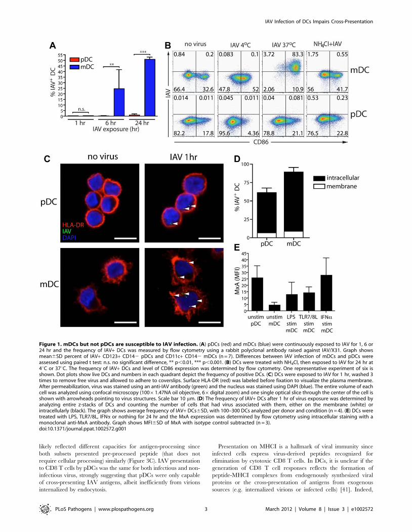
Figure 1. mDCs but not pDCs are susceptible to IAV infection. (A) pDCs (red) and mDCs (blue) were continuously exposed to IAV for 1, 6 or24 hr and the frequency of IAV+ DCs was measured by flow cytometry using a rabbit polyclonal antibody raised against IAV/X31. Graph showsmean6SD percent of IAV+ CD123+ CD142 pDCs and CD11c+ CD142 mDCs (n = 7). Differences between IAV infection of mDCs and pDCs wereassessed using paired t test: n.s. no significant difference, ** p,0.01, *** p,0.001. (B) DCs were treated with NH4Cl, then exposed to IAV for 24 hr at4uC or 37uC. The frequency of IAV+ DCs and level of CD86 expression was determined by flow cytometry. One representative experiment of six isshown. Dot plots show live DCs and numbers in each quadrant depict the frequency of positive DCs. (C) DCs were exposed to IAV for 1 hr, washed 3times to remove free virus and allowed to adhere to coverslips. Surface HLA-DR (red) was labeled before fixation to visualize the plasma membrane.After permeabilization, virus was stained using an anti-IAV antibody (green) and the nucleus was stained using DAPI (blue). The entire volume of eachcell was analyzed using confocal microscopy (10061.47NA oil objective, 66digital zoom) and one single optical slice through the center of the cell isshown with arrowheads pointing to virus structures. Scale bar 10 mm. (D) The frequency of IAV+ DCs after 1 hr of virus exposure was determined byanalyzing entire z-stacks of DCs and counting the number of cells that had virus associated with them, either on the membrane (white) orintracellularly (black). The graph shows average frequency of IAV+ DCs6SD, with 100–300 DCs analyzed per donor and condition (n = 4). (E) DCs weretreated with LPS, TLR7/8L, IFNa or nothing for 24 hr and the MxA expression was determined by flow cytometry using intracellular staining with amonoclonal anti-MxA antibody. Graph shows MFI6SD of MxA with isotype control subtracted (n = 3).doi:10.1371/journal.ppat.1002572.g001
IAV Infection of DCs Impairs Cross-Presentation
PLoS Pathogens | www.plospathogens.org 3 March 2012 | Volume 8 | Issue 3 | e1002572
DCs are well known to have an enhanced capacity for cross-
presentation, which requires that internalized antigens exit the
endosomal pathway for peptide cleavage in the cytosol and
subsequent loading onto MHCI molecules in the ER or elsewhere
[14]. Although HI IAV cannot infect DCs, it retains the capacity
to fuse with the endosomal membrane [32] thus providing an
intrinsic capacity to reach the cytosol, which is possibly the rate-
limiting step in the cross-presentation [42].
Strikingly, mDCs exposed to HI IAV induced a statistically
significant, two-fold more effective expansion of IAV-specific CD8
T cells than mDCs infected with IAV (Figures 3A–B). This was
surprising because it is generally assumed that presentation of
peptides from endogenously synthesized proteins is more efficient
than cross-presentation [19]. In addition, IAV infected DCs
expressed far greater amounts of IAV proteins than DCs exposed
to HI IAV. This was readily apparent (Figures 3D and 2A);
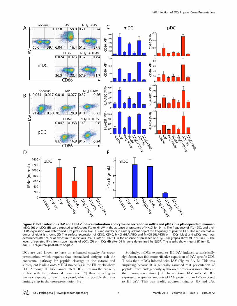
Figure 2. Both infectious IAV and HI IAV induce maturation and cytokine secretion in mDCs and pDCs in a pH-dependent manner.mDCs (A) or pDCs (B) were exposed to infectious IAV or HI IAV in the absence or presence of NH4Cl for 24 hr. The frequency of IAV+ DCs and theirCD86 expression was determined. Dot plots show live DCs and numbers in each quadrant depict the frequency of positive DCs. One representativedonor of eight is shown. (C) The surface expression of CD86, CD40, MHCI (HLA-ABC) and MHCII (HLA-DR) on mDCs (blue) and pDCs (red) wasdetermined after 24 hr of exposure to infectious IAV, HI IAV or TLR7/8L in the absence or presence of NH4Cl. Bar graphs show MFI6SD (n = 3). Thelevels of secreted IFNa from supernatants of pDCs (D) or mDCs (E) after 24 hr were determined by ELISA. The graphs show mean6SD (n = 9).doi:10.1371/journal.ppat.1002572.g002
IAV Infection of DCs Impairs Cross-Presentation
PLoS Pathogens | www.plospathogens.org 4 March 2012 | Volume 8 | Issue 3 | e1002572
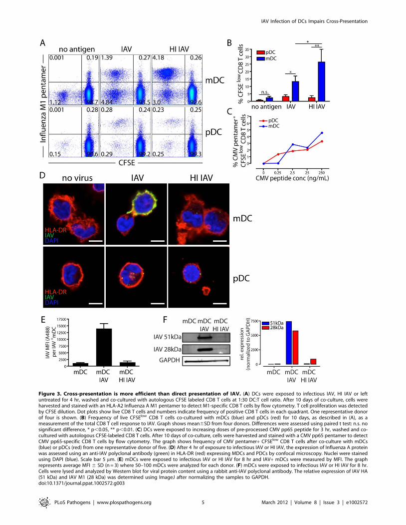
Figure 3. Cross-presentation is more efficient than direct presentation of IAV. (A) DCs were exposed to infectious IAV, HI IAV or leftuntreated for 4 hr, washed and co-cultured with autologous CFSE labeled CD8 T cells at 1:30 DC:T cell ratio. After 10 days of co-culture, cells wereharvested and stained with an HLA-A2 Influenza A M1 pentamer to detect M1-specific CD8 T cells by flow cytometry. T cell proliferation was detectedby CFSE dilution. Dot plots show live CD8 T cells and numbers indicate frequency of positive CD8 T cells in each quadrant. One representative donorof four is shown. (B) Frequency of live CFSElow CD8 T cells co-cultured with mDCs (blue) and pDCs (red) for 10 days, as described in (A), as ameasurement of the total CD8 T cell response to IAV. Graph shows mean6SD from four donors. Differences were assessed using paired t test: n.s. nosignificant difference, * p,0.05, ** p,0.01. (C) DCs were exposed to increasing doses of pre-processed CMV pp65 peptide for 3 hr, washed and co-cultured with autologous CFSE-labeled CD8 T cells. After 10 days of co-culture, cells were harvested and stained with a CMV pp65 pentamer to detectCMV pp65-specific CD8 T cells by flow cytometry. The graph shows frequency of CMV pentamer+ CFSElow CD8 T cells after co-culture with mDCs(blue) or pDCs (red) from one representative donor of five. (D) After 4 hr of exposure to infectious IAV or HI IAV, the expression of Influenza A proteinwas assessed using an anti-IAV polyclonal antibody (green) in HLA-DR (red) expressing MDCs and PDCs by confocal microscopy. Nuclei were stainedusing DAPI (blue). Scale bar 5 mm. (E) mDCs were exposed to infectious IAV or HI IAV for 8 hr and IAV+ mDCs were measured by MFI. The graphrepresents average MFI 6 SD (n = 3) where 50–100 mDCs were analyzed for each donor. (F) mDCs were exposed to infectious IAV or HI IAV for 8 hr.Cells were lysed and analyzed by Western blot for viral protein content using a rabbit anti-IAV polyclonal antibody. The relative expression of IAV HA(51 kDa) and IAV M1 (28 kDa) was determined using ImageJ after normalizing the samples to GAPDH.doi:10.1371/journal.ppat.1002572.g003
IAV Infection of DCs Impairs Cross-Presentation
PLoS Pathogens | www.plospathogens.org 5 March 2012 | Volume 8 | Issue 3 | e1002572
although infected cells stained heavily for IAV proteins, cells
exposed to HI IAV contained only 4–5 virions (defined as IAV+puncta) per cell (Figure S5). We determined the mean fluorescence
intensity (MFI) of the IAV staining in IAV+ mDCs and found that
mDCs infected with infectious IAV displayed 10-fold more IAV
staining than mDCs exposed to HI IAV (Figure 3E). To more
accurately determine the relative content of IAV proteins, we next
analyzed cell lysates of mDCs exposed to infectious or HI IAV by
Western blot. Due to continued synthesis in the infected cells, and
continued degradation of virions in the HI IAV exposed cells, a
quantitative comparison was no more than an estimate. Yet,
influenza proteins were present at 100–1000 fold higher amounts
in infected cells as compared to cells that had internalized HI IAV
(Figure 3F). The 28 kDa band most likely corresponds to the M1
protein, ,3000 copies of which are contained within each virion,
or 15,000 copies per cell after endocytosis of 5 virions. Assuming
all of the HI IAV fuse with the endosome membrane releasing all
of the incoming M1 into the cytosol, we estimate that the
uninfected DCs are at least 300-fold more efficient at stimulating
M1-specific CD8 T cells than infected DCs: i.e. despite vastly
greater amounts of cytosolic M1, IAV infected DCs process and
present M1-derived peptides to CD8 T cells less well. This
difference in antigen processing and presentation translates into
the observed difference in frequency of proliferating IAV-specific
CD8 T cells depicted in Figure 3B.
On the other hand, pDCs exposed to either infectious IAV or
HI IAV were comparable in their ability to expand IAV-specific
CD8 T cells (Figures 3A–B). Comparing presentation of HI IAV
by mDC and pDCs, pDCs were 10–20 fold less effective at cross-
presentation than mDCs (Figures 3A–B). This difference was
consistent over a range of IAV concentrations and DC:T cell
ratios (Figure S6).
Contrary to CD8 T cell responses, CD4 T cells responded
comparably to mDCs exposed to either infectious or HI IAV. As
with presentation on MHCI, mDCs were superior to pDCs for
MHCII-restricted presentation (Figure 4). Thus, IAV infection did
not diminish the efficiency of presentation to CD4 T cells, showing
that the effect on the MHCI pathway was selective.
IAV infected mDCs have an impaired capacity to cross-present other viral antigens
IAV infection often predisposes individuals to secondary
infections, suggesting that infection history affects the ability to
mount adaptive responses to new pathogens. Therefore, we
investigated if uninfected and IAV infected DCs were comparable
in their ability to present a second antigen to CD8 T cells and
support their activation and expansion. The most common
secondary infection in IAV infected individuals is Streptococcus
pneumoniae. However, in the absence of tools to look at potential T
cell responses against S. pneumoniae, we made use of existing
immunodominant CD8 T cell memory responses against EBV and
CMV in HLA-A2+ donors as model antigens. Since pDCs were
not susceptible to IAV infection, we focused on mDCs.
Uninfected and HI IAV exposed mDCs had similar capacities
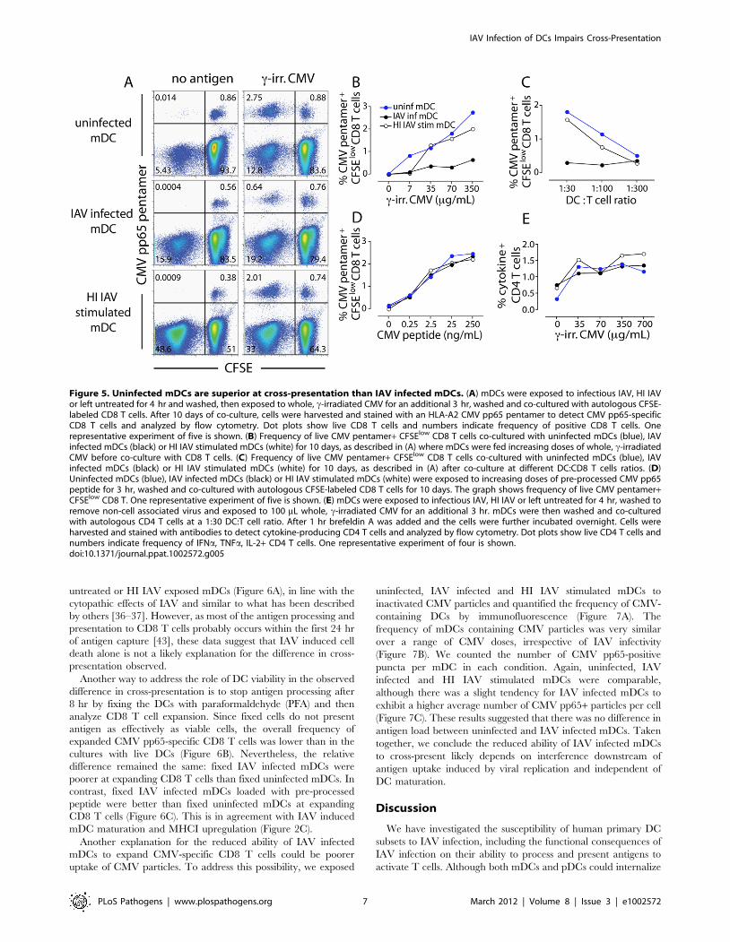
to cross-present CMV to antigen-specific CD8 T memory cells. In
contrast, IAV infected mDCs consistently produced several-fold
lower frequency of proliferating CMV pp65-specific CD8 T cells
(Figure 5A). The impaired ability of IAV infected mDCs to cross-
present inactivated CMV to CD8 T cells was apparent over a
range of CMV concentrations (Figure 5B) and DC:T cell ratios
(Figure 5C). Analysis of the CMV pentamer-negative, CFSElow
population in the absence of exogenous CMV also showed that the
overall T cell response was more pronounced to HI IAV (48.6%)
than to infectious IAV (15.9%) (Figure 5A, left panel). Further-
more, IAV infected and HI IAV stimulated mDCs loaded with
pre-processed CMV peptide were comparable or superior to
uninfected mDCs in their ability to expand CMV-specific CD8 T
cells, consistent with a defect in antigen-processing capacity rather
than in antigen-presentation (Figure 5D). Similar results were
observed for cross-presentation of HI EBV or EBV infected cell
extract (Figure S7). This suggests that IAV infected mDCs have an
impaired capacity to cross-present both different sources of antigen
(CMV and EBV) as well as different forms of antigen (inactivated
virus and virus infected cells) to CD8 T cells as compared to
uninfected mDCs.
We also compared the ability of uninfected, IAV infected, and
HI IAV stimulated mDCs to present CMV to autologous CD4 T
cells. Unlike cross-presentation to CD8 T cells, IAV infected and
HI IAV exposed mDCs stimulated CMV-specific CD4 responses
similarly (Figure 5E). This again indicates that IAV infected mDCs
can function as APCs in general but that IAV infection selectively
impairs the ability to cross-present antigen on MHCI to CD8 T
cells.
Neither differential cellular viability nor antigen loadexplain the decreased ability of IAV infected mDCs tocross-present
One explanation for the decreased ability of IAV infected
mDCs to cross-present could be IAV-induced DC death. This
explanation appeared unlikely since presentation of pre-processed
peptide was similar between uninfected and IAV infected mDCs
(Figure 5D), and IAV infected mDCs could activate CD4 T cells
(Figures 4A and 5A). Assessing the viability of the mDCs in the co-
cultures after 10 days, the time at which we measure T cell
activation, is a challenge since DCs are in great minority and the
majority of T cells that have not seen their cognate antigen have
died. To investigate the role of IAV mediated cell death, mDCs
were exposed to infectious IAV or HI IAV and the frequency of
dead mDCs was compared to untreated mDCs by Annexin V
staining. The viability of all mDCs was comparable at 2 hr and
6 hr after virus exposure (Figure 6A). At later time points (days),
the viability of IAV infected mDCs was reduced compared to
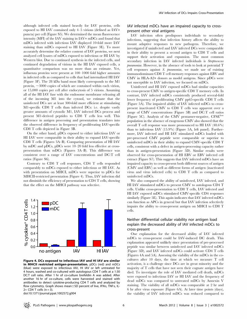
Figure 4. DCs exposed to infectious IAV and HI IAV are similarin MHCII restricted antigen-presentation. pDCs (red) and mDCs(blue) were exposed to infectious IAV, HI IAV or left untreated for4 hours, washed and co-cultured with autologous CD4 T cells at a 1:30DC:T cell ratio. After 1 hr of co-culture brefeldin A was added. Afteranother 16 hr of co-culture, cells were harvested and stained withantibodies to detect cytokine-producing CD4 T cells and analyzed byflow cytometry. Graph shows mean6SD percent of live, IFNa, TNFa, IL-2+ CD4 T cells (n = 3).doi:10.1371/journal.ppat.1002572.g004
IAV Infection of DCs Impairs Cross-Presentation
PLoS Pathogens | www.plospathogens.org 6 March 2012 | Volume 8 | Issue 3 | e1002572
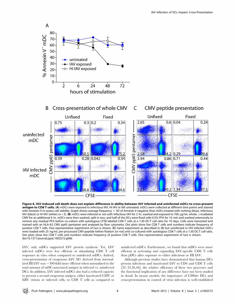
untreated or HI IAV exposed mDCs (Figure 6A), in line with the
cytopathic effects of IAV and similar to what has been described
by others [36–37]. However, as most of the antigen processing and
presentation to CD8 T cells probably occurs within the first 24 hr
of antigen capture [43], these data suggest that IAV induced cell
death alone is not a likely explanation for the difference in cross-
presentation observed.
Another way to address the role of DC viability in the observed
difference in cross-presentation is to stop antigen processing after
8 hr by fixing the DCs with paraformaldehyde (PFA) and then
analyze CD8 T cell expansion. Since fixed cells do not present
antigen as effectively as viable cells, the overall frequency of
expanded CMV pp65-specific CD8 T cells was lower than in the
cultures with live DCs (Figure 6B). Nevertheless, the relative
difference remained the same: fixed IAV infected mDCs were
poorer at expanding CD8 T cells than fixed uninfected mDCs. In
contrast, fixed IAV infected mDCs loaded with pre-processed
peptide were better than fixed uninfected mDCs at expanding
CD8 T cells (Figure 6C). This is in agreement with IAV induced
mDC maturation and MHCI upregulation (Figure 2C).
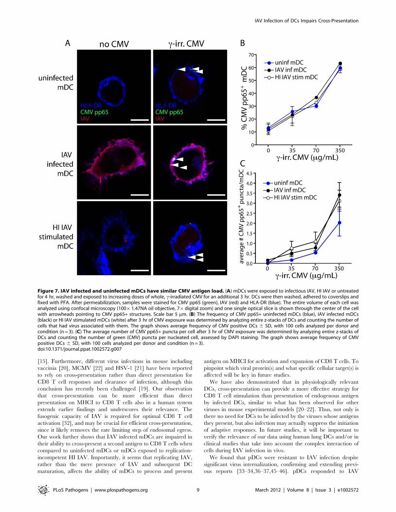
Another explanation for the reduced ability of IAV infected
mDCs to expand CMV-specific CD8 T cells could be poorer
uptake of CMV particles. To address this possibility, we exposed
uninfected, IAV infected and HI IAV stimulated mDCs to
inactivated CMV particles and quantified the frequency of CMV-
containing DCs by immunofluorescence (Figure 7A). The
frequency of mDCs containing CMV particles was very similar
over a range of CMV doses, irrespective of IAV infectivity
(Figure 7B). We counted the number of CMV pp65-positive
puncta per mDC in each condition. Again, uninfected, IAV
infected and HI IAV stimulated mDCs were comparable,
although there was a slight tendency for IAV infected mDCs to
exhibit a higher average number of CMV pp65+ particles per cell
(Figure 7C). These results suggested that there was no difference in
antigen load between uninfected and IAV infected mDCs. Taken
together, we conclude the reduced ability of IAV infected mDCs
to cross-present likely depends on interference downstream of
antigen uptake induced by viral replication and independent of
DC maturation.
Discussion
We have investigated the susceptibility of human primary DC
subsets to IAV infection, including the functional consequences of
IAV infection on their ability to process and present antigens to
activate T cells. Although both mDCs and pDCs could internalize
Figure 5. Uninfected mDCs are superior at cross-presentation than IAV infected mDCs. (A) mDCs were exposed to infectious IAV, HI IAVor left untreated for 4 hr and washed, then exposed to whole, c-irradiated CMV for an additional 3 hr, washed and co-cultured with autologous CFSE-labeled CD8 T cells. After 10 days of co-culture, cells were harvested and stained with an HLA-A2 CMV pp65 pentamer to detect CMV pp65-specificCD8 T cells and analyzed by flow cytometry. Dot plots show live CD8 T cells and numbers indicate frequency of positive CD8 T cells. Onerepresentative experiment of five is shown. (B) Frequency of live CMV pentamer+ CFSElow CD8 T cells co-cultured with uninfected mDCs (blue), IAVinfected mDCs (black) or HI IAV stimulated mDCs (white) for 10 days, as described in (A) where mDCs were fed increasing doses of whole, c-irradiatedCMV before co-culture with CD8 T cells. (C) Frequency of live CMV pentamer+ CFSElow CD8 T cells co-cultured with uninfected mDCs (blue), IAVinfected mDCs (black) or HI IAV stimulated mDCs (white) for 10 days, as described in (A) after co-culture at different DC:CD8 T cells ratios. (D)Uninfected mDCs (blue), IAV infected mDCs (black) or HI IAV stimulated mDCs (white) were exposed to increasing doses of pre-processed CMV pp65peptide for 3 hr, washed and co-cultured with autologous CFSE-labeled CD8 T cells for 10 days. The graph shows frequency of live CMV pentamer+CFSElow CD8 T. One representative experiment of five is shown. (E) mDCs were exposed to infectious IAV, HI IAV or left untreated for 4 hr, washed toremove non-cell associated virus and exposed to 100 mL whole, c-irradiated CMV for an additional 3 hr. mDCs were then washed and co-culturedwith autologous CD4 T cells at a 1:30 DC:T cell ratio. After 1 hr brefeldin A was added and the cells were further incubated overnight. Cells wereharvested and stained with antibodies to detect cytokine-producing CD4 T cells and analyzed by flow cytometry. Dot plots show live CD4 T cells andnumbers indicate frequency of IFNa, TNFa, IL-2+ CD4 T cells. One representative experiment of four is shown.doi:10.1371/journal.ppat.1002572.g005
IAV Infection of DCs Impairs Cross-Presentation
PLoS Pathogens | www.plospathogens.org 7 March 2012 | Volume 8 | Issue 3 | e1002572
IAV, only mDCs supported IAV protein synthesis. Yet, IAV
infected mDCs were less efficient at stimulating CD8 T cell
responses in vitro when compared to uninfected mDCs. Indeed,
cross-presentation of exogenous IAV M1 derived from internal-
ized HI IAV was ,300-fold more efficient when normalized to the
total amount of mDC-associated antigen in infected vs. uninfected
DCs. In addition, IAV infected mDCs also had a reduced capacity
to present a second exogenous antigen, either inactivated CMV or
EBV virions or infected cells, to CD8 T cells as compared to
uninfected mDCs. Furthermore, we found that mDCs were more
efficient at activating and expanding IAV-specific CD8 T cells
than pDCs after exposure to either infectious or HI IAV.
Although previous studies have demonstrated that human DCs
present infectious and inactivated IAV to CD4 and CD8 T cells
[32–33,36,44], the relative efficiency of these two processes and
the functional implication of any difference have not been studied
in detail. In mouse models, the importance of CD8a+ DCs and
cross-presentation in control of virus infection is well-established
Figure 6. IAV-induced cell death does not explain difference in ability between IAV infected and uninfected mDCs to cross-presentantigen to CD8 T cells. (A) mDCs were exposed to infectious IAV, HI IAV or left untreated. mDCs were collected at different time points and stainedwith Annexin V to assess cell viability. Graph shows average frequency 6 SD of Annexin V negative (live) mDCs treated with nothing (blue), infectiousIAV (black) or HI IAV (white) (n = 3). (B) mDCs were infected or not with infectious IAV for 2 hr, washed and exposed to 700 mg/mL whole, c-irradiatedCMV for an additional 6 hr. mDCs were then washed, split in two, and half of the DCs were fixed with 0.5% PFA for 10 min and washed extensively toremove any residual PFA before co-culture with autologous CFSE-labeled CD8 T cells at a 1:30 DC:T cell ratio for 10 days. Cells were harvested andstained with an HLA-A2 CMV pp65 pentamer and analyzed by flow cytometry. Dot plots show live CD8 T cells and numbers indicate frequency ofpositive CD8 T cells. One representative experiment of two is shown. (C) Same experiment as described in (B) but uninfected or IAV infected mDCswere loaded with 25 ng/mL pre-processed CMV peptide before fixation (or not) and co-cultured with autologous CD8 T cells at a 1:30 DC:T cell ratio.Dot plots show live CD8 T cells and numbers indicate frequency of positive CD8 T cells. One representative experiment of two is shown.doi:10.1371/journal.ppat.1002572.g006
IAV Infection of DCs Impairs Cross-Presentation
PLoS Pathogens | www.plospathogens.org 8 March 2012 | Volume 8 | Issue 3 | e1002572
[15]. Furthermore, different virus infections in mouse including
vaccinia [20], MCMV [22] and HSV-1 [21] have been reported
to rely on cross-presentation rather than direct presentation for
CD8 T cell responses and clearance of infection, although this
conclusion has recently been challenged [19]. Our observation
that cross-presentation can be more efficient than direct
presentation on MHCI to CD8 T cells also in a human system
extends earlier findings and underscores their relevance. The
fusogenic capacity of IAV is required for optimal CD8 T cell
activation [32], and may be crucial for efficient cross-presentation,
since it likely removes the rate limiting step of endosomal egress.
Our work further shows that IAV infected mDCs are impaired in
their ability to cross-present a second antigen to CD8 T cells when
compared to uninfected mDCs or mDCs exposed to replication-
incompetent HI IAV. Importantly, it seems that replicating IAV,
rather than the mere presence of IAV and subsequent DC
maturation, affects the ability of mDCs to process and present
antigen on MHCI for activation and expansion of CD8 T cells. To
pinpoint which viral protein(s) and what specific cellular target(s) is
affected will be key in future studies.
We have also demonstrated that in physiologically relevant
DCs, cross-presentation can provide a more effective strategy for
CD8 T cell stimulation than presentation of endogenous antigen
by infected DCs, similar to what has been observed for other
viruses in mouse experimental models [20–22]. Thus, not only is
there no need for DCs to be infected by the viruses whose antigens
they present, but also infection may actually suppress the initiation
of adaptive responses. In future studies, it will be important to
verify the relevance of our data using human lung DCs and/or in
clinical studies that take into account the complex interaction of
cells during IAV infection in vivo.
We found that pDCs were resistant to IAV infection despite
significant virus internalization, confirming and extending previ-
ous reports [33–34,36–37,45–46]. pDCs responded to IAV
Figure 7. IAV infected and uninfected mDCs have similar CMV antigen load. (A) mDCs were exposed to infectious IAV, HI IAV or untreatedfor 4 hr, washed and exposed to increasing doses of whole, c-irradiated CMV for an additional 3 hr. DCs were then washed, adhered to coverslips andfixed with PFA. After permeabilization, samples were stained for CMV pp65 (green), IAV (red) and HLA-DR (blue). The entire volume of each cell wasanalyzed using confocal microscopy (10061.47NA oil objective, 76digital zoom) and one single optical slice is shown through the center of the cellwith arrowheads pointing to CMV pp65+ structures. Scale bar 5 mm. (B) The frequency of CMV pp65+ uninfected mDCs (blue), IAV infected mDCs(black) or HI IAV stimulated mDCs (white) after 3 hr of CMV exposure was determined by analyzing entire z-stacks of DCs and counting the number ofcells that had virus associated with them. The graph shows average frequency of CMV positive DCs 6 SD, with 100 cells analyzed per donor andcondition (n = 3). (C) The average number of CMV pp65+ puncta per cell after 3 hr of CMV exposure was determined by analyzing entire z-stacks ofDCs and counting the number of green (CMV) puncta per nucleated cell, assessed by DAPI staining. The graph shows average frequency of CMVpositive DCs 6 SD, with 100 cells analyzed per donor and condition (n = 3).doi:10.1371/journal.ppat.1002572.g007
IAV Infection of DCs Impairs Cross-Presentation
PLoS Pathogens | www.plospathogens.org 9 March 2012 | Volume 8 | Issue 3 | e1002572
exposure by secreting large amounts of IFNa, but showed only
modest upregulation of co-stimulatory molecules compared to
TLR7/8L stimulation. In our hands, pDCs were less potent than
mDCs at inducing CD8 T cell activation after acquiring a large
antigen that requires processing into peptides before loading onto
MHCI. This was probably due to a lower expression of co-
stimulatory molecules and therefore weaker DC-T cell interaction
and/or a reduced capacity to process large antigens compared to
mDCs, rather than a lack of viral antigen available for
presentation, as pDCs carrying abundant IAV NP are unable to
activate IAV-specific T cells [30]. Previous studies have reported
that human pDCs are similar [36] or superior [35] in their ability
to present antigen to CD8 T cells as compared to human mDCs.
The lack of clear consensus may partly be explained by differences
in maturation/phenotype of the pDCs as well as length of
exposure, and dose of IAV, as it has recently been reported that
the timing of pDC stimulation and route of antigen uptake affect
the ability of pDCs to present antigens [47].
It is well documented that IAV infection renders infected
individuals more prone to secondary bacterial infections, but the
importance of CD8 T cell response to control and clear
extracellular bacterial infections is unclear. IAV infection has an
immunomodulatory effect that is thought to promote an increased
susceptibility to secondary infections [24,26]. The impact of IAV
induced immunomodulation combined with an impaired ability to
cross-present subsequently encountered antigens might act
together to compromise a proper immune response to secondary
pathogens. Systemic injection of TLR ligands results in reduced
cross-presentation of a subsequently encountered antigen [18].
While this was suggested to be a consequence of systemic DC
maturation, we recently showed that reduced antigen-presentation
in vivo after systemic TLR injection could also be a consequence
of the antigen not reaching DCs in the spleen due to alterations in
splenic blood flow [48]. Previous studies using monocyte-derived
DCs have shown that IAV infection induces suboptimal
maturation of the cells with respect to upregulation of co-
stimulatory molecules and secretion of cytokines as compared to
LPS stimulation [49]. Using recombinant IAV that did not encode
the multifunctional viral protein NS1, the authors found that NS1
has an inhibitory effect on expression of several genes involved in
monocyte-derived DC maturation and migration, including the
pro-inflammatory cytokines IL-6 and TNFa [49]. In our hands,
primary mDCs do not show a defect in their ability to upregulate
co-stimulatory molecules in response to IAV as compared to TLR
stimulation, but did indeed show lower secretion of pro-
inflammatory cytokines. In any event, altered DC maturation
seems unlikely to fully explain the defect in mDC cross
presentation of a second antigen, since mDCs stimulated with
HI IAV, which mature to the same extent as IAV infected mDCs,
were found to retain their ability to cross-present. In future studies,
it will be important to use recombinant IAV strains in which
different viral proteins have been mutated or deleted to study their
potential impact on DC maturation and antigen-presentation on
the protein level, as well as in functional assays as outlined in the
present study.
Finally, our findings shed light on how uninfected human DCs,
rather than IAV infected DCs, may be crucial for processing and
presentation of IAV antigen to initiate anti-viral immunity. Even if
infected DCs can present antigen, cross-presentation facilitated by
uninfected DCs may be sufficient or even required for induction of
anti-viral immune responses. While the in vivo situation is much
more complicated, the in vitro results presented here do create the
conceptual possibility that the same situation applies in vivo.
Indeed, it was unexpected that uninfected mDCs cross-present
viral antigens more efficiently than IAV infected DCs present
endogenously synthesized antigens. Thus, IAV infection not only
inhibits cross-presentation of subsequently encountered antigens,
but also acts to diminish direct presentation. As a result, it is now
of interest to determine the mechanism of both forms of inhibition.
Besides DC death, other potential contributors may include the
partial reduction in host cell protein synthesis following IAV
infection or a direct inactivation of the antigen processing
machinery, as observed for medium to large DNA viruses that
cause chronic infections [50–51]. As discussed above, the
multifunctional IAV protein NS1 is an important virulence factor
associated with the suppression of innate immunity [52–54]. The
major function of NS1 is to antagonize the type I IFN mediated
host response. Current evidence suggests that NS1 can limit IFNbproduction both on the pre- and post-transcriptional level. While
most IAV strains can utilize both strategies, some strains may have
lost one of these mechanisms naturally or as a consequence of
passage in the laboratory [53]. NS1 not only prevents the
activation of IRF3, a transcription factor involved in IFNbinduction (pre-transcriptional), but can also block the expression of
cellular genes such as MxA at the post-transcriptional level, and
thereby IFN gene expression. In contrast to more recent human
strains of IAV like A/TX/91 (TX), NS1 expressed by A/PR/8
(PR8), a widely used laboratory IAV strain, can only limit pre-
transcriptional events of IFNb induction [54]. Monocyte-derived
DCs infected with IAV/TX displayed higher viral replication but
reduced capacity to induce IFNc secretion in allogeneic naive
CD4 T cells compared to monocyte-derived DCs infected with
IAV/PR8 [55]. Monocyte-derived DCs infected with NS1 deleted
versions of the two virus strains were comparable in their ability to
induce IFNc secretion in allogeneic naive CD4 T cells [55],
suggesting that a more recent human isolate of IAV (TX) is a more
potent inhibitor of DC function than a laboratory adapted strain
(PR8). In addition, recent data using human lung epithelial cells
indicate that NS1 specifically suppresses the expression of several
genes involved in IFN-stimulated MHCI antigen presentation and
immune-proteasome activation during IAV infection [56]. Anoth-
er potential viral protein to consider in this context is the most
recently discovered IAV protein, PB1-F2 [57]. PB1-F2 is a
virulence factor described to contribute to pathogenesis of IAV as
well as secondary bacterial infections [58–60]. Taken together,
these studies have contributed significantly to our initial under-
standing of how individual IAV proteins may impact the immune
response to IAV and they also highlight the importance of
studying a wider selection of IAV strains. Whether NS1 and/or
PB1-F2 also affect the ability of IAV infected primary DCs to
cross-present is a relevant question that merits further investiga-
tion. A deeper understanding of how IAV infection of human DCs
impairs their function may prove to be useful for improved vaccine
design or therapeutic approaches to enhance endogenous
responses.
Materials and Methods
Ethics statementThis study was approved by the Genentech Institutional Review
Board. Written informed consent was obtained from all human
participants.
Isolation and culture of cellsOur procedures for isolation of subsets of DCs and T cells from
blood have been described previously [61]. Briefly, healthy blood
donors underwent automated leukapheresis and enriched popu-
lations of lymphocytes and monocytes were obtained by
IAV Infection of DCs Impairs Cross-Presentation
PLoS Pathogens | www.plospathogens.org 10 March 2012 | Volume 8 | Issue 3 | e1002572
counterflow centrifugal elutriation. DCs were isolated from
elutriated monocytes using magnetic bead isolation followed by
sequential separation on AutoMacs (Miltenyi Biotec). The BDCA-
4 and the CD1c isolation kits were used for isolation of pDCs and
mDCs, respectively. pDCs and mDCs were cultured at 16106
cells/ml in complete medium (RPMI 1640 Glutamax supple-
mented with 1% streptomycin and penicillin, 1% HEPES (all
Invitrogen), 10% fetal bovine serum (Gibco)) in the presence of
recombinant human IL-3 (10 ng/ml, R&D Systems) or GM-CSF
(2 ng/ml, PeproTech). T cells were isolated from elutriated
lymphocytes by negative selection and separation on AutoMacs.
T cells were cultured at 106106 cells/ml in complete medium and
rested overnight before use.
IAV strainsInfluenza A/NWS/33 and Influenza A/PR/8/34 strains
(ATCC) were propagated in MDCK cells. Supernatants were
concentrated by ultracentrifugation and resuspended in RPMI.
Influenza A/X31 was propagated in chicken eggs, purified and
concentrated on sucrose gradients (Virapur). Mock infected
supernatants and allantoic fluid were processed in the same
manner and used as controls to exclude any non-specific activation
of DCs (data not shown). TCID50 for all IAV strains was
determined by infecting a light monolayer of MDCKs in the
presence of trypsin and monitoring the cytopathic effect. DCs were
infected with 600,000 infectious particles (assessed in MDCK
plaque assay) of IAV per 1,000,000 DCs (0.6 MOI). This dose of
IAV resulted in 50–95% IAV+ mDCs after 24 hr of exposure.
Virus was replication incompetent after heat-inactivation at 56uCfor 30 min. Unless otherwise stated in the text, IAV refers to IAV/
X31.
IAV infection and stimulation of DCsDCs were exposed to IAV, washed twice in RPMI and infection
was monitored using an anti-IAV rabbit polyclonal (Pinda, Dr. Ari
Helenius, ETH Zurich, Switzerland) or anti-nucleoprotein (NP)
antibody (clone A3, Chemicon) and flow cytometry (FACSCanto
II, BD Biosciences). Alternatively, infected DCs were allowed to
adhere to alcian blue (Sigma) coated glass coverslips for 20 min at
37uC, fixed with 4% paraformaldehyde (PFA) (Electron micros-
copy sciences) for 20 min at room temperature and permeabilized
with 0.05% saponin (Sigma), stained with antibodies and analyzed
by immunofluorescence confocal microscopy (Leica TCS SP5,
Leica Microsystems). To prevent IAV infection, 20 mM NH4Cl
was added before IAV.
DC phenotype and cytokine secretionAfter IAV infection, DCs were harvested, washed twice and
surface stained with antibodies against (CD14 (MwP9), CD11c (B-
ly6), CD123 (9F5), CD86 (FUN-1), CD40 (5C3), HLA-ABC (W6/
32) all BD Biosciences) or HLA-DR (L243, Biolegend). DCs were
washed, fixed and analyzed by flow cytometry. Supernatants were
harvested and cytokines were measured by ELISA (IFNa; PBL
Interferon Source) or Luminex (Biorad). MxA expression was
determined using a mouse anti-MxA monoclonal antibody (clone
M143, Dr. Otto Haller, University of Freiburg, Germany) and
flow cytometry or immunofluorescence confocal microscopy.
Presentation of IAV to memory CD4 T by DCsAfter 4 hr of IAV exposure, DCs were washed and co-cultured
with autologous CD4 T cells at different DC:T cell ratios. After
1 hr, GolgiPlug containing Brefeldin A (BD Biosciences) was
added and the cells were further incubated overnight. Cells were
harvested and stained with surface antibodies against CD4 (SK3),
CD3 (SK7), CD8 (SK1), CD14 (all BD Biosciences) and HLA-DR,
followed by fixation and permeabilization for 10 min using BD
cytofix/cytoperm (BD Biosciences). Cells were stained intracellu-
larly with antibodies against IFNc (B27, BD), TNFa (MAb11, BD)
and IL-2 (MQ1-17H12, Caltag laboratories) and analyzed by flow
cytometry.
Presentation of IAV to memory CD8 T by DCsDCs isolated from HLA-A2+ donors were exposed to IAV or
loaded with 0.25–250 ng/mL pre-processed peptide for 4 hr,
washed and co-cultured with autologous CD8 T cells labeled with
0.25 mM CFSE (Molecular Probes). As a positive control, the
TCR superantigen Staphylococcal enterotoxin B (1 mg/ml, Sigma)
was used. HLA-A2 restricted HIV-1 gag pre-processed peptide
(SLYNTVATL) was used as an irrelevant pre-processed peptide
control (ProImmune). After 10 days, cells were harvested and
stained with HLA-A2 Influenza M1 (GILGFVFTL) pentamer
(ProImmune) for 15 min at room temperature followed by labeling
with antibodies against CD3, CD8, CD14, CD19 (SJ25C1),
CD11c (B-ly6) (BD Biosciences), fixation and analysis by flow
cytometry.
Presentation of CMV or EBV to memory CD8 T byuninfected or IAV infected mDCs
HLA-A2+ mDCs were exposed to IAV for 4 hr, washed and
loaded with 7–700 mg/mL of total protein whole, inactivated
CMV (Microbix) or 0.25–250 ng/mL pre-processed HLA-A2
CMV pp65 peptide (NLVPMVATV) for an additional 3 hr. DCs
were washed and co-cultured with autologous CD8 T cells labeled
with CFSE. After 10 days, cells were harvested and stained with
HLA-A2 CMV pp65 (NLVPMVATV) pentamer (ProImmune)
followed by labeling with antibodies against CD3, CD8, CD19,
CD11c, CD14, fixation and analysis by flow cytometry. Alterna-
tively, mDCs were loaded with 200 mg/mL total protein from
whole, heat-inactivated EBV (Virusys) or 200 mg/mL total protein
cell extract from EBV infected or control cells (Virusys) or 0.25–
250 ng/mL pre-processed HLA-A2 EBV BMLF-1 peptide
(GLCTLVAML) for 3 hr, washed and co-cultured with CD8 T
cells. After 10 days, cells were harvested and stained with HLA-A2
EBV BMLF-1 (GLCTLVAML) pentamer (ProImmune) and
surface antibodies as described above. The CMV and EBV
antigen preparations were titrated to find a dose that was not toxic
to the cells yet adequate to activate memory T cells.
Presentation of CMV to memory CD4 T by uninfected orIAV infected mDCs
After 4 hr IAV exposure, mDCs were washed and pulsed with
7–700 mg/mL of total protein from whole, inactivated CMV or
overlapping pre-processed peptides to CMV pp65, 15-mers
overlapping by 11 (2.5 mg of peptide per mL, ProImmune) for
3 hr. DCs were washed and co-cultured with autologous CD4 T
cells at a 1:30 DC:T cell ratio. After 2 hr, GolgiPlug was added
and the cells were incubated overnight. Cells were harvested and
stained with surface antibodies against CD4, CD3, HLA-DR,
CD14, CD8, followed by fixation and permeabilization. Cells were
subsequently stained with antibodies against IFNc, TNFa and IL-
2, and analyzed by flow cytometry.
IAV antigen load in mDCsAfter 8 hr of IAV exposure, mDCs were harvested and lysed in
SDS lysis buffer (1% SDS, 20 mM Tris pH 7.5 and protease
inhibitors (Roche)). DNA was shed mechanically and lysates were
IAV Infection of DCs Impairs Cross-Presentation
PLoS Pathogens | www.plospathogens.org 11 March 2012 | Volume 8 | Issue 3 | e1002572
snap frozen on dry ice. Lysates were run on a 4–12% Bis-Tris
reducing gel, transferred to a PVDF membrane and blotted for
viral proteins with the anti-IAV polyclonal Pinda. GAPDH was
used as loading control.
mDC viability after IAV infectionmDCs were exposed to infectious IAV or HI IAV or left
untreated. DCs were harvested, washed twice in ice-cold PBS,
resuspended in 16binding buffer and stained with Annexin V and
propidium iodide (BD Biosciences) and analyzed by flow
cytometry within one hour of processing.
CMV antigen load in uninfected and IAV infected mDCsAfter 4 hr of IAV exposure, DCs were washed and pulsed with
7–700 mg/mL of total protein whole, inactivated CMV (Microbix)
for 3 hr. DCs were washed twice in complete medium, adhered to
coverslips, fixed and permeabilized. DCs were stained with
antibodies against IAV (Pinda), CMV pp65 (clones 2+6, Leica)
and HLA-DR and mounted with Prolong Gold containing DAPI
(Molecular Probes). Samples were analyzed by immunofluores-
cence confocal microscopy.
Statistical analysesStatistical significance was assessed using paired t test and
considered significant at P value less than 0.05.
Supporting Information
Figure S1 Neither mDCs nor pDCs support productionof infectious IAV. (A) DCs were exposed to IAV in the absence
or presence of NH4Cl for 1 hr, washed 3 times to remove any free
virus and cultured for 24 hr with or without NH4Cl. Supernatants
were collected and TCID50 was determined by infecting a light
monolayer of MDCKs in the presence of trypsin and monitoring
the cytopathic effect. For comparison, the input IAV was included
in the assay. Graph shows mean6SD (n = 3). (B) Susceptibility of
mDCs and pDCs to different IAV strains. pDCs (red) and mDCs
(blue) were exposed to IAV/X31, IAV/PR8 or IAV/WS in the
absence or presence of NH4Cl for 24 hr. DCs were harvested and
stained with an anti-nucleoprotein antibody to assess the frequency
of IAV infected DCs by flow cytometry. Graph shows average
frequency of NP+ DCs 6 SD (n = 3).
(TIF)
Figure S2 pDCs constitutively express high levels of theanti-viral type I interferon inducible protein MxA, whilemDCs upregulate MxA expression upon maturation. (A)
Localization of MxA (green) in pDCs and mDCs after 24 hr of
culture with or without stimulation with LPS or IFNa was analyzed
by immunofluorescence and confocal microscopy. Images show
DCs in bright field and nuclei are stained with DAPI (blue). 636objective, 86 digital zoom. Scale bar 5 mm. (B) mDCs were
stimulated with LPS or IFNa or left untreated overnight. The
following day mDCs were exposed to IAV in the presence or
absence of NH4Cl for 6 hr and the frequency of IAV+ mDCs was
determined by intracellular staining and flow cytometry. Dot plots
show live CD11c+ CD142 mDCs and numbers indicate frequency
of positive mDCs. One representative donor of 3.
(TIF)
Figure S3 IFNa secretion from mDCs and pDCs inresponse to different IAV strains. pDCs (red) and mDCs
(blue) were exposed to IAV/X31, IAV/PR8 or IAV/WS in the
absence or presence of NH4Cl for 24 hr. Supernatants were
harvested and analyzed by ELISA to assess the concentration of
secreted IFNa. Graph shows average concentration of secreted
IFNa 6 SD (n = 3).
(TIF)
Figure S4 Cytokine secretion from mDCs and pDCs inresponse to IAV. pDCs (red) and mDCs (blue) were exposed to
infectious IAV, HI IAV or TLR7/8L in the presence or absence of
NH4Cl and the levels of secreted TNFa (A), IL-6 (B), MIP-1a (C),
IL-1b (D), IL-12 p70 (E) and IL-10 (F) were determined by
ELISA. The graphs show mean 6 SD (n = 3).
(TIF)
Figure S5 Number IAV structures per mDC. mDCs were
exposed to IAV for 1 hr, washed 3 times to remove free virus and
allowed to adhere to coverslips. Cells were surface stained for
HLA-DR, fixed and permeabilized and stained using an anti-IAV
antibody. The entire volume of each cell was analyzed using
confocal microscopy (10061.47NA oil objective, 66digital zoom)
and 3D reconstructed in Imaris before counting IAV+ puncta in
individual cells. The graph shows individual cells as circles, from
two independent experiments. The line indicates the average
number of IAV+ structures per mDC in each experiment.
(TIF)
Figure S6 mDCs are superior at activating IAV-specificCD8 T cells compare to pDCs. mDCs (A) and pDCs (B) were
exposed to increasing doses of infectious IAV, HI IAV or left
untreated for 4 hr, washed to remove free virus and co-cultured
with autologous CFSE labeled CD8 T cells at different DC:T cell
ratios. After 10 days of co-culture, cells were harvested and stained
with an HLA-A2 Influenza A M1 (GILGFVFTL) pentamer to
detect Influenza M1-specific CD8 T cells and analyzed by flow
cytometry. T cell proliferation was detected by CFSE dilution. Bar
graphs show one representative donor of two.
(TIF)
Figure S7 IAV infected mDCs cross-present EBV lessefficiently to CD8 T cells than uninfected mDCs. mDCs
were infected with infectious IAV or not for 4 hr, washed to
remove non-cell associated virus and exposed to (A–B) HI EBV,
(C–D) EBV infected or control cell extract, or (E–F) increasing
doses of EBV MBLF-1 peptide (GLCTLVAML) for an additional
3 hr. mDCs were then washed and co-cultured with autologous
CFSE-labeled CD8 T cells at (A–D) different or (E–F) 1:30 DC:T
cell ratios. After 10 days of co-culture, cells were harvested and
stained with an HLA-A2 EBV BMLF-1 pentamer to detect EBV
BMLF-1-specific CD8 T cells and analyzed by flow cytometry. T
cell proliferation was detected by CFSE dilution. (A, C, E) The
graph shows frequency of EBV pentamer+ CFSElow CD8 T cells
after co-culture with uninfected mDCs (blue) or IAV infected
mDCs (black). (B–F) Dot plots show live CD8 T cells and numbers
indicate frequency of positive CD8 T cells at (B) 1:100 or (D) 1:30
DC:T cell ratio or (F) co-cultured with mDCs loaded with 250 ng/
mL EBV peptide. One representative experiment of 3 is shown.
(TIF)
Acknowledgments
We would like to thank Laurie Gilmour for technical assistance with
elutriations and members of the Mellman lab for advice and fruitful
discussions, as well as Jessica Ma for critical reading of this manuscript.
Author Contributions
Conceived and designed the experiments: AS-S IM. Performed the
experiments: AS-S BC LC PB NN LD. Analyzed the data: AS-S CC.
Contributed reagents/materials/analysis tools: CC. Wrote the paper: AS-S
IM.
IAV Infection of DCs Impairs Cross-Presentation
PLoS Pathogens | www.plospathogens.org 12 March 2012 | Volume 8 | Issue 3 | e1002572
References
1. Cox NJ, Subbarao K (2000) Global epidemiology of influenza: past and present.
Annu Rev Med 51: 407–421.
2. Bender BS, Small PA, Jr. (1992) Influenza: pathogenesis and host defense. Semin
Respir Infect 7: 38–45.
3. La Gruta NL, Kedzierska K, Stambas J, Doherty PC (2007) A question of self-
preservation: immunopathology in influenza virus infection. Immunol Cell Biol
85: 85–92.
4. Cheung CY, Poon LL, Lau AS, Luk W, Lau YL, et al. (2002) Induction of
proinflammatory cytokines in human macrophages by influenza A (H5N1)viruses: a mechanism for the unusual severity of human disease? Lancet 360:
1831–1837.
5. Doherty PC, Topham DJ, Tripp RA, Cardin RD, Brooks JW, et al. (1997)Effector CD4+ and CD8+ T-cell mechanisms in the control of respiratory virus
infections. Immunol Rev 159: 105–117.
6. Banchereau J, Briere F, Caux C, Davoust J, Lebecque S, et al. (2000)
Immunobiology of dendritic cells. Annu Rev Immunol 18: 767–811.
7. Grouard G, Rissoan MC, Filgueira L, Durand I, Banchereau J, et al. (1997) Theenigmatic plasmacytoid T cells develop into dendritic cells with interleukin (IL)-3
and CD40-ligand. J Exp Med 185: 1101–1111.
8. Holt PG, Haining S, Nelson DJ, Sedgwick JD (1994) Origin and steady-state
turnover of class II MHC-bearing dendritic cells in the epithelium of theconducting airways. J Immunol 153: 256–261.
9. McWilliam AS, Nelson D, Thomas JA, Holt PG (1994) Rapid dendritic cell
recruitment is a hallmark of the acute inflammatory response at mucosalsurfaces. J Exp Med 179: 1331–1336.
10. Cavanagh LL, Bonasio R, Mazo IB, Halin C, Cheng G, et al. (2005) Activationof bone marrow-resident memory T cells by circulating, antigen-bearing
dendritic cells. Nat Immunol 6: 1029–1037.
11. Legge KL, Braciale TJ (2003) Accelerated migration of respiratory dendritic cellsto the regional lymph nodes is limited to the early phase of pulmonary infection.
Immunity 18: 265–277.
12. Flynn KJ, Belz GT, Altman JD, Ahmed R, Woodland DL, et al. (1998) Virus-
specific CD8+ T cells in primary and secondary influenza pneumonia. Immunity
8: 683–691.
13. Valkenburg SA, Rutigliano JA, Ellebedy AH, Doherty PC, Thomas PG, et al.
(2011) Immunity to seasonal and pandemic influenza A viruses. Microbes Infect13: 489–501.
14. Mellman I, Cresswell P (2010) Antigen processing and presentation. Curr Opin
Immunol 22: 78–80.
15. Hildner K, Edelson BT, Purtha WE, Diamond M, Matsushita H, et al. (2008)
Batf3 deficiency reveals a critical role for CD8alpha+ dendritic cells in cytotoxicT cell immunity. Science 322: 1097–1100.
16. Heath WR, Belz GT, Behrens GM, Smith CM, Forehan SP, et al. (2004) Cross-
presentation, dendritic cell subsets, and the generation of immunity to cellularantigens. Immunol Rev 199: 9–26.
17. Melief CJ (2003) Mini-review: Regulation of cytotoxic T lymphocyte responsesby dendritic cells: peaceful coexistence of cross-priming and direct priming?
Eur J Immunol 33: 2645–2654.
18. Wilson NS, Behrens GM, Lundie RJ, Smith CM, Waithman J, et al. (2006)
Systemic activation of dendritic cells by Toll-like receptor ligands or malaria
infection impairs cross-presentation and antiviral immunity. Nat Immunol 7:165–172.
19. Xu RH, Remakus S, Ma X, Roscoe F, Sigal LJ (2010) Direct presentation issufficient for an efficient anti-viral CD8+ T cell response. PLoS Pathog 6:
e1000768.
20. Gasteiger G, Kastenmuller W, Ljapoci R, Sutter G, Drexler I (2007) Cross-priming of cytotoxic T cells dictates antigen requisites for modified vaccinia virus
Ankara vector vaccines. J Virol 81: 11925–11936.
21. Jirmo AC, Nagel CH, Bohnen C, Sodeik B, Behrens GM (2009) Contribution of
direct and cross-presentation to CTL immunity against herpes simplex virus 1.
J Immunol 182: 283–292.
22. Snyder CM, Allan JE, Bonnett EL, Doom CM, Hill AB (2010) Cross-
presentation of a spread-defective MCMV is sufficient to prime the majority ofvirus-specific CD8+ T cells. PLoS One 5: e9681.
23. Beadling C, Slifka MK (2004) How do viral infections predispose patients to
bacterial infections? Curr Opin Infect Dis 17: 185–191.
24. Didierlaurent A, Goulding J, Patel S, Snelgrove R, Low L, et al. (2008) Sustained
desensitization to bacterial Toll-like receptor ligands after resolution ofrespiratory influenza infection. J Exp Med 205: 323–329.
25. Jamieson AM, Yu S, Annicelli CH, Medzhitov R (2010) Influenza virus-induced
glucocorticoids compromise innate host defense against a secondary bacterialinfection. Cell Host Microbe 7: 103–114.
26. Shahangian A, Chow EK, Tian X, Kang JR, Ghaffari A, et al. (2009) Type IIFNs mediate development of postinfluenza bacterial pneumonia in mice. J Clin
Invest 119: 1910–1920.
27. van der Sluijs KF, van Elden LJ, Nijhuis M, Schuurman R, Pater JM, et al.
(2004) IL-10 is an important mediator of the enhanced susceptibility to
pneumococcal pneumonia after influenza infection. J Immunol 172: 7603–7609.
28. Aldridge JR, Jr., Moseley CE, Boltz DA, Negovetich NJ, Reynolds C, et al.
(2009) TNF/iNOS-producing dendritic cells are the necessary evil of lethalinfluenza virus infection. Proc Natl Acad Sci U S A 106: 5306–11.
29. Belz GT, Smith CM, Kleinert L, Reading P, Brooks A, et al. (2004) Distinctmigrating and nonmigrating dendritic cell populations are involved in MHC
class I-restricted antigen presentation after lung infection with virus. Proc NatlAcad Sci U S A 101: 8670–8675.
30. GeurtsvanKessel CH, Willart MA, van Rijt LS, Muskens F, Kool M, et al.(2008) Clearance of influenza virus from the lung depends on migratory
langerin+CD11b2 but not plasmacytoid dendritic cells. J Exp Med 205:
1621–1634.
31. Wolf AI, Buehler D, Hensley SE, Cavanagh LL, Wherry EJ, et al. (2009)
Plasmacytoid dendritic cells are dispensable during primary influenza virusinfection. J Immunol 182: 871–879.
32. Bender A, Bui LK, Feldman MA, Larsson M, Bhardwaj N (1995) Inactivated
influenza virus, when presented on dendritic cells, elicits human CD8+ cytolyticT cell responses. J Exp Med 182: 1663–1671.
33. Bhardwaj N, Bender A, Gonzalez N, Bui LK, Garrett MC, et al. (1994)Influenza virus-infected dendritic cells stimulate strong proliferative and cytolytic
responses from human CD8+ T cells. J Clin Invest 94: 797–807.
34. Cella M, Salio M, Sakakibara Y, Langen H, Julkunen I, et al. (1999) Maturation,
activation, and protection of dendritic cells induced by double-stranded RNA.
J Exp Med 189: 821–829.
35. Di Pucchio T, Chatterjee B, Smed-Sorensen A, Clayton S, Palazzo A, et al.
(2008) Direct proteasome-independent cross-presentation of viral antigen byplasmacytoid dendritic cells on major histocompatibility complex class I. Nat
Immunol 9: 551–557.
36. Fonteneau JF, Gilliet M, Larsson M, Dasilva I, Munz C, et al. (2003) Activation
of influenza virus-specific CD4+ and CD8+ T cells: a new role for plasmacytoid
dendritic cells in adaptive immunity. Blood 101: 3520–3526.
37. Cella M, Facchetti F, Lanzavecchia A, Colonna M (2000) Plasmacytoid
dendritic cells activated by influenza virus and CD40L drive a potent TH1polarization. Nat Immunol 1: 305–310.
38. Pavlovic J, Zurcher T, Haller O, Staeheli P (1990) Resistance to influenza virus
and vesicular stomatitis virus conferred by expression of human MxA protein.J Virol 64: 3370–3375.
39. Diebold SS, Kaisho T, Hemmi H, Akira S, Reis e Sousa C (2004) Innateantiviral responses by means of TLR7-mediated recognition of single-stranded
RNA. Science 303: 1529–1531.
40. Pichlmair A, Schulz O, Tan CP, Naslund TI, Liljestrom P, et al. (2006) RIG-I-
mediated antiviral responses to single-stranded RNA bearing 59-phosphates.
Science 314: 997–1001.
41. Norbury CC, Sigal LJ (2003) Cross priming or direct priming: is that really the
question? Curr Opin Immunol 15: 82–88.
42. Vyas JM, Van der Veen AG, Ploegh HL (2008) The known unknowns of antigen
processing and presentation. Nat Rev Immunol 8: 607–618.
43. Delamarre L, Holcombe H, Mellman I (2003) Presentation of exogenous
antigens on major histocompatibility complex (MHC) class I and MHC class II
molecules is differentially regulated during dendritic cell maturation. J Exp Med198: 111–122.
44. Larsson M, Messmer D, Somersan S, Fonteneau JF, Donahoe SM, et al. (2000)Requirement of mature dendritic cells for efficient activation of influenza A-
specific memory CD8+ T cells. J Immunol 165: 1182–1190.
45. Bender A, Albert M, Reddy A, Feldman M, Sauter B, et al. (1998) The
distinctive features of influenza virus infection of dendritic cells. Immunobiology
198: 552–567.
46. Thitithanyanont A, Engering A, Ekchariyawat P, Wiboon-ut S, Limsalakpetch A,
et al. (2007) High susceptibility of human dendritic cells to avian influenza H5N1virus infection and protection by IFN-alpha and TLR ligands. J Immunol 179:
5220–5227.
47. Kool M, Geurtsvankessel C, Muskens F, Madeira FB, van Nimwegen M, et al.(2011) Facilitated antigen uptake and timed exposure to TLR ligands dictate the
antigen-presenting potential of plasmacytoid DCs. J Leukoc Biol 90: 1177–1190.
48. Platt CD, Ma JK, Chalouni C, Ebersold M, Bou-Reslan H, et al. (2010) Mature
dendritic cells use endocytic receptors to capture and present antigens. Proc NatlAcad Sci U S A 107: 4287–4292.
49. Fernandez-Sesma A, Marukian S, Ebersole BJ, Kaminski D, Park MS, et al.
(2006) Influenza virus evades innate and adaptive immunity via the NS1 protein.J Virol 80: 6295–6304.
50. Lilley BN, Ploegh HL (2005) Viral modulation of antigen presentation:manipulation of cellular targets in the ER and beyond. Immunol Rev 207:
126–144.
51. Yewdell JW, Bennink JR (1999) Mechanisms of viral interference with MHC
class I antigen processing and presentation. Annu Rev Cell Dev Biol 15:
579–606.
52. Garcia-Sastre A (2011) Induction and evasion of type I interferon responses by
influenza viruses. Virus Res 162: 12–18.
53. Hale BG, Randall RE, Ortin J, Jackson D (2008) The multifunctional NS1
protein of influenza A viruses. J Gen Virol 89: 2359–2376.
54. Kochs G, Garcia-Sastre A, Martinez-Sobrido L (2007) Multiple anti-interferonactions of the influenza A virus NS1 protein. J Virol 81: 7011–7021.
55. Haye K, Burmakina S, Moran T, Garcia-Sastre A, Fernandez-Sesma A (2009)The NS1 protein of a human influenza virus inhibits type I interferon
production and the induction of antiviral responses in primary human dendriticand respiratory epithelial cells. J Virol 83: 6849–6862.
IAV Infection of DCs Impairs Cross-Presentation
PLoS Pathogens | www.plospathogens.org 13 March 2012 | Volume 8 | Issue 3 | e1002572
56. Tisoncik JR, Billharz R, Burmakina S, Belisle SE, Proll SC, et al. (2011) The
NS1 protein of influenza A virus suppresses interferon-regulated activation ofantigen-presentation and immune-proteasome pathways. J Gen Virol 92:
2093–2104.
57. Chen W, Calvo PA, Malide D, Gibbs J, Schubert U, et al. (2001) A novelinfluenza A virus mitochondrial protein that induces cell death. Nat Med 7:
1306–1312.58. Conenello GM, Palese P (2007) Influenza A virus PB1-F2: a small protein with a
big punch. Cell Host Microbe 2: 207–209.
59. McAuley JL, Hornung F, Boyd KL, Smith AM, McKeon R, et al. (2007)
Expression of the 1918 influenza A virus PB1-F2 enhances the pathogenesis ofviral and secondary bacterial pneumonia. Cell Host Microbe 2: 240–249.
60. Varga ZT, Ramos I, Hai R, Schmolke M, Garcia-Sastre A, et al. (2011) The
influenza virus protein PB1-F2 inhibits the induction of type i interferon at thelevel of the MAVS adaptor protein. PLoS Pathog 7: e1002067.
61. Smed-Sorensen A, Lore K, Vasudevan J, Louder MK, Andersson J, et al. (2005)Differential susceptibility to human immunodeficiency virus type 1 infection of
myeloid and plasmacytoid dendritic cells. J Virol 79: 8861–8869.
IAV Infection of DCs Impairs Cross-Presentation
PLoS Pathogens | www.plospathogens.org 14 March 2012 | Volume 8 | Issue 3 | e1002572