Embed Size (px)
Citation preview
Infra-Red Absorption Spectra of Some Amino CompoundsAuthor(s): Lotte KellnerSource: Proceedings of the Royal Society of London. Series A, Mathematical and PhysicalSciences, Vol. 177, No. 971 (Mar. 18, 1941), pp. 447-456Published by: The Royal SocietyStable URL: http://www.jstor.org/stable/97467 .
Accessed: 05/05/2014 02:32
Your use of the JSTOR archive indicates your acceptance of the Terms & Conditions of Use, available at .http://www.jstor.org/page/info/about/policies/terms.jsp
.JSTOR is a not-for-profit service that helps scholars, researchers, and students discover, use, and build upon a wide range ofcontent in a trusted digital archive. We use information technology and tools to increase productivity and facilitate new formsof scholarship. For more information about JSTOR, please contact [email protected].
.
The Royal Society is collaborating with JSTOR to digitize, preserve and extend access to Proceedings of theRoyal Society of London. Series A, Mathematical and Physical Sciences.
http://www.jstor.org
This content downloaded from 62.122.77.88 on Mon, 5 May 2014 02:32:52 AMAll use subject to JSTOR Terms and Conditions
A study of sensitized explosions 447
Norrish, R. G. W. and Griffiths, J. G. A. I933 Proc. Roy. Soc. A, 139, 147. Rabinowitch, E. and Wood, W. C. 1936 J. Chem. Phys. 4, 497. Rice, 0. K., Allen, A. 0. and Campbell, H. C. 1935 J. Amer. Chem. Soc. 57, 2212. Rice, 0. K. and Campbell, H. C. I939 J. Chem. Phys. 7, 700. Ritchie, M. 1937 J. Chem. Soc. p. 857. Schumacher, H. J. 1930 J. Amer. Chem. Soc. 52, 2584. Smith, W., Ritchie, M. and Ludlam, E. B. 1937 J. Chem. Soc. p. 1680. Thompson, H. W. and Hinshelwood, C. N. 1929 Proc. Roy. Soc. A, 124, 219. Waddington, G. and Tolman, R. C. 1935 J. Amer. Chem. Soc. 57, 689. Wassiljewa, A. 1904 Phys. Z. 5, 737. Zeise, H. 1936 Z. Elektrochem. 42, 785.
Infra-red absorption spectra of some amino compounds
BY LOTTE KELLNER
(Communicated by W T. Astbury, F.R.S. Received 25 June 1940-Revised 21 November 1940)
An investigation of the infra-red absorption spectra of five amino com- pounds (glycine, diketopiperazine, tetramethyl-diketopiperazine, glycyl- glycine and urea) has been made in the region 2 8-36jcr. The substances were used in the form of thin crystalline layers deposited on quartz windows.
The spectra are discussed with regard to the molecular structure of the compounds under consideration. The number and position of the N-H frequencies in glycine and glycyl-glycine are in agreement with the assump- tion that these two molecules are in the zwitterion form in the crystal. The close similarity between the spectra of diketopiperazine and tetramethyl- diketopiperazine on the one hand, and the amino acids and urea on the other, proves that no lactam-lactim interchange occurs in diketopiperazine and its derivative. Both compounds are shown to possess a centre of sym- metry. It follows from the experimental evidence that in all the substances investigated resonance between the C-N and C O bonds takes place.
1. INTRODUCTION
The infra-red absorption spectra of five amino compounds (glycine, diketopiperazine, tetramethyl-diketopiperazine, glycyl-glycine and urea) have been studied between 2-8 and 3-6,t. Though the Raman spectra of glycine and urea are known (Kahovec and Kohlrausch 1936; Kohlrausch and Pongratz 1934), the infra-red spectra of none of the five substances have been observed in this region. It was the object of these investigations
This content downloaded from 62.122.77.88 on Mon, 5 May 2014 02:32:52 AMAll use subject to JSTOR Terms and Conditions
448 L. Kellner
to determine the N-H frequencies near 3,u and the presence or absence of 0-H bands, and to apply this knowledge to the elucidation of the molecular structure.
2. EXPERIMENTAL METHODS
The quartz spectrometer used for these experiments has been described previously (Kellner 1936). As quartz shows considerable absorption in the spectral region under consideration, the spectrometer slits had to be opened to approximately 0 7 mm. width, corresponding to a spectral range of 0022,t. The wave-length calibration was carried out in the same way as formerly described, but above 3 18,t Drummond's data (I934) for the refractive index of quartz were substituted for Rubens's values. Readings were taken at intervals of 001 ,. As the substances used are soluble, if at all, only in water or alcohol, both of which liquids have very intense absorp- tion bands in this region, it was necessary to study these compounds in the form of thin crystalline deposits on quartz windows. The two diketo- piperazines, which are nearly insoluble, were deposited from a suspension of the finely ground powder in absolute alcohol, while in the case of the other three substances a solution in alcohol or water was left to crystallize in a thin layer on the window. The thickness of this crystalline deposit was approximately 0 I mm. Part of the window was left free from the deposit. The window was then set in a holder before the spectrometer slit, and the light beam allowed to pass alternately through the uncovered and covered half of the window. The ratio of the two galvanometer deflexions obtained for the two positions of the quartz window gave the transmission of the compounds under investigation. Though the amount of scattered light in these layers is considerable, the absorption bands stand out distinctly from the general background. In the case of glycine and urea, two deposits were studied; one had been obtained from a solution in water and one from a solution in alcohol. The observed spectra were identical.
3. EXPERIMENTAL RESULTS
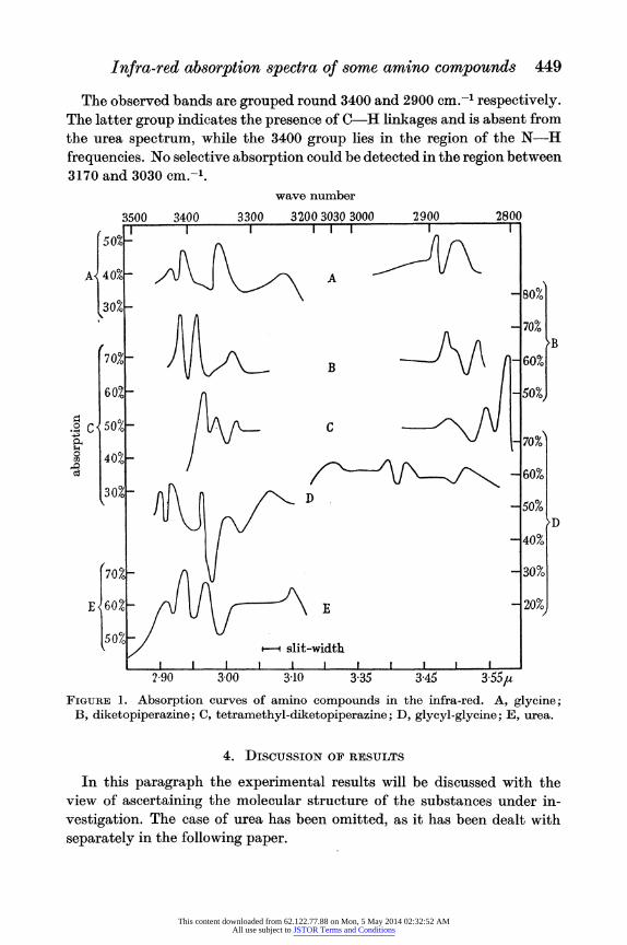
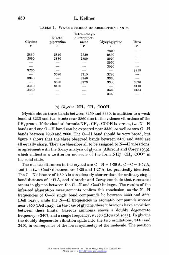
The results of the experiments are given in figure 1, curves I-V, and in table 1. Figure 1 shows the absorption of the five compounds as function of the wave-lengths. Each curve shows the mean results of five sets of measurements. In all cases, a band at 3300 cm.-l was found in addition to those shown in the diagrams and the table, indicating the presence of 0-H bonds. This band disappeared entirely when the substance was subjected to prolonged drying. It must be ascribed, therefore, to traces of adsorbed water in the crystals.
This content downloaded from 62.122.77.88 on Mon, 5 May 2014 02:32:52 AMAll use subject to JSTOR Terms and Conditions
Infra-red absorption spectra of some amino compounds 449
The observed bands are grouped round 3400 and 2900 cm.-L respectively. The latter group indicates the presence of C-H linkages and is absent from the urea spectrum, while the 3400 group lies in the region of the N-H frequencies. No selective absorption could be detected in the region between 3170 and 3030 cm.-'.
wave number
3500 3400 3300 3200 3030 3000 2900 2800 -rIII I . I I
I 0%-n115
.A 40%-
-~~ slit-width ~ ~ so
(702 lJ_ 0
E607.- / JU \ 0?
2 90 3 00 3-10 3-35 3-45 3.55}U
FIG[URE 1. Absorption curves of amino compounds in the infra-red. A, glycine; B, diketopiperazine; C, tetramethyl-diketopiperazine; D, glycyl-glycine; E, urea.
4. DISCUSSION OF RESULTS
In this paragraph the experimental results will be discussed with the view of ascertaininlg the molecular structure of the substances under in- vestigation. The case of urea has been omitted, as it has been dealt with separately in the following paper.
This content downloaded from 62.122.77.88 on Mon, 5 May 2014 02:32:52 AMAll use subject to JSTOR Terms and Conditions
450 L. Kellner
TABLE 1. WAVE NUMBERS OF ABSORPTION BANDS
Tetramethyl- Diketo- diketopiper-
Glycine piperazine azine Glycyl-glycine Urea v v v v v
- - 2800 2860 2840 2830 2860 2890 2880 2880 2920
2950 3020
3235 - 3218 3320 3315 3280
3340 - 3340 3330 3390 3373 3380 3376
3410 3420 - 3410 3440 - 3430 3434
3450
(a) Glycine, NHi2. CH2. COOH
Glycine shows three bands between 3450 and 3330, in addition to a weak band at 3235 and two bands near 2860 due to the valence vibrations of the CH2 group. If the classical formula NH2. (CH2. COOH is correct, two N-H bands and one 0-H band can be expected near 3330, as well as two C-H bands between 2950 and 2860. The 0-H band should be very broad, but figure 1 shows that the three observed bands between 3450 and 3330 are all equally sharp. They are therefore all to be assigned to N-H vibrations, in agreement with the X-ray analysis of glycine (Albrecht and Corey I939),
which indicates a zwitterion molecule of the form NH+ . CH2. COO in the solid state.
The nuclear distances in the crystal are C-N = -39A, C-C = 1V52A, and the two C=O distances are 1P25 and 1b27 A, i.e. practically identical. The C-N distance of 1 39 A is considerably shorter than the ordinary single bond distance of 1-47 A, and Albrecht and Corey conclude that resonance occurs in glycine between the C-N and C-0 linkages. The results of the infra-red absorption measurements confirm this conclusion, as the N-H frequencies of C-N single bond compounds lie between 3330 and 3220 (Bell I927), while the N-H frequencies in aromatic compounds appear near 3450 (Bell I 925). In the case of glycine, these vibrations have a position between these limits. Gaseous ammonia shows a doubly degenerate frequency, v 3407, and a single frequency, v 3336 (Howard I935). In glycine the doubly degenerate vibration splits into the two oscillations, 3440 and 3410, in consequence of the lower symmetry of the molecule. The position
This content downloaded from 62.122.77.88 on Mon, 5 May 2014 02:32:52 AMAll use subject to JSTOR Terms and Conditions
Infra-red absorption spectra of some amino compounds 451
of the C-H frequencies, v 2890 and v 2860, corresponds to the presence of a C-C single bond, in agreement with the C-C distance of 1P52 A. The band v 3235 has also been observed for urea, and may be assigned to the first overtone of the C-O0 vibration near 1660 cm.-1.
The Raman spectrum of glycine has been observed by Wright and Lee (I935), Kahovec and Kohlrausch (1936) and Edsall (1936). No trace of the N-H oscillations was found by these investigators, while Kahovec and Kohlrausch showed that they are present in the glycine esters which possess an ordinary NH2 group and no zwitterion structure. These results are in accordance with the assumption of ionic bonds in the NH+ group, as the Raman lines of ionic linkages are expected to have vanishing intensity. On the other hand, the infra-red bands will be very intense, as the dipole moment changes rapidly with the nuclear distance in heteropolar linkages.
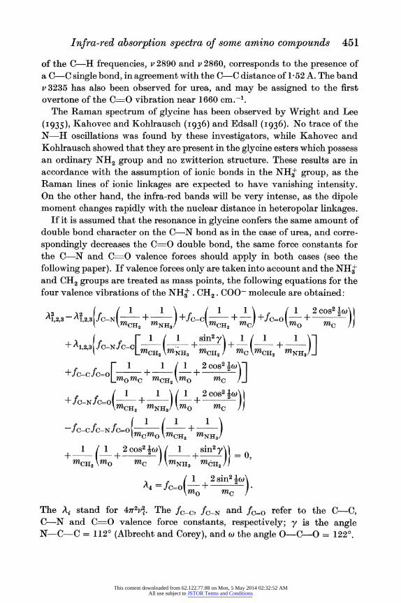
If it is assumed that the resonance in glycine confers the same amount of double bond character on the C-N bond as in the case of urea, and corre- spondingly decreases the C_O double bond, the same force constants for the C-N and C-O valence forces should apply in both cases (see the following paper). If valence forces only are taken into account and the NH+ and CH2 groups are treated as mass points, the following equations for the four valence vibrations of the NH+. CH2. COO- molecule are obtained:
A1,2,3-A1,2.3(fC-N( +m ) +f-(>c( +m) I
+fC=O 2 cs )4
+ A1,2,3fC-NfC-C[m' (:113 + i ) +1(1 +NH3)]
+f JCO L1 + 1 (1 + 2cos2 w)]
0 MC MC rn MO MC +Jec-N JC=O O~~+ ! 1+-2o~)
-fc-cfC-N fCo(rnlrn (rnCH2 +mNH3)
rn012\mO m0C ! nMNH3 MO J2/C -fC_CfC_Nfm= + C
A4 = fc-o( 2 51n2 lW)
The Ai stand for 4,g2Vg. The JC_c, fc-N and fC=o refer to the CC, C-N and C_O valence force constants, respectively; y is the angle N-C-C = 112? (Albrecht and Corey), and wo the angle O-C-O = 1220.
This content downloaded from 62.122.77.88 on Mon, 5 May 2014 02:32:52 AMAll use subject to JSTOR Terms and Conditions
452 L. Kellner
If the force constants, fc-N = 66 x 105 dynes/cm. and fc0 - 97 x 105 dynes/cm., evaluated from the urea spectrum (see the following paper), are used, together with the commonly accepted value, fC =-5 0 x 105
dynes/cm., the following valence vibrations are computed:
v1 = 840, v2 = 1322, V3= 1420, V4= 1778.
The calculated frequencies agree very well with the observed data, 870, 1320, 1400 and 1650, when it is taken into consideration that the angular forces, which contribute considerably to the oscillations in this spectral region, have been neglected. It is permissible to leave the N-H and C-H frequencies out of the calculations, as they lie so far away from the chain vibrations. Further confirmation of the resonance between the C-N and C=O bonds in glycine comes from the fact that the C-O frequency in the glycine esters and in the hydrochloride is considerably higher than in glycine itself (approximately 1720 as compared with 1650).
(b) 2, 5-diketopiperazine (glycine anhydride) (NH. CO. CH2)2
A short discussion of the spectrum of this substance has previously been given (Kellner I937), but the new evidence on the crystalline structure (Corey I938) and the near infra-red spectrum (Ellis and Bath I939) demands certain modifications of the former interpretation. The analysis of the crystal structure of glycine anhydride makes it evident that a type of resonance similar to that in glycine occurs between the C-N and C O bonds. The nuclear distances are OC-N = 133 A, C-O = l-25 A, OC-CH2 = l-47 A and N-CH2 = 1-41 A. The carbon, oxygen and nitrogen atoms lie in one plane; the molecule has a centre of symmetry and belongs to the symmetry group S2 (i). The hydrogen atoms are arranged symmetrically with respect to the symmetry plane. The relatively short carbon-carbon and nitrogen- methylene carbon distances seem to indicate that the C-C bonds are involved in the resonance, but this assumption is not borne out by the evidence of the infra-red bands. The two C-H frequencies, 2880 and 2840, are typical for CH2 groups attached to C-C single bonds. As the glycine anhydride molecule possesses two CH2 groups, four C-H frequencies may be expected, two of which, belonging to the symmetry classes A, and B., will be forbidden in the infra-red spectrum. The presence of only two C-H bands confirms, therefore, the existence of a centre of symmetry in the molecule. The two missing C-H vibrations should appear in the Raman spectrum. It follows from the symmetry requirements of the oscillations that the C-H frequencies of the classes A, and B. will be of the form
This content downloaded from 62.122.77.88 on Mon, 5 May 2014 02:32:52 AMAll use subject to JSTOR Terms and Conditions
Infra-red absorption spectra of some amino compounds 453
2 2 -g~ cs2iO 47T2V2 = _+ in02) wheref>1 iS the C-H valence force constant
and 0 the tetrahedral angle; and the two oscillations of A. and Bg will be of
fCtHh m 1+ 2 ). It will be seen that for the two MnH Mc
infra-red active bands, VAu > VBu. Therefore v 2880 is assigned to the symmetry class A., and v 2840 to the class Bu.
Ellis and Bath (I939), who have observed the absorption bands of di- ketopiperazine in the region of the first overtone, find two parallel bands at 1*703,u and 1.754,u and a perpendicular band at 1 7451t. They assign 1b703,t to the first overtone of the parallel C-H valence frequency, v,; 1V745,u to the first overtone of the perpendicular C-H valence frequency, v,; and 1b754,t to the third overtone of the parallel deformation frequency of the CH2 group, 4. This classification takes no account of the selection rules which apply to the symmetry group S2 (i). Only those overtones and com- bination tones have non-vanishing intensity in the infra-red or Raman spectrum which belong to the permitted symmetry type (Tisza I933).
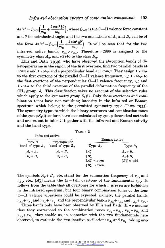
The symmetry types to which the binary overtones and combination tones of the group S2 (i) conform have been calculated by group theoretical methods and are set out in table 2, together with the infra-red and Raman activity and the band type.
TABLE 2 Infra-red active
Raman active Parallel Perpendicular
band of type Au band of type B, Type A, Type Bg
Ag+Au Au+B, [Al] Au+Bu Bg+Bu Ag+Bu [Bg] Ag+Bg
[A'] n even [B,] n odd
[Bu] n even
The symbols Ag + Bu, etc. stand for the summation frequency of vA. and VBu~ etc., [Ag] means the (n - 1)th overtone of the fundamental .*. It follows from the table that all overtones for which n is even are forbidden in the infra-red spectrum; but four binary combination tones of the four C-H valence vibrations could be expected, namely, the parallel bands
vAg + vAu and VB9 + VBu, and the perpendicular bands vAu + vBg and VAg + VBU .
Three bands only have been observed by Ellis and Bath. If we assume that they correspond to the combination tones g+ vAu, vB + vBu and
VA9+ VBU, they enable us, in connexion with the two fundamentals here observed, to evaluate the two inactive oscillations P,A and PB,, taking into
This content downloaded from 62.122.77.88 on Mon, 5 May 2014 02:32:52 AMAll use subject to JSTOR Terms and Conditions
454 L. Kellner
consideration that PA. < PB. It is found that PA4 = 2856 and PB = 3032 furnish the parallel combination tones P?_ +PAU = 5736 (5701 observed) and VBg + VB = 5872 (5872 observed), and the perpendicular bands
VB_+ AU = 5912 (not observed) and VA.?+Bu = 5696 (5731 observed). Fox and Martin (I 940) point out that in Ellis and Bath's interpretation the parallel combination tone is larger than the perpendicular, contrary to what is usually found. This objection would apply to the present classifica- tion as well, but the terms 'parallel' and 'perpendicular' refer here to the symmetry axis of the whole molecule, which is inclined to the symmetry axis of the single CH2 group which Fox and Martin have in mind.
Three more bands have been observed for diketopiperazine in the region of the N-H frequencies. The molecule should possess two N-H valence vibrations of the types AS and BW respectively. In VNH the two N-H bonds stretch in phase; in PB. they move out of phase. Only BvH should be infra-red active, and may be identified with v 3420. v 3320 is assigned to the permitted summation frequency of the two C-O vibrations vc4? + iBf?, as it can be inferred from the Raman spectrum of glycine that these oscilla- tions will be near 1650 cm.-'. This leaves v 3390 still unaccounted for. It seems indicated from its proximity to 3420 that it is identical with the forbidden N-H frequency VN-11. It is known that in the case of liquid benzene a few forbidden bands appear in the infra-red spectrum. The proximity of the N-H bands to those of glycine confirms the evidence of the crystal analysis (Corey I938), that in 2, 5-diketopiperazine resonance occurs between the C-N and C-O bonds, and that the molecule does not possess any 0-H linkages as had formerly been suggested (Sanborn 1932).
It follows that neither keto-enol nor lactam-lactim transformation occurs in the molecule.
(c) Tetramethyl-diketopiperazine [NH. CO. C(CH3)2]2
The spectrum of tetramethyl-diketopiperazine is very similar to that of 2, 5-diketopiperazine. The interpretation follows the same lines: v 3373 is identified with the asymmetric N-H frequency and v 3340 with the sym- metric N-H frequency. The two N-H vibrations are shifted to smaller frequencies as compared with glycine anhydride. The shift is practically the same for both bands. v 3315 can again be assigned to the combination tone of the two C-O vibrations. The similarity of the positions of the N-H and C -O oscillations to those of diketopiperazine leads to the conclusion that the same type of resonance between the C-N and C =O bonds takes place in tetramethyl-diketopiperazine. The number of C-H oscillations has increased to three. As the molecule possesses four CH3 groups, six C-H
This content downloaded from 62.122.77.88 on Mon, 5 May 2014 02:32:52 AMAll use subject to JSTOR Terms and Conditions
Infra-red absorption spectra of some amino compouncls 455
frequencies would be expected in the case of central symmetry, but they might overlap to such an extent that a smaller number is observed. The similarity between the spectra of diketopiperazine and its derivative makes it feasible to assume that these substances have the same symmetry.
(d) Glycyl-glycine NH2. CH2. CO . NH. CH2. COOH
Glycyl-glycine exhibits four bands in the region of the N-H frequencies, v 3450, 3430, 3380 and 3330; and four C-H frequencies, v 3020, 2950, 2920 and 2860, in addition to a band at v 3280. The appearance of four C-H vibrations is in agreement with the existence of four C-H linkages in the molecule, and the four N-H frequencies show that glycyl-glycine in the crystal is in the zwitterion form NH+. CR2. CO . CH2.NH. COO-. The position of the N-H vibrations between 2 9 and 3 0# again points to the probability that resonance occurs between the C-N and C-O bonds. Correspondingly, v 3280 may be assigned to the first overtone or a binary combination tone of the oscillations of the C O linkages involved in the resonance. The infra- red absorption spectrum of glycyl-glycine ethyl ester dissolved in CC14 has been observed by Buswell, Downing and Rodebush (1939). They report absorption bands at 3720, 3390, 3090, 2990 and 2960, and a band at 3280 which vanishes for a concentration of 0-008 mol. No ionic structure is to be expected in the glycyl-glycine ester, and 3390 therefore represents the N-H frequencies. The C-H frequencies are surprisingly high for C-C single bond compounds, even for very low concentrations. In the crystal their frequencies are reduced by 40-70 cm.-'. In the case of the ethyl ester, the absorption bands include the C-H oscillations of the ethyl group as well, so that a comparison between the two observations is not possible.
I wish to express my gratitude to Professor H. Dingle for putting the laboratory facilities of the Imperial College of Science and Technology, London, S.W. 7, at my disposal. I am furthermore very much indebted to Professor A. C. Chibnall and Dr W. T. Astbury for providing me with the five compounds.
REFERENCES
Albrecht and Corey I939 J. Amer. Chem. Soc. 61, 1087. Bell I925 J. Amer. Chem. Soc. 47, 3039. Bell I927 J. Amer. Chem. Soc. 49, 1837. Buswell, Downing and Rodebush I939 J. Amer. Chem. Soc. 61, 3252. Corey I938 J. Amer. Chem. Soc. 60, 1598. Drummond I934 Nature, Lond., 134, 937.
This content downloaded from 62.122.77.88 on Mon, 5 May 2014 02:32:52 AMAll use subject to JSTOR Terms and Conditions
456 L. Kellner
Edsall 1936 J. Chem. Phys. 4, 1. Ellis and Bath 1939 Phys. Rev. 55, 1098. Ellis and Bath 1939 J. Chem. Phys. 7, 862. Fox and Martin 1940 Proc. Roy. Soc. A, 175, 208. Howard 1935 J. Chem. Phys. 3, 207. Kahovec and Kohlrausch 1936 S.B. Akad. Wiss. Wien, Ilb, 145, 579. Kellner 1936 Proc. Roy. Soc. A, 157, 100. Kellner 1937 Nature, Lond., 140, 123. Kohlrausch and Pongratz 1934 Z. phys. Chem. B, 27, 176. Sanborn 1932 J. Phys. Chem. 36, 1799. Tisza 1933 Z. Phys. 82, 48. Wright and Lee 1935 Nature, Lond., 136, 300.
The vibrations and the molecular structure of urea and guanidonium
BY LOTTE KELLNER
(Communicated by W. T. Astbury, F.R.S.- Received 25 June 1940-Revised 21 November 1940)
The vibrations of urea and guanidonium have been calculated for a field containing valence and angle forces. The assumption is made that urea has the symmtery C2, and guanidonium C3h.
It is shown that it is possible to assign every observed frequency of these two substances to definite modes of vibration under these assumptions. The force constants have been evaluated and have been found to be
fc-N= 7a1 x 105 dynes/cm. for guanidonium, and fCN - 6-6 x 10⁵ dynes/cm. andfc=o = 9 7 x 10⁵ dynes/cm. for urea. These values are compatible with the hypothesis that quantum mechanical resonance occurs in both molecules, with the result that the C-N bond in urea has approximately 28 % double- bond character and the C=O linkage a corresponding single-bond character. The guanidonium ion shows complete resonance; each C-N bond has f double-bond character. Curves have been drawn to illustrate the relation
between the valence force constants and the bond character.
1. INTRODUCTION
It has been evident for some time, from the study of the chemical behaviour
as well as from the X-ray investigation of the crystalline structure of urea
and guanidonium, that these two molecules show quantum mechanical
resonance between several possible configurations. Pauling (1935) suggests resonance between a homopolar (figure 1, I) and two ionic (figure 1, II, III)
This content downloaded from 62.122.77.88 on Mon, 5 May 2014 02:32:52 AMAll use subject to JSTOR Terms and Conditions