Embed Size (px)
Citation preview
Inguinal Region & Secrotum
Abdomen, Pelvis & Perineum Unit Lecture 2األعسم. جليل حيدر د
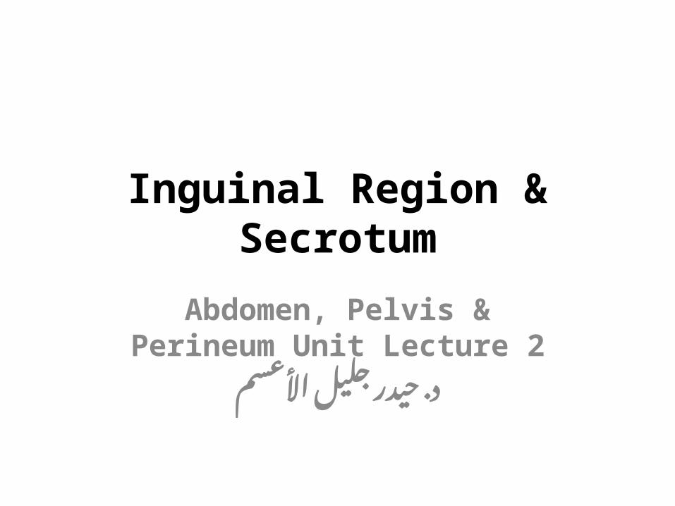
Inguinal CanalIt is an oblique passage through the lower part of the anterior abdominal wall. It is about 4 cm long in adult and extends from the deep inguinal ring, downward and medially to the superficial inguinal ring. It lies parallel to and immediately above the inguinal ligament.
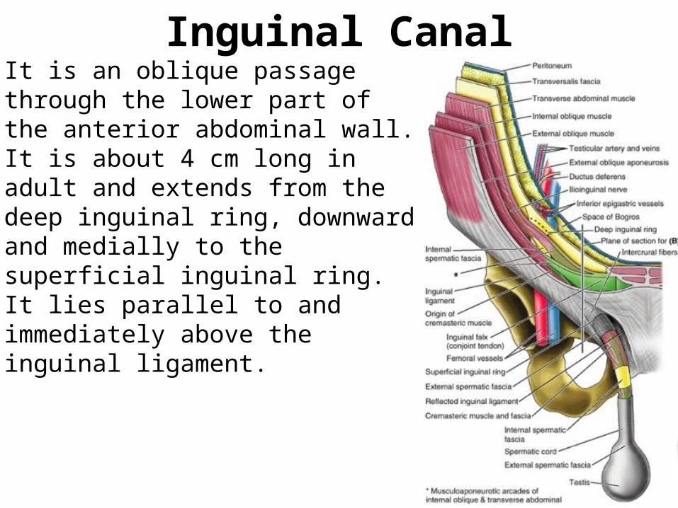
Inguinal Canal - RingsThe superficial inguinal ring: It is a triangular-shaped opening in the aponeurosis of external oblique muscle and lies immediately above and medial to pubic tubercle. Margins of the ring, sometimes called crura (medial & lateral), give attachment to external spermatic fascia. The crura are held by intercrural fibres at the apex of the ring.The deep inguinal ring: It is an oval opening in fascia transversalis, lies about 0.5 inch (1.3 cm) above inguinal ligament midway between anterior superior iliac spine and symphysis pubis (Mid-inguinal Point). Medial to it, there are inferior epigastric vessels.Margins of the ring give attachment to internal spermatic fascia (or internal covering of round ligament of uterus).
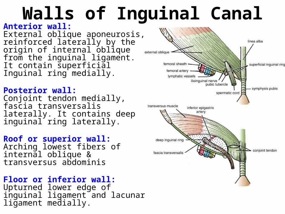
Walls of Inguinal CanalAnterior wall: External oblique aponeurosis, reinforced laterally by the origin of internal oblique from the inguinal ligament. It contain superficial Inguinal ring medially.
Posterior wall: Conjoint tendon medially, fascia transversalis laterally. It contains deep inguinal ring laterally.
Roof or superior wall: Arching lowest fibers of internal oblique & transversus abdominis
Floor or inferior wall: Upturned lower edge of inguinal ligament and lacunar ligament medially.
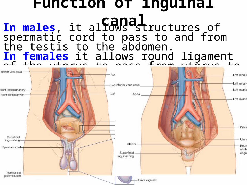
Function of inguinal canalIn males, it allows structures of spermatic cord to pass to and from the testis to the abdomen. In females it allows round ligament of the uterus to pass from uterus to labium majus.
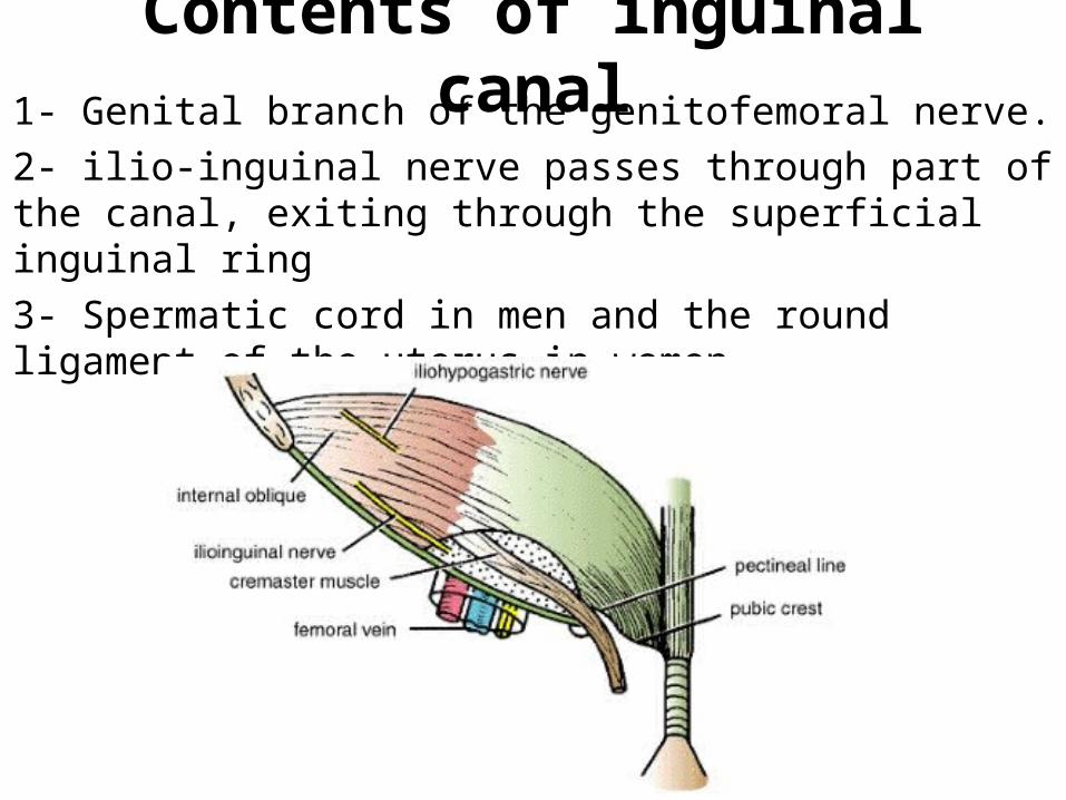
Contents of inguinal canal1- Genital branch of the genitofemoral nerve.2- ilio-inguinal nerve passes through part of the canal, exiting through the superficial inguinal ring3- Spermatic cord in men and the round ligament of the uterus in women.
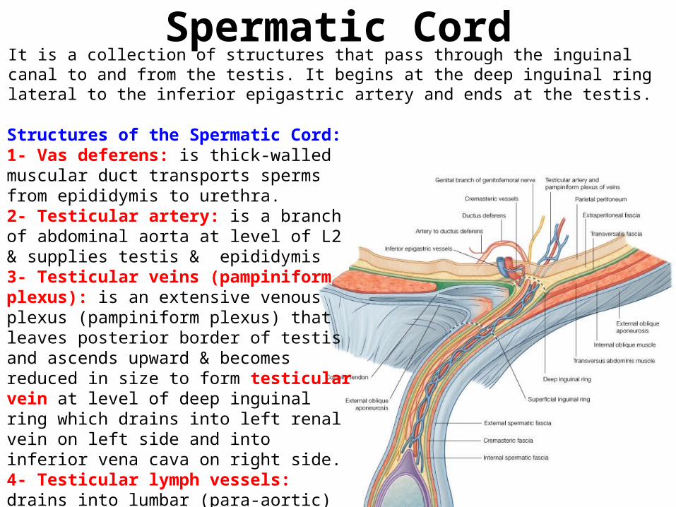
Spermatic CordIt is a collection of structures that pass through the inguinal canal to and from the testis. It begins at the deep inguinal ring lateral to the inferior epigastric artery and ends at the testis.Structures of the Spermatic Cord:1- Vas deferens: is thick-walled muscular duct transports sperms from epididymis to urethra.2- Testicular artery: is a branch of abdominal aorta at level of L2 & supplies testis & epididymis3- Testicular veins (pampiniform plexus): is an extensive venous plexus (pampiniform plexus) that leaves posterior border of testis and ascends upward & becomes reduced in size to form testicular vein at level of deep inguinal ring which drains into left renal vein on left side and into inferior vena cava on right side.4- Testicular lymph vessels: drains into lumbar (para-aortic) lymph nodes.5- Autonomic nerves:6- Remains of the processus vaginalis:7- Genital branch of genitofemoral nerve: which supplies the cremaster muscle
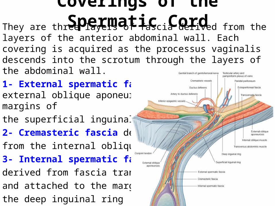
Coverings of the Spermatic CordThey are three layers of fascia derived from the layers of the anterior abdominal wall. Each covering is acquired as the processus vaginalis descends into the scrotum through the layers of the abdominal wall.1- External spermatic fascia derived from external oblique aponeurosis and attached to the margins of the superficial inguinal ring2- Cremasteric fascia derived from the internal oblique muscle3- Internal spermatic fascia derived from fascia transversalis and attached to the margins of the deep inguinal ring
ScrotumScrotum is an out-pouching of lower part of anterior abdominal wall. It contains testes, epididymides, & lower ends of spermatic cords. The wall of the scrotum has the following layers:1- Skin: is thin, wrinkled & pigmented and forms a single pouch. 2- Superficial fascia: is continuous with fatty and membranous layers of anterior abdominal wall; the fat is replaced by dartos muscle. This is innervated by sympathetic nerve fibers. It is attached to the perineal body and the posterior edge of the perineal membrane. At the sides it is attached to the ischiopubic rami. Both layers of superficial fascia contribute to median partition that crosses scrotum & separates testes.3- Spermatic fasciae: lie beneath superficial fascia and are derived from the three layers of the anterior abdominal wall.4- Tunica vaginalis: lies within spermatic fasciae and covers anterior, medial & lateral surfaces of each testis. It is the lower expanded part of processus vaginalis; normally, just before birth, it becomes disconnected from upper part of processus and peritoneal cavity.
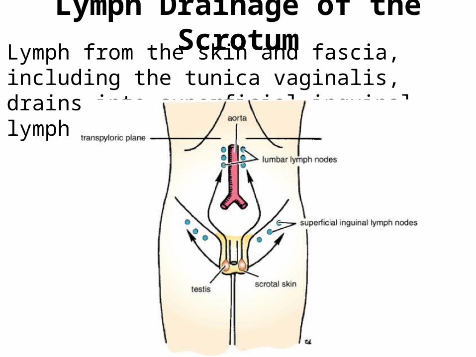
Lymph Drainage of the ScrotumLymph from the skin and fascia, including the tunica vaginalis, drains into superficial inguinal lymph nodes
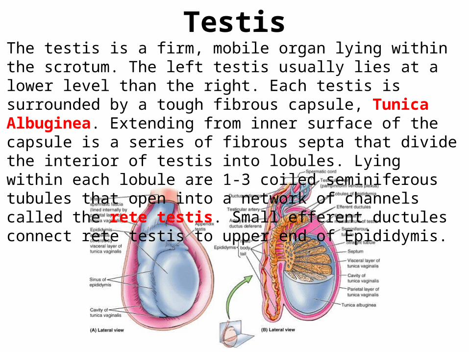
TestisThe testis is a firm, mobile organ lying within the scrotum. The left testis usually lies at a lower level than the right. Each testis is surrounded by a tough fibrous capsule, Tunica Albuginea. Extending from inner surface of the capsule is a series of fibrous septa that divide the interior of testis into lobules. Lying within each lobule are 1-3 coiled seminiferous tubules that open into a network of channels called the rete testis. Small efferent ductules connect rete testis to upper end of Epididymis.
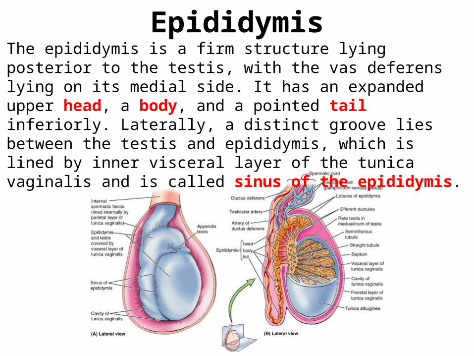
EpididymisThe epididymis is a firm structure lying posterior to the testis, with the vas deferens lying on its medial side. It has an expanded upper head, a body, and a pointed tail inferiorly. Laterally, a distinct groove lies between the testis and epididymis, which is lined by inner visceral layer of the tunica vaginalis and is called sinus of the epididymis.
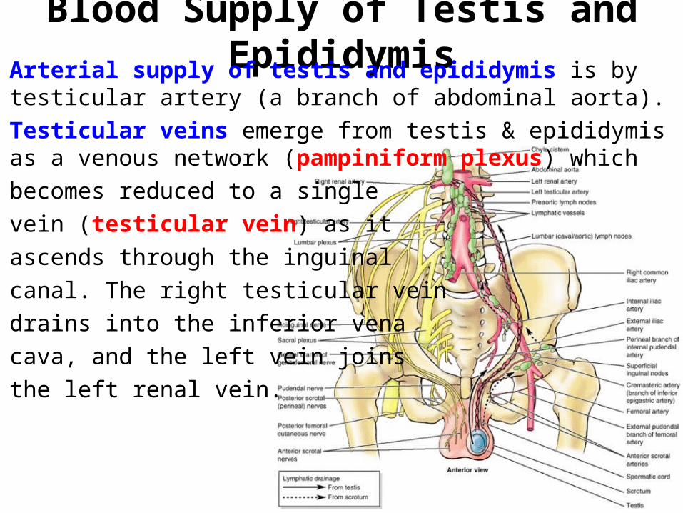
Blood Supply of Testis and EpididymisArterial supply of testis and epididymis is by testicular artery (a branch of abdominal aorta). Testicular veins emerge from testis & epididymis as a venous network (pampiniform plexus) whichbecomes reduced to a singlevein (testicular vein) as it ascends through the inguinalcanal. The right testicular veindrains into the inferior vena cava, and the left vein joins the left renal vein.

Lymph Drainage of Testis and Epididymis
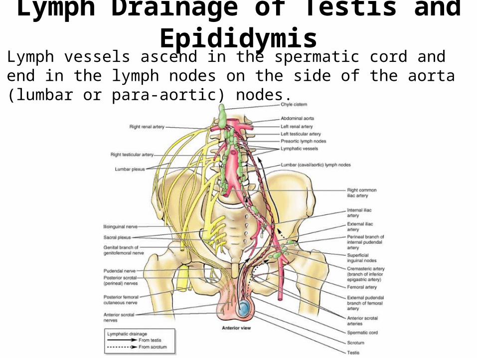
Lymph vessels ascend in the spermatic cord and end in the lymph nodes on the side of the aorta (lumbar or para-aortic) nodes.
Lymph Drainage of Testis and Epididymis
Lymph vessels ascend in the spermatic cord and end in the lymph nodes on the side of the aorta (lumbar or para-aortic) nodes.
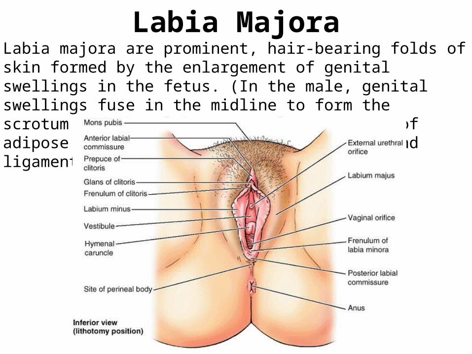
Labia MajoraLabia majora are prominent, hair-bearing folds of skin formed by the enlargement of genital swellings in the fetus. (In the male, genital swellings fuse in the midline to form the scrotum.) Within labia are a large amount of adipose tissue and terminal strands of round ligaments of uterus.

Thank You