Embed Size (px)
Citation preview
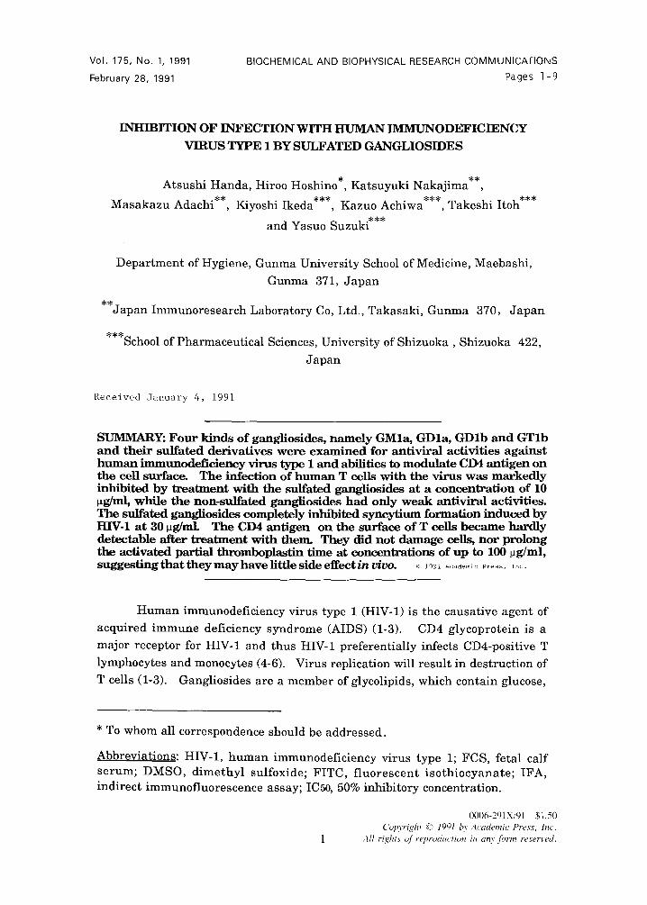
Vol. 175, No. 1, 1991
February 28, 1991
BIOCHEMICAL AND BIOPHYSICAL RESEARCH COMMUNICATIONS
Pages I - 9
INHIBITION OF INFECTION WITH HUMAN IMMUNODEFICIENCY
VIRUS TYPE 1 BY SULFATED GANGLIOSIDES
Atsushi Handa, Hiroo Hoshmo , Katsuyuki Nakajima**, * * ~ . . * * *
Masakazu Adachi , Kiyoshi lkeda , Kazuo Achiwa***, Takeshi Itoh***
and Yasuo Suzuki***
Department of Hygiene, Gunma University School of Medicine, Maebashi,
Gunma 371, Japan
**Japan Immunoresearch Laboratory Co, Ltd., Takasaki, Gunma 370, Japan
***School of Pharmaceutical Sciences, University of Shizuoka, Shizuoka 422,
Japan
Received January 4, 1991
SUMMARY: Four kinds of gangliosides, namely GMla, GDla, GDlb and GTlb and their sulfated derivatives were examined for antiviral activities against human immunodeficiency virus type 1 and abilities to modulate CD4 antigen on the cell surface. The infection of human T cells with the virus was markedly inhibited by treatment with the sulfated gangliosides at a concentration of 10 ~g/ml~ while the non-sulfated gangliosides had only weak antiviral activities. The sulfated gangliosides completely inhibited syncytium formation induced by HIV-1 at 30 pg/mL The CD4 antigen on the surface of T cells became hardly detectable after t reatment with then~ They did not damage cells, nor prolong the activated partial thromboplastin time at concentrations of up to 100 ~g/ml, s u g g e s t l n g t h a t t h e y m a y h a v e l i t t l e s i d e e f f e c t i n v i v o . ~ ~99~ ~ d ~ m ~ o ~ . . . . . ~ o .
Human immunodeficiency virus type 1 (HIV-1) is the causative agent of
acquired immune deficiency syndrome (AIDS) (1-3). CD4 glycoprotein is a
major receptor for HIV-1 and thus HIV-1 preferentially infects CD4-positive T
lymphocytes and monocytes (4-6). Virus replication will result in destruction of
T cells (1-3)• Gangliosides are a member of glycolipids, which contain glucose,
* To whom all correspondence should be addressed.
Abbreviations: HIV-1, human immunodeficiency virus type 1; FCS, fetal calf serum; DMSO, dimethyl sulfoxide; FITC, fluorescent isothiocyanate; IFA, indirect immunofluorescence assay; IC50, 50% inhibitory concentration.
0 0 0 6 - 2 9 1 X / 9 1 $ 1 . 5 0
Copyright ~:) 1991 by Academic Press, lnc. All ri~t ts of reproduction in any form reserved.
Vol. 175, No. 1, 1991 BIOCHEMICAL AND BIOPHYSICAL RESEARCH COMMUNICATIONS
galactose, fucose, hexosamines, ceramide and sialic acids. Gangliosides are
present in many tissue, especially rich in the brain. It was reported recently
that GM1 or GMla, one of gangliosides, binds to the CD4 antigen on the cell
surface of T cells, down-regulate its expression and inhibi t HIV-1 infection
when examined under serum-free culture conditions (7-9). We purified four
different gangliosides GMla, GDla , GDlb and GTlb from bovine brains and
their derivatives were prepared by sulfation. We examined these compounds
for anti-HIV-1 activities and their effects on expression of CD4 molecules on the
cell surface.
MATERIALS AND METHODS
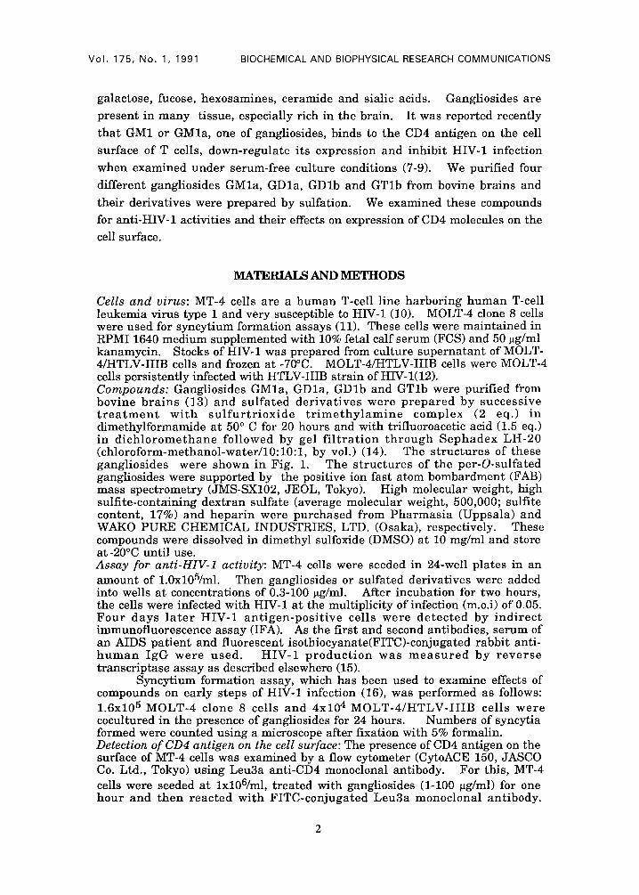
Cells and virus: MT-4 cells are a human T-cell line harbor ing h u m a n T-cell leukemia virus type 1 and very susceptible to HIV-1 (10). MOLT-4 clone 8 cells were used for syncytium formation assays (11). These cells were maintained in RPMI 1640 medium supplemented with 10% fetal calf serum (FCS) and 50 ~g/ml kanamycin. Stocks of HIV-1 was prepared from culture superna tant of MOLT- 4/HTLV-IIIB cells and frozen at -70°C. MOLT-4/HTLV-IIIB cells were MOLT-4 cells persistently infected with HTLV-IIIB strain of HIV-l(12). Compounds: Gangliosides GMla, GDla , GDlb and GTlb were purified from bovine b r a i n s (13) and su l f a t ed de r iva t ives were p r e p a r e d by success ive t r e a t m e n t w i t h s u l f u r t r i o x i d e t r i m e t h y l a m i n e complex (2 eq.) in dimethylformamide at 50 ° C for 20 hours and with trifluoroacetic acid (1.5 eq.) in d i c h l o r o m e t h a n e fo l lowed by gel f i l t r a t i o n t h r o u g h S e p h a d e x LH-20 (chloroform-methanol-water /10:10: l , by vol.) (14). The s t ruc tu res of these gangl iosides were shown in Fig. 1. The s t ruc tu res of the per-O-sulfated gangliosides were supported by the positive ion fast atom bombardment (FAB) mass spectrometry (JMS-SX102, JEOL, Tokyo). High molecular weight, high sulfite-containing dextran sulfate (average molecular weight, 500,000; sulfite content, 17%) and hepar in were purchased from P h a r m a s i a (Uppsala) and WAKO PURE CHEMICAL INDUSTRIES, LTD. (Osaka), respectively. These compounds were dissolved in dimethyl sulfoxide (DMSO) at 10 mg/ml and store at-20°C until use. Assay for anti-HIV-1 activity: MT-4 cells were seeded in 24-well plates in an amount of 1.0xl05/ml. Then gangliosides or sulfated derivatives were added into wells at concentrations of 0.3-100 ~g/ml. After incubation for two hours, the cells were infected with HIV-1 at the multiplicity of infection (m.o.i) of 0.05. F o u r days l a t e r HIV-1 a n t i g e n - p o s i t i v e cel ls were d e t e c t e d by i nd i r ec t immunofluorescence assay (IFA). As the first and second antibodies, serum of an AIDS pat ient and fluorescent isothiocyanate(FITC)-conjugated rabbi t anti- h u m a n IgG were u s e d . HIV-1 p r o d u c t i o n was m e a s u r e d by r e v e r s e transcriptase assay as described elsewhere (15).
Syncytium formation assay, which has been used to examine effects of compounds on early steps of HIV-1 infection (16), was performed as follows: 1.6x10⁵ MOLT-4 clone 8 cells and 4x10⁴ M O L T - 4 / H T L V - I I I B cel ls we re cocultured in the presence of gangliosides for 24 hours. Numbers of syncytia formed were counted using a microscope after fixation with 5% formalin. Detection of CD4 antigen on the cell surface: The presence of CD4 antigen on the surface of MT-4 cells was examined by a flow cytometer (CytoACE 150, JASCO Co. Ltd., Tokyo) using Leu3a anti-CD4 monoclonal antibody. For this, MT-4 cells were seeded at lxl06/ml, t reated with gangliosides (1-100 ~g/ml) for one hour and then reac ted wi th FITC-conjuga ted Leu3a monoclonal ant ibody.
2
Vol. 175, No. 1, 1 9 9 1 BIOCHEMICAL AND BIOPHYSICAL RESEARCH COMMUNICATIONS
o i / T " - f J / ~ c . ~ o ~ , . . , , ,
Z~, CH,OR' ~o, CH,OR' ~,._\j:...'~-~.k..o_ / I 1 / o o R o .
Gal GalNAc " ~ Gal CH2OR (~HNHCOR4 /~ }< / GIc ~HOR 1 0 / \ CH
c .
x I o., (c.,),, RIocH2C" \ 8R1 " ~ CH3
AcNH Cer R 1 R 2 R 3 R4: AIkyl
GMI~: H H H " 0 2 C ' ~
G D 1 ~: H H NeuAc
GD~b: H NeuAc H
GT~b: H NeuAc NeuAc R20
GMla-SO3H: SO3H SO3H SO3H R1OCH2 C-\ (~'~.t , ~ O R
G D i a'SO3H: SO3H SO3H NeuAc-S O3 H / - . . AcNH
GDlb-SO3H: SO3H NeuAc-SO3H SO3H GTlb_SO3H: SO3H NeuAc_SO3H NeuAc.SO3H NeuAc
Fig. 1. Structures of gangliosides and sulfated derivatives: Cer, ceramide; Glc, glucose; Gal, galactose; GalNAc, N-acetylgalactosamine; NeuAc, N- acetylneuraminic acid.
Af t e r i n c u b a t i o n for 30 min a t 4°C, the cell we re f ixed w i t h 1% paraformaldehyde and subjected to flow cytometry.
Activated partial thromboplastin time (APTT): APTT of p lasma from a normal subject was examined in the presence of su l fa ted gangl iosides by us ing an au toma ta t ed machine. For this, p la ter in plus act ivator was obtained from Organon Teknika Corporation (Durham). H u m a n plasma (100 ~l) was mixed with platerin plus activator (100 ~1) and tes t compounds and then APTT was measured .
RESULTS
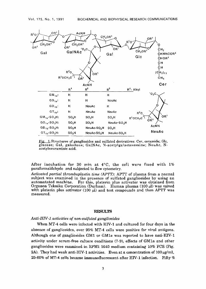
Anti-HIV-1 activities of non-sulfated gangliosides When MT-4 cells were infected with HIV-1 and cultured for four days in the
absence of gangliosides, over 95% MT-4 cells were positive for viral antigens.
Although one of gangliosides GM1 or GMla was reported to have anti-HIV-1
activity under serum-free culture conditions (7-9), effects of G M l a and other
gangliosides were examined in RPMI 1640 medium containing 10% FCS (Fig.
2A). They had weak anti-HIV-1 activities. Even at a concentration of 100 ~g/ml,
20-60% of MT-4 cells became immunofluorescent after HIV-1 infection. Fifty %
VOI. 175, No. 1, 1991 BIOCHEMICAL AND BIOPHYSICAL RESEARCH COMMUNICATIONS
o_ E d3
tu > T
100
8 0
6 0 ¸
4 0
2 0 ¸
GMla GDla GDlb
c
>
I
'1 ' ; ' ; o ' 3'o ' - ; o o
Drug concentration ( p g/ml)
10O
6 0
4 0
2O
0
0
\
i
0 GMla-SO3 H • GDla S O 3 H ,I, GD1b-SO 3 H
GTlb-SO 3 H • Dextran sulfate
3 10 30
Drug c o n c e n t r a t i o n ( J.l g / m l )
Fig. 2.Effects of gangliosides and sulfated derivatives on infection of MT-4 cells with HIV-1. MT-4 cells were infected with HIV-1 in the presence of non- sulfated (A) or sulfated (B) gangliosides. Expression of HIV-1 antigen was detected by IFA after cultivation for four days.
100
i n h i b i t o r y c o n c e n t r a t i o n s (IC50's) for G M l a , G D l a , G D l b and G T l b were
e s t imated from dose-response curves and shown in Table I.
Anti-HIV-1 activities of sulfated gangliosides Because sul fated der ivat ives of po lysacchar ides have been reported to
have m u c h stronger anti-HIV-1 activit ies than non-sul fated polysaccharide (17-
Table I. Effects ofgangliosides and sulfated derivatives on HIV-1 infection and CD4 expression on the surface of MT-4 cells
Compound HIV-1 infection (ICso) CD4 expression (%)
Non-sulfated Sulfated Non-sulfated Sulfated
GMla 30* 0.8 66** 18
GDla >100 0.8 69 16
GD lb 10 0.9 65 18
GTlb 10 2.0 65 10
Dextran ND*** 0.5 ND ND
* Concentrations (~g/ml) of compounds at which 50% of MT-4 cells expressed HIV-1 antigens.
** CD4-positive cells (%) detected by flow cytometry after treatment with gangliosides at 100 ~g/ml for one hour.
*** ND, not done.
Vo l . 175, No. 1, 1991 BIOCHEMICAL AND BIOPHYSICAL RESEARCH COMMUNICATIONS
250 -
O G M l a - S O 3 H • G D l a - S O 3 H ,,& G D l b - S O 3 H
----E}--- G T l b - S O 3 H 200
.__ 1 5 0
"5
1 0 0
Z
5 0
o ~'o 1;o 0 1 3 10
D r u g c o n c e n t r a t i o n ( p , g / m l )
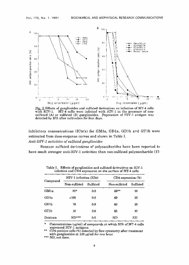
Fig. 3. Inhibition of syncytium formation induced by HIV-1 by sulfated gangliosides. MOLT-4 cells were cocuttivated with HIV-l-producing MOLT- 4/HTLV-IIIB cells for 24 hours in the presence of sulfated gangliosides. Syncytia in wells were counted using an inverted microscope.
22), the gangl ios ides were su l fa ted and the i r ant i -HIV-1 act iv i t ies were
examined (Fig. 2B). The sulfated gangliosides had potent anti-HIV-1 activities:
Infection with HIV-1 was completely inhibited at 10 ~g/ml. IC5o's for sulfated
GMla, GDla, GDlb and GTlb and dextran sulfate were est imated (Table I).
Thus anti-HIV-1 activities of gangliosides were enhanced five to hundred times
by sulfation. HIV-1 production by MT-4 cells after infection as detected by
reverse t ranscr iptase assays was also markedly inhibi ted in the presence of
sulfated gangliosides (data not shown).
Next whether the sulfated gangliosides inhibited syncytium formation
was examined in the culture medium containing 10% FCS (Fig. 3). Syncytium
format ion was inhibi ted almost completely at a concentrat ion of 30 ~g/ml.
These results suggested tha t the sulfated gangliosides would act on at least an
early step of HIV-1 infection, namely, adsorption or penetration.
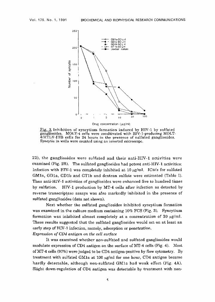
Expression of CD4 antigen on the cell surface
It was examined whether non-sulfated and sulfated gangliosides would
modulate expression of CD4 antigen on the surface of MT-4 cells (Fig. 4). Most
of MT-4 cells (97%) were judged to be CD4 antigen-positive by flow cytometry. By
t reatment with sulfated GMla at 100 ~g/ml for one hour, CD4 ant igen became
ha rd ly detectable, a l though non-sul fa ted G M l a had weak effect (Fig. 4A).
Slight down-regulation of CD4 antigen was detectable by t rea tment with non-
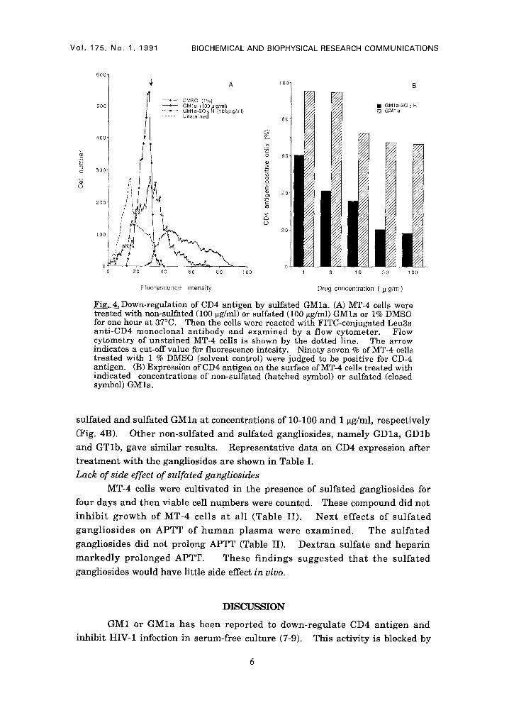
Vol . 175, No. 1, 1991 BIOCHEMICAL AND BIOPHYSICAL RESEARCH COMMUNICATIONS
6007
500
400
300
200
100
I
A 100
DMSO (1%) G M l a (100 p g/ml)
• GMla-SO 3 H (lOOp g/ml) U n s t a i n e d
0 0 20 40 60 80 100
80
o~
60
o_
40 ¸ o3
O 2 0
O I
B
• GMla -SO3 H [ ] G M l a
10 30 100
Fluorescence Intensity Drug concentration ( p g/ml)
Fig. 4. Down-regulation of CD4 antigen by sulfated GMla. (A) MT-4 cells were treated with non-sulfated (100 ~g/ml) or sulfated (100 ~g/ml) GMla or 1% DMSO for one hour at 37°C. Then the cells were reacted with FITC-conjugated Leu3a anti-CD4 monoclonal antibody and examined by a flow cytometer. Flow cytometry of unstained MT-4 cells is shown by the dotted line. The arrow indicates a cut-off value for fluorescence intesity. Ninety seven % of MT-4 cells treated with 1% DMSO (solvent control) were judged to be positive for CD-4 antigen. (B) Expression of CD4 antigen on the surface of MT-4 cells treated with indicated concentrations of non-sulfated (hatched symbol) or sulfated (closed symbol) GMla.
sulfated and sulfated GM l a at concentrations of 10-100 and 1 ~tg/ml, respect ively
(Fig. 4B). Other non-sulfated and sulfated gangliosides, namely GDla, GDlb
and GTlb, gave similar results. Representat ive data on CD4 expression after
t r ea tmen t with the gangliosides are shown in Table I.
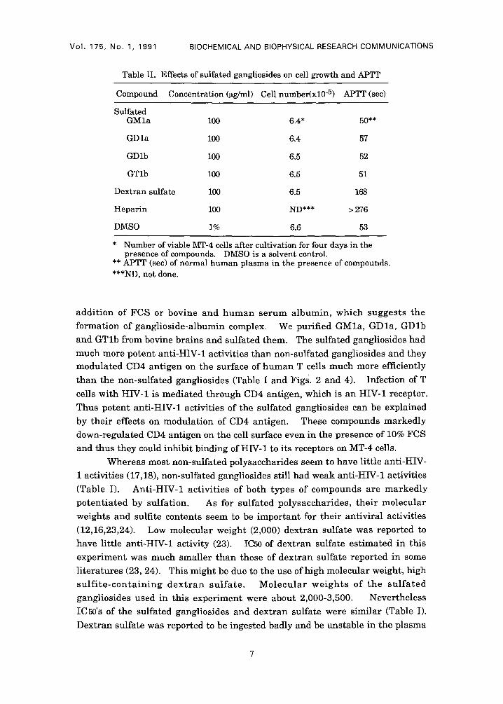
Lack of side effect of sulfated gangliosides MT-4 cells were cult ivated in the presence of sulfated gangliosides for
four days and then viable cell numbers were counted. These compound did not
i n h i b i t g r o w t h of MT-4 cells a t all (Table II). Nex t ef fec ts of su l f a t ed
gang l io s ides on APTT of h u m a n p l a s m a were examined . The su l f a t ed
gangliosides did not prolong APTT (Table II). Dext ran sulfate and hepar in
m a r k e d l y pro longed APTT. These f indings sugges ted t h a t the su l fa ted
gangliosides would have little side effect in vivo.
DISCUSSION
GM1 or G M l a has been repor ted to down-regula te CD4 ant igen and
inhibit HIV-1 infection in serum-free culture (7-9). This activity is blocked by
Vol . 175, No. 1, 1991 BIOCHEMICAL AND BIOPHYSICAL RESEARCH COMMUNICATIONS
Table II. Effects of sulfated gangliosides on cell growth and APTT
Compound Concentration (~g/ml) Cell number(xl0 -5) APTT (sec)
Sulfated GMla 100 6.4* 50**
GDla 100 6.4 57
GDlb 100 6.5 52
GTlb 100 6.5 51
Dextran sulfate 100 6.5 168
Heparin 100 ND*** > 276
DMSO 1% 6.6 53
* Number of viable MT-4 cells after cultivation for four days in the presence of compounds. DMSO is a solvent control.
** APTT (sec) of normal human plasma in the presence of compounds. ***ND, not done.
addi t ion of FCS or bovine and h u m a n serum albumin, which suggests the
formation of ganglioside-albumin complex. We purified GMla, GDla, GDlb
and GTlb from bovine brains and sulfated them. The sulfated gangliosides had
much more potent anti-HIV-1 activities than non-sulfated gangliosides and they modulated CD4 antigen on the surface of human T cells much more efficiently
than the non-sulfated gangliosides (Table I and Figs. 2 and 4). Infection of T
cells with HIV-1 is mediated through CD4 antigen, which is an HIV-1 receptor.
Thus potent anti-HIV-1 activities of the sulfated gangliosides can be explained
by their effects on modulation of CD4 antigen. These compounds markedly
down-regulated CD4 antigen on the cell surface even in the presence of 10% FCS
and thus they could inhibit binding of HIV-1 to its receptors on MT-4 cells.
Whereas most non-sulfated polysaccharides seem to have little anti-HIV-
1 activities (17,18), non-sulfated gangliosides still had weak anti-HIV-1 activities
(Table I). Anti-HIV-1 activit ies of both types of compounds are markedly
potent ia ted by sulfation. As for sulfated polysaccharides, their molecular
weights and sulfite contents seem to be important for their antiviral activities
(12,16,23,24). Low molecular weight (2,000) dextran sulfate was reported to
have little anti-HIV-1 activity (23). IC50 of dextran sulfate est imated in this
experiment was much smaller than those of dextran sulfate reported in some
l i teratures (23, 24). This might be due to the use of high molecular weight, high
s u l f i t e - c o n t a i n i n g d e x t r a n su l fa te . Molecu la r we igh t s of the s u l f a t e d
gangliosides used in this experiment were about 2,000-3,500. Nevertheless
IC50's of the sulfated gangliosides and dextran sulfate were similar (Table I).
Dextran sulfate was reported to be ingested badly and be unstable in the plasma
Vol. 175, No. 1, 1991 BIOCHEMICAL AND BIOPHYSICAL RESEARCH COMMUNICATIONS
of rats: Its half life was 20-480 min (25). It remains to be investigated whether
sulfated gangliosides have similar disadvantages. Many sulfated polysaccharides having anti-HIV-1 activities are known to
prolong APTT (16, 20-22). On the other hand, the sulfated gangliosides did not prolong it (Table II), indicating that they would not have anti-coagulant activity in vivo. They did not affect growth of T cells in vitro (Table II). The sulfated gangliosides had potent anti-HIV-1 activities even when T cells were cultured in the presence of FCS. These properties of the sulfated gangliosides may be
therapeut ical ly advantageous if we should consider the possibility of their clinical use. GM1 was really administered to humans to treat neurological
disorders (Alzheimer's disease), its half life being 60-75 hours in blood (26).
Gangliosides have been reported to influence biological activities, such as cellular interactions or induction of differentiation of leukemic cells (27, 28). It also remains to be investigated whether sulfation of gangliosides will affect these activities.
REFERENCI~
1. Gallo,R.C., Salahuddin,S.Z., Popovic,M., Shearer,G.C., Kaplan, M., Haynes, B.F., Palker,T.J., Redfield,R., Oleske, J., Safai,B., White,G., Foster, P., and Markham,P.D. (1984) Science 224, 500-502
2. Barre-Sinoussi,F., Chermann,J.C., Rey,F., Nugeyre,M.T., Chamaret,S., Gruest,J. , Dauguet,C., Axler-Blin,C., Vezinet-Brun,F., Rouzioux,C., Rozenbaum,W., and Montagnier,L. (1983) Science 220, 868-870
3. Levy,J.A., Hoffman,A.D., Kramer,S.M., Landis,J.A., Shimabukuro,J.M., and Oshiro,L.S. (1984) Science 225, 840-842
4. Dalgleish,A.G., Beverley,P.C.L., Clapham,P.R., Crawford,D.H., Greaves, M.F., and Weiss,R.A. (1984) Nature 312, 763-767
5. Klatzmann,D., Champagne,E., Chamaret,S., Gruest,J., Guetard, D., Hercend,T., Gluckman,J.C., Montagnier,L. (1984)Nature 312, 767-768
6. Salahuddin,S.Z., Rose,R.M., Groopman,J.E., Markham,P.D., and Gal]o, R.C. (1986)Blood 68, 281-284
7. Chieco-Bianchi,L., Calabro,M.L., Panozzo,M., Rossi,A.D., Amadori,A., Callegaro,L., and Siccardi,A. (1989) AIDS 3, 501-507
8. Nakakuma,H., Kawaguchi,T., Koito,A., Hattori,T., Kagimoto,T., and Takatsuki,I~ (1989) Jpn. J. Cancer Res. (Ga nn) 80, 702-705
9. Kawaguchi,T., Nakakuma,H., Kagimoto,T., Shirono,K., Horikawa, K., Hidaka,M., Iwamori,M., Nagai,Y., and Takatsuki K. (1989) Biochem.Biophys.Res.Commun. 158, 1050-1059
10. Harada,S., Koyanagi,Y., and Yamamoto,N. (1985)Science 229, 563-566 11. Nakashima,H., Tochikura,T., Kobayashi,N., Matsuda,A., Ueda,T., and
Yamamoto,N. (1987) Virology 159, 169-173 12. Nagumo,T., and Hoshino,H. (1988)Jpn.J.Cancer Res.(Gann) ~9, 9-11 13. Hirabayashi,Y., Hyogo,A., Nakao,T., Tsuchiya,K., Suzuki,Y., Matsumoto,
M., Kon,K., and Ando,S. (1990)J.Biol.Chem. 265, 8144-8151. 14. Idegami,K., Ikeda,K., and Achiwa,K. (1990) Chem.Pharm.Bull. 38, 1766-
1768. 15. Jacks,T., Power,M.D., Masiarz,F.R., Luciw,P.A., Barr,P.J., and Varmus,
H.E. (1988)Nature 331, 280-283 16. Nakashima,H., Tanabe,A., Tochikura,T.S., and Yamamoto,N. (1988)
J.Clin. Microbiol. 26, 1229-1232
VoI. 175, No. 1, 1991 BIOCHEMICAL AND BIOPHYSICAL RESEARCH COMMUNICATIONS
17. Baba,M., Pauwels,R., Balzarini,J., Arnout,J., Desmyter,J., and Clercq,E.D. (1988) Proc.Natl.Acad. Sci. USA, 85, 6132-6136
18. Tochikura,T.S., Nakashima,H., and Yamamoto,N. (1989) J. Acq.ImmunDef.Synd., 2, 441-447
19. Schols,D., Pauwels,R., Desmyter,J., and Clercq,E.D. (1990) Virology 175, 556-561
20. Baba,M., Clercq,E.D., Schols,D., Pauwels,R., Snoeck,R., Boeckel,C.V., Dedem,G.V., Kraaijeveld,N., Hobbelen,P., Ottenhemjm,H., and Hollander, F.D. (1990)J.Infect.Dis. 161, 208-213
21. Mitsuya,H., Looney,D.J., Kuno,S., Ueno,R., Wong-Staal,F., and Broder,S. (1988) Science 24{}, 646-649
22. Bagasra,O., and Lischner,H.W. (1988) J.Infect.Dis. 158, 1084-1087 23. Montefiori,D.C., Robinson,W.E., Modliszewski,A., Rowland,J.M.,
Schuffman,S.S., and Mitchell,W.M. (1990) J.Antimicrob.Chemother. 25, 313-318.
24. Baba,M., Schols,D., Pauwels,R., Nakashima,H., and Clercq.E.D. (1990) J.Acq.Immun.Def.Synd. 3, 493-499
25. Hartman, N.R., Johns, D.G., and Mitsuya,H. (1990) AIDS Res. Hum. Retroviruses 6, 805-812
26. Svennerholm,L., Gottfries,C.G., Blennow,K., Fredman,P., Karlsson,I., Mansson,J-E., Toffano,G., and Wallin,A. (1990)Acta Neurol.Scand. 81, 48- 53
27. Nojiri,H.,Takaku,F.,Terui,Y.,Miura,Y.,and Saito,M.(1986) Proc.Natl.Acad. Sci. USA, 83,782-786
28. Grassi,F., Lopalco,L., Lanza,P., Ciccomascolo,F., Cazzola,F., Di Martino, A., Kirschner,G., Callegaro,L., Chieco-Banchi,L., and Siccardi,A.G. (1990)Eur.J.Immunol., 20, 145-50