Embed Size (px)
Citation preview
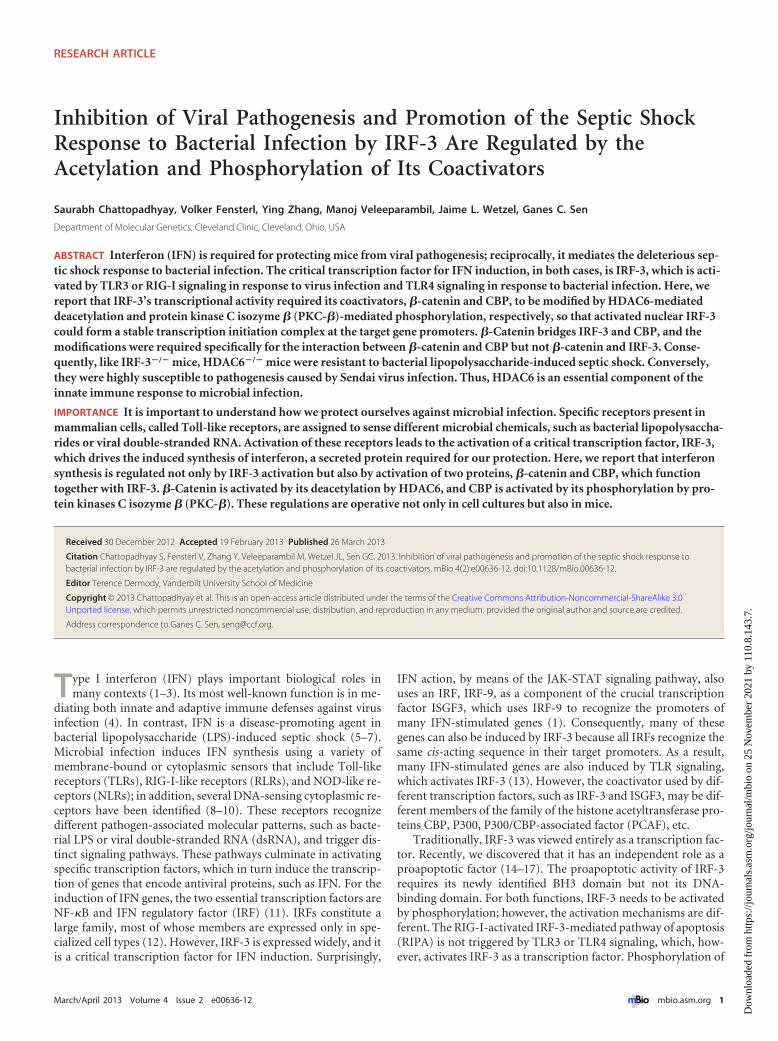
Inhibition of Viral Pathogenesis and Promotion of the Septic ShockResponse to Bacterial Infection by IRF-3 Are Regulated by theAcetylation and Phosphorylation of Its Coactivators
Saurabh Chattopadhyay, Volker Fensterl, Ying Zhang, Manoj Veleeparambil, Jaime L. Wetzel, Ganes C. Sen
Department of Molecular Genetics, Cleveland Clinic, Cleveland, Ohio, USA
ABSTRACT Interferon (IFN) is required for protecting mice from viral pathogenesis; reciprocally, it mediates the deleterious sep-tic shock response to bacterial infection. The critical transcription factor for IFN induction, in both cases, is IRF-3, which is acti-vated by TLR3 or RIG-I signaling in response to virus infection and TLR4 signaling in response to bacterial infection. Here, wereport that IRF-3’s transcriptional activity required its coactivators, �-catenin and CBP, to be modified by HDAC6-mediateddeacetylation and protein kinase C isozyme � (PKC-�)-mediated phosphorylation, respectively, so that activated nuclear IRF-3could form a stable transcription initiation complex at the target gene promoters. �-Catenin bridges IRF-3 and CBP, and themodifications were required specifically for the interaction between �-catenin and CBP but not �-catenin and IRF-3. Conse-quently, like IRF-3�/� mice, HDAC6�/� mice were resistant to bacterial lipopolysaccharide-induced septic shock. Conversely,they were highly susceptible to pathogenesis caused by Sendai virus infection. Thus, HDAC6 is an essential component of theinnate immune response to microbial infection.
IMPORTANCE It is important to understand how we protect ourselves against microbial infection. Specific receptors present inmammalian cells, called Toll-like receptors, are assigned to sense different microbial chemicals, such as bacterial lipopolysaccha-rides or viral double-stranded RNA. Activation of these receptors leads to the activation of a critical transcription factor, IRF-3,which drives the induced synthesis of interferon, a secreted protein required for our protection. Here, we report that interferonsynthesis is regulated not only by IRF-3 activation but also by activation of two proteins, �-catenin and CBP, which functiontogether with IRF-3. �-Catenin is activated by its deacetylation by HDAC6, and CBP is activated by its phosphorylation by pro-tein kinases C isozyme � (PKC-�). These regulations are operative not only in cell cultures but also in mice.
Received 30 December 2012 Accepted 19 February 2013 Published 26 March 2013
Citation Chattopadhyay S, Fensterl V, Zhang Y, Veleeparambil M, Wetzel JL, Sen GC. 2013. Inhibition of viral pathogenesis and promotion of the septic shock response tobacterial infection by IRF-3 are regulated by the acetylation and phosphorylation of its coactivators. mBio 4(2):e00636-12. doi:10.1128/mBio.00636-12.
Editor Terence Dermody, Vanderbilt University School of Medicine
Copyright © 2013 Chattopadhyay et al. This is an open-access article distributed under the terms of the Creative Commons Attribution-Noncommercial-ShareAlike 3.0Unported license, which permits unrestricted noncommercial use, distribution, and reproduction in any medium, provided the original author and source are credited.
Address correspondence to Ganes C. Sen, [email protected].
Type I interferon (IFN) plays important biological roles inmany contexts (1–3). Its most well-known function is in me-
diating both innate and adaptive immune defenses against virusinfection (4). In contrast, IFN is a disease-promoting agent inbacterial lipopolysaccharide (LPS)-induced septic shock (5–7).Microbial infection induces IFN synthesis using a variety ofmembrane-bound or cytoplasmic sensors that include Toll-likereceptors (TLRs), RIG-I-like receptors (RLRs), and NOD-like re-ceptors (NLRs); in addition, several DNA-sensing cytoplasmic re-ceptors have been identified (8–10). These receptors recognizedifferent pathogen-associated molecular patterns, such as bacte-rial LPS or viral double-stranded RNA (dsRNA), and trigger dis-tinct signaling pathways. These pathways culminate in activatingspecific transcription factors, which in turn induce the transcrip-tion of genes that encode antiviral proteins, such as IFN. For theinduction of IFN genes, the two essential transcription factors areNF-�B and IFN regulatory factor (IRF) (11). IRFs constitute alarge family, most of whose members are expressed only in spe-cialized cell types (12). However, IRF-3 is expressed widely, and itis a critical transcription factor for IFN induction. Surprisingly,
IFN action, by means of the JAK-STAT signaling pathway, alsouses an IRF, IRF-9, as a component of the crucial transcriptionfactor ISGF3, which uses IRF-9 to recognize the promoters ofmany IFN-stimulated genes (1). Consequently, many of thesegenes can also be induced by IRF-3 because all IRFs recognize thesame cis-acting sequence in their target promoters. As a result,many IFN-stimulated genes are also induced by TLR signaling,which activates IRF-3 (13). However, the coactivator used by dif-ferent transcription factors, such as IRF-3 and ISGF3, may be dif-ferent members of the family of the histone acetyltransferase pro-teins CBP, P300, P300/CBP-associated factor (PCAF), etc.
Traditionally, IRF-3 was viewed entirely as a transcription fac-tor. Recently, we discovered that it has an independent role as aproapoptotic factor (14–17). The proapoptotic activity of IRF-3requires its newly identified BH3 domain but not its DNA-binding domain. For both functions, IRF-3 needs to be activatedby phosphorylation; however, the activation mechanisms are dif-ferent. The RIG-I-activated IRF-3-mediated pathway of apoptosis(RIPA) is not triggered by TLR3 or TLR4 signaling, which, how-ever, activates IRF-3 as a transcription factor. Phosphorylation of
RESEARCH ARTICLE
March/April 2013 Volume 4 Issue 2 e00636-12 ® mbio.asm.org 1
Dow
nloa
ded
from
http
s://j
ourn
als.
asm
.org
/jour
nal/m
bio
on 2
5 N
ovem
ber
2021
by
110.
8.14
3.7.
specific Ser residues of IRF-3 and then IRF-3’s dimerization andnuclear translocation were known to be the only regulated steps ofits action as a transcription factor (18, 19) until Nusinzon andHorvath (20) discovered that histone deacetylase activity was re-quired as well. They identified HDAC6 as an enzyme that modu-lates IRF-3 action. When �-catenin, an acetylated protein, wasidentified as a coactivator of IRF-3 for its transcriptional activity(21), a potential mechanism for HDAC6 involvement became ap-parent. Indeed, when our work was in progress, Zhu et al. (22)reported that RIG-I activation by Sendai virus (SeV) leads to pro-tein kinase C isozyme � (PKC-�)-mediated activation of HDAC6,causing deacetylation of �-catenin, which results in IRF-3-mediated gene induction.
Here, we report that �-catenin/CBP interaction, not IRF-3/�-catenin interaction, is regulated by HDAC6 and PKC isozyme �;inhibition of either enzyme prevented IRF-3 from forming a stabletranscription initiation complex. Furthermore, we demonstratethat the above-described regulation is critical in vivo. Like IRF-3�/� mice, HDAC6�/� mice were highly susceptible to SeV infec-tion but resistant to LPS-induced septic shock.
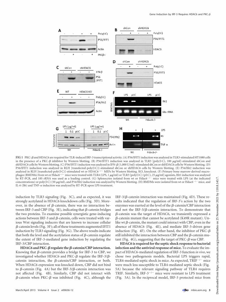
RESULTSPKC-� and HDAC6 are required for IRF-3-mediated gene in-duction by TLR4 and TLR3. Whereas TLR3 signals exclusivelyfrom the endosomal membrane, TLR4 signals from both theplasma membrane and the endosomal membrane, and the endo-somal TLR3 and TLR4 signaling pathways partially overlap (23).The two major transcription factors activated by signaling fromboth TLRs are NF-�B and IRF-3, which induce transcription oftwo sets of genes individually or of a third set, which includesIFN-�, by acting together. To examine whether PKC is requiredfor TLR3 signaling, we used the human cell line HT1080 and auniversal PKC inhibitor, Gö6976. Microarray gene expressionprofiling revealed that induction of IRF-3-driven genes (seeFig. S1A, left panel, in the supplemental material), but not NF-�B-driven genes (Fig. S1A, right panel), was strongly inhibited. West-ern blotting for selective proteins, such as P56/IFIT1 (Fig. S1B),confirmed the conclusion. To distinguish among the various iso-forms of PKC, isoform-specific inhibitors were used. A PKC-�-specific inhibitor was as effective as the universal inhibitor(Fig. 1A), indicating that PKC-� was required for IRF-3-drivengene induction. For testing the requirement of HDAC6 in TLR3signaling, its expression was knocked down by a specific shorthairpin RNA (shRNA). It caused a loss of induction of IFIT1 byTLR3 (Fig. 1B), an effect that was shared by many IRF-3-inducedgenes (Fig. S1C). However, there was no global inhibition of geneinduction; induction of an NF-�B-driven gene, A20, by tumornecrosis factor alpha (TNF-�) was unimpaired (Fig. S1D). Morestrikingly, although the induction of IFIT1 by TLR3 signaling wasinhibited in the absence of HDAC6 (Fig. 1B), induction of thesame gene by beta interferon (IFN-�) was unimpaired (Fig. 1C).This result indicates that the requirement of HDAC6 is signalingpathway specific. Requirement of HDAC6 was further investi-gated in the cytoplasmic RIG-I-like helicase (RLH)-mediatedtranscriptional activation of IRF-3. Induction of IRF-3-dependentgenes by RLH activation was inhibited in HDAC6 knockdownhuman cells (Fig. 1D) and HDAC6�/� mouse embryonic fibro-blasts (MEFs) (Fig. 1E). To expand the significance of our obser-vations, we used primary myeloid cells from wild-type (wt) andHDAC6�/� mice (24). Splenocytes or bone marrow-derived mac-
rophages (BMDMs) were treated with dsRNA for triggering TLR3signaling or with LPS for triggering TLR4 signaling. Both path-ways induced mouse Ifit1 mRNA in wt BMDMs but not inHDAC6�/� cells (Fig. 1F). Similar results were obtained insplenocytes when mouse Ifit2 induction was measured at the pro-tein level (Fig. 1G). Although Ifit1 and Ifit2 induction by TLR4signaling was impaired in HDAC6�/� cells, the induction of NF-�B-driven genes, such as those for interleukin 6 (IL-6) andTNF-�, was unaffected (Fig. 1H; see also Fig. S1E in the supple-mental material). These results demonstrate that the need forHDAC6 was not only signaling pathway specific but also tran-scription factor specific.
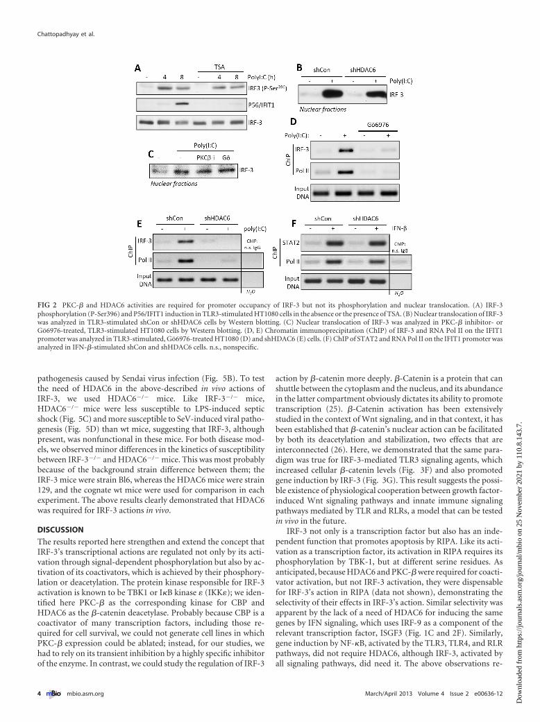
PKC-� and HDAC6 regulate target gene promoter occu-pancy by IRF-3, not its activation and nuclear translocation.Once we identified the affected genes to be IRF-3 driven, we in-quired at which step of its activation or action PKC-� and HDAC6were needed. Inhibition of HDAC6 activity by trichostatin A(TSA), a universal HDAC inhibitor, did not impair IRF-3 activa-tion by TLR3 signaling, as measured by its specific phosphoryla-tion, although, as expected, IFIT1 induction was inhibited (Fig.2A). Similarly, HDAC6 knockdown did not impair nuclear trans-location of activated IRF-3 (Fig. 2B). The same was true for cellstreated with general or the PKC-�-specific inhibitors (Fig. 2C).Although activated IRF-3 was present in the nuclei of PKC-inhibited cells after TLR3 stimulation, it did not occupy the pro-moter of the IFIT1 gene, as revealed by chromatin immunopre-cipitation (ChIP) assay. Both IRF-3 and polymerase II (Pol II)were present at the IFIT1 promoter region in cells treated withpoly(I·C) but only in the absence of the PKC inhibitor (Fig. 2D).Similar results were obtained by comparing wt and HDAC6knockdown cells; upon TLR3-stimulation by poly(I·C), IRF-3 andPol II were bound to the promoter of the IFIT1 gene only in wtcells (Fig. 2E). Remarkably, when cells were stimulated withIFN-�, which uses ISGF3, not IRF-3, to induce the IFIT1 gene,STAT2, a component of ISGF3, and Pol II were bound to thepromoter even in the cells lacking HDAC6 (Fig. 2F). Thus, theresults from the ChIP assays mirrored those from the gene expres-sion analyses. The results presented in Fig. 2 demonstrated thatboth PKC-� and HDAC6 were required, not for IRF-3 activationand nuclear translocation, but to form a stable initiation complexat the promoters of target genes. Moreover, the effects appeared tobe transcription factor specific and not mediated by any altera-tions of chromatin structures or the transcription machinery, be-cause the same gene could be induced normally by a differenttranscription factor, one that recognizes the same promoter se-quence.
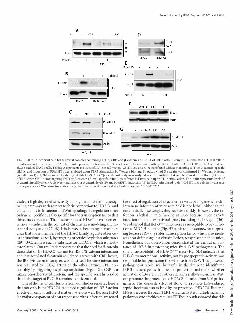
HDAC6 is needed for IRF-3/CBP interaction, which is medi-ated by �-catenin. As a transcription factor, IRF-3 uses CBP as thecoactivator. As revealed by their coimmunoprecipitation (Co-IP),the two proteins physically interacted upon IRF-3 activation byTLR3 signaling; this interaction did not occur in cells treated withan HDAC inhibitor (Fig. 3A) or in HDAC6 knockdown cells (Fig.3B). To identify the substrate of HDAC6 in this context, we didextensive mass spectrometric analysis of IRF-3 isolated from un-stimulated and stimulated cells for its possible acetylation anddeacetylation; these efforts could not demonstrate that IRF-3 isacetylated under any situation (results not shown). However, inlight of the discovery of the need of �-catenin for IRF-3 activity(21), we focused our attention on the acetylation status of�-catenin. We confirmed that �-catenin was needed for IFIT1
Chattopadhyay et al.
2 ® mbio.asm.org March/April 2013 Volume 4 Issue 2 e00636-12
Dow
nloa
ded
from
http
s://j
ourn
als.
asm
.org
/jour
nal/m
bio
on 2
5 N
ovem
ber
2021
by
110.
8.14
3.7.
induction by TLR3 signaling (Fig. 3C), and as expected, it wasstrongly acetylated in HDAC6 knockdown cells (Fig. 3D). More-over, in the absence of �-catenin, there was no interaction be-tween IRF-3 and CBP (Fig. 3E), indicating that �-catenin bridgesthe two proteins. To examine possible synergistic gene-inducingactions between IRF-3 and �-catenin, cells were treated with var-ious Wnt signaling inducers that are known to increase cellular�-catenin levels (Fig. 3F); all of these treatments augmented IFIT1induction by TLR3 signaling (Fig. 3G). The above results indicatethat both the level and the acetylation status of �-catenin regulatethe extent of IRF-3-mediated gene induction by regulating theIRF-3/CBP interaction.
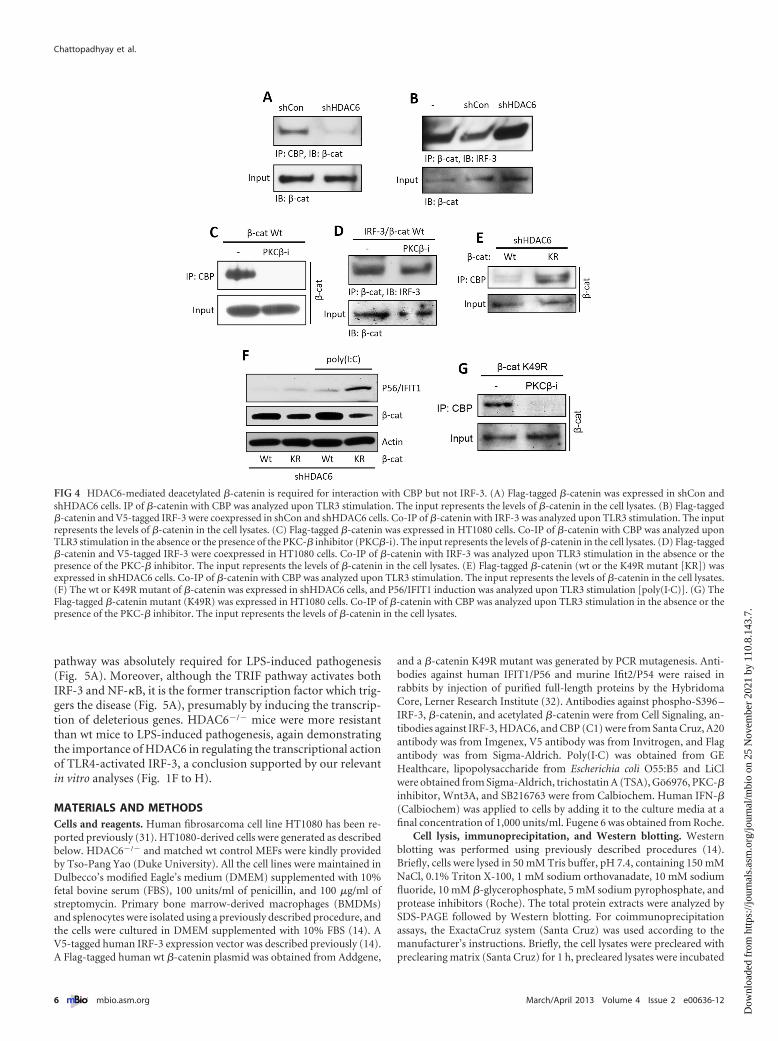
HDAC6 and PKC-� regulate the �-catenin/CBP interaction.Knowing that �-catenin provides a bridge for IRF-3 to CBP, weinvestigated whether HDAC6 and PKC-� regulate the IRF-3/�-catenin interaction, the �-catenin/CBP interaction, or both.When HDAC6 expression was knocked down, CBP did not bindto �-catenin (Fig. 4A) but the IRF-3/�-catenin interaction wasnot affected (Fig. 4B). Similarly, CBP did not interact with�-catenin when PKC-� was inhibited (Fig. 4C), although the
IRF-3/�-catenin interaction was maintained (Fig. 4D). These re-sults indicated that the regulation of IRF-3’s action by the twoenzymes was exerted at the level of the �-catenin/CBP interactionand not the IRF-3/�-catenin interaction. To demonstrate that�-catenin was the target of HDAC6, we transiently expressed a�-catenin mutant that cannot be acetylated (K49R mutant). Un-like wt �-catenin, the mutant could interact with CBP, even in theabsence of HDAC6 (Fig. 4E), and mediate IRF-3-driven geneinduction (Fig. 4F). On the other hand, the inhibitor of PKC-�still inhibited the interaction between CBP and the �-catenin mu-tant (Fig. 4G), suggesting that the target of PKC-� was CBP.
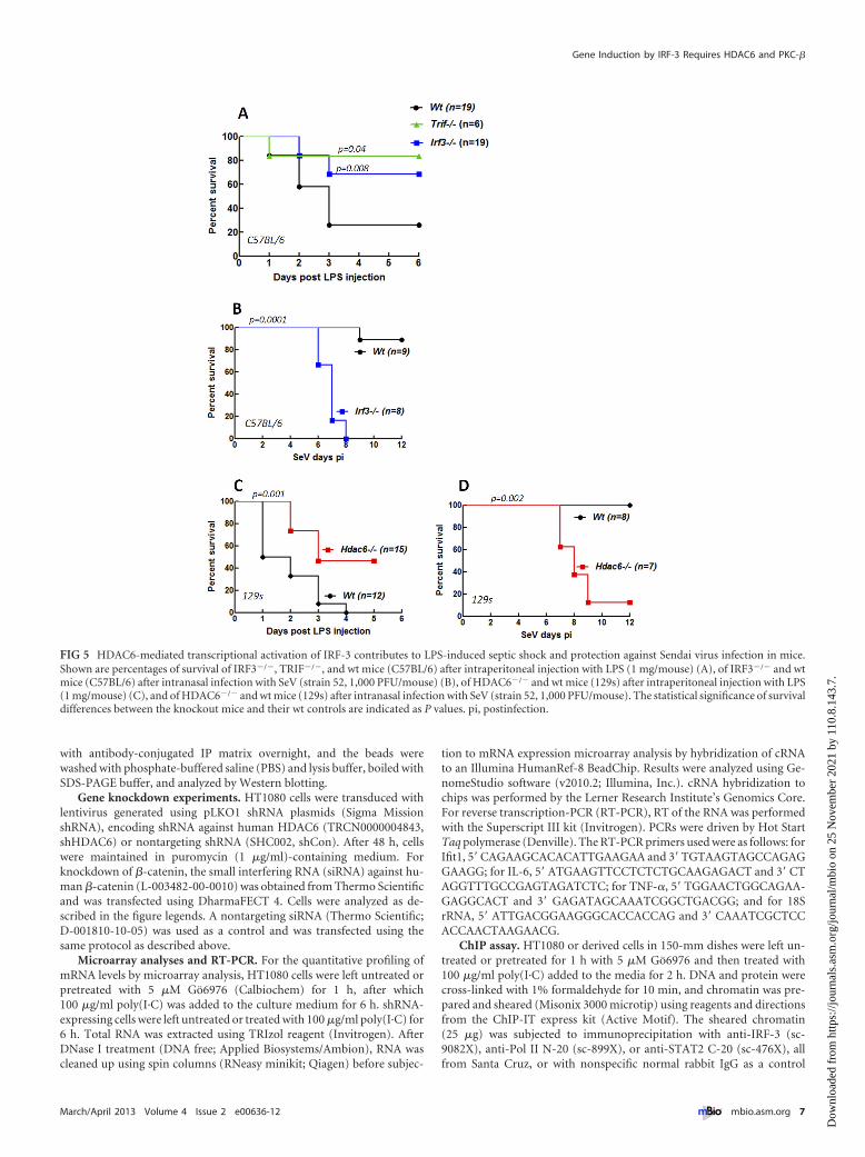
HDAC6 is required for the septic shock response to bacterialinfection and the antiviral response of mice. To evaluate the im-pact of HDAC6-mediated regulation of IRF-3 function in vivo, wechose two pathogenesis models. Bacterial LPS triggers rapid,TLR4-mediated septic shock in mice. As expected, TRIF�/� micewere much less susceptible to TLR4-mediated septic shock (Fig.5A) because the relevant signaling pathway of TLR4 requiresTRIF. Similarly, IRF-3�/� mice were resistant to LPS treatment(Fig. 5A). In the reciprocal model, IRF-3 protected mice from
FIG 1 PKC-� and HDAC6 are required for TLR-induced IRF-3 transcriptional activity. (A) P56/IFIT1 induction was analyzed in TLR3-stimulated HT1080 cellsin the presence of a PKC-� inhibitor by Western blotting. (B) P56/IFIT1 induction was analyzed in TLR3 [poly(I·C), 100 �g/ml]-stimulated shCon andshHDAC6 cells by Western blotting. (C) P56/IFIT1 induction was analyzed in IFN-� (1,000 U/ml)-stimulated shCon or shHDAC6 cells by Western blotting. (D)P56/IFIT1 induction was analyzed in RLH [transfected-poly(I·C)]-stimulated shCon or shHDAC6 cells by Western blotting. (E) P54/Ifit2 induction wasanalyzed in RLH [transfected-poly(I·C)]-stimulated wt or HDAC6�/� MEFs by Western blotting. KO, knockout. (F) Primary bone marrow-derived macro-phages (BMDMs) from wt or Hdac6�/� mice were treated with TLR4 (LPS, 1 �g/ml) or TLR3 [poly(I·C) (pI:C), 25 �g/ml] agonists, Ifit1 induction was analyzedby RT-PCR, and 18S rRNA was used as a loading control. (G) Splenocytes isolated from wt or Hdac6�/� mice were treated with LPS (at the indicatedconcentrations) or poly(I·C) (50 �g/ml), and P54/Ifit2 induction was analyzed by Western blotting. (H) BMDMs were isolated from wt or Hdac6�/� mice, andIL-6 (Il6) and TNF-� induction was analyzed by RT-PCR upon LPS treatment.
Gene Induction by IRF-3 Requires HDAC6 and PKC-�
March/April 2013 Volume 4 Issue 2 e00636-12 ® mbio.asm.org 3
Dow
nloa
ded
from
http
s://j
ourn
als.
asm
.org
/jour
nal/m
bio
on 2
5 N
ovem
ber
2021
by
110.
8.14
3.7.
pathogenesis caused by Sendai virus infection (Fig. 5B). To testthe need of HDAC6 in the above-described in vivo actions ofIRF-3, we used HDAC6�/� mice. Like IRF-3�/� mice,HDAC6�/� mice were less susceptible to LPS-induced septicshock (Fig. 5C) and more susceptible to SeV-induced viral patho-genesis (Fig. 5D) than wt mice, suggesting that IRF-3, althoughpresent, was nonfunctional in these mice. For both disease mod-els, we observed minor differences in the kinetics of susceptibilitybetween IRF-3�/� and HDAC6�/� mice. This was most probablybecause of the background strain difference between them; theIRF-3 mice were strain Bl6, whereas the HDAC6 mice were strain129, and the cognate wt mice were used for comparison in eachexperiment. The above results clearly demonstrated that HDAC6was required for IRF-3 actions in vivo.
DISCUSSION
The results reported here strengthen and extend the concept thatIRF-3’s transcriptional actions are regulated not only by its acti-vation through signal-dependent phosphorylation but also by ac-tivation of its coactivators, which is achieved by their phosphory-lation or deacetylation. The protein kinase responsible for IRF-3activation is known to be TBK1 or I�B kinase � (IKK�); we iden-tified here PKC-� as the corresponding kinase for CBP andHDAC6 as the �-catenin deacetylase. Probably because CBP is acoactivator of many transcription factors, including those re-quired for cell survival, we could not generate cell lines in whichPKC-� expression could be ablated; instead, for our studies, wehad to rely on its transient inhibition by a highly specific inhibitorof the enzyme. In contrast, we could study the regulation of IRF-3
action by �-catenin more deeply. �-Catenin is a protein that canshuttle between the cytoplasm and the nucleus, and its abundancein the latter compartment obviously dictates its ability to promotetranscription (25). �-Catenin activation has been extensivelystudied in the context of Wnt signaling, and in that context, it hasbeen established that �-catenin’s nuclear action can be facilitatedby both its deacetylation and stabilization, two effects that areinterconnected (26). Here, we demonstrated that the same para-digm was true for IRF-3-mediated TLR3 signaling agents, whichincreased cellular �-catenin levels (Fig. 3F) and also promotedgene induction by IRF-3 (Fig. 3G). This result suggests the possi-ble existence of physiological cooperation between growth factor-induced Wnt signaling pathways and innate immune signalingpathways mediated by TLR and RLRs, a model that can be testedin vivo in the future.
IRF-3 not only is a transcription factor but also has an inde-pendent function that promotes apoptosis by RIPA. Like its acti-vation as a transcription factor, its activation in RIPA requires itsphosphorylation by TBK-1, but at different serine residues. Asanticipated, because HDAC6 and PKC-� were required for coacti-vator activation, but not IRF-3 activation, they were dispensablefor IRF-3’s action in RIPA (data not shown), demonstrating theselectivity of their effects in IRF-3’s action. Similar selectivity wasapparent by the lack of a need of HDAC6 for inducing the samegenes by IFN signaling, which uses IRF-9 as a component of therelevant transcription factor, ISGF3 (Fig. 1C and 2F). Similarly,gene induction by NF-�B, activated by the TLR3, TLR4, and RLRpathways, did not require HDAC6, although IRF-3, activated byall signaling pathways, did need it. The above observations re-
FIG 2 PKC-� and HDAC6 activities are required for promoter occupancy of IRF-3 but not its phosphorylation and nuclear translocation. (A) IRF-3phosphorylation (P-Ser396) and P56/IFIT1 induction in TLR3-stimulated HT1080 cells in the absence or the presence of TSA. (B) Nuclear translocation of IRF-3was analyzed in TLR3-stimulated shCon or shHDAC6 cells by Western blotting. (C) Nuclear translocation of IRF-3 was analyzed in PKC-� inhibitor- orGo6976-treated, TLR3-stimulated HT1080 cells by Western blotting. (D, E) Chromatin immunoprecipitation (ChIP) of IRF-3 and RNA Pol II on the IFIT1promoter was analyzed in TLR3-stimulated, Go6976-treated HT1080 (D) and shHDAC6 (E) cells. (F) ChIP of STAT2 and RNA Pol II on the IFIT1 promoter wasanalyzed in IFN-�-stimulated shCon and shHDAC6 cells. n.s., nonspecific.
Chattopadhyay et al.
4 ® mbio.asm.org March/April 2013 Volume 4 Issue 2 e00636-12
Dow
nloa
ded
from
http
s://j
ourn
als.
asm
.org
/jour
nal/m
bio
on 2
5 N
ovem
ber
2021
by
110.
8.14
3.7.
vealed a high degree of selectivity among the innate immune sig-naling pathways with respect to their connection to HDAC6 andconsequently to �-catenin and Wnt signaling; the regulation is notonly gene specific but also specific for the transcription factor thatdrives its expression. The nuclear roles of HDACs have been ex-tensively studied in the context of chromatin remodeling and hi-stone deacetylation (27, 28). It is, however, becoming increasinglyclear that some members of the HDAC family regulate other cel-lular functions, as well, by targeting other deacetylation substrates(29). �-Catenin is such a substrate for HDAC6, which is mostlycytoplasmic. Our results demonstrated that the need for �-catenindeacetylation by HDAC6 was not for IRF-3/�-catenin interactionand that acetylated �-catenin could not interact with CBP; hence,the IRF-3/�-catenin complex was inactive. The same interactionwas regulated by PKC-� acting on the other partner, CBP, pre-sumably by triggering its phosphorylation (Fig. 4G). CBP is ahighly phosphorylated protein, and the specific Ser/Thr residuethat is the target of PKC-� remains to be identified.
One of the major conclusions from our studies reported here isthat not only is the HDAC6-mediated regulation of IRF-3 actioneffective in cells in culture, it matters in vivo as well. Because IRF-3is a major component of host response to virus infection, we tested
the effect of regulation of its action in a virus pathogenesis model.Intranasal infection of mice with SeV is not lethal. Although themice initially lose weight, they recover quickly. However, the in-fection is lethal in mice lacking MDA-5 because it senses SeVinfection and induces antiviral genes, including the IFN gene (30).We observed that IRF-3�/� mice were as susceptible to SeV infec-tion as MDA-5�/� mice (Fig. 5B); this result is somewhat surpris-ing because IRF-7, a sister transcription factor which also medi-ates host defense against virus infection, was present in these mice.Nonetheless, our observation demonstrated the central impor-tance of IRF-3 in protecting mice from SeV pathogenesis. Thesimilar susceptibility of HDAC6�/� mice (Fig. 5D) indicated thatIRF-3’s transcriptional activity, not its proapoptotic activity, wasresponsible for protecting the wt mice from SeV. This powerfulpathogenesis model will be useful in the future to identify theIRF-3-induced genes that mediate protection and to test whetheractivation of �-catenin by other signaling pathways, such as Wnt,can promote the protection of HDAC6�/� mice from SeV patho-genesis. The opposite effect of IRF-3 to promote LPS-inducedseptic shock was also assisted by the presence of HDAC6. BacterialLPS is triggered through TLR4, which activates multiple signalingpathways, one of which requires TRIF; our results showed that this
FIG 3 HDAC6-deficient cells fail to recruit complex containing IRF-3, CBP, and �-catenin. (A) Co-IP of IRF-3 with CBP in TLR3-stimulated HT1080 cells inthe absence or the presence of TSA. The input represents the levels of IRF-3 in cell lysates. IB, immunoblotting. (B) Co-IP of IRF-3 with CBP in TLR3-stimulatedshCon and shHDAC6 cells. The input represents the levels of IRF-3 in cell lysates. (C) HT1080 cells were transfected with nontargeting (NT) or �-catenin-specificsiRNA, and induction of P56/IFIT1 was analyzed upon TLR3 stimulation by Western blotting. Knockdown of �-catenin was confirmed by Western blotting(middle panel). (D) �-Catenin acetylation (acetylated K49 [Ac-K49]-specific antibody) was analyzed in shCon and shHDAC6 cells by Western blotting. (E) Co-IPof IRF-3 with CBP in nontargeting (NT) or �-catenin (�-cat)-specific, siRNA-transfected HT1080 cells upon TLR3 stimulation. The input represents levels of�-catenin in cell lysates. (F, G) Western analyses of �-catenin levels (F) and P56/IFIT1 induction (G) in TLR3-stimulated [poly(I·C)] HT1080 cells in the absenceor the presence of Wnt signaling activators (as indicated). Actin was used as a loading control. SB, SB216763.
Gene Induction by IRF-3 Requires HDAC6 and PKC-�
March/April 2013 Volume 4 Issue 2 e00636-12 ® mbio.asm.org 5
Dow
nloa
ded
from
http
s://j
ourn
als.
asm
.org
/jour
nal/m
bio
on 2
5 N
ovem
ber
2021
by
110.
8.14
3.7.
pathway was absolutely required for LPS-induced pathogenesis(Fig. 5A). Moreover, although the TRIF pathway activates bothIRF-3 and NF-�B, it is the former transcription factor which trig-gers the disease (Fig. 5A), presumably by inducing the transcrip-tion of deleterious genes. HDAC6�/� mice were more resistantthan wt mice to LPS-induced pathogenesis, again demonstratingthe importance of HDAC6 in regulating the transcriptional actionof TLR4-activated IRF-3, a conclusion supported by our relevantin vitro analyses (Fig. 1F to H).
MATERIALS AND METHODSCells and reagents. Human fibrosarcoma cell line HT1080 has been re-ported previously (31). HT1080-derived cells were generated as describedbelow. HDAC6�/� and matched wt control MEFs were kindly providedby Tso-Pang Yao (Duke University). All the cell lines were maintained inDulbecco’s modified Eagle’s medium (DMEM) supplemented with 10%fetal bovine serum (FBS), 100 units/ml of penicillin, and 100 �g/ml ofstreptomycin. Primary bone marrow-derived macrophages (BMDMs)and splenocytes were isolated using a previously described procedure, andthe cells were cultured in DMEM supplemented with 10% FBS (14). AV5-tagged human IRF-3 expression vector was described previously (14).A Flag-tagged human wt �-catenin plasmid was obtained from Addgene,
and a �-catenin K49R mutant was generated by PCR mutagenesis. Anti-bodies against human IFIT1/P56 and murine Ifit2/P54 were raised inrabbits by injection of purified full-length proteins by the HybridomaCore, Lerner Research Institute (32). Antibodies against phospho-S396 –IRF-3, �-catenin, and acetylated �-catenin were from Cell Signaling, an-tibodies against IRF-3, HDAC6, and CBP (C1) were from Santa Cruz, A20antibody was from Imgenex, V5 antibody was from Invitrogen, and Flagantibody was from Sigma-Aldrich. Poly(I·C) was obtained from GEHealthcare, lipopolysaccharide from Escherichia coli O55:B5 and LiClwere obtained from Sigma-Aldrich, trichostatin A (TSA), Go6976, PKC-�inhibitor, Wnt3A, and SB216763 were from Calbiochem. Human IFN-�(Calbiochem) was applied to cells by adding it to the culture media at afinal concentration of 1,000 units/ml. Fugene 6 was obtained from Roche.
Cell lysis, immunoprecipitation, and Western blotting. Westernblotting was performed using previously described procedures (14).Briefly, cells were lysed in 50 mM Tris buffer, pH 7.4, containing 150 mMNaCl, 0.1% Triton X-100, 1 mM sodium orthovanadate, 10 mM sodiumfluoride, 10 mM �-glycerophosphate, 5 mM sodium pyrophosphate, andprotease inhibitors (Roche). The total protein extracts were analyzed bySDS-PAGE followed by Western blotting. For coimmunoprecipitationassays, the ExactaCruz system (Santa Cruz) was used according to themanufacturer’s instructions. Briefly, the cell lysates were precleared withpreclearing matrix (Santa Cruz) for 1 h, precleared lysates were incubated
FIG 4 HDAC6-mediated deacetylated �-catenin is required for interaction with CBP but not IRF-3. (A) Flag-tagged �-catenin was expressed in shCon andshHDAC6 cells. IP of �-catenin with CBP was analyzed upon TLR3 stimulation. The input represents the levels of �-catenin in the cell lysates. (B) Flag-tagged�-catenin and V5-tagged IRF-3 were coexpressed in shCon and shHDAC6 cells. Co-IP of �-catenin with IRF-3 was analyzed upon TLR3 stimulation. The inputrepresents the levels of �-catenin in the cell lysates. (C) Flag-tagged �-catenin was expressed in HT1080 cells. Co-IP of �-catenin with CBP was analyzed uponTLR3 stimulation in the absence or the presence of the PKC-� inhibitor (PKC�-i). The input represents the levels of �-catenin in the cell lysates. (D) Flag-tagged�-catenin and V5-tagged IRF-3 were coexpressed in HT1080 cells. Co-IP of �-catenin with IRF-3 was analyzed upon TLR3 stimulation in the absence or thepresence of the PKC-� inhibitor. The input represents the levels of �-catenin in the cell lysates. (E) Flag-tagged �-catenin (wt or the K49R mutant [KR]) wasexpressed in shHDAC6 cells. Co-IP of �-catenin with CBP was analyzed upon TLR3 stimulation. The input represents the levels of �-catenin in the cell lysates.(F) The wt or K49R mutant of �-catenin was expressed in shHDAC6 cells, and P56/IFIT1 induction was analyzed upon TLR3 stimulation [poly(I·C)]. (G) TheFlag-tagged �-catenin mutant (K49R) was expressed in HT1080 cells. Co-IP of �-catenin with CBP was analyzed upon TLR3 stimulation in the absence or thepresence of the PKC-� inhibitor. The input represents the levels of �-catenin in the cell lysates.
Chattopadhyay et al.
6 ® mbio.asm.org March/April 2013 Volume 4 Issue 2 e00636-12
Dow
nloa
ded
from
http
s://j
ourn
als.
asm
.org
/jour
nal/m
bio
on 2
5 N
ovem
ber
2021
by
110.
8.14
3.7.
with antibody-conjugated IP matrix overnight, and the beads werewashed with phosphate-buffered saline (PBS) and lysis buffer, boiled withSDS-PAGE buffer, and analyzed by Western blotting.
Gene knockdown experiments. HT1080 cells were transduced withlentivirus generated using pLKO1 shRNA plasmids (Sigma MissionshRNA), encoding shRNA against human HDAC6 (TRCN0000004843,shHDAC6) or nontargeting shRNA (SHC002, shCon). After 48 h, cellswere maintained in puromycin (1 �g/ml)-containing medium. Forknockdown of �-catenin, the small interfering RNA (siRNA) against hu-man �-catenin (L-003482-00-0010) was obtained from Thermo Scientificand was transfected using DharmaFECT 4. Cells were analyzed as de-scribed in the figure legends. A nontargeting siRNA (Thermo Scientific;D-001810-10-05) was used as a control and was transfected using thesame protocol as described above.
Microarray analyses and RT-PCR. For the quantitative profiling ofmRNA levels by microarray analysis, HT1080 cells were left untreated orpretreated with 5 �M Gö6976 (Calbiochem) for 1 h, after which100 �g/ml poly(I·C) was added to the culture medium for 6 h. shRNA-expressing cells were left untreated or treated with 100 �g/ml poly(I·C) for6 h. Total RNA was extracted using TRIzol reagent (Invitrogen). AfterDNase I treatment (DNA free; Applied Biosystems/Ambion), RNA wascleaned up using spin columns (RNeasy minikit; Qiagen) before subjec-
tion to mRNA expression microarray analysis by hybridization of cRNAto an Illumina HumanRef-8 BeadChip. Results were analyzed using Ge-nomeStudio software (v2010.2; Illumina, Inc.). cRNA hybridization tochips was performed by the Lerner Research Institute’s Genomics Core.For reverse transcription-PCR (RT-PCR), RT of the RNA was performedwith the Superscript III kit (Invitrogen). PCRs were driven by Hot StartTaq polymerase (Denville). The RT-PCR primers used were as follows: forIfit1, 5= CAGAAGCACACATTGAAGAA and 3= TGTAAGTAGCCAGAGGAAGG; for IL-6, 5= ATGAAGTTCCTCTCTGCAAGAGACT and 3= CTAGGTTTGCCGAGTAGATCTC; for TNF-�, 5= TGGAACTGGCAGAA-GAGGCACT and 3= GAGATAGCAAATCGGCTGACGG; and for 18SrRNA, 5= ATTGACGGAAGGGCACCACCAG and 3= CAAATCGCTCCACCAACTAAGAACG.
ChIP assay. HT1080 or derived cells in 150-mm dishes were left un-treated or pretreated for 1 h with 5 �M Gö6976 and then treated with100 �g/ml poly(I·C) added to the media for 2 h. DNA and protein werecross-linked with 1% formaldehyde for 10 min, and chromatin was pre-pared and sheared (Misonix 3000 microtip) using reagents and directionsfrom the ChIP-IT express kit (Active Motif). The sheared chromatin(25 �g) was subjected to immunoprecipitation with anti-IRF-3 (sc-9082X), anti-Pol II N-20 (sc-899X), or anti-STAT2 C-20 (sc-476X), allfrom Santa Cruz, or with nonspecific normal rabbit IgG as a control
FIG 5 HDAC6-mediated transcriptional activation of IRF-3 contributes to LPS-induced septic shock and protection against Sendai virus infection in mice.Shown are percentages of survival of IRF3�/�, TRIF�/�, and wt mice (C57BL/6) after intraperitoneal injection with LPS (1 mg/mouse) (A), of IRF3�/� and wtmice (C57BL/6) after intranasal infection with SeV (strain 52, 1,000 PFU/mouse) (B), of HDAC6�/� and wt mice (129s) after intraperitoneal injection with LPS(1 mg/mouse) (C), and of HDAC6�/� and wt mice (129s) after intranasal infection with SeV (strain 52, 1,000 PFU/mouse). The statistical significance of survivaldifferences between the knockout mice and their wt controls are indicated as P values. pi, postinfection.
Gene Induction by IRF-3 Requires HDAC6 and PKC-�
March/April 2013 Volume 4 Issue 2 e00636-12 ® mbio.asm.org 7
Dow
nloa
ded
from
http
s://j
ourn
als.
asm
.org
/jour
nal/m
bio
on 2
5 N
ovem
ber
2021
by
110.
8.14
3.7.
(Sigma). After un-cross-linking and proteinase K digestion of proteins inthe precipitation reaction or of input chromatin as a control, coprecipi-tated genomic IFIT1 promoter DNA was detected by PCR amplificationof the 125-bp Interferon Stimulated Response Element (ISRE) plus TATAbox region of the IFIT1 promoter. Primers used were 5= GAATTCCGCTAGCTTTAGTTTCAC and 3= CCCCAAGACAGTGTTATATAAGGG.
Mice, LPS treatment, and Sendai virus infection. IRF-3�/� mice(C57BL/6 background) were obtained from Riken Bio Resource Center,Japan (with permission from Tadatsugu Taniguchi, University of Tokyo),TRIF�/� mice (C57BL/6 background) were obtained from Jackson Lab-oratories, and HDAC6�/� mice (129s background) were kindly providedby Timothy McKinsey (University of Colorado, Denver, CO). For LPStreatment, 8- to 10-week-old mice were injected with LPS (1 mg/mouse)intraperitoneally, and their survival was monitored for 1 week. Sendaivirus (strain 52) was obtained from ATCC and was grown by CharlesRiver Laboratories. For virus infections, 1,000 PFU of SeV in 35 �l ofendotoxin-free PBS was intranasally administered to isoflurane-anesthetized 8- to 10-week-old mice. The mice were monitored daily fortheir weight loss and disease symptoms. All the animal procedures wereperformed according to the protocols approved by the institutional ani-mal care and use committee.
Microarray data accession numbers. All microarray data have beendeposited in the NCBI Gene Expression Omnibus (GEO) database (http://www.ncbi.nlm.nih.gov/geo) under accession numbers GSE43217 andGSE43218.
SUPPLEMENTAL MATERIALSupplemental material for this article may be found at http://mbio.asm.org/lookup/suppl/doi:10.1128/mBio.00636-12/-/DCSupplemental.
Figure S1, PDF file, 0.2 MB.
ACKNOWLEDGMENT
This work was supported by National Institutes of Health grant AI-073303.
REFERENCES1. Fensterl V, Sen GC. 2009. Interferons and viral infections. Biofactors
35:14 –20.2. Borden EC, Sen GC, Uze G, Silverman RH, Ransohoff RM, Foster GR,
Stark GR. 2007. Interferons at age 50: past, current and future impact onbiomedicine. Nat. Rev. Drug Discov. 6:975–990.
3. Nagarajan U. 2011. Induction and function of IFN� during viral andbacterial infection. Crit. Rev. Immunol. 31:459 – 474.
4. Schoggins JW, Rice CM. 2011. Interferon-stimulated genes and theirantiviral effector functions. Curr. Opin. Virol. 1:519 –525.
5. Karaghiosoff M, Steinborn R, Kovarik P, Kriegshäuser G, Baccarini M,Donabauer B, Reichart U, Kolbe T, Bogdan C, Leanderson T, Levy D,Decker T, Müller M. 2003. Central role for type I interferons and Tyk2 inlipopolysaccharide-induced endotoxin shock. Nat. Immunol. 4:471– 477.
6. Sakaguchi S, Negishi H, Asagiri M, Nakajima C, Mizutani T, TakaokaA, Honda K, Taniguchi T. 2003. Essential role of IRF-3 inlipopolysaccharide-induced interferon-beta gene expression and endo-toxin shock. Biochem. Biophys. Res. Commun. 306:860 – 866.
7. Trinchieri G. 2010. Type I interferon: friend or foe? J. Exp. Med. 207:2053–2063.
8. Brennan K, Bowie AG. 2010. Activation of host pattern recognitionreceptors by viruses. Curr. Opin. Microbiol. 13:503–507.
9. Arpaia N, Barton GM. 2011. Toll-like receptors: key players in antiviralimmunity. Curr. Opin. Virol. 1:447– 454.
10. Yan N, Chen ZJ. 2012. Intrinsic antiviral immunity. Nat. Immunol.13:214 –222.
11. Hiscott J. 2007. Convergence of the NF-kappaB and IRF pathways in the
regulation of the innate antiviral response. Cytokine Growth Factor Rev.18:483– 490.
12. Honda K, Taniguchi T. 2006. IRFs: master regulators of signalling byToll-like receptors and cytosolic pattern-recognition receptors. Nat. Rev.Immunol. 6:644 – 658.
13. Sen GC, Sarkar SN. 2005. Transcriptional signaling by double-strandedRNA: role of TLR3. Cytokine Growth Factor Rev. 16:1–14.
14. Chattopadhyay S, Marques JT, Yamashita M, Peters KL, Smith K, DesaiA, Williams BR, Sen GC. 2010. Viral apoptosis is induced by IRF-3-mediated activation of Bax. EMBO J. 29:1762–1773.
15. Chattopadhyay S, Yamashita M, Zhang Y, Sen GC. 2011. The IRF-3/Bax-mediated apoptotic pathway, activated by viral cytoplasmic RNA andDNA, inhibits virus replication. J. Virol. 85:3708 –3716.
16. Chattopadhyay S, Fensterl V, Zhang Y, Veleeparambil M, YamashitaM, Sen GC. 2013. Role of interferon regulatory factor 3-mediated apo-ptosis in the establishment and maintenance of persistent infection bySendai virus. J. Virol. 87:16 –24.
17. White CL, Chattopadhyay S, Sen GC. 2011. Phosphatidylinositol3-kinase signaling delays Sendai virus-induced apoptosis by preventingXIAP degradation. J. Virol. 85:5224 –5227.
18. Hiscott J. 2007. Triggering the innate antiviral response through IRF-3activation. J. Biol. Chem. 282:15325–15329.
19. Servant MJ, Grandvaux N, tenOever BR, Duguay D, Lin R, Hiscott J.2003. Identification of the minimal phosphoacceptor site required for invivo activation of interferon regulatory factor 3 in response to virus anddouble-stranded RNA. J. Biol. Chem. 278:9441–9447.
20. Nusinzon I, Horvath CM. 2006. Positive and negative regulation of theinnate antiviral response and beta interferon gene expression by deacety-lation. Mol. Cell. Biol. 26:3106 –3113.
21. Yang P, An H, Liu X, Wen M, Zheng Y, Rui Y, Cao X. 2010. Thecytosolic nucleic acid sensor LRRFIP1 mediates the production of type Iinterferon via a beta-catenin-dependent pathway. Nat. Immunol. 11:487– 494.
22. Zhu J, Coyne CB, Sarkar SN. 2011. PKC alpha regulates Sendai virus-mediated interferon induction through HDAC6 and �-catenin. EMBO J.30:4838 – 4849.
23. McGettrick AF, O’Neill LA. 2010. Localisation and trafficking of Toll-likereceptors: an important mode of regulation. Curr. Opin. Immunol. 22:20 –27.
24. Gao YS, Hubbert CC, Lu J, Lee YS, Lee JY, Yao TP. 2007. Histonedeacetylase 6 regulates growth factor-induced actin remodeling and en-docytosis. Mol. Cell. Biol. 27:8637– 8647.
25. Valenta T, Hausmann G, Basler K. 2012. The many faces and functionsof �-catenin. EMBO J. 31:2714 –2736.
26. Li Y, Zhang X, Polakiewicz RD, Yao TP, Comb MJ. 2008. HDAC6 isrequired for epidermal growth factor-induced beta-catenin nuclear local-ization. J. Biol. Chem. 283:12686 –12690.
27. Hildmann C, Riester D, Schwienhorst A. 2007. Histone deacetylases—animportant class of cellular regulators with a variety of functions. Appl.Microbiol. Biotechnol. 75:487– 497.
28. Yang XJ, Seto E. 2007. HATs and HDACs: from structure, function andregulation to novel strategies for therapy and prevention. Oncogene 26:5310 –5318.
29. Glozak MA, Sengupta N, Zhang X, Seto E. 2005. Acetylation anddeacetylation of non-histone proteins. Gene 363:15–23.
30. Gitlin L, Benoit L, Song C, Cella M, Gilfillan S, Holtzman MJ, ColonnaM. 2010. Melanoma differentiation-associated gene 5 (MDA5) is involvedin the innate immune response to Paramyxoviridae infection in vivo.PLoS Pathog. 6(1):e1000734. http://dx.doi.org/10.1371/journal.ppat.1000734.
31. Peters K, Chattopadhyay S, Sen GC. 2008. IRF-3 activation by Sendaivirus infection is required for cellular apoptosis and avoidance of persis-tence. J. Virol. 82:3500 –3508.
32. Terenzi F, White C, Pal S, Williams BR, Sen GC. 2007. Tissue-specificand inducer-specific differential induction of ISG56 and ISG54 in mice. J.Virol. 81:8656 – 8665.
Chattopadhyay et al.
8 ® mbio.asm.org March/April 2013 Volume 4 Issue 2 e00636-12
Dow
nloa
ded
from
http
s://j
ourn
als.
asm
.org
/jour
nal/m
bio
on 2
5 N
ovem
ber
2021
by
110.
8.14
3.7.