Embed Size (px)
Citation preview
Initial Experience with the Radiotracer Anti-1-Amino-3-18F-Fluorocyclobutane-1-CarboxylicAcid with PET/CT in Prostate Carcinoma
David M. Schuster1, John R. Votaw1, Peter T. Nieh2, Weiping Yu1, Jonathon A. Nye1, Viraj Master2, F. DuBois Bowman3,Muta M. Issa2, and Mark M. Goodman1
1Division of Nuclear Medicine, Department of Radiology, Emory University, Atlanta, Georgia; 2Department of Urology, EmoryUniversity, Atlanta, Georgia; and 3Department of Biostatistics, Emory University, Atlanta, Georgia
Conventional imaging techniques have serious limitations in thedetection, staging, and restaging of prostate carcinoma. Anti-1-amino-3-18F-fluorocyclobutane-1-carboxylic acid (anti-18F-FACBC)is a synthetic L-leucine analog that has excellent in vitro uptake withinthe DU-145 prostate carcinoma cell line and orthotopically implantedprostate tumor in nude rats. There is little renal excretion comparedwith 18F-FDG. The present study examines anti-18F-FACBC uptakein patients with newly diagnosed and recurrent prostate carcinoma.Methods: Fifteen patients with a recent diagnosis of prostate carci-noma (n 5 9) or suspected recurrence (n 5 6) underwent 65-mindynamic PET/CT of the pelvis after intravenous injection of 300–410MBq anti-18F-FACBC followed by static body images. Each studywas evaluated qualitatively and quantitatively. Maximum standardizeduptake values were recorded in the prostate or prostate bed, andwithin lymph nodes at 4.5 min (early) and 20 min (delayed), and corre-lated with clinical, imaging and pathologic follow-up. Time–activitycurves were also generated for benign and malignant tissue.Results:In the 8 patients with newly diagnosed prostate carcinoma whounderwent dynamic scanning, visual analysis correctly identified thepresence or absence of focal neoplastic involvement in 40 of 48 pros-tate sextants. Pelvic nodal status correlated with anti-18F-FACBCfindings in 7 of 9 patients and was indeterminate in 2 of 9. In all 4patients in whom there was proven recurrence, visual analysis wassuccessful in identifying disease (1 prostate bed, 3 extraprostatic). In3 of these patients, 111In-capromab-pendetide had no significantuptake at nodal and skeletal foci. Malignant lymph node uptake inboth the staging and restaging patients was significantly higher thanbenign nodal uptake. Though uptake faded with time, in all 6 patientswith either lymph node metastases or recurrent prostate bed carci-noma, there was intense persistent uptake at 65 min. Conclu-sion: Anti-18F-FACBC is a promising radiotracer for imagingprostate carcinoma. Radiotracer uptake was demonstrated in pri-mary and metastatic disease. Future research should investigatethe mechanism of radiotracer uptake in normal and pathologic tis-sue and develop a clinical imaging strategy for initial staging andrestaging.
Key Words: anti-18F-FACBC; prostate; neoplasia; PET
J Nucl Med 2007; 48:56–63
Prostate carcinoma is the most common malignancy inmen, comprising 33% of newly diagnosed cancers in theUnited States in 2005 (1). Accurately detecting the pres-ence of disease confined to the prostate bed versus that ofextraprostatic spread to lymph nodes or the skeletal systemhas profound treatment implications.
Currently, there is no definitive imaging technique in theinitial staging and restaging of prostate carcinoma. Whereasultrasound and MRI have proven useful for local (T) stag-ing, they have limitations and are not universally used (2–4).For local recurrence in the prostate bed, ultrasound-guidedtransrectal biopsy is often used but it is invasive and subjectto sampling error (5,6). For lymph node assessment, routineCT and MRI are considered to have poor accuracy (3,7).
Molecular imaging techniques such as 111In-capromabpendetide (ProstaScint; Cytogen Corp.) have been used, butsensitivity is only 62%–75% (3,8,9). 18F-FDG PET is notof sufficient diagnostic accuracy for routine clinical usefor prostate carcinoma (5,9–14). Other PET radiotracers,including 11C-acetate, 11C-choline, 11C-methionine, and18F-fluorocholine (18F-FCH), may characterize different met-abolic aspects of prostate carcinoma but have demonstratedmixed results for clinical evaluation (4,5,10,15–25). Ofcourse, the 11C-based radiotracers are impractical without anon-site cyclotron. 18F-FCH also seems to have high urinaryexcretion starting at 5 min after acquisition (5,19,20,25–28).Thus, there is a need for a better radiotracer, with little or nourinary excretion, for the evaluation and staging of patientswith prostate carcinoma.
Anti-1-amino-3-18F-fluorocyclobutane-1-carboxylic acid(anti-18F-FACBC) is a synthetic L-leucine analog that hasexcellent in vitro uptake within the DU-145 prostate carci-noma cell line and within orthotopically implanted prostatetumors in nude rats (29). The uptake of anti-18F-FACBC islikely mediated via the sodium-independent L large-neutral
Received Jul. 20, 2006; revision accepted Oct. 12, 2006.For correspondence or reprints contact: David M. Schuster, MD, Depart-
ment of Radiology, Emory University Hospital, Room E145, 1364 Clifton Rd.,Atlanta, GA 30322.
E-mail: [email protected] ª 2007 by the Society of Nuclear Medicine, Inc.
56 THE JOURNAL OF NUCLEAR MEDICINE • Vol. 48 • No. 1 • January 2007
amino acid transport system (LAT) (30,31). There is little re-nal excretion compared with 18F-FDG (29). The present studyexamines anti-18F-FACBC uptake in patients with newly diag-nosed and suspected recurrent prostate carcinoma.
MATERIALS AND METHODS
Preparation of Anti-18F-FACBCThe preparation of anti-18F-FACBC has been previously re-
ported (32). The decay-corrected radiochemical yield of the de-sired product was 24% and its radiochemical purity was 99% at80 min after the end of bombardment. The injected mass wasapproximately 5 mg, or 38 mmol, and the specific activity was136.9–192.4 GBq/mmol (3.7–5.2 Ci/mmol).
Patient SelectionAll studies were performed under the auspices of the Emory
University Institutional Review Board, Radioactive Drug ResearchCommittee, and the Atlanta Veterans Affairs Medical CenterResearch and Development Committee. Inclusion criteria includedany patient with histologically confirmed prostate carcinomaeligible for prostatectomy and possible lymph node biopsy/dis-section or with a prior history of histologically confirmed prostatecarcinoma with suspected recurrence or metastatic disease. Onepatient had been recruited under a prior protocol for imaging renalmasses who also happened to have prostate carcinoma. Thispatient underwent dynamic imaging of the abdomen at the levelof the kidneys (otherwise the same protocol as in this study). Allnewly diagnosed patients were imaged 4–8 wk after the initialprostate biopsy. Prostate-specific antigen (PSA) values wereobtained within 4 mo of the anti-18F-FACBC scan. Patient demo-graphics are as follows: mean age 6 SD is 62.0 6 8.8 y witha range of 45–76 y; mean PSA 6 SD is 15.0 6 18.6 ng/mL with arange of 1.9–71 ng/mL; and median Gleason score is 7 with arange of 6–10. The original Gleason scores are used for thepatients with suspected recurrence.
PET Imaging ProtocolAll scanning was conducted on a Discovery DLS or DST in-
tegrated PET/CT scanner (GE Healthcare), and the images wereinterpreted on a combination of an AW workstation with VolumeViewer Plus (GE Healthcare) as well as a program developed bythe authors on the IDL platform (RSI Inc.) running on a Pentium 4(Intel Corp.) computer (operating system, Microsoft; computer hard-ware, IBM). All patients fasted for 4–6 h before the anti-18F-FACBCscan. This is our standard practice for 18F-FDG PET, though it hasnot been established if a fasting or nonfasting state optimizesanti-18F-FACBC imaging. The patient first underwent a CT scan ofthe abdomen and pelvis (80–120 mA) without oral or intravenouscontrast for anatomic correlation and attenuation correction ofemission data. The patient then received a bolus of anti-18F-FACBC(300–410 MBq) injected intravenously over 1–2 min. PET con-sisted of a 15-frame dynamic sequence lasting 65 min (number offrames · time [min]: 6 · 0.5, 4 · 3, and 5 · 10) followed by a staticscan of the abdomen and pelvis at 4 min per bed position.
Image AnalysisThe PET/CT studies were assessed by 1 experienced nuclear
radiologist in a prospective manner. For patients with newly diag-nosed prostate carcinoma, the prostate was visually divided into6 sextants (right and left apex, mid, and base). Three equallyspaced regions of interest (ROIs) conforming to the axial dimen-
sions of the sextant were drawn, and the pixel with the greateststandardized uptake value (SUV) in the ROI was noted. Thehighest of these 3 SUVs was recorded as the maximum SUV(SUVmax) for that sextant at the 4.5-min midpoint (3-min framespanning a dynamic acquisition from 3 to 6 min) and at the 20-minmidpoint (10-min frame at 15–25 min). The 4.5-min (early) and20-min (delayed) frames were chosen based on preliminary anal-ysis of the time–activity curves. In addition, each sextant wasvisually assessed for the presence of focal activity. A region wasconsidered positive if there was asymmetric focal activity exceed-ing prostate background activity, similar to the criteria used byYamaguchi et al. in studying 11C-choline PET (18). We augmentedvisual interpretation by deriving a ratio of sextant SUVmax dividedby muscle mean SUV (SUVmean) in each patient. The SUVmean wasobtained by drawing a 3-cm ROI in each gluteus muscle. SUVmean
in muscle was chosen because it remained fairly constant with timeand best-simulated typical soft-tissue background. Ideally, we wouldhave chosen a prostatectomy-proven normal sextant as backgroundbut this was not possible in most patients.
A lymph node or treated prostate bed was considered visuallypositive if there was sustained focal activity over expected softtissue or blood pool. Intensity was recorded as follows: mild(above blood pool but less than muscle), moderate (above musclebut less than marrow), and intense (above marrow). Values ofSUVmax were also obtained from these lymph nodes or for thoseof .1 cm in short axis.
Time–Activity CurvesROIs were defined by hand on transaxial planes over areas of
focal uptake or drawn on the CT scan in regions of mild or diffuseuptake. The ROIs included prostate, benign and malignant lymphnodes, bone marrow (ilium), blood pool (femoral artery), muscle(gluteus), bowel (sigmoid), and bladder. Regions of ,2 cm indiameter used CT for correction of partial-volume and tissuespillover effects (33).
Correlation of Imaging to Clinical DataAnti-18F-FACBC imaging findings were correlated with path-
ologic, clinical, biochemical, and imaging follow-up for up to 1 yafter scanning. For local disease, this included pathologic analysisof prostatectomy as well as prostate biopsy tissue. A lymph nodewas considered clinically positive if there was pathologic proof ofneoplasia or progressive increase in lymph node size in thepresence of elevated PSA. The absence of lymph node involve-ment was confirmed if there was a negative surgical lymph nodedissection or biopsy or, if lymph node dissection was not per-formed, PSA declining to nadir (,0.05) after definitive therapy, orif there were no enlarged lymph nodes on follow-up imaging at6 mo. If a pelvic lymph node dissection was not performed and thepatient was placed on hormonal therapy, lymph node status wasconsidered indeterminate.
Statistical AnalysisNonparametric statistical methods were applied for analyses,
given the limited sample size in our study. We assessed allbetween-group differences in SUVmax using the Wilcoxon ranksum test. Statistical significance was determined using a type-Ierror rate of a 5 0.05, and specific P values are reported with theresults. The statistical analyses were performed using SAS version9.1 software. Statistical significance was reported only if n . 2 ineach comparison group.
ANTI-18F-FACBC IN PROSTATE CARCINOMA • Schuster et al. 57
RESULTS
Initial Staging
Eight patients underwent dynamic prostate imaging andwere correlated with either sextant biopsy (n 5 4) or histo-logic examination of the surgically removed prostate (n 5
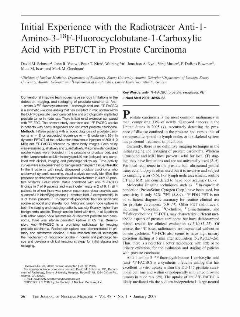
4). Thirty-nine sextants were positive for carcinoma (1 witha minute focus) and 9 were negative (1 with chronicinflammation). Visual analysis predicted the presence orabsence of carcinoma in 40 of 48 sextants. Table 1 is alisting of sextant values of SUVmax and ratios of SUVmax tomuscle SUVmean for each patient. A typical patient exampleis presented in Figure 1.
In correlation of lymph node status, 7 of 9 patients hadexcellent concordance of anti-18F-FACBC pelvic nodalfindings with clinical follow-up. Two of 9 were indetermi-nate. Table 2 is a summary of lymph node concordance and
Figure 2 is an example of a patient with lymph node me-tastases. Both patients with malignant lymph nodes hadintense persistent uptake, even at 65 min, though less in-tense than on earlier sequences.
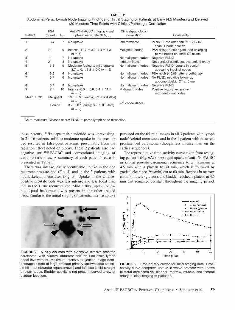
The representative time–activity curve taken from patient3 (Fig. 3) shows rapid uptake of anti-18F-FACBC in prostatecarcinoma to a maximum at 4.5 min, followed by gradualclearance (9.5%/min) beyond 20 min. Regions in marrow(ilium), muscle (gluteus), and bladder reached a plateau at4.5 min, which remained constant throughout the imagingperiod. This activity time course was consistent across thepatient population for malignant prostate and normal tissues.
Suspected Recurrence
On a per-patient basis, the anti-18F-FACBC scan detectedneoplasia in 4 of 4 patients with proven recurrence. In 3 of
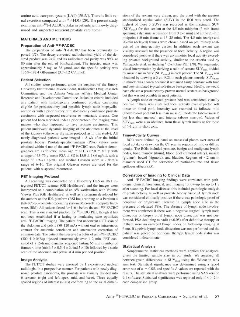
TABLE 1Anti-18F-FACBC Pathologic Malignant vs. Benign Sextant SUVmax and SUVmax/Muscle SUVmean Ratios at Early
(4.5 Minutes) and Delayed (20 Minutes) Time Points for Initial Staging of Patients
Patient
Time
point Malignant SUVmax Benign SUVmax
Malignant
sextant/muscle ratio
Benign
sextant/muscle ratio
1 Early 7, 9.4, 5.6, 9.3, 9.2, 3.8 9.1, 12.2, 7.3, 12.1, 11.9, 4.9
Late 4.8, 5.1, 3.9, 5.9, 5.3, 3.6 4.5, 4.8, 3.7, 5.6, 5.0, 3.4
2 Early 7.4, 7.6, 11.2, 7.4, 7.7, 8.1 11.9, 12.3, 18.1, 11.9, 12.4, 13.1Late 4.3, 4.9, 4.8, 4.3, 4.5, 4.3 4.5, 5.1, 5.0, 4.5, 4.7, 4.5
3 Early 6.8, 6.5, 5.8, 6.9, 5.6 5.1 14.5, 13.8, 12.3, 14.7, 11.9 10.9
Late 5.7, 5.4, 5.2, 5.5, 4.3 4.6 7.6, 7.2, 6.9, 7.3, 5.7 6.1
4 Early 2.2, 2.5, 1.6, 2.1, 2.0, 1.8 4.4, 5.0, 3.2, 4.2, 4.0, 3.6Late 2.8, 2.8, 1.9, 2.4, 2.2, 1.7 4.0, 4.0, 2.7, 3.4, 3.1, 2.4
5 Early 4.5, 5.8, 4.4 3.6, 4.6, 3.3 4.1, 5.3, 4.0 3.3, 4.2, 2.8
Late 6.0, 5.3, 5.0 3.2, 3.2, 3.0 3.8, 3.4, 3.2 2.0, 2.3, 1.8
6 Early 6.7, 6.7, 4.1 5.8, 5.5, 4.6 6.8, 6.8, 4.1 5.9, 5.6, 4.6Late 5.6, 4.3, 3.0 3.4, 3.5, 3.2 3.9, 3.0, 2.1 2.4, 2.5, 2.3
7 Early 6.5, 8.9, 5.5, 5.0, 8.2, 6.6 13.5, 18.5, 11.5, 10.4, 17.1, 13.8
Late 5.0, 4.7, 3.9, 5.0, 5.7, 4.7 6.8, 6.4, 5.3, 6.8, 7.7, 6.48 Early 6.6, 6.3, 4.4, 4.5 4.5, 4.8 6.5, 6.2, 4.3, 3.5 4.4, 4.7
Late 4.3, 4.2, 4.3, 4.4 4.1, 3.9 3.0, 2.9, 3.0, 2.4 2.8, 2.7
n (mean 6 SD) 39 9 39 9
Early 6.0 6 2.3* 4.6 6 0.8* 9.4 6 4.6y 5.2 6 2.3y
Late 4.4 6 1.1z 3.6 6 0.5z 4.6 6 1.6§ 2.8 6 1.3§
*P 5 0.05.yP 5 0.01.zP 5 0.01.§P 5 0.0005.
FIGURE 1. Coronal PET (A) and CTfused (B) anti-18F-FACBC images of 63-y-old male patient with pathologicallyproven bilateral prostate carcinoma (arrowsin A). Note little bladder activity (white ar-rows in B).
58 THE JOURNAL OF NUCLEAR MEDICINE • Vol. 48 • No. 1 • January 2007
these patents, 111In-capromab-pendetide was unrevealing.In 2 of 6 patients, mild-to-moderate uptake in the prostatebed resulted in false-positive scans, presumably from theradiation effect noted on biopsy. These 2 patients also hadnegative anti-18F-FACBC and conventional imaging ofextraprostatic sites. A summary of each patient’s case ispresented in Table 3.
There was intense, easily identifiable uptake in the onerecurrent prostate bed (Fig. 4) and in the 3 patients withnodal/skeletal metastases (Fig. 5). Uptake in the 2 false-positive prostate beds was less intense and less focal thanthat in the 1 true recurrent site. Mild diffuse uptake belowblood-pool background was present in the other treatedbeds. Similar to the initial staging of patients, intense uptake
persisted on the 65-min images in all 3 patients with lymphnode/skeletal metastases and in the 1 patient with recurrentprostate bed carcinoma (though less intense than on theearlier sequences).
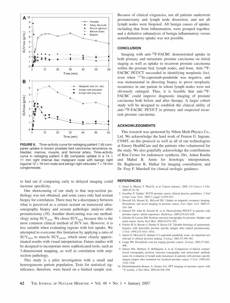
The representative time–activity curve taken from restag-ing patient 1 (Fig. 6A) shows rapid uptake of anti-18F-FACBCin known prostate carcinoma recurrence to a maximum at4.5 min with a plateau to 30 min, which is followed bygradual clearance (9%/min) out to 60 min. Regions in marrow(ilium), muscle (gluteus), and bladder reached a plateau at 4.5min that remained constant throughout the imaging period.
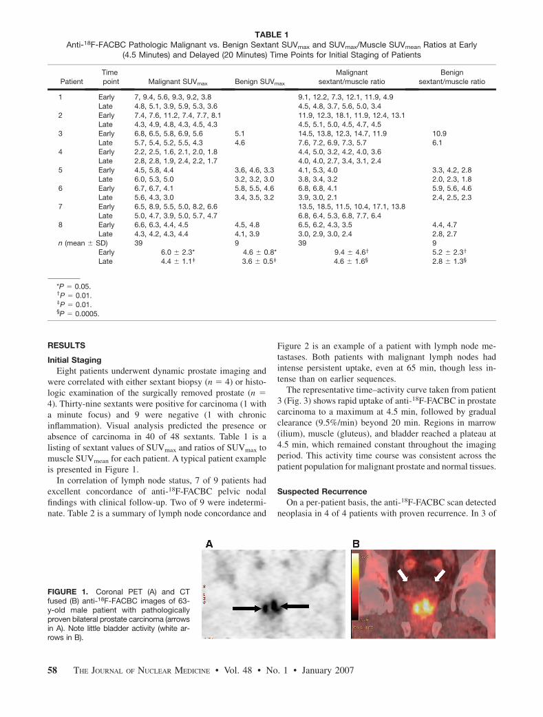
TABLE 2Abdominal/Pelvic Lymph Node Imaging Findings for Initial Staging of Patients at Early (4.5 Minutes) and Delayed
(20 Minutes) Time Points with Clinical/Pathologic Correlation
Patient
PSA
(ng/mL) GS
Anti-18F-FACBC imaging visual
uptake: early; late SUVmax
Clinical/pathologic
correlation Comments
1 8.4 7 No uptake Indeterminate PLND 11 mo after anti-18F-FACBC
scan, 1 node positive
2 71 9 Intense: 11.7 6 3.2; 4.4 6 1.3(n 5 5)
Malignant nodes PSA rising to 290 ng/mL and enlargingpelvic nodes on serial CT scans
3 11 7 No uptake No malignant nodes Negative PLND
4 21 8 No uptake Indeterminate Not surgical candidate, systemic therapy
5 6.3 9 Moderate fading to mild uptake:3.7 6 0.1, 3.2 6 0.0 (n 5 2)
No malignant nodes Negative PLND; uptake in benign-appearing inguinal nodes
6 16.2 6 No uptake No malignant nodes PSA nadir (,0.05) after cryotherapy
7 5.7 6 No uptake No malignant nodes No PLND; negative follow-up
abdomen/pelvic CT at 6 mo8 5.7 8 No uptake No malignant nodes Negative PLND
9 2.7 10 Intense: 8.3 6 0.8, 8.4 6 11.1
(n 5 3)
Malignant nodes Positive biopsy, extensive
retroperitoneal nodesMean 6 SD Malignant 10.5 6 3.0 (early); 5.9 6 2.4 (late)
(n 5 8)7/9 concordance
Benign 3.7 6 0.1 (early); 3.2 6 0.0 (late)
(n 5 2)
GS 5 maximum Gleason score; PLND 5 pelvic lymph node dissection.
FIGURE 2. A 73-y-old man with extensive invasive prostatecarcinoma, with bilateral obturator and left iliac chain lymphnodal involvement. Maximum-intensity-projection image dem-onstrates extent of large prostate primary (arrowheads) as wellas bilateral obturator (open arrows) and left iliac (solid straightarrows) nodes. Bladder activity is not present (curved arrow atbladder location).
FIGURE 3. Time–activity curves for initial staging data. Time–activity curve compares uptake in whole prostate with knownbilateral carcinoma vs. bladder, marrow, muscle, and femoralartery in initial staging of patient 3.
ANTI-18F-FACBC IN PROSTATE CARCINOMA • Schuster et al. 59
The time–activity curve for patient 3 (Fig. 6B) compares
uptake in a malignant right internal iliac lymph node with
benign right inguinal and obturator nodes. The clearance of
the benign regions is more abrupt between 4.5 and 10 min
(30%/min) and then slows to that of the malignant region.
DISCUSSION
On the basis of in vitro and in vivo studies (29,34,35), wehypothesized that anti-18F-FACBC would be able to serveas a valuable adjunct in the initial staging of prostatecarcinoma, as well as detecting the presence of local versus
TABLE 3Imaging Findings for Suspected Recurrence in Prostate Beds and in Lymph Nodes at Early (4.5 Minutes) and Delayed
(20 Minutes) Time Points with Clinical/Pathologic Correlation
Patient History
PSA
(ng/mL)
Anti-18F-FACBC uptake: early; late SUVmax Presence of
carcinoma CommentsProstate bed Lymph nodes
1 Subtotal
prostatectomy
43.5 Focal intense:
5.8; 6.4
No uptake Bed: positive;
LN: indeterminate
Positive biopsy bed;
no other follow-up2 Brachytherapy,
XRT, salvage
cryotherapy
11.7 Focal moderate
seminal vesicle:
4.1; 3.0
Intense:
9.6 6 6.1;
6.3 6 4.5
(n 5 2)
Bed: indeterminate;
LN: positive
No biopsy bed; PSA
rising and enlarging
nodes on serial
CT scans3 Brachytherapy 4.1 Diffuse mild
uptake: 3.9; 2.7
Intense:
5.7; 5.3
(n 5 1)*
Bed: negative;
LN: positive
Radiation-effect bed;
positive laparoscopic
LN sampling*4 Brachytherapy,
XRT
1.9 Focal mild–
moderate: 4.1; 3.8
No uptake Bed: negative;
LN: indeterminate
Radiation effect bedy
5 Brachytherapy 6.8 Linear moderate
central: 4.6; 3.9
No uptakez Bed: negative;
LN: indeterminate
Radiation effect bedyz
6 Prostatectomy,
salvage
radiotherapy
9.5 Diffuse mild
uptake: 2.8; 2.7
Intense:
10.5 6 4.0;
12.3 6 5.0
(n 5 3)
Bed: indeterminate;
LN: positive
Positive sternal biopsy,
nodes enlarging on
CT and PSA rising;
prostate bed notbiopsied§
Mean 6 SD Malignant 5.8 (early); 6.4
(late) (n 5 1)
9.4 6 4.2 (early);
9.1 6 5.1 (late)k
(n 5 6) 4/4 concordance (3 had negative111In-capromab-pendetide)Clinically or
pathologically
proven
Benign 4.2 6 0.4 (early);
3.5 6 0.7 (late)
(n 5 3)
2.8 6 0.8 (early);
1.4 6 0.3 (late)k
(n 5 4)
*Patient 3 also had moderate fading to mild uptake in benign-appearing inguinal node (SUVmax 5 3.7 early, 1.8 late) and biopsy-proven
benign obturator nodal grouping (SUVmax 5 3.3 early, 1.5 late).yPatients 4 and 5 had subsequently decreasing PSA. Postbrachytherapy prolonged PSA bump is clinically suggested but more follow-up
is needed.zPatient 5 had moderate fading to mild uptake in 2 benign-appearing inguinal nodes (SUVmax 5 2.2 and 2 early, 1.1 and 1.2 late).§Patient 6 had skeletal metastases that were also intensely positive on anti-18F-FACBC scan.kP 5 0.01 on early and delayed images.
LN 5 lymph nodes.
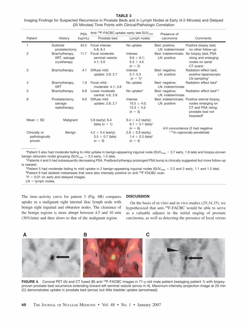
FIGURE 4. Coronal PET (A) and CT fused (B) anti-18F-FACBC images in 71-y-old male patient (restaging patient 1) with biopsy-proven prostate bed recurrence extending toward left seminal vesicle (arrow in A). Maximum-intensity-projection image at 20 min(C) demonstrates uptake in prostate bed (arrow) but little bladder uptake (arrowhead).
60 THE JOURNAL OF NUCLEAR MEDICINE • Vol. 48 • No. 1 • January 2007
metastatic prostate carcinoma in the setting of biochemi-cally recurrent disease. The present study demonstratesencouraging but preliminary initial results.
Anti-18F-FACBC seems to share characteristics with otherPET radiotracers used for prostate scanning. Similar to11C-acetate and 18F-FCH (4,20,36), we observed a certaindegree of nonspecific uptake, including relatively intense inci-dental bowel uptake as well as occasional low-level uptakein benign inguinal lymph nodes.
We also noted regions of false positivity and negativitywithin the prostate. Though malignant sextants had statis-tically significant higher SUVmax than benign sextants, therewas little separation between benign and malignant sextantvalues of SUVmax. Yet, visual analysis revealed that therewas agreement of 40 of 48 sextants with pathologic results,and ratios of sextant uptake to mean muscle SUVmax dem-onstrated improved, though still incomplete, separation ofmalignant from benign tissue. Some pathologically identi-fied benign prostate tissue, including that with inflamma-tion, had focal uptake but these were visually less intensethan malignant disease. Future studies will be designed tobetter examine the question of uptake in inflammatoryversus neoplastic foci within the same prostate.
Uptake within the prostate in areas of inflammation,hyperplasia, and high-grade prostatic intraepithelial neo-plasia has also been reported with 18F-FCH, 11C-choline,and 11C-acetate (4,5,21,24,37). Kwee et al. (25) noted bet-ter discrimination between malignant and benign prostaticregions using early and 1-h delayed prostate imaging with18F-FCH. It remains to be seen whether anti-18F-FACBCimaging may be useful for primary tumors and to separateareas of inflammation and hyperplasia from neoplasia.
Anti-18F-FACBC demonstrates potential clinical benefitfor nodal staging. Formal lymph node dissection is a highlymorbid procedure, and anti-18F-FACBC may prove valu-able in directing biopsy. For both staging and restaging,
SUVmax of malignant nodes was significantly higher thanthat of benign nodes and anti-18F-FACBC helped discrim-inate prostatic from extraprostatic involvement. It is alsointeresting to note that 3 patients with negative 111In-capromab-pendetide studies had positive anti-18F-FACBCimaging, proven to be true positive.
Choline-based compounds have also shown similar prom-ise in nodal staging and in detecting recurrence (4,15).Although intense bladder activity has been reported 5 minafter injection with 18F-FCH (5,19,26), anti-18F-FACBCdemonstrates relatively little, though variable, renal excre-tion and bladder activity. Even the most intense bladderactivity did not interfere with scan interpretation as hasbeen reported with choline-based PET radiotracers. Yet, themechanism of anti-18F-FACBC uptake in prostate carci-noma cells is not well understood and is the basis ofongoing studies. Preliminary data suggest a major role foran LAT transporter (30,31).
The time–activity curves of Figures 3 and 6 show rapiduptake of anti-18F-FACBC to 4.5 min, which is followedquickly by a fast and then slow washout rate out to 60 min.Tracer retention in background structures for all casesapproaches a plateau after the first 4.5 min. More detailedanalysis of the uptake mechanisms awaits further study, butmetabolite analysis of arterial plasma samples in humansubjects indicates that anti-18F-FACBC does not undergo me-tabolism (Mark M. Goodman, unpublished data, July 2006).
These findings also suggest that anti-18F-FACBC may beless useful for delayed imaging in some patients because ofwashout of radiotracer. Yet, all 6 patients with either locallyrecurrent prostate carcinoma or lymph node metastases didhave persistent and often striking uptake, even at 1 h, whichmay reflect increased uptake in neoplastic cells prone tometastasize or recur. More comprehensive studies must beundertaken to further determine the significance of thesefindings, to define the optimal time course of imaging, and
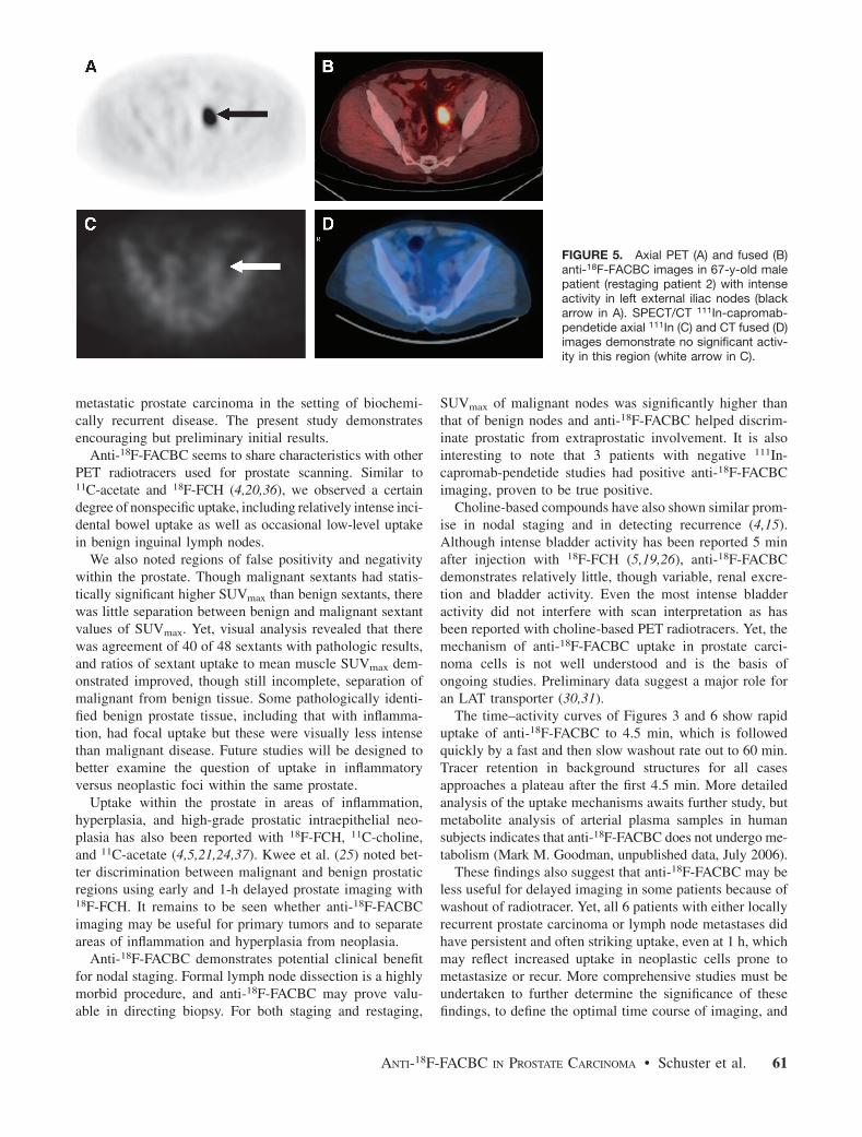
FIGURE 5. Axial PET (A) and fused (B)anti-18F-FACBC images in 67-y-old malepatient (restaging patient 2) with intenseactivity in left external iliac nodes (blackarrow in A). SPECT/CT 111In-capromab-pendetide axial 111In (C) and CT fused (D)images demonstrate no significant activ-ity in this region (white arrow in C).
ANTI-18F-FACBC IN PROSTATE CARCINOMA • Schuster et al. 61
to find out if comparing early to delayed imaging couldincrease specificity.
One shortcoming of our study is that step-section pa-thology was not obtained, and some cases only had sextantbiopsy for correlation. There may be a discrepancy betweenwhat is perceived as a certain sextant on transrectal ultra-sonography biopsy and sextant pathologic analysis afterprostatectomy (38). Another shortcoming was our method-ology using SUVmax. We chose SUVmax because this is themost common clinical method of SUV use. However, it isless suitable when evaluating regions with low uptake. Weattempted to overcome this limitation by applying a ratio ofSUVmax to muscle SUVmean, which more closely approx-imated results with visual interpretation. Future studies willbe designed to incorporate more sophisticated tools, such as3-dimensional mapping, as well as correlation with step-section pathology.
This study is a pilot investigation with a small andheterogeneous patient population. Tests for statistical sig-nificance, therefore, were based on a limited sample size.
Because of clinical exigencies, not all patients underwentprostatectomy and lymph node dissection, and not alllymph nodes were biopsied. All benign causes of uptake,including that from inflammation, were grouped together,and a definitive subanalysis of benign inflammatory versusnoninflammatory uptake was not possible.
CONCLUSION
Imaging with anti-18F-FACBC demonstrated uptake inboth primary and metastatic prostate carcinoma on initialstaging as well as uptake in recurrent prostate carcinomawithin the prostate bed, lymph nodes, and bone. Anti-18F-FACBC PET/CT succeeded in identifying neoplastic foci,even when 111In-capromab-pendetide was negative, andwas instrumental in directing biopsy to prove neoplasticrecurrence in one patient in whom lymph nodes were notobviously enlarged. Thus, it is feasible that anti-18F-FACBC could improve diagnostic imaging of prostatecarcinoma both before and after therapy. A larger cohortstudy will be designed to establish the clinical utility ofanti-18F-FACBC PET/CT in primary and suspected recur-rent prostate carcinoma.
ACKNOWLEDGMENTS
This research was sponsored by Nihon Medi-Physics Co.,Ltd. We acknowledge the hard work of Fenton G. Ingram,CNMT, on this protocol as well as all of our technologistsat Emory HealthCare and the patients who volunteered forthe study. We also gratefully acknowledge the contributionsof Ron Crowe for radiotracer synthesis, Drs. Adam Kuehnand Mahul B. Amin for histologic interpretation,Dr. Raghuveer K. Halkar for imaging consultation, andDr. Fray F. Marshall for clinical urologic guidance.
REFERENCES
1. Jemal A, Murray T, Ward E, et al. Cancer statistics, 2005. CA Cancer J Clin.
2005;55:10–30.
2. Scardino P. Update: NCCN prostate cancer clinical practice guidelines. J Natl
Compr Canc Netw. 2005;3 (suppl 1):S29–S33.
3. Brassell SA, Rosner IL, McLeod DG. Update on magnetic resonance imaging,
ProstaScint, and novel imaging in prostate cancer. Curr Opin Urol. 2005;15:
163–166.
4. Schmid DT, John H, Zweifel R, et al. Fluorocholine PET/CT in patients with
prostate cancer: initial experience. Radiology. 2005;235:623–628.
5. Schoder H, Larson SM. Positron emission tomography for prostate, bladder, and
renal cancer. Semin Nucl Med. 2004;34:274–292.
6. Fowler JE Jr, Brooks J, Pandey P, Seaver LE. Variable histology of anastomotic
biopsies with detectable prostate specific antigen after radical prostatectomy.
J Urol. 1995;153:1011–1014.
7. Sartor O, McLeod D. Indium-111-capromab pendetide scans: an important test
relevant to clinical decision making. Urology. 2001;57:399–401.
8. Lange PH. ProstaScint scan for staging prostate cancer. Urology. 2001;57:402–
406.
9. Seltzer MA, Barbaric Z, Belldegrun A, et al. Comparison of helical comput-
erized tomography, positron emission tomography and monoclonal antibody
scans for evaluation of lymph node metastases in patients with prostate specific
antigen relapse after treatment for localized prostate cancer. J Urol. 1999;162:
1322–1328.
10. Dimitrakopoulou-Strauss A, Strauss LG. PET imaging of prostate cancer with11C-acetate. J Nucl Med. 2003;44:556–558.
FIGURE 6. Time–activity curve for restaging patient 1 (A) com-pares uptake in known prostate bed carcinoma recurrence vs.bladder, marrow, muscle, and femoral artery. Time–activitycurve in restaging patient 3 (B) compares uptake in a 14 ·11 mm right internal iliac malignant node with benign rightinguinal 12 · 18 mm node and benign right obturator 7 · 18 mmconglomerate.
62 THE JOURNAL OF NUCLEAR MEDICINE • Vol. 48 • No. 1 • January 2007
11. Shreve PD, Grossman HB, Gross MD, Wahl RL. Metastatic prostate cancer:
initial findings of PET with 2-deoxy-2-[F-18]fluoro-D-glucose. Radiology. 1996;
199:751–756.
12. Schoder H, Herrmann K, Gonen M, et al. 2-[18F]Fluoro-2-deoxyglucose positron
emission tomography for the detection of disease in patients with prostate-
specific antigen relapse after radical prostatectomy. Clin Cancer Res. 2005;11:
4761–4769.
13. Larson SM, Morris M, Gunther I, et al. Tumor localization of 16b-18F-fluoro-5a-
dihydrotestosterone versus 18F-FDG in patients with progressive, metastatic
prostate cancer. J Nucl Med. 2004;45:366–373.
14. Hofer C, Laubenbacher C, Block T, Breul J, Hartung R, Schwaiger M. Fluorine-
18-fluorodeoxyglucose positron emission tomography is useless for the detection
of local recurrence after radical prostatectomy. Eur Urol. 1999;36:31–35.
15. de Jong IJ, Pruim J, Elsinga PH, Vaalburg W, Mensink HJ. Preoperative staging
of pelvic lymph nodes in prostate cancer by 11C-choline PET. J Nucl Med.
2003;44:331–335.
16. Toth G, Lengyel Z, Balkay L, Salah MA, Tron L, Toth C. Detection of prostate
cancer with 11C-methionine positron emission tomography. J Urol. 2005;173:
66–69.
17. Kwee SA, Coel MN, Lim J, Ko JP. Prostate cancer localization with 18fluorine
fluorocholine positron emission tomography. J Urol. 2005;173:252–255.
18. Yamaguchi T, Lee J, Uemura H, et al. Prostate cancer: a comparative study of11C-choline PET and MR imaging combined with proton MR spectroscopy. Eur
J Nucl Med Mol Imaging. 2005;32:742–748.
19. DeGrado TR, Coleman RE, Wang S, et al. Synthesis and evaluation of 18F-
labeled choline as an oncologic tracer for positron emission tomography: initial
findings in prostate cancer. Cancer Res. 2001;61:110–117.
20. Price DT, Coleman RE, Liao RP, Robertson CN, Polascik TJ, DeGrado TR.
Comparison of [18F]fluorocholine and [18F]fluorodeoxyglucose for positron
emission tomography of androgen dependent and androgen independent prostate
cancer. J Urol. 2002;168:273–280.
21. Yoshida S, Nakagomi K, Goto S, Futatsubashi M, Torizuka T. 11C-Choline
positron emission tomography in prostate cancer: primary staging and recurrent
site staging. Urol Int. 2005;74:214–220.
22. Fricke E, Machtens S, Hofmann M, et al. Positron emission tomography with11C-acetate and 18F-FDG in prostate cancer patients. Eur J Nucl Med Mol
Imaging. 2003;30:607–611.
23. Oyama N, Akino H, Kanamaru H, et al. 11C-Acetate PET imaging of prostate
cancer. J Nucl Med. 2002;43:181–186.
24. Farsad M, Schiavina R, Castellucci P, et al. Detection and localization of prostate
cancer: correlation of 11C-choline PET/CT with histopathologic step-section
analysis. J Nucl Med. 2005;46:1642–1649.
25. Kwee SA, Wei H, Sesterhenn I, Yun D, Coel MN. Localization of primary prostate
cancer with dual-phase 18F-fluorocholine PET. J Nucl Med. 2006;47:262–269.
26. Hara T, Kosaka N, Kishi H. Development of 18F-fluoroethylcholine for cancer
imaging with PET: synthesis, biochemistry, and prostate cancer imaging. J Nucl
Med. 2002;43:187–199.
27. Heinisch M, Dirisamer A, Loidl W, et al. Positron emission tomography/
computed tomography with F-18-fluorocholine for restaging of prostate cancer
patients: meaningful at PSA
28. de Jong IJ, Pruim J, Elsinga PH, Vaalburg W, Mensink HJ. 11C-Choline positron
emission tomography for the evaluation after treatment of localized prostate
cancer. Eur Urol. 2003;44:32–38.
29. Oka S, Hattori R, Kurosaki F, et al. A preliminary study of anti-1-amino-3-18F-
fluorocyclobutyl-1-carboxylic acid for the detection of prostate cancer. J Nucl
Med. 2007;48:46–55.
30. Martarello L, McConathy J, Camp VM, et al. Synthesis of syn- and anti-1-
amino-3-[18F]fluoromethyl-cyclobutane-1-carboxylic acid (FMACBC), potential
PET ligands for tumor detection. J Med Chem. 2002;45:2250–2259.
31. McConathy J, Martarello L, Simpson NE, et al. Uptake profiles of six 18F-
labeled amino acids for tumor imaging: comparison of in vitro and in vivo uptake
of branched chain and cyclobutyl amino acids by 9L gliosarcoma tumor cells.
J Nucl Med. 2002;43 (suppl):41P.
32. McConathy J, Voll RJ, Yu W, Crowe RJ, Goodman MM. Improved synthesis of
anti-[18F]FACBC: improved preparation of labeling precursor and automated
radiosynthesis. Appl Radiat Isot. 2003;58:657–666.
33. Kessler RM, Ellis JR Jr, Eden M. Analysis of emission tomographic scan data:
limitations imposed by resolution and background. J Comput Assist Tomogr.
1984;8:514–522.
34. Oka S, Hattori R, Kurosaki F, et al. Biodistribution and microPET imaging of
anti-[18F]FACBC using orthotopic prostate tumor transplantation model in rats.
J Nucl Med. 2005;46 (suppl):381P.
35. Schuster DM, Votaw JR, Nieh PT, et al. Initial experience with the radiotracer
anti 1 amino 3 [18F]fluorocyclobutane-1-carboxylic acid (anti-[18F]FACBC) with
PET/CT in newly diagnosed prostate cancer. J Nucl Med. 2006;47 (suppl):224P.
36. Oyama N, Miller TR, Dehdashti F, et al. 11C-Acetate PET imaging of prostate
cancer: detection of recurrent disease at PSA relapse. J Nucl Med. 2003;44:549–555.
37. Kato T, Tsukamoto E, Kuge Y, et al. Accumulation of [11C]acetate in normal
prostate and benign prostatic hyperplasia: comparison with prostate cancer. Eur J
Nucl Med Mol Imaging. 2002;29:1492–1495.
38. Wefer AE, Hricak H, Vigneron DB, et al. Sextant localization of prostate cancer:
comparison of sextant biopsy, magnetic resonance imaging and magnetic reso-
nance spectroscopic imaging with step section histology. J Urol. 2000;164:400–
404.
ANTI-18F-FACBC IN PROSTATE CARCINOMA • Schuster et al. 63


![Disclaimer - Seoul National University...myocardial infarction, myocarditis, and cardiomyopathy[23,24].By using an appropriate radiotracer to target inflammatory lesions, the efficiency](https://img.pdfslide.net/doc/110x75/60eea402f5de20202350ebd2/disclaimer-seoul-national-university-myocardial-infarction-myocarditis-and.jpg)


![[XLS] · Web viewApproaches used in the Mackenzie laboratory include theoretical modeling of transporter function, structure/function relationships, electrophysiology, radiotracer](https://img.pdfslide.net/doc/110x75/5b0875c87f8b9a992a8c4f95/xls-viewapproaches-used-in-the-mackenzie-laboratory-include-theoretical-modeling.jpg)


![arXiv:1403.7565v1 [hep-ph] 28 Mar 2014 fileforward-backward asymmetry of top anti-top quark pairs in Tevatron experiments with a proton anti-proton initial state. Recently it was shown](https://img.pdfslide.net/doc/110x75/5d4dc7da88c993af7b8bbb7a/arxiv14037565v1-hep-ph-28-mar-2014-asymmetry-of-top-anti-top-quark-pairs-in.jpg)