Embed Size (px)
Citation preview
Institute of Clinical Microbiology
Faculty of Medicine
University of Szeged
Characterization of medically important Pseudomonas aeruginosa isolates
Csilla Ratkai
Szeged, 2011

LIST OF PUBLICATIONS
This thesis is based on the following publications:
I. Ratkai C, Quinteira S, Grosso F, Monteiro N, Nagy E, Peixe L.: Controlling for false
positives: interpreting MBL Etest and MBL combined disc test for the detection of
metallo-beta-lactamases. J Antimicrob Chemother. 2009;64:657-8
II. Ratkai C., Nagy E., Peixe L., Bertalan V., Hajdu E.: Isolation and characterization of
an imported extremely-resistant Pseudomonas aeruginosa producing three different
extended-spectrum β-lactamases and hyperproducing two multidrug-efflux pumps. J
Inf 2010, 61:511-512
III. Ratkai C, Peixe LV, Grosso F, Freitas AR, Antunes P, Fodor E, Hajdú E, Nagy E.:
Successful application of the DiversiLab repetitive-sequence-based PCR typing
system for confirmation of the circulation of a multiresistant Pseudomonas
aeruginosa clone in different hospital wards. Diagn Microbiol Infect Dis. 2010 ;67:202-
206.
IV. Ratkai C., Nagy E., Peixe L., Szabó Á., Hajdu E.: Characterisation of Pseudomonas
aeruginosa strains isolated from patients suffering from cystic fibrosis in South-East
Hungary
under publication
TABLE OF CONTENTS
1. Introduction……………………………………………………………………………….…1
1.1. Features of Pseudomonas aeruginosa......................................................................1
1.2. Antibiotic resistance in P. aeruginosa……………………………………………2
1.2.1. Restricted uptake: porins, efflux pumps…………………...……..……..2
1.2.2. Drug inactivation: enzymatic resistance…………………………...……5
1.2.2.1. Problems with MBL detection..................................................10
1.2.3. Changes in targets: mutational resistance………………………............10
1.2.4. Multi-, and pandrug resistance of P. aeruginosa - clinical impact.…....12
1.3. Determination of different genotypes of P. aeruginosa – bacterial typing
methods………………………………………………………………...……………..13
1.3.1. Pulsed-field-gel-electrophoresis (PFGE)………….............................…14
1.3.2. Multilocus sequence typing (MLST).......................................................14
1.3.3. Repetitive-element-based PCR (rep-PCR) assays...................................15
1.4. Cystic fibrosis and the role of P. aeruginosa in this clinical entity...........………15
2. Aims of the study………………………………………………………………........……..20
3. Methods and materials…………………………………………………………….....…….21
3.1. Bacterial isolates…………………………………………………………..……..21
3.1.1. Multidrug-resistant P. aeruginosa isolates recovered from non-cystic-
fibrosis patients ……………………………………………………………….21
3.1.1.1. Isolates of the epidemiological analysis; environmental
screening................................................................................................21
3.1.2. P. aeruginosa isolates recovered from cystic fibrosis patients...............25
3.2. Determination of different antibiotic-resistance mechanisms………………........25
3.2.1. Resistance mechanisms in multidrug resistant P. aeruginosa isolates
recovered from non-cystic-fibrosis patients…………………………………..25
3.2.1.1. Detection of ESBLs..................................................................25
3.2.1.1.1. Phenotypical methods................................................25
3.2.1.1.2. Genotypical methods…………………………….....25
3.2.1.2. Detection of metallo-lactamases..............................................26
3.2.1.2.1. Phenotypical methods................................................26
3.2.1.2.2. Genotypical methods.................................................26
3.2.1.3. Detection of integrons..............................................................27

3.2.1.4. Efflux pump, porin channel examinations................................27
3.2.2. Detection of different antibiotic-resistance mechanisms in P. aeruginosa
isolates recovered from cystic fibrosis patients.................................................28
3.3. Molecular typing methods......................................................................................31
3.3.1. Molecular typing of multidrug-resistant P. aeruginosa isolates recovered
from non-cystic-fibrosis patients.......................................................................31
3.3.1.1. Pulsed-field gel-electrophoresis................................................31
3.3.1.2. Repetitive-element-based PCR, DiversiLab.............................31
3.3.1.3. Repetitive-element-based (ERIC) PCR assay………………..31
3.3.1.4. Multi-locus sequence typing………………………………….32
3.3.2. Molecular typing of P. aeruginosa isolates recovered from cystic fibrosis
patients...............................................................................................................32
4. Results………………………………………………………………………………….......33
4.1. Antibiotic resistance...............................................................................................33
4.1.1. Antibiotic resistance in multidrug-resistant P. aeruginosa isolates
recovered from non-cystic-fibrosis patients......................................................33
4.1.1.1. ESBL detection.........................................................................33
4.1.1.2. MBL detection..........................................................................34
4.1.1.3. Detection of integrons..............................................................35
4.1.1.4. Efflux pump, porin channel examinations................................36
4.1.2. Antibiotic resistance in P. aeruginosa isolates recovered from cystic
fibrosis patients..................................................................................................37
4.2. Bacterial typing......................................................................................................38
4.2.1. Molecular typing of multidrug-resistant P. aeruginosa isolates recovered
from non-cystic-fibrosis patients.......................................................................38
4.2.1.1. Investigation of multidrug resistant isolates of the
epidemiological analysis; environmental screening..............................40
4.2.2. Molecular typing of P. aeruginosa isolates recovered from cystic fibrosis
patients...............................................................................................................46
5. Discussion…………………………………………………………………………….....…47
5.1. Antibiotic resistance...............................................................................................47
5.1.1. Antibiotic resistance of multidrug-resistant P. aeruginosa isolates
recovered from non-cystic-fibrosis patients......................................................47

5.1.2. Antibiotic resistance of P. aeruginosa isolates recovered from CF
patients………………………………………………………………………...48
5.2. Comparison of three different molecular typing methods.....................................49
5.2.1. Molecular typing of multidrug-resistant P. aeruginosa isolates recovered
from non-cystic-fibrosis patients.......................................................................49
5.2.2 Molecular typing of P. aeruginosa isolates recovered from CF
patients...............................................................................................................53
6. Conclusions...........................................................................................................................55
7. Acknowledgement................................................................................................................56
8. References.............................................................................................................................57
Abbreviations
bp base pair
CC clonal complex
CF cystic fibrosis
CLSI Clinical and Laboratory Standard Institute
CFTR cystic fibrosis transmembrane regulator
EDTA ethylene-diamin-tetraacetic acid
ESBL extended spectrum β-lactamase
GIM german imipenemase
IMP imipenemase
IS insertion sequence
ICU intensive care unit
IM internal medicine ward
MBL metallo- β-lactamase
MLST multilocus-sequence typeing
Mex multidrug efflux
MDR multidrug resistant
MICU medical ICU
NE neurology ward
NS neurosurgical ward
OXA oxacillinase
Opr outer membrane protein
PFGE pulsed-field gel-electrophoresis
Rep-PCR repetitive-element-based PCR
RT-PCR Real-time PCR
SICU general surgical ICU
SICU2 trauma and neurosurgical ICU
SICU3 cardiac sugical ICU
SPM São-Paolo metallo- β-lactamase
ST sequence type
Tn transposon
VIM Verona-imipenemase
VAP ventilation-associated pneumonia
1. INTRODUCTION
1.1. Features of Pseudomonas aeruginosa
Pseudomonas aerugionosa (P. aruginosa) is a non-fermentative, aerobic Gram-
negative rod, measuring 0.5 to 0.8 µm by 1.5 to 3.0 µm. Almost all strains are motile by
means of a single polar flagellum. It normally lives in moist environments, and uses a wide
range of organic compounds for growth, thus giving it an exceptional ability to colonize
ecological niches where nutrients are limited, from water and soil to plant and animal tissues.
Typical biochemical features of P. aeruginosa isolates are: positive oxidase test, growth at 42
°C, hydrolysis of arginine and gelatine, and nitrate reduction. P. aeruginosa strains produce
two types of soluble pigments, pyoverdin and pyocyanin. The latter blue pigment is produced
abundantly in media of low-iron content and functions in iron metabolism in the bacterium.
Pyocyanin (from "pyocyaneus") refers to "blue pus", which is characteristic for suppurative
infections caused by P. aeruginosa [1]. The bacterium is ubiquitous in soil, variety of aqueous
solutions, including disinfectants, soaps, eye drops, as well as sinks and respiratory
equipments. P. aeruginosa is a highly adaptable organism. It has a large genome containing
6.26 Mbp (encoding 5567 genes) compared to 4.64 Mbp (4279 genes) in Escherichia coli [3].
An approximate calculation of the number of genes needed for cell growth and division in a
minimal medium, is around 1500. P. aeruginosa therefore has considerable additional genetic
capacity. This explains its highly adaptable nature, including the ability to develop resistance
against antibiotics.
Occasionally, P. aeruginosa can colonise human body sites, with a preference for
moist areas, such as the perineum, axilla, ear, nasal mucosa and throat, as well, as stools. The
prevalence of colonisation in healthy individuals is usually low, higher colonisation rates can
be encountered following hospitalisation, especially among patients treated with broad-
spectrum antibiotics. Normally, for an infection to occur, some disruption of the physical
barriers (skin or mucous membrane), or by-passing of them (invasive devices), and/or an
underlying dysfunction of the immune defence mechanisms is necessary. Therefore, P.
aeruginosa is mostly a nosocomial pathogen. Infections associated with this bacterium are
nosocomial respiratory tract infections including ventilator-associated pneumonia (VAP),
dermatitis, soft tissue infections, bacteraemia, bone and joint infections, gastrointestinal
infections and a variety of systemic infections, particularly in immunosuppressed patients
(AIDS), or patients with severe burns or cancer. Community acquired infections caused by P.
aeruginosa are uncommon. The most frequent ones are: urinary tract infections, otitis externa,
1
folliculitis acquired in swimming pools, keratitis due to wearing contact lenses. The mucoid
phenotype of P. aeruginosa frequently chronically colonises and infects patients with cystic
fibrosis causing damage of the lung tissue and decreased pulmonary function. P. aeruginosa
has an abundance of virulence factors, including flagella, pili, lipopolysaccharides, alginate,
alkaline protease, elastase, phospholipase C, exotoxin A, quorum sensing mechanisms, type
III secretion system, pyocianin, pyoverdin, and produces a number of toxic proteins which not
only cause extensive tissue damage, but also interfere with the human immune system's
defence mechanisms [1].
1.2. Antibiotic resistance in P. aeruginosa
P. aeruginosa is one of the main organisms responsible for drug-resistant nosocomial
infections, and is one of the leading causes of bacteraemia and pneumonia in hospitalised
patients. In addition being intrinsically resistant to several antimicrobial agents, P. aeruginosa
can easily develop resistance to all conventional antipseudomonal antibiotics via different
mechanisms [2] (Table 1.).
The aminoglycosides inhibit protein synthesis by binding to the 30S subunit of the
ribosome. Quinolones bind to the A subunit of DNA gyrase enzyme, which maintains the
ordered structure of the chromosome inside the cells. The β-lactams inhibit the peptidoglycan-
assembling transpeptidases located on the outer face of the cytoplasmic membrane. Finally
the polymyxins bind to phospholipids in the cytoplasmic membrane, destroying its barrier
function. There are three basic mechanisms by which P. aeruginosa resists the action of the
above antimicrobial agents: restricted uptake and efflux; enzymatic drug inactivation and
mutations in the targets [3].
1.2.1. Restricted uptake: porins, efflux pumps
All of the antibiotics have to cross the cell wall, to reach their targets. Failure of
antibiotics to accumulate in the organism is due to the combination of restricted permeability
of the outer membrane and the efficient removal of antibiotic molecules by efflux pumps. The
outer membrane of P. aeruginosa is an important barrier of the penetration of antibiotics (has
low permeability, approximately 8% that of E. coli) [4], excluding the larger molecules. Small
hydrophilic molecules, such as β-lactams and quinolons can only cross the outer membrane
by passing through the channels provided by porin proteins. Investigation of porins has
revealed that they contain transmembrane anti-parallel β-strands that wrap into a barrel, and
2

this β-barrel is embedded into the outer membrane bilayer (Figure 1.). The central area of the
barrels of the general and specific porins contains stretches of amino acids from one of the
interconnecting regions, that folds back into the channel region and give this region many of
its important characteristics.
Figure 1. The structure of the outer membrane porin OprD. (Source of picture is:
opm.phar.umich.edu/protein.php?pdbid=2odj)
In P. aeruginosa there are 163 known or predicted porin proteins based on its genome
sequence, 64 are found as part of 3 families of porins: the OprD-specific porin family, the
TonB-dependent gated porin family, and the OprM efflux family. OprF is the major channel
of P. aeruginosa for larger substrates, and is considered to be general, nonspecific porin [4].
Although mutants lacking OprF have been reported, loss of OprF has not been found to be a
major cause of antibiotic resistance. Other proteins, that function as substrate-specific porins
can serve as general channels for small substrates. OprD is a specialized porin which has a
specific role in the uptake of e.g. positively charged molecules such as lysine, or certain
carbapenems. Lack of OprD in P. aeruginosa is due to a mutation, which can occur at
relatively high frequency (10-7), and can be due to deletion, substitution or insertions that
cause inactivation of the oprD gene. Loss of OprD is frequently associated with resistance to
imipenem, which requires this porin to cross the outer membrane. Interestingly, meropenem is
not affected by loss of OprD, indicating that the different carbapenems cross the outer
membrane by different channels. The aminoglycosides and colistin do not cross the outer
membrane via porin channels. Instead they promote their own uptake by binding to the
3

lipopolysaccharide (LPS) on the outer face of the membrane, which destroys the permeability
barrier of the outer membrane and allows the antibiotics to penetrate through the wall to the
cytoplasmic membrane. Resistance to aminoglycosides and colistin could be due to the
overexpression of OprH protein, which protects the LPS from binding the antibiotics,
although this type of resistance mechanism was observed only in laboratory strains [3].
Acquired resistance to polymyxins has been occasionally described in P. aeruginosa isolates
from cystic fibrosis patients treated for a long time with this drug, and seems to be related to
mutations in the outer membrane structure [5].
The multidrug efflux system of P. aeruginosa are composed of three protein
components, an energy-dependent pump located in the cytoplasmic membrane, an outer
membrane porin and a peripheral cytoplasmic membrane linker (sometimes called as
membrane fusion protein). Each of these proteins is highly conserved, at around 20% or
greater identity level. P. aeruginosa has 18 outer membrane proteins with putative function in
efflux. Eleven of these, including OprM, OprN, OprJ, fall into one phylogenetic subclass, and
are parts of multiple antibiotic efflux systems [4]. The MexAB-OprM system (Figure 2.) is
constitutively expressed in virtually all isolates, and substrates for this pump include
fluoroquinolones, tetracycline, chloramphenicol, and β-lactams (including carbenicillin,
piperacillin, ceftazidime, cefepime, and aztreonam). Imipenem does not appear to be a
substrate for MexAB-OprM, but because of its hydrophobic side chain, meropenem can be
affected by this system. The expression of MexAB-OprM system is negatively controlled by
the repressor MexR. Mutational inactivation of a second regulatory gene nalC increases the
expression of the PA3719 protein, which then increases MexAB-OprM expression. A third
regulatory gene of the mexAB-oprM operon nalD, has also been identified; mutations in this
gene have been observed in clinical isolates overexpressing MexAB-OprM [4].
4

Figure 2. Schematic representation of the assembly of efflux pump MexAB-OprM in the cell
wall of P. aeruginosa. The pump is composed of three protein components: an energy-
dependent pump located in the cytoplasmic membrane (MexB), an outer membrane porin
(OprM) and a peripheral cytoplasmic membrane linker (MexA).
Fluoroquinolones and the antipseudomonal β-lactams (piperacillin, cefepime, and
meropenem, but not carbenicillin, aztreonam, ceftazidime, or imipenem) are substrates for the
MexCD-OprJ system, although this efflux pump is not typically expressed under normal
growth conditions. A third efflux system, MexEF-OprN, can export fluoroquinolones,
trimethoprim, and chloramphenicol. Finally, MexXY-OprM may contribute to
fluoroquinolone, aminoglycoside and selected β-lactam (piperacillin, cefepime, and
meropenem but not carbenicillin, ceftazidime, or imipenem) resistance. MexXYOprM
expression can be induced with growth in the presence of tetracycline or aminoglycosides [4].
1.2.2. Drug inactivation: enzymatic resistance
All P. aeruginosa strains have the ampC gene of the inducible β-lactamase. However,
induction alone probably does not account for resistance, instead overexpression of the
enzyme due to spontaneous mutation in its regulatory gene, which has occurred particularly
where ceftazidime therapy was used. Although the enzyme is normally located in the
periplasm, it has been detected in sputum during antipseudomonal treatment [3]. The enzyme
is probably released from high-level producers in the lungs, and would protect low-level
5
producers by reducing the local concentration of certain β-lactam antibiotics, like cephalotin
or ampicillin.
Many other acquired β-lactamases and aminoglycoside modifying enzymes have been
noted in P. aeruginosa. The most frequently acquired β-lactamases are PSE-1 and PSE-4.
Like classical TEM and OXA enzymes, which also occur in P. aeruginosa, these enzymes are
not effective against carbapenems, oxyimino-aminothiazolyl cephalosporins (ceftazidime,
cefepime, cefpirome) and monobactams. However, β-lactamases that give wider resistance
are emerging in P. aeruginosa, such as PER-1 β-lactamases, TEM, SHV and OXA ESBLs
and carbapanamases and the metallo- β-lactamases [2,10] (Table 1.).
PER-1, an Ambler’s molecular class A β-lactamase confers high-level resistance to
ceftazidime, but it has little in vitro effect on piperacillin. It was recovered in 1991 from
France, later was recognized to be widespread in Turkey, and disseminated in Italy, Poland,
Japan and Romania [6]. Up to this day only a few sporadic cases of PER-1 producer P.
aeruginosa has been detected in Hungary, interestingly almost all of them were imported
cases [7,8,article II].
The presence of OXA-type ESBLs is quite frequent among pseudomonads [11] (Table
1.). Like TEM and SHV ESBLs, the OXA ESBLs have minor sequence substitutions that
greatly extend their hydrolytic spectra. They confer resistance to carboxypenicillins and
ureidopenicillins, but not to ceftazidime. There are five groups of these enzymes, namely
OXA group I (including OXA-5; OXA-7; OXA-10 and its derivates: OXA-11, OXA-14,
OXA-16, OXA-17, OXA-74; OXA-13 and its derivates: OXA-19, OXA-28), OXA group II
(including OXA-2; OXA-3; OXA-15; OXA-20), OXA group III (including OXA-1, OXA-4,
OXA-30, ESBLs, OXA-31), OXA group IV (defined by OXA-9) and OXA group V
(containing LCR-1). In addition, OXA-18 does not belong to any of these groups [11].
Carbapenems, such as imipenem and meropenem have a very broad spectrum of
activity, and the drugs resist hydrolysis by most of the β-lactamases, including ESBLs, and
derepressed chromosomal ampC β-lactamases. Resistance to carbapenems could be mediated
by different enzymes [12,13], namely most of the metallo- β-lactamases together with some
class A and D enzymes. β-lactameses of molecular classes A, C and D use a catalytically
active serine residue for inactivation of the β-lactam drug. OXA-type carbapenemases [12]
belong to molecular class D, and are usually encoded by chromosomal genes. Some of these
carbapenemases are widely distributed in P. aeruginosa, and although these enzymes show
only week carbapenemase activity, carbapenem resistance may results from a combination of
an OXA-type carbapenemase and secondary resistance mechanism, such as porin deficiency
6

or efflux pumps. Class B metallo-β-lactameses (MBLs), however are enzymes with versatile
hydrolytic capabilities, namely the ability to hydrolyze all β-lactam antibiotics, with the
exception of monobactams. Four groups of MBLs have so far been reported in P. aeruginosa,
namely IMP (imipenemase), VIM (Verona imipenemase), SPM (São Paolo metallo-β-
lactamese), ans GIM (German imipenemase). These four classes of enzymes together with the
SIM isolated from Acinetobacter comprise the MBLs (Table 1.).
The common feature of MBLs is the principal zinc binding motif histidine-X-
histidine-X-aspartic acid in the active site, which coordinates the arrangement of two H2O
molecules that are important in the hydrolysis. Hence, chelation of zinc by EDTA or
mercaptopropionic acid, impairs β-lactam hydrolysis and restores susceptibility to
carbapenems. MBLs have a wide and plastic active site, which let all β-lactams to accomodate
in there, except aztreonam. β-lactamase inhibitors such as clavulanic acid, tazobactam and
sulbactam are also hydrolised by MBLs.
Of the four classes of MBLs only the VIM and IMP classes have so far been found to
have three subgroups, and several enzyme variants (Figure 3.), which are as follows: VIM-7
group, VIM-2 group, VIM-1 group and IMP-12, IMP-1, IMP-2 group, IMP-1 group [14].
Figure 3. Phylogenetic relationship between the five groups of acquired MBLs, and the
subgroups of VIM and IMP.
While GIM and SPM have so far been reported from a restricted geographical area,
the IMP and VIM enzymes have reached a worldwide dissemination [14]. The IMP enzymes
7
were originally detected in Asia, but later spread to Europe, to the United States and to
Australia, while the VIM gene was first found in Europe, and shortly after emerged to other
continents. Despite the worldwide dissemination of these two groups of enzymes, the
tendency of the dominancy of the IMP enzyme in Asia, and the VIM enzyme in Europe
prevails [14].
Significant differences in the hydrolytic capacity of the different MBL groups are
observed. SPM-1 is a very efficient, while GIM-1 is rather a weak carbapenemase. Within the
IMP group it has been observed, that IMP-6 and IMP-3 have low capacity of imipenem
hydrolysis, while within the VIM group VIM-1 hydrolyse meropenem, ceftazidime and
piperacillin more efficiently, than VIM-2 [14]. Still, the majority of MBL producers are
highly resistant to carbapenems and cephalosporins.
The genes encoding the MBLs are almost always located on class 1 integrons [15,16].
Integrons are genetic elements that although unable to move themselves, contain gene
cassettes that can be mobilized to other integrons or to secondary sites in the bacterial
genome. The majority of integrons described belong to class 1. These integrons consist of two
conserved regions (5’CS and 3’CS) and a variable region, where different gene cassettes can
be inserted. The 5’CS part contains an integrase gene (intI), an adjacent recombination site
(attI) and a common promoter. The 3’CS region usually consist of a partially deleted gene,
encoding a quaternary ammonium compound efflux pump (qacEΔ1) fused with a
sulphonamide resistance gene (sulI). In the variable region different β-lactamases, usually
MBLs, and/or aminoglycoside-resistance genes, between the genes recombination sites (also
known as 59-base elements) are found. The 59-base elements are not highly conserved, and
contain imperfect inverted repeats. Integration of new gene cassettes, which is mediated by
the integrase can take place either between the gene cassettes and attI, or between two gene
cassettes 59-base elements. The integrase excises the gene cassettes as covalently closed
circular molecules. The recombination takes place close to one end of the 59-base element.
Due to the integration of circular gene cassette, part of the 59-base element ends up at the
5’side of the coding sequence of the gene cassette to which it belongs [15,16] (Figure 4).
8
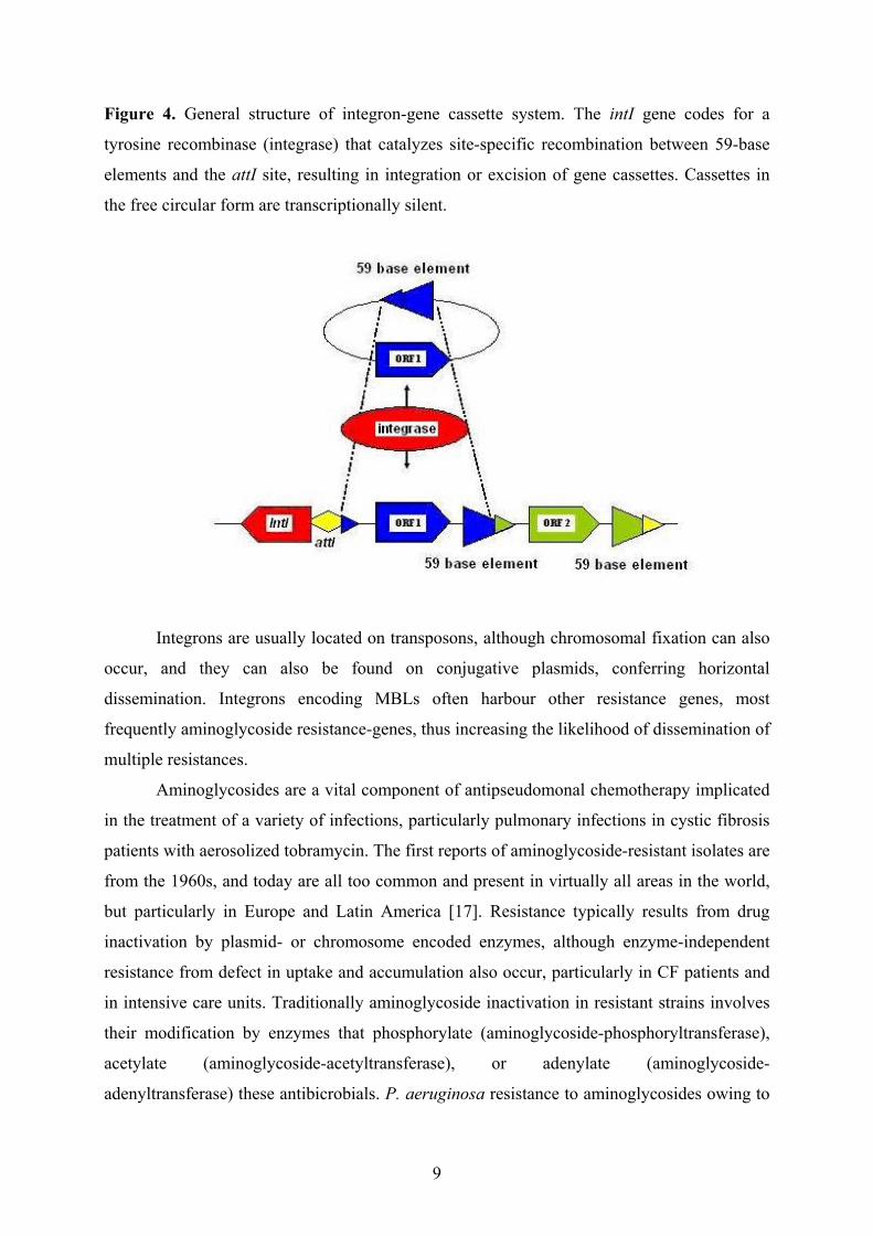
Figure 4. General structure of integron-gene cassette system. The intI gene codes for a
tyrosine recombinase (integrase) that catalyzes site-specific recombination between 59-base
elements and the attI site, resulting in integration or excision of gene cassettes. Cassettes in
the free circular form are transcriptionally silent.
Integrons are usually located on transposons, although chromosomal fixation can also
occur, and they can also be found on conjugative plasmids, conferring horizontal
dissemination. Integrons encoding MBLs often harbour other resistance genes, most
frequently aminoglycoside resistance-genes, thus increasing the likelihood of dissemination of
multiple resistances.
Aminoglycosides are a vital component of antipseudomonal chemotherapy implicated
in the treatment of a variety of infections, particularly pulmonary infections in cystic fibrosis
patients with aerosolized tobramycin. The first reports of aminoglycoside-resistant isolates are
from the 1960s, and today are all too common and present in virtually all areas in the world,
but particularly in Europe and Latin America [17]. Resistance typically results from drug
inactivation by plasmid- or chromosome encoded enzymes, although enzyme-independent
resistance from defect in uptake and accumulation also occur, particularly in CF patients and
in intensive care units. Traditionally aminoglycoside inactivation in resistant strains involves
their modification by enzymes that phosphorylate (aminoglycoside-phosphoryltransferase),
acetylate (aminoglycoside-acetyltransferase), or adenylate (aminoglycoside-
adenyltransferase) these antibicrobials. P. aeruginosa resistance to aminoglycosides owing to
9
enzymatic N-acetylation has been known for a long time. Acetylation of aminoglycosides can
occur at the 1, 3, 6`, and 2`amino groups and involves all medically useful compounds.
Inactivation of aminoglycosides such as kanamycin, neomycin, and streptomycin by a
phosphotransferase, that modify the 3`-OH of these antimicrobials, is well known since the
1980s. The adenylation of streptomycin and gentamycin has been described first 20 years ago.
While the specificity of aminoglycoside-modifying enzymes has in the past tended to
compromise the use of only selected aminoglycosides, leaving the others still effective,
increased the prevalence of strains harboring multiple aminoglycoside-modifying enzymes
[17].
1.2.2.1. Problems with MBL-detection
The international epidemiology of MBL-producing P. aeruginosa is not quite clear.
The prevalence of these enzymes is still unknown in most countries, due to the lack of proper
screening recommendations. Several methodologies have been proposed for the routine
detection of MBL-producing P. aeruginosa, including microdilution test, iodometric
polyacrylamide gel method, particularly the MBL E-test and the MBL combined disc test [18-
25], although there are concerns regarding the latter two method’s reliability. All of these
methods have to be available for routine diagnosis; most of these are phenotypic methods,
based on the fact that the activity of MBLs can be experimentally inhibited by metal
chelators, such as EDTA, due to the Zn2+ in the active site. Recently, in order to increase the
reliability of these tests, some authors suggested the inclusion of ceftazidime-resistance beside
meropenem- and/or imipenem-resistance to the MBL-screening criteria [19]. Nevertheless, in
very specific circumstances such as the case of mucoid P. aeruginosa isolates, these methods
are stated as inefficient. Relying on EDTA for MBL inactivation, these methods are prone to
false-positive results since the chelating agent itself can increase membrane permeability, thus
increasing the chance of a bactericidal effect. Given the significance of MBL-producing P.
aeruginosa isolates, reliable and rapid detection of MBLs would be crucial for patient
management and appropriate infection control procedures.
1.2.3 Changes in targets: mutational resistance
This mechanism of resistance results from mutational changes in target enzymes
which result in maintenance of their vital role in cell metabolism, but resistance to the action
of selective inhibition by antibiotics. In P. aeruginosa it is most commonly seen in the
quinolones through mutation in the gyrA gene, encoding the A subunit of the target enzyme,
10
DNA gyrase [26]. Changes in the structure of the ribosome 30S subunit, which is the target of
the aminoglycosides, influence streptomycin sensitivity, while alteration in the penicillin-
binding proteins of P. aeruginosa could result resistance to β-lactams. Mutations leading to
the increased production of the AmpC β-lactamase can occur at frequencies of 10-7-10-9, and
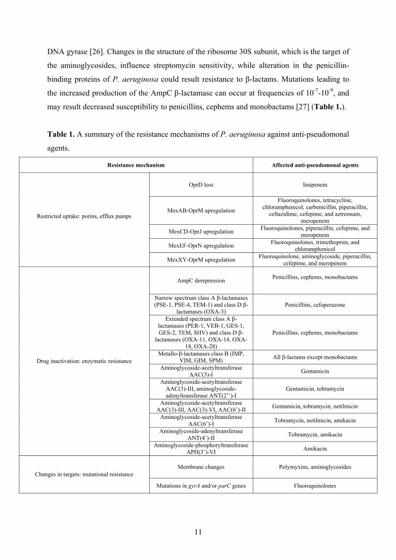
may result decreased susceptibility to penicillins, cephems and monobactams [27] (Table 1.).
Table 1. A summary of the resistance mechanisms of P. aeruginosa against anti-pseudomonal
agents.
Resistance mechanism Affected anti-pseudomonal agents
OprD loss Imipenem
MexAB-OprM upregulation
Fluoroquinolones, tetracycline, chloramphenicol, carbenicillin, piperacillin,
ceftazidime, cefepime, and aztreonam, meropenem
MexCD-OprJ upregulation Fluoroquinolones, piperacillin, cefepime, and meropenem
MexEF-OprN upregulation Fluoroquinolones, trimethoprim, and chloramphenicol
Restricted uptake: porins, efflux pumps
MexXY-OprM upregulation Fluoroquinolone, aminoglycoside, piperacillin, cefepime, and meropenem
AmpC derepression Penicillins, cephems, monobactams
Narrow spectrum class A β-lactamases (PSE-1, PSE-4, TEM-1) and class D β-
lactamases (OXA-3) Penicillins, cefoperazone
Extended spectrum class A β-lactamases (PER-1, VEB-1, GES-1, GES-2, TEM, SHV) and class D β-
lactamases (OXA-11, OXA-14, OXA-18, OXA-28)
Penicillins, cephems, monobactams
Metallo-β-lactamases class B (IMP, VIM, GIM, SPM) All β-lactams except monobactams
Aminoglycoside-acetyltransferase AAC(3)-I Gentamicin
Aminoglycoside-acetyltransferase AAC(3)-III, aminoglycoside-adenyltransferase ANT(2’’)-I
Gentamicin, tobramycin
Aminoglycoside-acetyltransferase AAC(3)-III, AAC(3)-VI, AAC(6’)-II Gentamicin, tobramycin, netilmicin
Aminoglycoside-acetyltransferase AAC(6’)-I Tobramycin, netilmicin, amikacin
Aminoglycoside-adenyltransferase ANT(4’)-II Tobramycin, amikacin
Drug inactivation: enzymatic resistance
Aminoglycoside-phosphoryltransferase APH(3’)-VI Amikacin
Membrane changes Polymyxins, aminoglycosides Changes in targets: mutational resistance
Mutations in gyrA and/or parC genes Fluoroquinolones
11
1.2.4. Multi-, and pandrug resistance of P. aeruginosa - clinical impact
As a consequence of all the above mentioned resistance mechanisms the repertoire of
antimicrobial agents that can be used against P. aeruginosa infections is relatively limited.
The most important antipseudomonal agents include some β-lactams, (ticarcillin,
ureidopenicillins, piperacillin, cefoperazone, ceftazidime, cefepime, aztreonam, imipenem and
meropenem), aminoglycosides (gentamicin, tobramycin, netilmicin and amikacin) and
fluoroquinolones (of which ciprofloxacin remains the most active compound). Polymyxins
(polymyxin B and polymyxin E - colistin) are also active, but due to their higher toxicity, are
usually considered only for multi-, or pandrug resistant strains, although newer studies shows
a better safety profile of polymyxins, than indicated in older reports [28]. The term multidrug
resistant (MDR) P. aeruginosa is defined by most authors as being resistant to at least 3
antipseudomonal antibiotic-groups. The term pandrug-resistant P. aeruginosa has been
introduced for isolates being resistant to all antipseudomonal agents, except the polymyxins.
Concerning the β-lactam–β-lactamase inhibitor combinations, piperacillin–tazobactam is
preferable to ticarcillin-clavulanate for the treatment of P. aeruginosa infections, because of
the more favourable pharmacokinetics of tazobactam, the superior antipseudomonal activity
of piperacillin, and the fact that unlike tazobactam, clavulanate usually induces the production
of AmpC enzyme and could antagonize the antimicrobial effect of ticarcillin [27].
In vitro susceptibility data are essential for the selection of antimicrobial
chemotherapy for P. aeruginosa infections, because of the frequency and variability of
acquired resistance mechanisms of the clinical isolates. As P. aeruginosa infection could be
lethal, empirical regimens adequate for P. aeruginosa coverage should always be initiated
prior to the results of cultures and susceptibility testing when infections by this pathogen are
suspected. For the empirical therapy several aspects should be considered, such as the nature
and source of infection, pharmacokinetic parameters, underlying risk factors and diseases.
Antibiotic monotherapy is usually recommended for non-complicated urinary tract infections,
whereas combination therapy with at least two different anti-pseudomonal agents is normally
recommended for the treatment of severe infections, such as nosocomial pneumonia,
bacteraemia [27]. The preferred combination remains the combination of aminoglycosides
with β-lactams. In cystic fibrosis patients, early aggressive combination therapy is currently
recommended for initial colonization episodes to delay as long as possible the chronic P.
aeruginosa infection. Once chronic infection is developed, maintenance chemotherapy based
on the administration of antipseudomonal agents at regular intervals can significantly improve
the survival of these patients [27].
12
Several factors indicate that the emergence and spread of drug-resistant P. aeruginosa
can be related to the overuse of antimicrobial agents, although the risk differs for the different
agents. A strong association between use and resistance has been documented for
carbapenems. In a cohort study comparing the relative risks for emergence of resistant P.
aeruginosa in patients treated with different anti-pseudomonal agents, imipenem was found to
be associated with a significantly higher risk of emergence of resistance. On the other hand
ceftazidime, piperacillin and ciprofloxacin had the lowest risk to develop resistance [27].
1.3. Determination of different genotypes of P. aeruginosa – bacterial typing methods
Bacterial typing has its own vocabulary, although the use of this terminology is not
always consistent, and can be confusing sometimes. The terms “isolate” and “strain” is often
used interchangeably, but not always appropriately. An isolate is a population of bacterial
cells in pure culture derived from a single colony. In clinical microbiology, isolates are
usually derived from the primary culture of a clinical specimen obtained from an individual
patient. A strain is the descendant of a single isolate in pure culture, usually derived from a
single initial colony on a solid growth medium. A strain may be considered an isolate or
group of isolates that can be distinguished from other isolates of the same genus and species
by phenotypic and genotypic characteristics. Cultures of a particular microorganism, isolated
at the same time from multiple body sites of a patient, and indistinguishable by typing, also
considered to be a single strain [29].
The ability to quickly and reliably differentiate among related bacterial isolates is
essential for epidemiological surveillance systems. There are several typing methods used in
laboratories today. These range from methods based on simple phenotypic features to DNA
sequencing [29]. Previously, the comparison of phenotypic characters, such as colony
morphology, color, odor, antibiogram-based typing, biotyping, serotyping and the ability of
growing in the presence of specific substances, were used for differentiation. Today these
methods are becoming obsolete, because they require strict standardization of experimental
conditions, since phenotypes are quite susceptible to the environmental conditions. Instead,
more reliable genotyping methods have been developed [29]. Genotypic assays are based on
the comparison of genomic DNA differences in the composition of bacterial DNA (e.g., the
presence or absence of plasmids), in the overall structure (e.g., restriction endonuclease
profiles), or in the precise nucleotide sequences of one or more genes or intergenic regions.
13
1.3.1. Pulsed-field-gel-electrophoresis (PFGE)
A variety of molecular genetic methods have been used to type P. aeruginosa strains,
each varying in their discriminatory potentials. Pulsed-field gel electrophoresis (PFGE)
allows separation of large DNA fragments generated with restriction endonucleases in
agarose gels, by periodic alteration of the angle of the electric field’s direction. PFGE has a
remarkable discriminatory power and reproducibility; therefore it is commonly used, and is
the gold-standard method for the comparative typing of most bacterial species including P.
aeruginosa. However, 2-4 days are required to obtain results, and relatively expensive PFGE
equipment is needed. Gels need to be analyzed closely and carefully, even after digitalization
and computerized processing [30,31]. When compared by PFGE, two isolates differing by
one mutational event (single nucleotide substitution, insertions, and deletions) may differ by
zero. When there are no observed band differences, the isolate should be termed as
“indistinguishable” rather than “identical”, and assigned to the same type. Subtypes will be
assigned to isolates that differ by one to eight bands. Isolates differ by at least nine bands
should be assigned as different types [29]. Tenover et al.[32] suggested that in case of health-
care associated outbreaks, isolates differing by one to four bands should be assigned as
“closely related”, and therefore “probably part of the outbreak”, while isolates differing five
to eight bands are “possibly related”, and therefore “possibly part of the outbreak”.
1.3.2. Multilocus sequence typing (MLST)
In recombining populations it is essential to obtain information regarding isolate
characterization from multiple chromosomal locations that are unlikely to be co-inherited in a
single genetic event. Additionally, it is recommended to avoid parts of the genome that are
evolving rapidly due to strong selection pressures, such as antibiotic use or immunological
selection. The approach to use housekeeping genes was established for multi-locus enzyme
electrophoresis (MLEE). Although MLEE played a central role in bacterial epidemiology, it is
technically cumbersome and has not been adopted for routine surveillance. The multilocus
sequence typing (MLST) was built on the success of MLEE targeting the variation present at
multiple housekeeping loci, however in the case of MLST typing is achieved by nucleotide
sequence determination of the gene fragments. In 2004, Curran and colleagues adopted the
MLST scheme for P. aeruginosa which is based on the allelic differences in the following
housekeeping genes: acsA, nuoD, trpE, mutL, guaA, aroE, ppsA [33]. The advantage of the
method is mainly the interpretative, no cost software (e.g., eBURST), and the freely available
database on the internet. This method is recently becoming more popular as a bacterial typing
14
method to compare clones spreading in different geographical areas [30,34,35]. The
implications for population genetics and dynamics are more significant than those for
bacterial epidemiology, since polymorphism in the slowly evolving genes, which are its
targets, may not be high enough for useful epidemiological comparisons. Additionally, the
genes in question are unlikely to have any direct relevance to virulence or drug resistance.
1.3.3. Repetitive-element-based PCR (rep-PCR) assays
Rep-PCR assays use primers targeting highly conserved, non-coding repetitive
sequence elements in the bacterial genome, and is an established approach for subspecies
classification and strain delineation [36,31,37]. Recently, rep-PCR has been adapted to an
automated format by Healy and colleagues [38], known as DiversiLab system (BioMerieux)
to provide a reliable PCR-based typing method for clinical laboratories. The standardized rep-
PCR and quality-controlled reagents in a kit format, automated detection and analysis using
micro-fluids for rapid detection, and digitized the corresponding information in a software
package allows simplistic data archiving, retrieval, and reporting, producing the automated
microbial DNA typing system [38].
The surveillance systems of nosocomial infections have to be rapid, reliable, and able
to differentiate among related and unrelated bacterial isolates, especially in highly critic areas,
such as Intensive Care Units (ICUs). ICUs are often the scene of sporadic appearance of
multiresistant pathogens, such as P. aeruginosa. An outbreak can be defined as a temporal
increase in the incidence of infection or colonization by a certain bacterial strain, caused by
enhanced, patient-to-patient transmission [29]. Often the sporadic isolation of multiresistant
strains does not draw the attention in time to determine the nosocomial spread of one clone.
The collection of multiresistant, potential nosocomial pathogens for further typing may help
to discover hidden reservoirs, and the circulation of outbreak clones in different wards. Only a
few typing methods evaluate outbreaks in real time, provide widespread epidemiological data,
and have data-archiving capability, all of which are required to build libraries and share data
among laboratories.
1.4. Cystic fibrosis and the role of P. aeruginosa in this clinical entity
Cystic fibrosis (CF) is an autosomal recessive disorder that occurs approximately in
1:2000 Caucasian children [39], in Hungary the rate is 1:4000. The disease is also present in
Hispanic and black population, but at a much lower rate. The highest incidence of CF patients
15
in the world, 1:1353, is found in Ireland [40]. Cystic fibrosis is caused by abnormal
functioning of the CFTR protein, the Cystic Fibrosis Transmembrane Regulator, which is a
cAMP induced chloride channel, and is expressed on the apic membrane of the cells. The
gene encoding the CFTR protein is expressed mainly in epithelial cells in the airway, kidneys,
pancreas, bile ducts, reproductive organs, and the bowel. The main function of CFTR protein
is to transport chloride ions across the cell membrane. CFTR has two nucleotide-binding
domains, both of them is capable to bind and hydrolyze ATP, which helps to open and close
the channel. An alteration of the cftr gene causes the disease. The alteration in cftr gene,
therefore the amino acid residues, can lead to a change in diameter size of the pore, causing
loss in function of the channel. The mutations can occur in different areas of the cftr gene,
with different consequences in the synthesis of the CFTR protein. Each mutation is associated
with a different phenotype, and results in a different prognosis for the patient. In Europe, the
most common mutation is the ΔF508, which refers to a deletion of a phenylalanin at position
508 in the protein.
The initial diagnosis of the disease is made via sweat test, since CF patients produce
higher levels of sodium chloride in their sweat (>60 mmol/L). Many newborns are screened
for CF if they present with meconium ileus. Due to the reduction in pancreatic enzymes the
patient may have a reduced ability to absorb fats, which has an effect on the growth of the CF
patients. If the screening sweat test is positive, additional confirmatory tests are carried out
via molecular methods to detect the exact alteration of the cftr gene. The different mutations
cause different symptoms. The pathophysiology of this disease is poorly understood, and
symptoms range from gastrointestinal or nutritional abnormalities, reproductive problems as
an adult, chronic sinusitis, endobronchial disease. In the lungs the loss of the CFTR protein
function affects the volume and viscosity of the airway surface liquid, which cause poor
mucociliary clearance. The high salt concentration of the CF lungs causes inactivation of
natural antimicrobials, such as defensins. There is an ongoing debate whether the CF lung
becomes first infected by bacteria followed by inflammation, or vice versa, but it is a fact that
bacteria infect the respiratory tract usually early in the course of cystic fibrosis disease, often
fail to be eradicated. The bacterial infections together with an aggressive host inflammatory
response are the key players in the irreversible airway damage from which most patients die.
These infections usually lead to chronic colonisation of the lung with exacerbations over time.
There is a pattern of the colonisation of the CF lung, which usually involves Staphylococcus
aureus in infancy, followed by Haemophilus influenzae, as a child, P. aeruginosa,
Stenotrophomonas maltophilia, Achromobacter xylosoxidans in teenage years, Burkholderia
16
cepacia complex, non-tuberculous mycobacteria in early twenties, and several newer species,
including moulds, are becoming more common [41].
Staphylococcus aureus. S. aureus is relatively common in early childhood, and is
often the first bacterium infects the CF lung. There is ongoing debate about preventing the
infection with antibiotic is useful. Small colony variants have been described and are
commonly associated with persisting infections and co-infections with other pathogens, such
as P. aeruginosa. Resistant strains, particularly MRSA (methicillin resistant S. aureus) cause
more problems for CF patients and centres.
Pseudomonas aeruginosa. With the advent of effective anti-staphylococcal
antimicrobial therapy, P. aeruginosa emerged as the most important bacterial pathogen in
lung disease of CF patients, with a high prevalence in adults, up to 80%. P. aeruginosa
possesses and develops a wide range of strategies by which it evades host defences and
ensures the survival within the CF respiratory tract.
Bacteria use specific adhesins to bind to receptors on the cell surface. P. aeruginosa
has several classes of adhesins, including pili and flagellae, which are also used for motility.
The majority of Pseudomonas strains isolated from recently infected patients with CF are
pilated [41], which supports the role of adherence at this stage. Once chronic infection has
been developed, the pilin genes are down-regulated. The receptor for both pili and flagellae
has been identified as a disaccharide, GalNAcβ1-4Gal. These receptors are more abundant on
the surface of CF respiratory epithelial cells, which may be related to the abnormal function
of sialyltransferase enzymes within CF cells, and that causes greater adherence of P.
aeruginosa to CF cells, than to others. It has long been described, that adherence of the
organism to this receptor led to an NFκB-mediated increase in expression of pro-
inflammatory cytokines, such as interleukin-8. This adherence hypothesis explains the high
prevalence of P. aeruginosa (and S. aureus, which also binds to this receptor) as opposed to
other common respiratory organisms, such as S. pneumoniae, and explains the inflammatory
response also [41].
After first infecting the host, P. aeruginosa has to survive host defences, and repeated
courses of antibiotic treatment. The bacterium has a repertoire of immunoevasive strategies
includes the secretion of exoproducts, antibiotic-resistance proteins, and phenotypic changes,
which make them virtually unrecognizable from the bacteria, that was first isolated. Elastase
and alkaline-protease protect against immune destruction by cleaving immunoglobulins,
17
complement components, and cytokins. Exotoxin A inhibits phagocytosis and supresses the
cell-mediated immune response. The siderophores, such as pyocianin, break down
intercellular tight junctions, slow the ciliary beat frequency, therefore affect mucociliary
clearance. P. aeruginosa strains found in the CF lung are unusually hypermutable, they can
react promptly to their environment, not only by switching genes on and off, but by an
increased frequency of mutation events within the genome. One such mutation triggers
conversion to a mucoid phenotype. Over-producing mucoid exopolysaccharide (alginate)
surrounds and protect them from external affects, such as mucociliary clearance, immune
response or antibiotics. Another highly successful survival strategy involves the formation of
biofilms (Figure 5.). Many bacteria protect themselves in this way, on biological surfaces in
diseases such as endocarditis and osteomyelitis, or on synthetic materials such as catheters,
prosthetic valves, and on environmental structures such as rocks in rivers. In the CF lung P.
aeruginosa and Burkholderia cepacia isolates exist in biofilms, which protects them from
phagocytosis, and prevents penetration of antobiotics. This phenotypic change plays a major
role in the persistence of pseudomonas infection in the majority of CF patients, despite best
medical attempts at eradication [41].
Figure 5. Steps thought to be involved in biofilm formation. I.: Single planktonic bacterial
cells are inhaled, and settle onto a surface, such as respiratory mucosa. II.: They multiply into
microcolonies. III.: These microcolonies evolve into a mushroom-shape structure, and biofilm
matrix is produced (in yellow). IV.: On occasion the biofilm matrix breaks, a shower of
organism leave the biofilm, which might cause respiratory exacerbations.
IV. III. II. I.
Molecular epidemiology of P. aeruginosa revealed the species to have an epidemic
population structure, where highly successful clones arise occasionally. One of the most
18

successful clones of P. aeruginosa is clone C, which had been found to spread throughout
Europe in the aquatic environment and in clinical samples [42,43,44]. Clone C strains were
found to be especially successful in infecting patients with cystic fibrosis. In a survey of CF
population of the Medical school of Hannover, 30 % of the CF patients found to harbour
clone C isolates [42], and it was also common in France [44], United Kingdom [46], Sweden
[45], Canada [42], Belgium [45] and in Germany [44] as well. However, Vosahlikova et al.
observed a high diversity among isolates from Czech CF patients [45], the prevalence of the
cluster genotypes was significantly lower than that of the epidemic strains found in other
centres, there were no indications of wide spread of a predominant epidemic strain. Similar
results have been found in Vancouver, British Columbia and Porto Alegre (Brazil) [45].
The factors which determine the successful transmission and colonization of
pathogens in environmental niches are mainly unknown as it is a complex interplay between
virulence potential and environmental spread. Romling et al. [42] investigated several
features of clone C P. aeruginosa isolates, which can determine the capability for spreading
and causing infections, especially in CF patients. Neither enhanced biofilm formation, nor
enhanced antibiotic resistance was observed within clone C isolates, and previous studies
revealed, that they do not produce pyocianin, or bacteriocins either, so there is still no answer
to that very complex and still remains a challenging question.
Burkholderia cepacia complex. Burkholderia cepacia complex led to severe
outbreaks in CF subjects worldwide during the 1980s, with substantial morbidity and
mortality. Currently 3-4% of CF patients are infected with this organism, although the
prevalence in certain centres are much higher. There are 17 species that is now referred to as
the Burkholderia cepacia complex. Within cystic fibrosis B. cenocepacia, B. multivorans, B.
vietnamiensis are responsible for the majority of the infections. Identifying the Burkholderia
cepacia complex species can be difficult, since they are easily confused with other non-
fermenting Gram-negative rods. Infections with Burkholderia cepacia complex is an
independent negative prognostic indicator in CF. Patients could experience rapid respiratory
decline, necrotising pneumonia, or develop the so called “cepacia syndrome”, in which
organisms invade systemically, and cause endotoxic shock, multi-organ failure, and in many
cases, death. What makes Burkholderia cepacia complex unique and dangerous for a CF
patient are as follows: the high potential of transmissibility, antibiotic multi-resistance, host-
defence survival strategies, producing biofilms and the associations with severe clinical
outcome.
19

2. AIMS OF THE STUDY
The aims of this study were to investigate clinically relevant P. aeruginosa isolates, namely:
- to determine the different resistance mechanisms, especially those causing resistance to
carbapenems, in multi- and pan-resistant isolates,
- to study the molecular relatedness between isolates, comparing different molecular typing
methods,
- to investigate P. aeruginosa isolates infecting/colonising patients suffering from cystic
fibrosis.
20

3. METHODS AND MATERIALS
3.1. Bacterial isolates
3.1.1. Multidrug-resistant P. aeruginosa isolates recovered from non-cystic-fibrosis
patients
From January of 2004 until November 2008 altogether 4761 P. aeruginosa isolates
were recovered from non-cystic-fibrosis patients, hospitalized in different hospital wards in
South-Hungary (Szeged). 1200 (25.69 %) of them were isolated at an Intensive Care Unit.
2199 (47.07 %) were isolated from respiratory samples, 824 (17.64 %) from urogenital tract
specimens, 328 (7.02 %) from blood cultures, 1107 (23.69 %) from wound cultures, 25 (0.5
%) from cerebrospinal fluids, 158 from other clinical samples. Of the 4671 clinical isolates
1502 (32 %) were imipenem and/or meropenem resistant, 364 (7 %) were multiresistant.
After excluding isolates obtained from the same patients, 51 isolates were selected for this
study for further analyses. The selection criteria were as follows: all 51 isolates were
multidrug resistant, being non-susceptible to at least three different antipseudomonal
antibiotic groups multidrug resistant, including carbapenems (imipenem and meropenem MIC
≥ 16 mg/L), and with one exception all exhibited ceftazidime resistance, which was used as a
marker for potential MBL production, as suggested by Samuelsen et al [19]. The investigated
isolates are listed in Table 2. Within this timeframe, three different outbreaks/case
accumulations were detected; 5, 7 and 29 patients were included respectively. The isolates
from the outbreak including the 29 patients were examined in the epidemiological analysis,
listed in chapter 3.1.1.1.
Identification of the isolates by VITEK 2 system (bioMérieux) and susceptibility test
by disk-diffusion method and E-test if required were performed, according to CLSI
guidelines. After isolation, the strains were stored at -70 °C in CryoBank medium (Copan
diagnostic Inc., California, USA) for further investigation. P. aeruginosa ATCC 27853 was
used for quality control.
3.1.1.1. Isolates of the epidemiological analysis; environmental screening
During the period of 17 months from June 2007 to November 2008 multiresistant P.
aeruginosa strains were isolated from 29 different patients during their stay in different wards
including the medical ICU (MICU), general surgical ICU (SICU1), surgical ICU for trauma
patients and neurosurgery (SICU2) and cardiac surgery ICU (SICU3) in the University
21
22
Hospital of Szeged, Hungary (isolates are listed in Table 2. marked with *). All the wards are
situated in different buildings. Twenty-eight isolates had the same antibiotic-resistance profile
(only susceptible to amikacin, netilmicin and polymixin B), while the remaining one isolate
(54688) had slightly different profile (only susceptible to amikacin and polymixin B). These
isolates have been tested during the epidemiological analysis.
Environmental sampling was carried out in the MICU in August 2008, in connection
with the regular hygienic control of the ward. A total of 12 sites and the tap water were
sampled from the patients’ environment, including tubes of the mechanical ventilation
system, blood gas machines, monitors, equipment trolley, sinks, computers of the clinicians,
hands of nurses. The sites were sampled by swabs moistened by sterile saline solution.
Sampling of the tap water was performed according to the Hungarian Government Decree
201/2001. (X. 25.).

Table 2. List of the examined multidrug resistant P. aeruginosa isolates, the date and site of isolation, and the results of antibiotic
susceptibility testing.
Strain Date of isolation
Hospital ward Site of isolation
TZP CFP CAZ FEP IPM MEM CN TOB AK NET CIP LEV MXF PB
82114 26.10.2004 UH urine R R R R R R R R I R R R R S 7025 24.01.2005 UH urine R R R R R R R R I R R R R S
72982 07.10.2005 MICU tracheal asp. R R R R R R R R I R R R R S 78065 24.10.2005 MICU tracheal asp. R R I R R R R R I R R R R S 79577 02.11.2005 SICU2 nasoph.swab I R R R R R R R I I R R R S 15939 17.11.2006 SB bile R R R R R R R R R R R R R S 95490 14.12.2004 MICU tracheal asp. R R R R R R S S S S S S S S 95149 12.12.2004 MICU tracheal asp. R R R R R R S S R R S S S S 2740 11.01.2005 MICU tracheal asp. R R R R R R S S R I S S S S
12064 11.02.2005 MICU tracheal asp. R R R R R R S S S S S S S S 9541 03.02.2005 MICU tracheal asp. R R R R R R R S S S S S S S
23611 27.03.2005 MICU tracheal asp. R R R R R R R I S R S S S S 30831 25.04.2005 MICU tracheal asp. R R R R R R R I R R S S R S 83712 16.11.2006 SICU2 tracheal asp. R R R R R R S S S S R R R S 49010 28.06.2005 MICU tracheal asp. R R R R R R R R R R R R R S 57006 02.08.2005 MICU blood culture R R R R R R R R R R R R R S 64007 27.11.2007 MICU tracheal asp. R R I I R R R R S S R R R S 7238 26.01.2005 PS urine R R R R R R S S S S S S S S 8768 01.02.2005 PN urine S I I R R R R S R R S S S S
22495 22.03.2005 PICU tracheal asp. S I I I R R R I R R S S R S 8107 30.01.2006 PICU nasoph. swab S I S I R R R S R S S S R S 44744 20.06.2006 PICU tracheal asp. R R R I R R R R R R S S S S
Antibiotics: TZP: piperacillin-tazobactam, CFP: cefoperazon, CAZ: ceftazidime, FEP: cefepime, IPM: imipenem, MEM: meropenem, CN: gentamycin, TOB: tobramycin, AK: amikacin, CIP: ciprofloxacin, LEV: levofloxacin, MXF: moxifloxcain, PB: polimyxin B. Hospital wards: MICU: medical ICU; SICU1: general surgical ICU; SICU2: trauma and neurosurgical ICU; SICU3: cardiac sugical ICU; NS: neurosurgical ward; NE: neurology ward; IM: internal medicine ward, PS: pediatric surgery; PN: pediatric nephrology; PICU: pediatric ICU, SB: surgery; UH: urology. Isolates marked with * are selected for the epidemiological analysis (chapter 3.1.1.1)
23
24
Table 2. cont’d Strain Date of
isolation Hospital ward Site of isolation
TZP CFP CAZ FEP IPM MEM CN TOB AK NET CIP LEV MXF PB
32174* 13.06.2007 MICU tracheal asp. R R R I R R R R S S R R R S 38979* 20.07.2007 SICU3 tracheal asp. R R R I R R R R S S R R R S 64152* 27.11.2007 MICU tracheal asp. R I I I R R R R S S R R R S
56* 02.01.2008 MICU blood culture R R R I R R R R S S R R R S 1321* 08.01.2008 MICU blood culture R R R R R R R R S S R R R S
45611* 09.07.2008 SICU2 tracheal asp. R R R R R R R R S S R R R S 47671* 17.07.2008 SICU1 wound R R R I R R R R S S R R R S 48968* 24.07.2008 SICU2 tracheal asp. R R I R R R R R S S R R R S 48974* 23.07.2008 SICU2 tracheal asp. R R I R R R R R S S R R R S 48964* 24.07.2008 SICU2 nasoph.swab R R I R R R R R S S R R R S 50626* 28.07.2008 MICU tracheal asp. R R R R R R R R S S R R R S 52432* 04.08.2008 MICU tracheal asp. R R R I R R R R S S R R R S 51356* 05.08.2008 SICU2 MiniBal R R R R R R R R S S R R R S 53818* 12.08.2008 SICU2 nasoph. swab R R R R R R R R S S R R R S 54688* 15.08.2008 SICU2 nasoph. swab R R R R R R R R S R R R R S 57258* 27.08.2008 SICU3 nasoph. swab R R I R R R R R S S R R R S 57866* 29.08.2008 MICU tracheal asp. R R I I R R R R S S R R R S 59371* 04.09.2008 NE tracheal asp. R R I R R R R R S S R R R S 59985* 05.09.2008 SICU1 blood culture R R R R R R R R S S R R R S 63272* 18.09.2008 SICU1 tracheal asp. R I R R R R R R S S R R R S 66401* 28.09.2008 MICU tracheal asp. R R R R R R R R S S R R R S 67609* 07.10.2008 MICU tracheal asp. R I I I R R R R S S R R R S 68241* 07.10.2008 SICU1 tracheal asp. R R I R R R R R S S R R R S 70436* 11.10.2008 MICU tracheal asp. R R R R R R R R S S R R R S 71228* 15.10.2008 SICU2 nasoph. swab R R R R R R R R S S R R R S 71032* 17.10.2008 SICU1 drain amylase R R R R R R R R S S R R R S 73127* 19.10.2008 MICU tracheal asp. R I I R R R R R S S I R R S 73558* 22.10.2008 IM urine R R R R R R R R S S R R R S 73608* 22.10.2008 SICU2 nasoph. swab R R R R R R R R S S R R R S
3.1.2. P. aeruginosa isolates recovered from cystic fibrosis patients
From 2003 to 2007 149 P. aeruginosa were isolated from 16 patients suffering from
cystic fibrosis, attending the CF centre in the Children’s Hospital of Szeged, Hungary. In this
period of time all isolates from the 16 patients were stored for further analysis, regardless
their antibiotic susceptibility. 78 isolates (52%) had mucoid colony morphology.
Identification of the isolates by VITEK 2 system (bioMérieux) and susceptibility test by disk-
diffusion method and E-test if required were performed, according to CLSI guidelines. After
isolation, the strains were stored at -70 °C in CryoBank medium (Copan diagnostic Inc.,
California, USA) for further investigation.
To verify whether strains found in different patients may have been transmitted from
patient-to patient, we tracked possible contacts among the different patients with the MedSol
patients’ data system used in the hospital, and by contacting the patient’s clinicians.
3.2. Determination of different antibiotic-resistance mechanisms
3.2.1. Resistance mechanisms in multidrug-resistant P. aeruginosa isolates recovered
from non-cystic-fibrosis patients
3.2.1.1. Detection of ESBLs
3.2.1.1.1. Phenotypical methods
Conventional double-disk synergy test (DDST) was performed to detect ESBLs in a
representative P. aeruginosa strain from each genotype using amoxicillin-clavulanate,
expanded spectrum cephalosporins (cefotaxie, ceftazidime), aztreonam and cefepime.
Modifications of DDST were applied according to Jiang et. al [50], namely shorter distance
(20 mm instead of 30 mm) between the discs, and 250 µg/ml cloxacillin and Phe-Arg β-
naphthylamide dihydrochloride (MC-207,110) was applied into the Mueller-Hinton agar.
Using cloxacillin, that inhibits the activities of the AmpC enzyme, and MC-207,110 that
inhibits the efflux pumps produced by P. aeruginosa increases the specificity of this test.
3.2.1.1.2. Genotypical methods
The genes of the extended spectrum β-lactamases (PER-1 [51], PER-2, TEM, SHV,
GES, VEB-1, OXA groups [11] were sought by PCR methods in a representative P.
25
aeruginosa strain from each genotype (the primers and PCR conditions used are listed in
Table 4.), confirmed by sequencing and restriction-fragment-length-polymorphism (RFLP)
assay was performed on PCR products, as described previously [11]. To investigate the
location of the PER-1 β-lactamase gene plasmid purification (QIAGEN Plasmid Midi Prep,
QIAGEN Inc), transformation assay using Stratagene XL-10 Gold Ultracomponent cells
(Agilent Technologies, USA) were used according to the manufacturer’s instruction. PCR
detection of the Tn1213 specific IS element were performed with the primers and conditions
used by Poirel et al [51].
3.2.1.2. Detection of metallo-lactamases
3.2.1.2.1. Phenotypical methods
In contrast to serine β-lactamases, MBLs can be experimentally inhibited with metal
chelators, such as EDTA or 2-mercapto-propionic acid. Based on this phenomenon several
methodologies have been proposed for the routine detection of MBL-producing Pseudomonas
aeruginosa, particularly the MBL E-test [18,20] and the MBL combined disc test [19,47]. The
MBL E-test (AB BIODISK, Solna, Sweden) consists of a plastic strip containing an
imipenem gradient in one end, and imipenem+EDTA in the other end. The MBL E-test is
considered positive in case of a reduction of the imipenem MIC by ≥3 twofold dilution in the
presence of EDTA. MBL-combined disc-test (using 750 µg EDTA) on Mueller–Hinton agar
media were performed, as described by Yong et. al [47]. MDL combined disc test was
considered positive if the zone diameter difference between imipenem+EDTA and imipenem
discs was larger than 7 mm.
Bioassay and carbapenem-hydrolysis assays with cell-free extracts were performed in
a representative P. aeruginosa strain from each genotype, in order to detect carbapenemase
enzyme activity as described previously [25,48].
3.2.1.2.2. Genotypical methods
MBL presence or absence was confirmed by PCR in a representative P. aeruginosa
strain from each genotype, aimed the blaSPM-1, blaGIM-1, and the most common blaVIM and
blaIMP genes [49]. PCR master mix was prepared according to the standard protocol: 1 x PCR
buffer, MgCl2 2,5 mM, dNTP 200 µM, primers 1-1 µM, Taq polymerase 1 U/reaction, sterile
water. The primers and PCR conditions used are listed in Table 3. Strain HMV-2, a blaVIM-2
26

producer, kindly provided by Prof. Luis Martinez-Martinez, was used as positive control for
VIM detection.
3.2.1.3. Detection of integrons
Acquired metallo-β-lactamases (MBLs) and some other antiobiotic resistance markers
(especially aminoglycoside-resistance genes) are mostly encoded by integron-borne genes.
Therefore class 1 integrons were sought, using PCR (primers that were used are listed in
Table 3.), characterized by sequencing, and restriction fragment-length polymorphism
(RFLP) using 10 μl integron-PCR product, 2 μl AluI restriction enzyme, 1,5 μl NEB2 buffer,
1,5 μl water, incubated overnight at 37°C, and examined with 2% gel-electrophoresis run at
70V for 2hrs.
3.2.1.4. Efflux pump, porin channel examinations
The expression of the chromosomal genes encoding the oprD, mexB, mexX, mexC was
studied in a representative isolate from each genotype with real-time reverse transcriptase
PCR assays according to Quale et al. [52]. RNA was isolated using the method by Palagyi-
Meszaros et al [53]. Briefly, the isolate was grown in 60 mL of liquid medium in a hypovial
to A600 nm = 1–1.5; 15 mL of culture was centrifuged at 15 000 g for 2 min, the pellet was
suspended in 300 µL of SET buffer [20% sucrose, 50 mm EDTA (pH 8.0) and 50 mm Tris ⁄
HCl (pH 8.0)] and 300 µL of SDS buffer was added [20% SDS, 1% (NH4)2SO4, pH 4.8];
500 µL of saturated NaCl was added next, the sample was centrifuged at 20 000 g for 10 min
and the clear supernatant was transferred into a new tube. 2-Propanol (70% of the total
volume of the supernatant) was added to the solution and the mixture was centrifuged at 20
000 g for 20 min. The pellet was washed twice with 1 mL of 70% ethanol. The dried pellet
was suspended in 20 µL of diethylpyrocarbonate-treated water.
The expression of mRNA for the genes of interest was optimised to that of the
housekeeping gene rpsL. This gene is known to be expressed consistently in P. aeruginosa
[54]. Normalized expression of each gene was calibrated to the mRNA expression of the
reference strain P. aeruginosa PAO1, results are given as the relative expression of the
mRNA compared to that of P. aeruginosa PAO1. The following values were considered to
represent overexpression compared to the control strain: for mexB ≥3-fold; for mexX≥10-fold;
mexC≥2-fold; and reduced expression of oprD≤0,7-fold [52].
27
28
3.2.2. Detection of different antibiotic-resistance mechanisms in P. aeruginosa isolates
recovered from cystic fibrosis patients
From the 149 isolates 26 were resistant to at least one carbapenem. These imipenem
and/or meropenem resistant isolates were selected for the following tests: MBL detection
using the PCR method as described in section 3.3.1.2. PCR detection of class 1 integrons as
described in section 3.3.1.3. (primers and PCR conditions are listed in Table 3).
From the 149 isolates 5 were resistant to ceftazidime and to cefepime. ESBL genes
were searched from these isolates as described in section 3.3.1.1. (primers and PCR
conditions are listed in Table 4).
Seven isolates were selected for RT-PCR analysis as described in section 3.3.1.4.,
examining efflux pumps and porins based on the following criteria:
- isolates resistant only to imipenem were tested for OprD deficiency;
- isolates resistant to imipenem and/or meropenem and cefepime, but not to ceftazidime
were tested for OprD deficiency and for overexpression of MexXY-OprM;
- isolates resistant imipenem and/or meropenem, cefepime and to ceftazidime were tested
for OprD deficiency and for overexpression of MexXY-OprM, MexAB-OprM, MexCD-
OprJ.

Table 3. Primers used for the detection of the MBL genes, and searching for Class I integrons.
Primer Sequence of primer Size of product Reaction conditions Homology with genes:
VIM B 5' ATG GTG TTT GGT CGC ATA TC 3' 94°C 2´, (94°C 1´, 51°C 1´, 72°C 3´) 35x,
VIM F 5' TGG GCC ATT CAG CCA GAT C 3' 261 bp 72°C 7´ VIM 2,18,1,3,6,13,4,11,14,10,9,12 MBL
VIP 1 5' ACT CAC CCC CAT GGA GTT TT 3' Multiplex PCR:
VIP 2 5' ACG ACT GAG CGA TTT GTG TG 3' 800 bp 94°C 2´ VIM 2,18,3,6,11,14,8,10,9,12 MBL
IMP-F 5' CTA CCG CAG AGT CTT TG 3' (94°C 2´, 55°C 1´, 72°C 3´) 35x
IMP-R 5' AAC CAG TTT TGC CTT ACC AT 3' 600 bp 72°C 7´ IMP 6,10,4,1,7,5,25 MBL
VIM-DIA F 5' CAG ATT GCC GAT GGT GTT TGG 3' Multiplex PCR:
VIM-DIA R 5' AGG TGG GCC ATT CAG CCA GA 3' 523 bp 96°C 2´ VIM 2,18,1,3,6,11,14,8,10,9 MBL
IMP-DIA F 5' GGA ATA GAG TGG CTT AAT TCT C 3' (94°C 1´, 52°C 1´, 72°C 1´) 30x
IMP-DIA R 5' GTG ATG CGT CYC CAA YTT CAC T 3' 361 bp 72°C 10´ IMP 6,10,1,4,8,25,13,9,22,24,8,9,18,5,7,21,19,20,15,14,11
GIM R 5' ACT CAT GAC TCC TCA CGA GG 3' 94°C 5´, (94°C 20´´, 57°C 45´´, 72°C 30´´) 35x
GIM F 5' AGA ACC TTG ACC GAA CGC AG 3' 753 bp 72°C 6´ German imipenemase
SPM F 5'-CCTACAATCTAACGGCGACC-3' 94°C 10´, (94°C 1´, 55°C 1´, 72°C 1´) 38x
SPM R 5'-TCGCCGTGTCCAGGTATAAC-3' 629 bp 72°C 10´ São-Paolo metallo beta lactamase
Integron 5'CS 5' GGC ATC CAA GCA GCA AG 3' 94°C 6´, (94°C 1´, 53°C 1´, 72°C 1´) 35x
3'CS 5' AAT GCG GAT GTT GCG ATT AC 3' variable 72°C 16´ class 1 integron
Int 1 F 5' GCC ACT GCG CGG TTA CCA CC 3' Multiplex PCR:
Int 1 R 5' GGC CGA GCA GAT CCT GCA CG 3' 898 bp Integrase 1
sul 1 F 5' CGG CGT GGG CTA CCT GAA CG 3' 94°C 5´,
sul 1 R 5' GCC GAT CGC GTG AAG TTC CG 3' 433 bp (94°C 30´´, 69°C 30´´, 72°C 1´) 30x sulfonamide resistance gene 1
sul 2 R 5' GCG TTT GAT ACC GGC ACC CGT 3' 72°C 8´
sul 2 F 5' GCG CTC AAG GCA GAT GGC ATT 3' 293 bp sulfonamide resistance gene 2
29

30
Table 4. Primers used for the detection of the different ESBL genes in P. aeruginosa.
Primer Sequence of primer
Size of product Reaction conditions Homology with genes:
PER-1-F 5’ATG AAT GTC ATT ATA AAA GC 3’ 94 °C 10´, (94 °C 1´, 50 °C 1´, 72 °C 1´) 32x
PER-1-R 5’AAT TTG GGC TTA GGG CAG AA 3’ 933 bp 72 °C 7´ PER-1 ESBL
PER-2 F 5’TGT GTT TTC ACC GCT TCT GCT CTG 3’ 94 °C 10´, (94 °C 1´, 56 °C 1´, 72 °C 1´) 32x
PER-2 R 5’CAG CTC AAA CTG ATA AGC CGC TTG 3’ ~900 bp 72 °C 7´ PER-2 ESBL
VEB-1 F 5’CGA CTT CCA TTT CCC GAT GC 3’ 94 °C 10´, (94 °C 1´, 56 °C 1´, 72 °C 1´) 32x
VEB-1 R 5’GGA CTC TGC AAC AAA TAC GC 3’ 642 bp 72 °C 7´ VEB -1 ESBL
GES-1 F 5’ATG CGC TTC ATT CAC GCA C 3’ 94 °C 10´, (94 °C 1´, 56 °C 1´, 72 °C 1´) 32x
GES-1 R 5’CTA TTT GTC CGT GCT CAG G 3’ ~800 bp 72 °C 7´ GES ESBL
TEM-1 F 5’ATG AGT ATT CAA CAT TTC CG 3’ 94 °C 12´, (94 °C 1´, 58 °C 1´, 72 °C 1´) 35x
TEM-1 R 5’CTG ACA GTT ACC AAT GCT TA 3’ 867 bp 72 °C 10´ TEM ESBL
OXA III F 5’TTT TCT GTT GTT TGG GTT TT 3’ 96 °C 5´, (96 °C 30´´, 55 °C 30´´, 72 °C 1´) 30x
OXA III R 5’TTT CTT GGC TTT TAT GCT TG 3’ 427 bp 72 °C 5´ OXA III group (OXA 1, 4, 30, 31) ESBL
OXA I F 5' TCA ACA AAT CGC CAG AGA AG 3' 96 °C 5´, (96 °C 30´´, 53 °C 30´´, 72 °C 1´) 30x
OXA I R 5' TCC CAC ACC AGA AAA ACC AG 3' 276 bp 72 °C 5´ OXA I group (OXA 5,7,10,11,14,16,17,13,19,28,47) ESBL
OXA II F 5' AAG AAA CGC TAC TCG CCT GC 3' 96 °C 5´, (96 °C 30´´, 58 °C 30´´, 72 °C 1´) 30x
OXA II R 5' CCA CTC AAC CCA TCC TAC CC 3' 427 bp 72 °C 5´ OXA II group (OXA 1,4,30,31) ESBL
OXA-5 F 5' AGC CGC ATA TTT AGT TCT AG 3' 96 °C 5´, (96 °C 30´´, 53 °C 30´´, 72 °C 1´) 30x
OXA-5 R 5' ACC TCA GTT CCT TTC TCT AC 3' 664 bp 72 °C 5´ OXA-5 ESBL
OXA-18 F 5' CGA TTA CGG CAA CAA GGA 3' 96 °C 5´, (96 °C 30´´, 53 °C 30´´, 72 °C 1´) 30x
OXA-18 R 5' TTA GGC GGG CGA AGA CGA 3' 322 bp 72 °C 5´ OXA-18 ESBL
OXA-20 F 5' AGA GCG GTG ACT ACT GGA TA 3' 96 °C 5´, (96 °C 30´´, 53 °C 30´´, 72 °C 1´) 30x
OXA-20 R 5' AAA GCA TTG ACG GAT TGA AG 3' 308 bp 72 °C 5´ OXA-20 ESBL
SHV 1 5’GGG TTA TTC TTA TTT GTC GC 3’ 96 °C 10´, (94 °C 1´, 56 °C 1´, 72 °C 1´) 30x
SHV 2 5’TTA GCG TTG CCA GTG CTC 3’ ~900 bp 72 °C 10´ SHV ESBL

3.3. Molecular typing methods
3.3.1. Molecular typing of multidrug-resistant P. aeruginosa isolates recovered from
non-cystic-fibrosis patients
3.3.1.1. Pulsed-field gel-electrophoresis
PFGE analysis was performed as described previously by Deplano et al. with a slight
modification [55]. All 51 multidrug resistant clinical isolates were digested with SpeI, and the
fragments were separated by electrophoresis in 1% agarose gels with a CHEF- DR III System
(Bio-Rad Laboratories) for 12-12 h, with the switch times ranging from 4 s to 12 s, and from
15 s to 36 s in a Tris-borate-EDTA buffer containing 50 µM thiourea. Gels for PFGE were
stained in an aqueous solution containing 0.5 µg ethidium-bromide and photographed under
UV translumination. Patterns of PFGE were visually analysed according to the criteria
described previously [29,32].
3.3.1.2. Repetitive-element-based PCR, DiversiLab
The 51 multi-resistant P. aeruginosa isolates were fingerprinted using the automated,
rep-PCR based DiversiLab system (bioMériux), version 3.3. DNA was extracted from the
bacteria using the Mo Bio Ultra CleanTM Microbial DNA Isolation Kit (MoBio Laboratories
Inc) kit according to the manufacturer’s instructions. Fingerprinting of the isolates was
performed with the Pseudomonas kit (DiversiLab) following the instructions of the rep-PCR
worksheet. After the amplification, the amplicons were loaded on a microfluids chip and
detected, using the Agilent 2100 Bioanalyzer. The results were sent via internet to the
hospital’s secured DiversiLab website. The analysis of the data was performed using the
DiversiLab software using the Pearson correlation coefficient. Reports were automatically
generated, included dendograms, virtual gel images, similarity matrix, scatter plots and
selectable demographic fields.
3.3.1.3. Repetitive-element-based (ERIC) PCR assay
ERIC (enterobacterial repetitive intergenic consensus) PCR was performed on a
selection of multi-drug resistant isolates to confirm the PFGE and/or DiversiLab results. The
reactions were performed as described by Syrmis et al. [58]. The results were analyzed
visually.
31
3.3.1.4. Multi-locus sequence typing
Four multidrug resistant isolates (82114, 49010, 56, 95149) were selected for MLST
analysis based on the antibiotic-resistance profile and the typing results obtained with PFGE
and DiversiLab. The clonal relatedness at global level of the above multidrug-resistant clinical
isolates was investigated as described previously by Curran et al [33]. Briefly, the genomic
DNA was purified using the Mo Bio Ultra CleanTM Microbial DNA Isolation Kit according to
the manufacturer’s instructions. DNA amplification and sequencing of the seven
housekeeping genes (acsA, nuoD, trpE, mutL, guaA, aroE, ppsA) were performed. The
nucleotide sequences were determined by using previously published primers [30,33] and
were compared to existing sequences in the MLST database (www.pubmlst.org/paeruginosa).
The eBURST algorithm (http://eburst.mlst.net/) was used for phylogenetic analysis [56,57].
3.3.2. Molecular typing of P. aeruginosa isolates recovered from cystic fibrosis patients
For bacterial typing with PFGE and DiversiLab methods 44 isolates were selected
from the 149 isolates, based on different antibiotic susceptibility profile. Six isolates were
randomly selected from different patients for MLST analysis. The methods were used for
bacterial typing are detailed in section 3.2.1.
32
4. RESULTS
4.1. Antibiotic resistance
4.1.1. Anibiotic resistance in multidrug-resistant P. aeruginosa isolates recovered from
non-cystic-fibrosis patients
From 2004 to 2008 altogether 51 multidrug resistant P. aeruginosa have been selected
for further analyses from different, non-cystic fibrosis patients. All isolates were resistant to
imipenem and meropenem, and additionally at least for two more antipseudomonal antibiotic
groups. The rate of susceptibility to the different antipseudomonal antibiotics were as follows:
piperacillin/tazobactam: 25 %, cefoperazone: 0 %, ceftazidime: 8,3 %, cefepime: 0 %,
imipenem: 0 %, meropenem: 0%, gentamycin: 16 %, tobramycin: 25 %, amikacin: 41,6 %,
netilmicin: 41,6 %, ciprofloxacin: 50 %, levofloxacin: 50 %, moxifloxacin: 33,3 %,
polymyxin B: 100 %. Isolates 82114, 15939 and 49010 were pandrug-resistant, being
susceptible only for polymyxin B. Molecular typing methods revealed 12 different genotypes;
a representative strain from each type has been selected for further testing, such as ESBL
detection, MBL detection, efflux-pump analyses.
4.1.1.1. ESBL detection
With phenotypical ESBL-detecting methods positive results was observed in case of
isolate 49010. This strain was obtained from a 20 year old poly-traumatised patient
transported from Temesvár, Romania, where she had been treated for two months after a car
accident, into our Surgical ICU. The microbiological results of the endotracheal aspirate,
wound sample, blood culture and urine sample were positive for this extremely-drug resistant
P. aeruginosa isolate [article II]. PCR experiments revealed the presence of blaPER-1, blaOXA-I,
II group –type genes in this isolate. Sequencing of the coding region identified the genes of PER-
1 and OXA-74 ESBL enzymes, the latter corresponds to the blaOXA-I group. The product of
blaOXA-II group PCR was investigated with RFLP analysis [11], which revealed blaOXA-2 gene.
To investigate the location of the PER-1 β-lactamase gene plasmid purification,
transformation assay were used. The pan-resitant strain did not appear to harbour plasmids,
and failed to transfer ceftazidime resistance marker by conjugation to other bacterial hosts,
suggesting that the blaPER-1 gene was chromosomally located. In order to learn more about the
genetic environment of the blaPER-1 gene, IS elements characteristic to the composite
transposon Tn1213, were searched for and found with PCR. The Tn1213 was first described
33
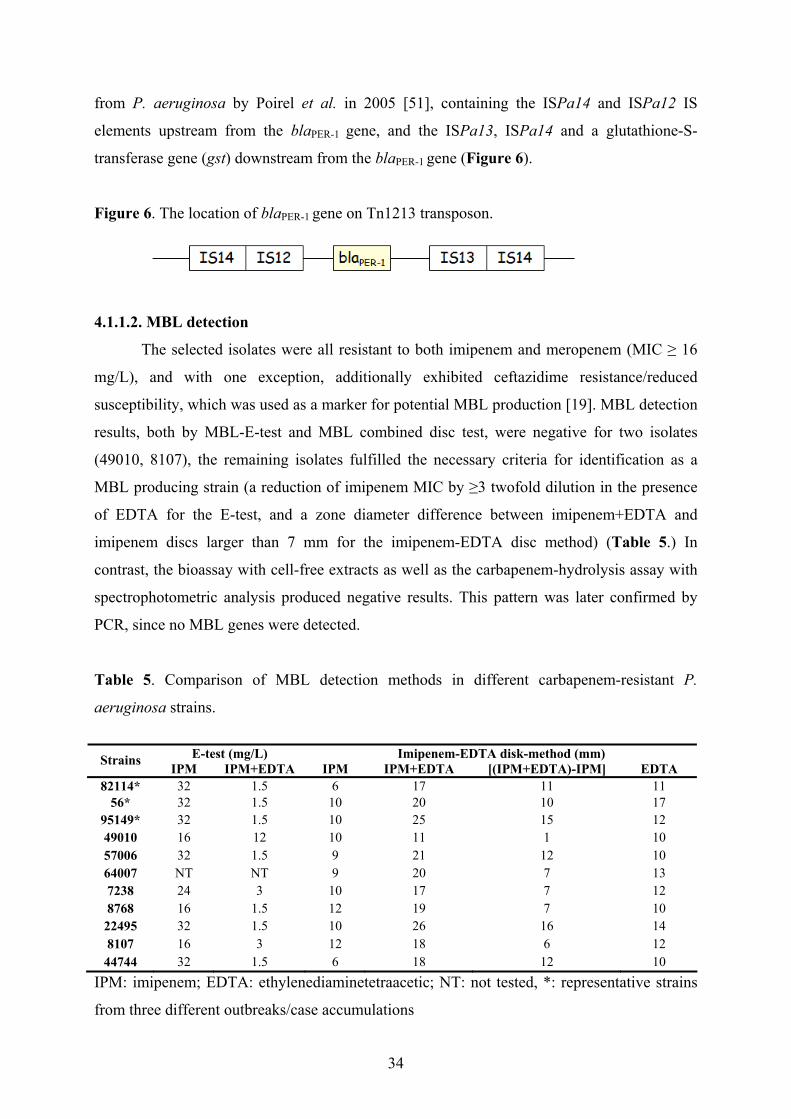
from P. aeruginosa by Poirel et al. in 2005 [51], containing the ISPa14 and ISPa12 IS
elements upstream from the blaPER-1 gene, and the ISPa13, ISPa14 and a glutathione-S-
transferase gene (gst) downstream from the blaPER-1 gene (Figure 6).
Figure 6. The location of blaPER-1 gene on Tn1213 transposon.
4.1.1.2. MBL detection
The selected isolates were all resistant to both imipenem and meropenem (MIC ≥ 16
mg/L), and with one exception, additionally exhibited ceftazidime resistance/reduced
susceptibility, which was used as a marker for potential MBL production [19]. MBL detection
results, both by MBL-E-test and MBL combined disc test, were negative for two isolates
(49010, 8107), the remaining isolates fulfilled the necessary criteria for identification as a
MBL producing strain (a reduction of imipenem MIC by ≥3 twofold dilution in the presence
of EDTA for the E-test, and a zone diameter difference between imipenem+EDTA and
imipenem discs larger than 7 mm for the imipenem-EDTA disc method) (Table 5.) In
contrast, the bioassay with cell-free extracts as well as the carbapenem-hydrolysis assay with
spectrophotometric analysis produced negative results. This pattern was later confirmed by
PCR, since no MBL genes were detected.
Table 5. Comparison of MBL detection methods in different carbapenem-resistant P.
aeruginosa strains.
E-test (mg/L) Imipenem-EDTA disk-method (mm) Strains IPM IPM+EDTA IPM IPM+EDTA [(IPM+EDTA)-IPM] EDTA
82114* 32 1.5 6 17 11 11 56* 32 1.5 10 20 10 17
95149* 32 1.5 10 25 15 12 49010 16 12 10 11 1 10 57006 32 1.5 9 21 12 10 64007 NT NT 9 20 7 13 7238 24 3 10 17 7 12 8768 16 1.5 12 19 7 10
22495 32 1.5 10 26 16 14 8107 16 3 12 18 6 12
44744 32 1.5 6 18 12 10 IPM: imipenem; EDTA: ethylenediaminetetraacetic; NT: not tested, *: representative strains
from three different outbreaks/case accumulations
34
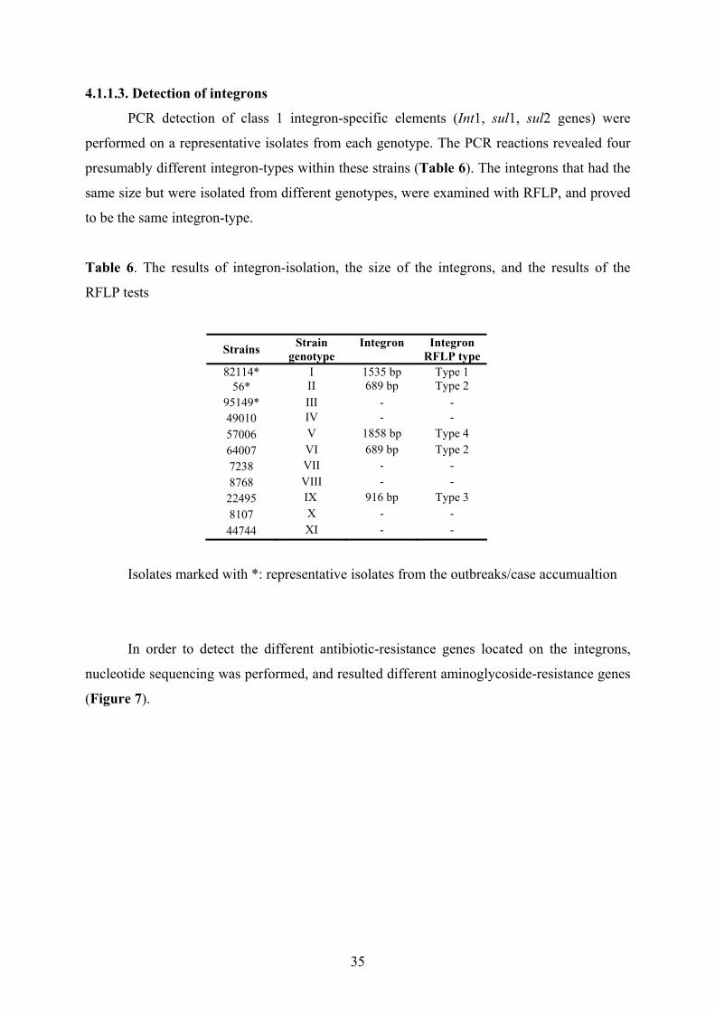
4.1.1.3. Detection of integrons
PCR detection of class 1 integron-specific elements (Int1, sul1, sul2 genes) were
performed on a representative isolates from each genotype. The PCR reactions revealed four
presumably different integron-types within these strains (Table 6). The integrons that had the
same size but were isolated from different genotypes, were examined with RFLP, and proved
to be the same integron-type.
Table 6. The results of integron-isolation, the size of the integrons, and the results of the
RFLP tests
Strains Strain genotype
Integron Integron RFLP type
82114* I 1535 bp Type 1 56* II 689 bp Type 2
95149* III - - 49010 IV - - 57006 V 1858 bp Type 4 64007 VI 689 bp Type 2 7238 VII - - 8768 VIII - -
22495 IX 916 bp Type 3 8107 X - -
44744 XI - -
Isolates marked with *: representative isolates from the outbreaks/case accumualtion
In order to detect the different antibiotic-resistance genes located on the integrons,
nucleotide sequencing was performed, and resulted different aminoglycoside-resistance genes
(Figure 7).
35

Figure 7. The structure of the four different class 1 integrons isolated from multidrug resistant
P. aeruginosa. The following aminoglycoside-resistance genes were encoded: aadB:
aminoglycoside-2’’-O-adenyltransferase, confers resistance to gentamycin, tobramycin,
kanamycin; aadA7: aminoglycoside-3’-adenyltransferase, confers resistance to streptomycin,
spectinomycin; aadA13: aminoglycoside (3’’)-O-adenyltransferase, confer resistance to
streptomycin, spectinomycin; aacA7: aminoglycoside 6'-acetyltransferase, confers resistance
to amikacin, isepamicin; aadA6: aminoglycoside-adenyltransferase, confers high level
resistance to streptomycin, spectinomycin.
4.1.1.4. Efflux pump, porin channel examinations
A representative isolate from each genotype was examined for the expression-level of
the following efflux-pumps and porins: MexAB-OprM, MexXY-OprM, MexCD-OprJ and
OprD. The expression of mRNA of the genes of interest was detected by RT-PCR assays, the
results are shown in Table 7. In case of two strains (64007, 44744) despite of repeating the
RT-PCR tests, inconclusive results were seen. Significant increase in the expression of
MexAB-OprM genes was only seen in the case of a pandrug-resistant isolate (49010).
Upregulation of the MexXY-OprM genes occurred in 4 strains (82114, 56, 49010, 22495),
there was a particularly high, 128 fold increase in case of the above mentioned pandrug-
resistant isolate (49010). In case of the isolate 57006, the increase was 9,17 fold, close to the
10 fold level, which is considered to be positive. Overexpression of the MexCD-OprJ genes
was detected in case of two isolates (95149, 22495). With the exception of two (49010, 8107),
all the multidrug-resistant strains down-regulated the expression of the OprD gene, in five
cases the decrease-level were less than 0,1 fold compared to the PAO1 control strain.
36
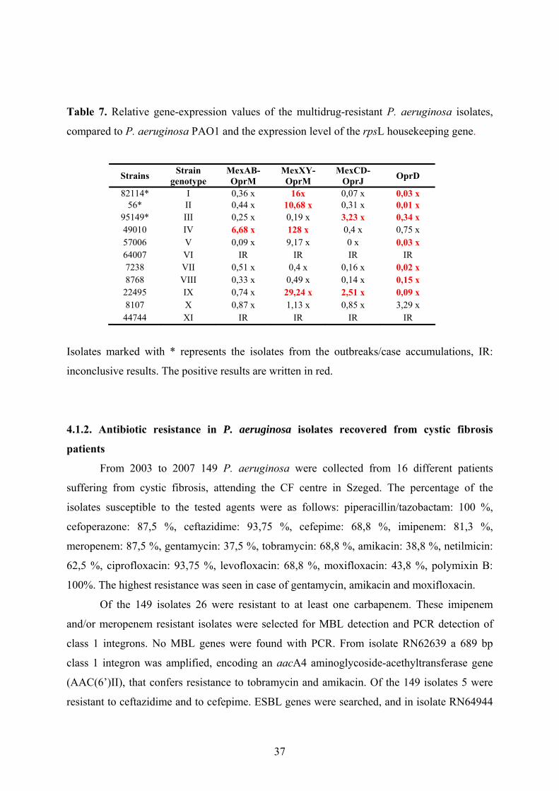
Table 7. Relative gene-expression values of the multidrug-resistant P. aeruginosa isolates,
compared to P. aeruginosa PAO1 and the expression level of the rpsL housekeeping gene.
Strains Strain genotype
MexAB-OprM
MexXY-OprM
MexCD-OprJ OprD
82114* I 0,36 x 16x 0,07 x 0,03 x 56* II 0,44 x 10,68 x 0,31 x 0,01 x
95149* III 0,25 x 0,19 x 3,23 x 0,34 x 49010 IV 6,68 x 128 x 0,4 x 0,75 x 57006 V 0,09 x 9,17 x 0 x 0,03 x 64007 VI IR IR IR IR 7238 VII 0,51 x 0,4 x 0,16 x 0,02 x 8768 VIII 0,33 x 0,49 x 0,14 x 0,15 x
22495 IX 0,74 x 29,24 x 2,51 x 0,09 x 8107 X 0,87 x 1,13 x 0,85 x 3,29 x
44744 XI IR IR IR IR
Isolates marked with * represents the isolates from the outbreaks/case accumulations, IR:
inconclusive results. The positive results are written in red.
4.1.2. Antibiotic resistance in P. aeruginosa isolates recovered from cystic fibrosis
patients
From 2003 to 2007 149 P. aeruginosa were collected from 16 different patients
suffering from cystic fibrosis, attending the CF centre in Szeged. The percentage of the
isolates susceptible to the tested agents were as follows: piperacillin/tazobactam: 100 %,
cefoperazone: 87,5 %, ceftazidime: 93,75 %, cefepime: 68,8 %, imipenem: 81,3 %,
meropenem: 87,5 %, gentamycin: 37,5 %, tobramycin: 68,8 %, amikacin: 38,8 %, netilmicin:
62,5 %, ciprofloxacin: 93,75 %, levofloxacin: 68,8 %, moxifloxacin: 43,8 %, polymixin B:
100%. The highest resistance was seen in case of gentamycin, amikacin and moxifloxacin.
Of the 149 isolates 26 were resistant to at least one carbapenem. These imipenem
and/or meropenem resistant isolates were selected for MBL detection and PCR detection of
class 1 integrons. No MBL genes were found with PCR. From isolate RN62639 a 689 bp
class 1 integron was amplified, encoding an aacA4 aminoglycoside-acethyltransferase gene
(AAC(6’)II), that confers resistance to tobramycin and amikacin. Of the 149 isolates 5 were
resistant to ceftazidime and to cefepime. ESBL genes were searched, and in isolate RN64944
37

an OXA-10 ESBL were found. No other β-lactamase or aminoglycoside-resistance genes
have been detected.
Seven isolates were selected for RT-PCR analysis, examining efflux pumps and
porins, based on the criteria detailed in section 3.3.2. One isolate showed inconclusive results.
OprD deficiency was detected in case of 5 of the 6 cases, hyperproduction of the MexXY-
OprM efflux pump was detected in 3 isolates, overexpression of MexAB-OprM was detected
in one isolate at a relatively high level, 148 fold. No isolates hyper-produced the MexCD-
OprJ efflux pump.
4.2. Bacterial typing
4.2.1. Molecular typing of multidrug-resistant P. aeruginosa isolates recovered from
non-cystic-fibrosis patients
From 2004 to 2008 51 multidrug-resistant, non-mucoid P. aeruginosa isolates were
selected for further analyses from different non-cystic fibrosis patients. Within this timeframe
at least three different outbreaks/case accumulations were detected; in one of the outbreaks 29
different patients were included, which isolates were examined in the epidemiological
analysis (in section 4.2.1.1). The remaining 22 multidrug resistant isolates were typed by
macrorestriction analyses (PFGE with SpeI digestion), which method is considered to be the
gold standard method of bacterial typing. With this method the isolates proved to belong to 11
different pulso-types. The isolates were also typed by the automated typing system,
DiversiLab, with this method 14 different genotypes were determined. The only difference
between the PFGE and the DiversiLab typing results was among 3 isolates that were occurred
in a smaller outbreak in the MICU department (PFGE pulso-type is designated as C,
DiversiLab Clone 3, Table 8). To confirm the PFGE or DiversiLab results, another molecular
typing method, namely the ERIC PCR was used for these isolates, which method confirmed
the PFGE results, namely that these isolates belong to the same genotype. Isolate 83712 was
determined as pulso-type C’ (C variant) with PFGE; with DiversiLab it showed 87%
homology with Clone 3 isolates; and with ERIC PCR it belonged to type 3, although the
ERIC results were analyzed visually, which does not allow to differentiate between slight
differences.
Based on the above different molecular typing methods, the 22 multidrug resistant P.
aeruginosa isolates belong to 11 different genotypes. Three representatives of the 11
38
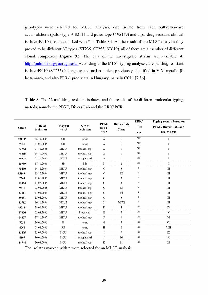
genotypes were selected for MLST analysis, one isolate from each outbreaks/case
accumulations (pulso-type A 82114 and pulso-type C 95149) and a pandrug-resistant clinical
isolate: 49010 (isolates marked with * in Table 8 ). As the result of the MLST analysis they
proved to be different ST types (ST235, ST253, ST619), all of them are a member of different
clonal complexes (Figure 8.). The data of the investigated strains are available at:
http://pubmlst.org/paeruginosa. According to the MLST typing analyses, the pandrug resistant
isolate 49010 (ST235) belongs to a clonal complex, previously identified in VIM metallo-β-
lactamase-, and also PER-1 producers in Hungary, namely CC11 [7,56].
Table 8. The 22 multidrug resistant isolates, and the results of the different molecular typing
metods, namely the PFGE, DiversiLab and the ERIC PCR.
Strain Date of isolation
Hospital ward
Site of isolation
PFGE pulso-type
DiversiLab
Clone
ERIC
PCR
type
Typing results-based on
PFGE, DiversiLab, and
ERIC PCR
82114* 26.10.2004 UH urine A 1 NT I
7025 24.01.2005 UH urine A 1 NT I
72982 07.10.2005 MICU tracheal asp. A 1 NT I
78065 24.10.2005 MICU tracheal asp. A 1 NT I
79577 02.11.2005 SICU2 nasoph.swab A 1 NT I
15939 17.11.2006 SB bile B’ 2 NT II
95490 14.12.2004 MICU tracheal asp. C 3 c III
95149* 12.12.2004 MICU tracheal asp. C 12 c III
2740 11.01.2005 MICU tracheal asp. C 3 c III
12064 11.02.2005 MICU tracheal asp. C 3 c III
9541 03.02.2005 MICU tracheal asp. C 13 c III
23611 27.03.2005 MICU tracheal asp. C 14 c III
30831 25.04.2005 MICU tracheal asp. C 3 c III
83712 16.11.2006 SICU2 tracheal asp. C´ 3-87% c III
49010* 28.06.2005 MICU tracheal asp. D 4 NT IV
57006 02.08.2005 MICU blood cult. E 5 NT V
64007 27.11.2007 MICU tracheal asp. F 6 NT VI
7238 26.01.2005 PS urine G 7 NT VII
8768 01.02.2005 PN urine H 8 NT VIII
22495 22.03.2005 PICU tracheal asp. I 9 NT IX
8107 30.01.2006 PICU nasoph.swab J 10 NT X
44744 20.06.2006 PICU tracheal asp. K 11 NT XI
The isolates marked with * were selected for an MLST analysis.
39

Figure 8 . The eBURST diagram of P. aeruginosa isolates generated by software available at
http://eburst.mlst.net with the three examined isolates. Isolate 82114 is ST619, belonging to
an unknown clonal complex circled in blue; isolate 95149 is ST253 belonging to an unknown
clonal complex circled in pink; isolate 49010 is ST235 belonging to clonal complex 11 cirled
in red.
4.2.1.1. Investigation of multidrug resistant isolates of the epidemiological analysis;
environmental screening
From June 2007 to November 2008 29 multiresistant clinical P. aeruginosa were
obtained from an outbreak, that showed resistance to all antibiotics except polymixin B,
amikacin, and + netilmicin. In addition to similar resistance patterns, they had similar colony
morphology as well. The 29 different patients, 14 females and 15 males, were admitted to
different clinical wards including four ICUs (Table 9). Twenty patients developed infection
based on clinical evidence, such as isolation of the strain from blood culture or from lower
respiratory tract specimens (endotracheal aspirates or protected bronchoalveolar lavage)
together with fever, increased procalcitonin and/or CRP levels, increased white-blood cell
sedimentation and granulocytes in the lower respiratory tract specimens. Nine patients
displayed simple colonization. The first patient, from whom the multiresistant P. aeruginosa
was isolated, was admitted to the MICU from another hospital ward (cardiac surgical ICU).
40
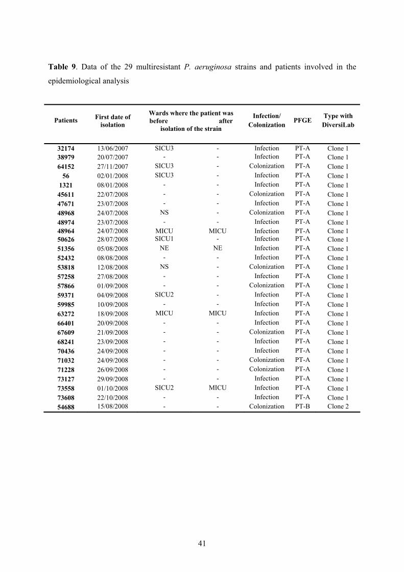
Table 9. Data of the 29 multiresistant P. aeruginosa strains and patients involved in the
epidemiological analysis
Patients First date of isolation
Wards where the patient was before after
isolation of the strain
Infection/ Colonization
PFGE Type with DiversiLab
32174 13/06/2007 SICU3 - Infection PT-A Clone 1 38979 20/07/2007 - - Infection PT-A Clone 1 64152 27/11/2007 SICU3 - Colonization PT-A Clone 1
56 02/01/2008 SICU3 - Infection PT-A Clone 1 1321 08/01/2008 - - Infection PT-A Clone 1
45611 22/07/2008 - - Colonization PT-A Clone 1 47671 23/07/2008 - - Infection PT-A Clone 1 48968 24/07/2008 NS - Colonization PT-A Clone 1 48974 23/07/2008 - - Infection PT-A Clone 1 48964 24/07/2008 MICU MICU Infection PT-A Clone 150626 28/07/2008 SICU1 - Infection PT-A Clone 1 51356 05/08/2008 NE NE Infection PT-A Clone 1 52432 08/08/2008 - - Infection PT-A Clone 1 53818 12/08/2008 NS - Colonization PT-A Clone 1 57258 27/08/2008 - - Infection PT-A Clone 1 57866 01/09/2008 - - Colonization PT-A Clone 1 59371 04/09/2008 SICU2 - Infection PT-A Clone 1 59985 10/09/2008 - - Infection PT-A Clone 1 63272 18/09/2008 MICU MICU Infection PT-A Clone 1 66401 20/09/2008 - - Infection PT-A Clone 1 67609 21/09/2008 - - Colonization PT-A Clone 1 68241 23/09/2008 - - Infection PT-A Clone 1 70436 24/09/2008 - - Infection PT-A Clone 1 71032 24/09/2008 - - Colonization PT-A Clone 1 71228 26/09/2008 - - Colonization PT-A Clone 1 73127 29/09/2008 - - Infection PT-A Clone 1 73558 01/10/2008 SICU2 MICU Infection PT-A Clone 1 73608 22/10/2008 - - Infection PT-A Clone 1 54688 15/08/2008 - - Colonization PT-B Clone 2
41
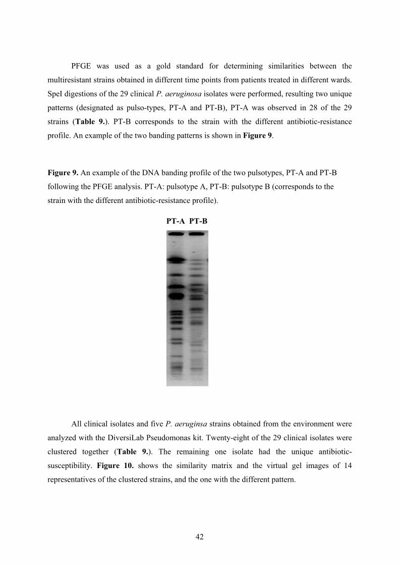
PFGE was used as a gold standard for determining similarities between the
multiresistant strains obtained in different time points from patients treated in different wards.
SpeI digestions of the 29 clinical P. aeruginosa isolates were performed, resulting two unique
patterns (designated as pulso-types, PT-A and PT-B), PT-A was observed in 28 of the 29
strains (Table 9.). PT-B corresponds to the strain with the different antibiotic-resistance
profile. An example of the two banding patterns is shown in Figure 9.
Figure 9. An example of the DNA banding profile of the two pulsotypes, PT-A and PT-B
following the PFGE analysis. PT-A: pulsotype A, PT-B: pulsotype B (corresponds to the
strain with the different antibiotic-resistance profile).
PT-A PT-B
All clinical isolates and five P. aeruginsa strains obtained from the environment were
analyzed with the DiversiLab Pseudomonas kit. Twenty-eight of the 29 clinical isolates were
clustered together (Table 9.). The remaining one isolate had the unique antibiotic-
susceptibility. Figure 10. shows the similarity matrix and the virtual gel images of 14
representatives of the clustered strains, and the one with the different pattern.
42
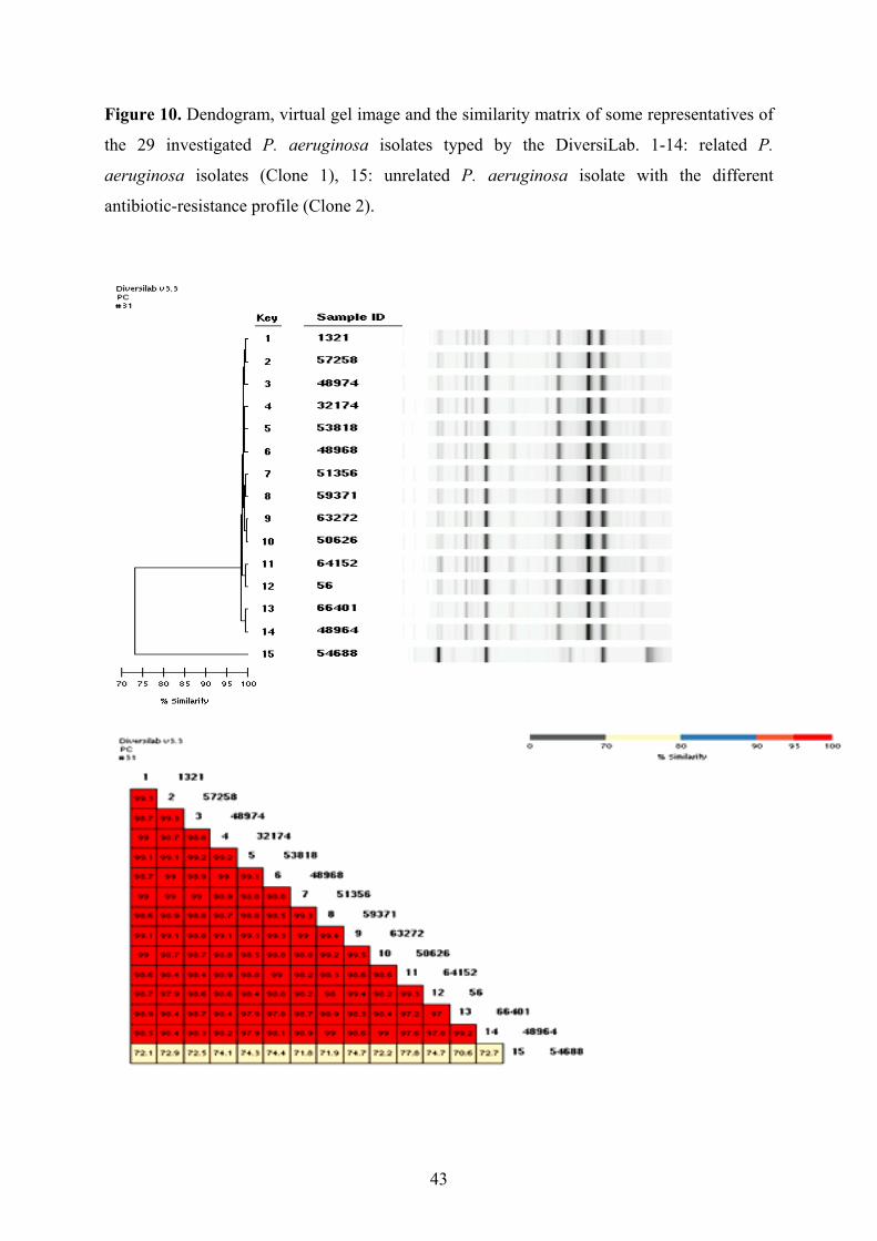
Figure 10. Dendogram, virtual gel image and the similarity matrix of some representatives of
the 29 investigated P. aeruginosa isolates typed by the DiversiLab. 1-14: related P.
aeruginosa isolates (Clone 1), 15: unrelated P. aeruginosa isolate with the different
antibiotic-resistance profile (Clone 2).
43
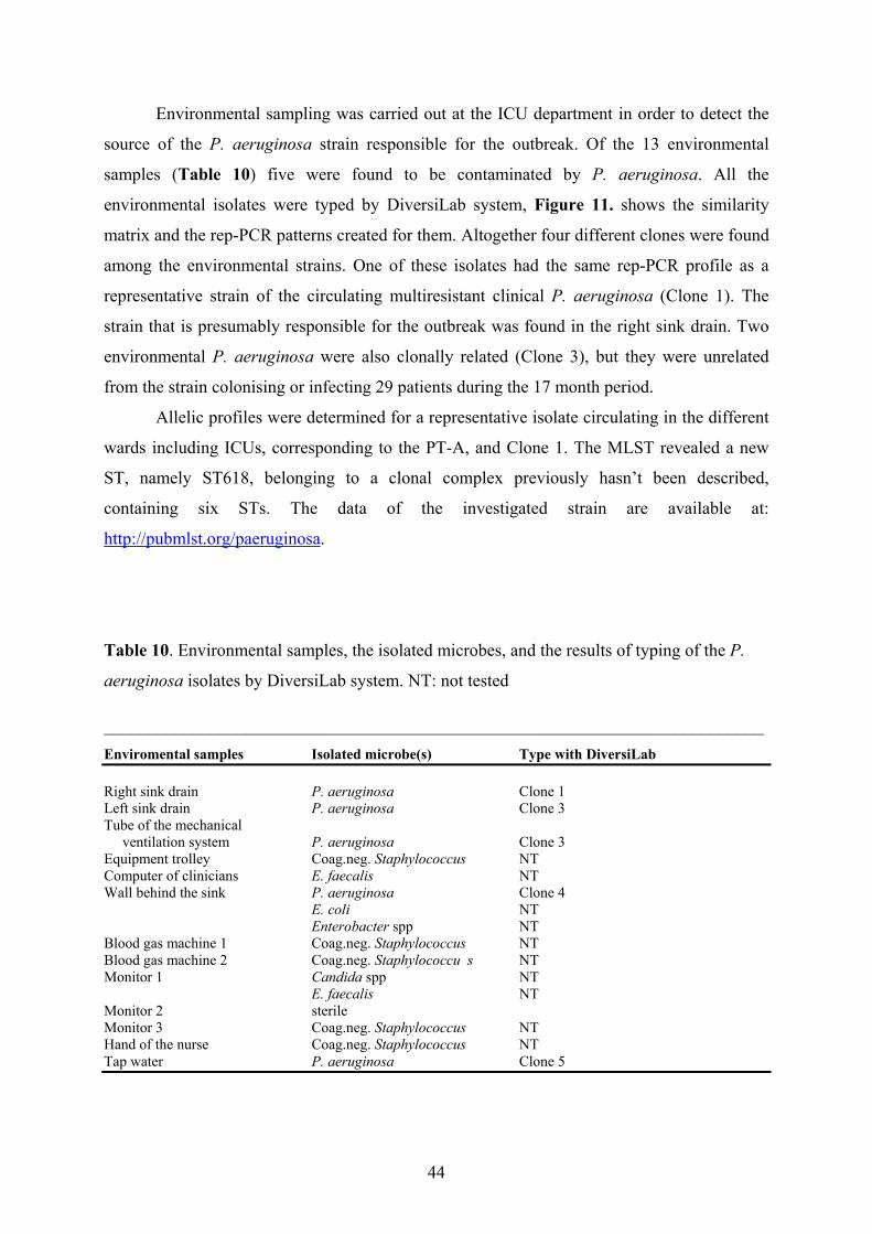
Environmental sampling was carried out at the ICU department in order to detect the
source of the P. aeruginosa strain responsible for the outbreak. Of the 13 environmental
samples (Table 10) five were found to be contaminated by P. aeruginosa. All the
environmental isolates were typed by DiversiLab system, Figure 11. shows the similarity
matrix and the rep-PCR patterns created for them. Altogether four different clones were found
among the environmental strains. One of these isolates had the same rep-PCR profile as a
representative strain of the circulating multiresistant clinical P. aeruginosa (Clone 1). The
strain that is presumably responsible for the outbreak was found in the right sink drain. Two
environmental P. aeruginosa were also clonally related (Clone 3), but they were unrelated
from the strain colonising or infecting 29 patients during the 17 month period.
Allelic profiles were determined for a representative isolate circulating in the different
wards including ICUs, corresponding to the PT-A, and Clone 1. The MLST revealed a new
ST, namely ST618, belonging to a clonal complex previously hasn’t been described,
containing six STs. The data of the investigated strain are available at:
http://pubmlst.org/paeruginosa.
Table 10. Environmental samples, the isolated microbes, and the results of typing of the P.
aeruginosa isolates by DiversiLab system. NT: not tested
___________________________________________________________________________ Enviromental samples Isolated microbe(s) Type with DiversiLab Right sink drain P. aeruginosa Clone 1 Left sink drain P. aeruginosa Clone 3 Tube of the mechanical ventilation system P. aeruginosa Clone 3 Equipment trolley Coag.neg. Staphylococcus NT Computer of clinicians E. faecalis NT Wall behind the sink P. aeruginosa Clone 4 E. coli NT Enterobacter spp NT Blood gas machine 1 Coag.neg. Staphylococcus NT Blood gas machine 2 Coag.neg. Staphylococcu s NT Monitor 1 Candida spp NT E. faecalis NT Monitor 2 sterile Monitor 3 Coag.neg. Staphylococcus NT Hand of the nurse Coag.neg. Staphylococcus NT Tap water P. aeruginosa Clone 5
44
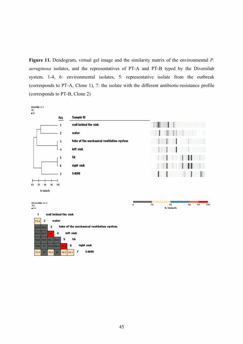
Figure 11. Dendogram, virtual gel image and the similarity matrix of the environmental P.
aeruginosa isolates, and the representatives of PT-A and PT-B typed by the Diversilab
system. 1-4, 6: environmental isolates, 5: representative isolate from the outbreak
(corresponds to PT-A, Clone 1), 7: the isolate with the different antibiotic-resistance profile
(corresponds to PT-B, Clone 2)
45
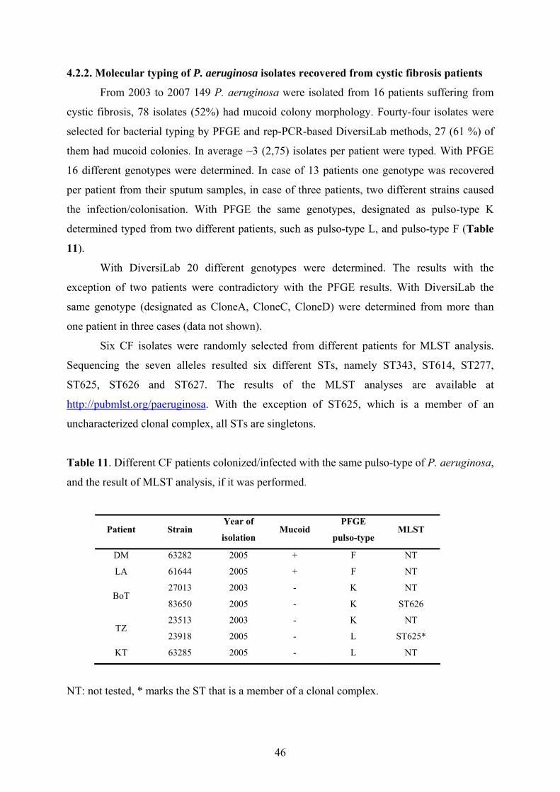
4.2.2. Molecular typing of P. aeruginosa isolates recovered from cystic fibrosis patients
From 2003 to 2007 149 P. aeruginosa were isolated from 16 patients suffering from
cystic fibrosis, 78 isolates (52%) had mucoid colony morphology. Fourty-four isolates were
selected for bacterial typing by PFGE and rep-PCR-based DiversiLab methods, 27 (61 %) of
them had mucoid colonies. In average ~3 (2,75) isolates per patient were typed. With PFGE
16 different genotypes were determined. In case of 13 patients one genotype was recovered
per patient from their sputum samples, in case of three patients, two different strains caused
the infection/colonisation. With PFGE the same genotypes, designated as pulso-type K
determined typed from two different patients, such as pulso-type L, and pulso-type F (Table
11).
With DiversiLab 20 different genotypes were determined. The results with the
exception of two patients were contradictory with the PFGE results. With DiversiLab the
same genotype (designated as CloneA, CloneC, CloneD) were determined from more than
one patient in three cases (data not shown).
Six CF isolates were randomly selected from different patients for MLST analysis.
Sequencing the seven alleles resulted six different STs, namely ST343, ST614, ST277,
ST625, ST626 and ST627. The results of the MLST analyses are available at
http://pubmlst.org/paeruginosa. With the exception of ST625, which is a member of an
uncharacterized clonal complex, all STs are singletons.
Table 11. Different CF patients colonized/infected with the same pulso-type of P. aeruginosa,
and the result of MLST analysis, if it was performed.
Patient Strain Year of
isolation Mucoid
PFGE
pulso-type MLST
DM 63282 2005 + F NT
LA 61644 2005 + F NT
27013 2003 - K NT BoT
83650 2005 - K ST626
23513 2003 - K NT TZ
23918 2005 - L ST625*
KT 63285 2005 - L NT
NT: not tested, * marks the ST that is a member of a clonal complex.
46
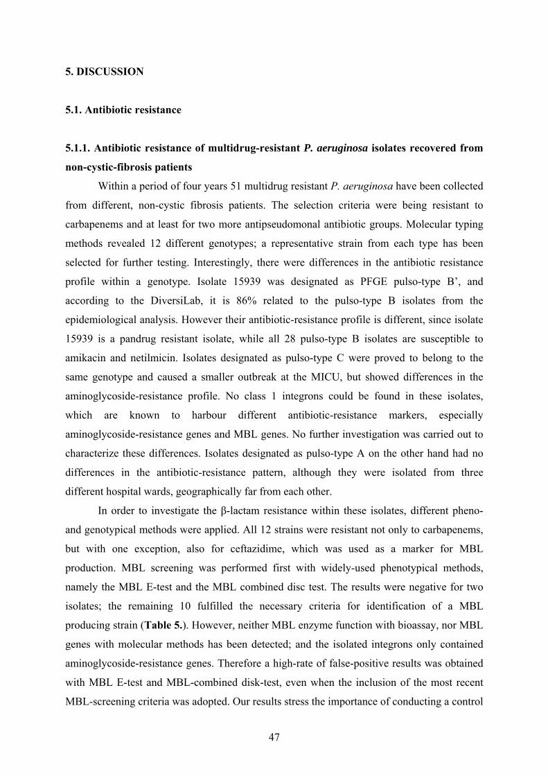
5. DISCUSSION
5.1. Antibiotic resistance
5.1.1. Antibiotic resistance of multidrug-resistant P. aeruginosa isolates recovered from
non-cystic-fibrosis patients
Within a period of four years 51 multidrug resistant P. aeruginosa have been collected
from different, non-cystic fibrosis patients. The selection criteria were being resistant to
carbapenems and at least for two more antipseudomonal antibiotic groups. Molecular typing
methods revealed 12 different genotypes; a representative strain from each type has been
selected for further testing. Interestingly, there were differences in the antibiotic resistance
profile within a genotype. Isolate 15939 was designated as PFGE pulso-type B’, and
according to the DiversiLab, it is 86% related to the pulso-type B isolates from the
epidemiological analysis. However their antibiotic-resistance profile is different, since isolate
15939 is a pandrug resistant isolate, while all 28 pulso-type B isolates are susceptible to
amikacin and netilmicin. Isolates designated as pulso-type C were proved to belong to the
same genotype and caused a smaller outbreak at the MICU, but showed differences in the
aminoglycoside-resistance profile. No class 1 integrons could be found in these isolates,
which are known to harbour different antibiotic-resistance markers, especially
aminoglycoside-resistance genes and MBL genes. No further investigation was carried out to
characterize these differences. Isolates designated as pulso-type A on the other hand had no
differences in the antibiotic-resistance pattern, although they were isolated from three
different hospital wards, geographically far from each other.
In order to investigate the β-lactam resistance within these isolates, different pheno-
and genotypical methods were applied. All 12 strains were resistant not only to carbapenems,
but with one exception, also for ceftazidime, which was used as a marker for MBL
production. MBL screening was performed first with widely-used phenotypical methods,
namely the MBL E-test and the MBL combined disc test. The results were negative for two
isolates; the remaining 10 fulfilled the necessary criteria for identification of a MBL
producing strain (Table 5.). However, neither MBL enzyme function with bioassay, nor MBL
genes with molecular methods has been detected; and the isolated integrons only contained
aminoglycoside-resistance genes. Therefore a high-rate of false-positive results was obtained
with MBL E-test and MBL-combined disk-test, even when the inclusion of the most recent
MBL-screening criteria was adopted. Our results stress the importance of conducting a control
47
test using a disc containing solely EDTA, since the increased inhibition zone around the disk
containing IPM-EDTA was most likely due to the membrane permeabilizing effect of EDTA,
rather then a chelating action upon any hypothetical MBLs. The same effect could be
observed in case of the E-test method. Among our clinical P. aeruginosa isolates the
susceptibility to EDTA was common, the inhibition zones around the disks containing only
EDTA were varying from 10 mm to 17 mm. These false-positive results unambiguously
preclude the recommendation for using the E-test or the combined disk-test as a
carbapenemase-screening method [article I]. An easy-to-perform bioassay using cell-free
extracts, where the interference of EDTA is avoided, is preferable for routine carbapenemase-
activity screening for clinical microbiology laboratory.
In fact, the resistant phenotype to carbapenems observed in the examined strains could
be linked to the overexpression of efflux pumps involved in the extrusion of carbapenems
and/or OprD deficiency, rather than the production of MBLs, as it was suspected by the
interpretation of the MBL E-test and MBL combined disc test. One of the examined
genotypes hyper-produced MexAB-OprM efflux pump, four strains over-expressed the genes
of MexXY-OprM and two of them the MexCD-OprJ. Eight strains had significantly down-
regulated the genes of the OprD porin channel (Table 7.). In a pandrug resistant strain
(49010), which was resistant to all anti-pseudomonal antibiotics with the exception of
polymyxin-B, beside the overexpression of two different efflux-pump genes, the production
of three ESBL enzymes did extensively contribute to β-lactam resistance [13].
5.1.2. Antibiotic resistance of P. aeruginosa isolates recovered from CF patients
Within a period of four years 149 P. aeruginosa were isolated from 16 different
patients suffering from cystic fibrosis, attending the CF centre in Szeged. In this period of
time all isolates from the 16 patients were stored for further analysis, regardless their
antibiotic susceptibility.
Among these isolates the highest resistance was seen in case of gentamycin, amikacin
and moxifloxacin (62,5 %, 61,2 %, 56,2 % respectively). Otherwise the strains had a good in
vitro susceptibility, which results do not follow the international tendency [9,59,60]. Among
our CF isolates were no pan- or multidrug resistant P. aeruginosa, and no developing
resistance was observed due to the antimicrobial treatments [article IV].
No MBLs were found, only one class 1 integron, and an OXA-10 ESBL was found
among the isolates. Isolate RN64944 producing the OXA-10 ESBL was resistant to cefepime,
48
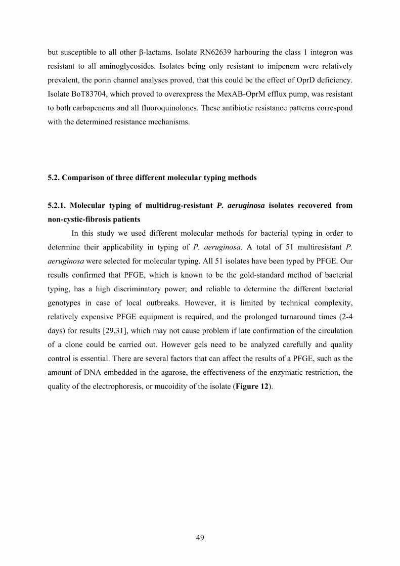
but susceptible to all other β-lactams. Isolate RN62639 harbouring the class 1 integron was
resistant to all aminoglycosides. Isolates being only resistant to imipenem were relatively
prevalent, the porin channel analyses proved, that this could be the effect of OprD deficiency.
Isolate BoT83704, which proved to overexpress the MexAB-OprM efflux pump, was resistant
to both carbapenems and all fluoroquinolones. These antibiotic resistance patterns correspond
with the determined resistance mechanisms.
5.2. Comparison of three different molecular typing methods
5.2.1. Molecular typing of multidrug-resistant P. aeruginosa isolates recovered from
non-cystic-fibrosis patients
In this study we used different molecular methods for bacterial typing in order to
determine their applicability in typing of P. aeruginosa. A total of 51 multiresistant P.
aeruginosa were selected for molecular typing. All 51 isolates have been typed by PFGE. Our
results confirmed that PFGE, which is known to be the gold-standard method of bacterial
typing, has a high discriminatory power; and reliable to determine the different bacterial
genotypes in case of local outbreaks. However, it is limited by technical complexity,
relatively expensive PFGE equipment is required, and the prolonged turnaround times (2-4
days) for results [29,31], which may not cause problem if late confirmation of the circulation
of a clone could be carried out. However gels need to be analyzed carefully and quality
control is essential. There are several factors that can affect the results of a PFGE, such as the
amount of DNA embedded in the agarose, the effectiveness of the enzymatic restriction, the
quality of the electrophoresis, or mucoidity of the isolate (Figure 12).
49

Figure 12. Examples for technical errors of PFGE. A: not enough DNA in the gel, B: too
much DNA in the gel, C: mucoid P. aeruginosa isolate.
A B C
Beside PFGE, all 51 P. aeruginosa were typed with the repetitive-element-PCR-based
DiversiLab instrument as well, and contradictory results only occurred in case of three
isolates (Table 8.). ERIC PCR was used in case of these isolates and confirmed the PFGE
results, namely that the isolates belonged to the same genotype (PFGE pulso-type C). In case
of these isolates the DiversiLab virtual gel image showed too many bands with similar sizes,
during the PCR reaction too many products have been amplified due to an unknown reason
(Figure 13).( The same technical problem occurred in case of the CF isolates, see section
5.2.2.).
50
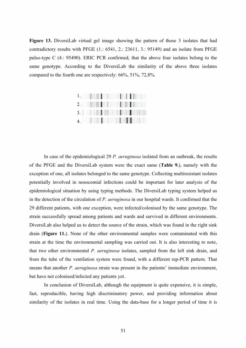
Figure 13. DiversiLab virtual gel image showing the pattern of those 3 isolates that had
contradictory results with PFGE (1.: 6541, 2.: 23611, 3.: 95149) and an isolate from PFGE
pulso-type C (4.: 95490). ERIC PCR confirmed, that the above four isolates belong to the
same genotype. According to the DiversiLab the similarity of the above three isolates
compared to the fourth one are respectively: 66%, 51%, 72,8%.
1.
2.
3.
4.
In case of the epidemiological 29 P. aeruginosa isolated from an outbreak, the results
of the PFGE and the DiversiLab system were the exact same (Table 9.), namely with the
exception of one, all isolates belonged to the same genotype. Collecting multiresistant isolates
potentially involved in nosocomial infections could be important for later analysis of the
epidemiological situation by using typing methods. The DiversiLab typing system helped us
in the detection of the circulation of P. aeruginosa in our hospital wards. It confirmed that the
29 different patients, with one exception, were infected/colonised by the same genotype. The
strain successfully spread among patients and wards and survived in different environments.
DiversiLab also helped us to detect the source of the strain, which was found in the right sink
drain (Figure 11.). None of the other environmental samples were contaminated with this
strain at the time the environmental sampling was carried out. It is also interesting to note,
that two other environmental P. aeruginosa isolates, sampled from the left sink drain, and
from the tube of the ventilation system were found, with a different rep-PCR pattern. That
means that another P. aeruginosa strain was present in the patients’ immediate environment,
but have not colonised/infected any patients yet.
In conclusion of DiversiLab, although the equipment is quite expensive, it is simple,
fast, reproducible, having high discriminatory power, and providing information about
similarity of the isolates in real time. Using the data-base for a longer period of time it is
51

possible to follow and compare outbreak isolates locally. Therefore it can be an effective tool
for a microbiological laboratory during epidemiological analyses [article III].
A representative isolate from each outbreak/case accumulation (82114, 95149, 56),
and the 49010 pandrug resistant isolate, which is proved to produce three different ESBLs and
hyperproduces two efflux-pumps, were selected for an MLST analysis. P. aeruginosa 82114
isolated from a case accumulation (of 5 cases) proved to be ST619, a member of a clonal
complex that has not been described previously. P. aeruginosa 95149 isolated from an
outbreak of 8 cases proved to be ST253, which is also a member of a clonal complex. P.
aeruginosa 56 isolated from an outbreak of 28 cases, is an ST618, also a member of an
unknown clonal complex (Figure 8.). It is known that P. aeruginosa has a non-clonal
population structure punctuated by successful epidemic clones, or clonal complexes [33].
Therefore, our MLST results draw the attention of the epidemiologists on account of the
gathering evidence that some clonal lineages are more likely spread among patients and cause
infections/outbreaks.
With MLST analysis the pandrug resistant isolate (49010) is proved to be ST235, a
member of the CC11 clonal complex. Interestingly, a PER-1 producer strain was isolated
from an outbreak in Poland [6], with the same ST, and also from Hungary and Serbia [7].
Although, our strain was isolated from a Romanian patient, regarding the virulence of the
CC11 members, and the geographical closeness of Hungary, Romania, Serbia and Poland, we
can not exclude the possibility that the same strain is circulating in these countries.
In this study we compared three different typing methods for the molecular typing of
clinical P. aeruginosa isolates. By using the MLST method we can gain information about the
clonal relationship of the bacterial isolates, as it has been proved previously [30,33]. PFGE
and rep-PCR-based methods nevertheless have higher discriminatory power, than MLST [30],
making it possible to also differentiate between isolates with similar antibiotic patterns, and to
find the environmental presence of the same epidemic strain. Therefore the implication of
MLST for population genetics and dynamics may be more significant than those for bacterial
epidemiology, since polymorphism in the slowly evolving genes, which are its targets, may
not be high enough for useful epidemiological comparisons.
52

5.2.2 Molecular typing of P. aeruginosa isolates recovered from CF patients
Based on the different antibiotic susceptibility profile 44 isolates were selected for
bacterial typing. With the PFGE method 16 different genotypes were determined. In case of
13 patients one genotype were recovered per patient from their sputum samples, so these
patients were colonised/infected with the same strain from 2003 to 2007. In case of three
patients, two different genotypes could be isolated from their sputum, so they were
colonised/infected with different strains. Overall a high diversity was observed, which
corresponds to the international literature [45]. The isolates were also typed by the DiversiLab
system. The virtual gel image showed too many bands with similar sizes in almost all of the
strains due to an unknown reason (Figure 14). Results did not correspond neither with the
antibiotic susceptibility pattern, nor with the PFGE results, which is considered to be the gold
standard method of bacterial typing.
Figure 14. The virtual gel image of some of the examined CF isolates shows the presence of
too many bands in case of almost all of the isolates.
The same genotype (designated as CloneA, CloneC, CloneD, data not shown) were
determined from more than one patient in three cases. PFGE analysis could not confirm these
results, and also the clinical background studies showed that these patients were not
hospitalised at the same time or did not participate the same CF camp, so the cross-infection
could be excluded. Considering all the above results, we concluded that during the molecular
53

typing of CF isolates the PFGE method is found to be more accurate and reliable than the
DiversiLab system.
Until recently, CF patients were thought to be colonised with their own unique P.
aeruginosa strain acquired from the environment. Cross-infection was considered a relatively
infrequent source of acquisition. With PFGE the same genotype (designated as pulso-type F,
data not shown) was isolated from patients DM and LA from 2005 (Table 11.), when they
participated in the same CF-camp. Pulso-type L was isolated from patients TZ and KT from
2005. At that time, they were hospitalised at the same time in the same hospital ward. Pulso-
type K recovered from patients BoT and TZ. The strains were isolated in 2003, and according
to their clinical background the two patients attended the same CF-camp in Budapest,
Hungary. Interestingly, the MLST results suggest that the latter strain, being a member of a
clonal complex, and not a singleton, could be a member of an epidemic clone (Table 11).
However the remaining five strains examined with MLST proved to be singletons with this
method, which suggests that the prevalence of the cluster genotypes is significantly lower
among these CF isolates. With our results we proved, that the transmission of a certain strain
from patient to patient in a hospital ward or in a CF camp is expected, but not inevitable.
Nevertheless the incidence of the cross-infections in the CF centre in Szeged were relatively
low due to the infection control measures applied to prevent patient-to-patient contacts, such
as scheduling appointments with the clinicians at different time points, trying to hospitalize
the patients in different hospital rooms [article IV].
54

6. CONCLUSIONS
Based on the present study we can conclude, that in case of P. aruginosa isolates obtained
from non-CF patients:
- The presence of multi- or pandrug resistant isolates among patients hospitalised in the
University Hospital of Szeged between 2004-and 2008 was relatively low (7 %).
- Routine detection of the different antibiotic resistance mechanisms, especially the ones
with epidemiological consequences, such as MBL production, is vital for the hospital
laboratories, therefore a cheap, fast and reliable method is required. MBL-E-test is used
worldwide, but in this study is proved to be inaccurate due to the effects of EDTA.
- Determination of the different resistance mechanisms against antipseudomonal β-lactams
revealed that in contrast with the international tendency, MBL production in clinical P.
aeruginosa isolates is not widespread in our University Hospital.
- In case of hospital outbreaks a fast, reliable molecular typing method is required in order
to localize the outbreak, and possibly to detect the source of the isolate responsible. In the
present study DiversiLab typing system is proved to be as accurate and reliable as the
gold-standard PFGE. Additional advantage of the system is the long-term, internet-based
database.
- Following the international tendency our epidemic P. aeruginosa strains proved to be
members of clonal complexes based on the MLST analysis.
Based on the present study we can conclude that the P. aeruginosa isolates obtained from CF
patients:
- have an overall good in vitro susceptibility, which results are in contrast with the
international tendency. Among our isolates there were no MBL producer, no multi-or
pandrug resistant isolate, and no therapy-induced resistance was observed.
- During the molecular typing of CF isolates the DiversiLab system is found to be too
discriminative and not as reliable as the PFGE method.
- These isolates have a high genetic diversity, no clonality was observed, which
corresponds to the international literature.
- With the exception of a few sporadic cases of transmission, there were no highly
epidemic strain isolated, which is expected to be a result of the good infection control
measures taken by the clinicians, nurses of the CF centre, and of the good patient
compliance.
55
7. ACKNOWLEDGEMENTS
I wish to express my special thanks to my supervisor Prof. Erzsébet Nagy for her continuous
support, scientific guidance and enthusiasm in her supervision of this study.
I am very grateful to Edit Hajdú for her knowledge, advices and handling of the clinical part
of this study.
I express my grateful thanks to all the staff members of the Institute of Clinical Microbiology,
University of Szeged, especially to Judit Deák, Edit Urbán, József Sóki, Mária Lohr, Éva
Dósa, Szilvia Batki for their helpful cooperation and their support.
Special thanks are due to Prof Luise Peixe, Sandra Quinteira, Filipa Grosso, Ana Freitas,
Patricia Antunes and all the members of the Laboratory of Microbiology, Faculty of
Pharmacy, University of Porto, without whom this task would never have been fulfilled.
I wish to express my appreciation to all my colleagues of the microbiology laboratory at
ICON for supporting me. Special thanks to Tracy Mullen, Desmond McNulty and Ann-Marie
Croghan for their helpful advices and encouragement.
Special thanks are due to Tímea Balogh, Gyula Orosz and the staff members of Department of
Biotechnology, Faculty of Sciences University of Szeged, for permission and help to carry out
the RNA extraction assays
I am especially grateful to my mother, father and sister for their love and support. Special
thanks to my boyfriend for the continuous encouragement and prompting.
This work was supported by a fellowship from Federation of European Microbiological
Societies (FEMS) FRF 2007-2.
56

8. REFERENCES
1. Murray R. P., Baron E. J., Pfaller M. A., Tenover F. C., Yolken L. H.: Manual of
clinical microbiology. 7th edition. 33: 517-526
2. Livermore D. M.: Multiple mechanisms of antimicrobial resistance in Pseudomonas
aerugionosa: our worst nightmare? Clin Inf Dis. 2002, 34:634-640
3. Lambert P. A.: Mechanisms of antibiotic resistance in Pseudomonas aeruginosa. J R
Soc Med. 2002, 95:22-26
4. Hancock R. E. W., Brinkman F. S.L.: Function of Pseudomonas aeruginosa in uptake
and efflux. Annu Rev Microbiol. 2002, 56:17-38
5. Wang C. Y., Jerng J. S., Cheng K. Y., Lee L. N., Yu C., J., Hsueh P. R., Yang P. C.:
Pandrug resistant Pseudomonas aeruginosa among hospitalized patients: clinical
features, risk-factors, and outcomes. Clin Microb Inf. 2006, 12:63-68
6. Empel J, Filczak K, Mrówka A, Hryniewicz W, Livermore DM, Gniadkowski W.
Outbreak of Pseudomonas aeruginosa infections with PER-1 extended-spectrum β-
lactamase in Warsaw, Poland: further evidence for an international clonal complex. J
Clin Microbiol 2007; 45: 1829-2834.
7. Libisch B, Poirel L, Lepsanovic Z, Mirovic V, Balogh B, Pászti J et al. Identification
of PER-1 extended-spectrum beta-lactamase producing Pseudomonas aeruginosa
clinical isolates of the international clonal complex CC11 from Hungary and Serbia.
FEMS Immunol Med Microbiol 2008; 54: 330-8.
8. Szabo D, Szentandrassy J, Juhasz Zs, Katona K, Nagy K, Rokusz L. Imported PER-1
producing Pseudomonas aeruginosa, PER-1 producing Acinetobacter baumanii and
VIM-2 producing Pseudomonas aeruginosa strains in Hungary. Ann Clin Microbiol
Antimicrob 2008; 30: 7-12.
9. Wolter DJ, Acquazzino D, Goering RV, Sammut P, Khalaf N, Hanson ND.:
Emergence of carbapenem resistance in Pseudomonas aeruginosa isolates from a
patient with cystic fibrosis in the absence of carbapenem therapy. Clin Infect Dis. 2008
46:137-41
10. Livermore D.: β-lactamases in laboratory and clinical resistance. Clin Microb
Reviews. 1995, 8:557-584
11. Bert F., Branger C., Lambert-Zechovsky N.: Identification of PSE and OXA β-
lactamase genes in Pseudomonas aeruginosa using PCR-restriction fragment-length
polymorphism. J Antimicrob Chemother. 2002, 50:11-18
57

12. Walther-Rassmussen J., Høiby N.: OXA-type carbapenemases. J Antimicrob
Chemother, 2006, 57:373-383
13. Livermore D. M., Woodford N.: Carbapenemasese: a problem in waiting. Current
Opinion Microbiol 2000, 3:489-495
14. Walsch T.R., Toleman M. A., Poirel L., Nordmann P.:Metallo-beta-lactamases: the
quiet before the storm? Clin Microbiol Rev 2005, 18: 306-325
15. Bennett T. M.: Integrons and gene cassettes: a genetic construction kit for bacteria. J
Antimicrob Chemother 1999, 43:1-4
16. Fluit A. C., Schmitz F. J.: Class 1integrons, Gene cassettes, mobility, and
epidemiology. Eur J Clin Microbiol Infect Dis, 1999, 18:761-770
17. Poole K.: Aminoglycoside reistance in Pseudomonas aeruginosa. Antimicrob Agents
Chemother. 2005, 49: 479-487
18. Bergés L, Rodriguez-Villalobos H, Deplano A et al. Prospective evaluation of
imipenem/EDTA combined disc and E-test for detection of metallo-β-lactamases. J
Antimicrob Chemother 2007; 59:812-3
19. Samuelsen Ø, Buarø L, Giske C et al. Evaluation of phenotypic tests for the detection
of metallo-β-lactamase-producing Pseudomonas aeruginosa in a low prevalence
country. J Antimicrob Chemother 2008; 61:827-830
20. Walsh T, Bolmström A, Quarnström A et al. Evaluation of a new Etest for detecting
metallo-β-lactamases in routine clinical testing. J Clin Microbiol 2002; 40:2755-59
21. Chu TW, Cheung TKM, Ngan JYW et al. EDTA susceptibility leading to false-
detection of metallo-β-lactamases in Pseudomonas aeruginosa by Etest and
imipenem-EDTA disc method. Int J Antim Agents 2005; 26:340-1
22. Vedel G.: Simple method to determine β-lactam resistance phenotypes in
Pseudomonas aeruginosa using the disc agar diffusion test. J Antimicrob Chemother
2005, 56:657-664
23. Eftekhar F., Rafiee R.: An overlay gel method for identification and isolation of
bacterial β-lactamases. J Microb Methods, 2006, 64:132-134
24. Migliavacca R., Docquier J. D., Mugnaioli C., Amicosante G., Daturi R., Lee K.,
Rossolini G., M., Pagani L.: Simple microdilution test for detection of metallo-β-
lactamase production in Pseudomonas aeruginosa. J Clin Micro, 2002, 40:4388-4390
25. Tan J., Pitout J. D. D., Guttman D. S.: New and sensitive assay for determining
Pseudomonas aeruginosa metallo-beta-lactamase resistance to imipenem. J Clin
Micro, 2008, 46:1870-1872
58

26. Jalal S., Ciofu O., Høiby N., Gotoh N., Wretlind B.: Molecular mechanism of
fluoroquinolone resistance in Pseudomonas aeruginosa isolates from cystic fibrosis
patients. Antimicrob Agents Chemother, 2000, 44:710-712
27. Rossolini G. M., Mantengoli E.: Treatment and control of severe infections caused by
multiresistant Pseudomonas aeruginosa, Clin Microbiol Inf , 2005, 11:17-32
28. Li J., Nation R. L., Turnidge J. D., Milne R. W., Coulthard K., Rayner C. R.: Colistin:
the re-emerging antibiotic for multidrug resistant Gram-negative bacterial infections.
Lancet Infect Dis, 2006, 6:589-601
29. Belkum A., Tassios P. T., Dijkshoorn L., Haeggman S., Cookson B., Fry N. K.,
Fussing V., Green J., Feil E., Gerner-Smidt P., Brisse S., Struelens M.: Guidelines for
the validation and application of typing methods for use in bacterial epidemiology.
Clin Microbiol Infect. 2007. 13:1-46
30. Johnson J. K., Arduino S. M., Stine O. C., Johnson J. A., Harris A. D. Multilocus
sequence typing compared to pulsed-field gel electrophoresis for molecular typing of
Pseudomonas aeruginosa. J Clin Microbiol 2007, 45: 3707-3712
31. Syrmis M. W., O’Carrol M. R., Sloots T. P., Coulter C., Wainwright C. E., Bell S. C.,
Nissen M. D.: Rapid genotyping of Pseudomonas aeruginosa isolates harboured by
adult and paediatric patients with cystic fibrosis using repetitive-element-based PCR
assays. J Med Microbiol 2004, 53: 1089-1096
32. Tenover F. C., Arbeit R. D., Goering R. V., Mickelsen P. A., Murray B. E., Persing D.
H., Swaminathan B.: Interpreting chromosomal DNA restriction patterns produced by
pulsed-field gel electrophoresis: criteria for bacterial strain typing. J Clin Microbiol
1995, 33:2233-2239
33. Curran B., Jonas D., Grundmann H., Pitt T., Dowson C. G.: Development of a
multilocus sequence typing scheme for the opportunistiv pathogen Pseudomonas
aeruginosa. J Clin Microbiol 2004, 42: 5644-5649
34. Urwin R., Maiden M. C. J.: Multi-locus sequence typing: a tool for global
epidemiology. Trends Microbiol 2003, 11: 479-487
35. Turner K. M. E., Feil E. J.: The secret life of a multilocus sequence type. Int J
Antimocrob Agents 2007, 29: 129-135
36. Fontana C., Favaro M., Minelli S., Bossa M. C., Testore G. P., Leonardis F., Natoli S.,
Favalli C.: Acinetobacter baumannii in intensive care unit: A novel system to study
clonal relationship among the isolates. BMC Infect Dis 2008, 8: 79
59
37. Caretto E., Barbarini D., Farina C., Grosini A., Nicoletti P., Manso E.: Use of the
DiversiLab semiautomated repetitive-sequence-based polymerase chain reaction for
epidemiologic analysis on Acinetobacter baumannii isolates in different Italian
hospitals. Diagn Microbiol Infect Dis 2008, 60: 1-7
38. Healy M, Huong J, Bittner T, Lising M., Frye S., Raza S., Schrock R., Manry J.,
Renwick A., Nieto R., Woods C., Versaloovic J., Lupski J. R.: Microbial DNA typing
by automated repetitive-sequence-based PCR. J Clin Microbiol 2005, 43: 199-207
39. Gilligan P.H.: Microbiology of airway disease in patients with cystic fibrosis. Clin
Microbiol Rev 1991, 4:35-51
40. Farrel P., Joffe S., Foley L., Canny G.J., Mayne P., Rosenberg M.: Diagnosis of cystic
fibrosis in the Republic of Ireland: epidemiology and costs. Ir Med J 2007, 100:557-
560
41. Davies J.C., Bilton D.: Bugs, biofilms, and resistance in cystic fibrosis. Respir Care
2009, 54:628-640
42. Römling U., Kader A., Sriramulu D.D., Simm R., Kronvall G.: Worldwide
distribution of Pseudomonas aeruginosa clone C strains in the aquatic environment
and cystic fibrosis patients. Environ Microbiol 2005, 7:1029-1038
43. Römling U., Wingender J., Muller H., Tummler B.: A major Pseudomonas aeruginosa
clone common to patients and aquatic habitats. Appl Environ Microbiol 1994,
60:1734-1738
44. Dinesh S.D., Grundmann H., Pitt T.L., Römling U.: Europen-wide distribution of
Pseudomonas aeruginosa clone C. Clin Microbiol Infect 2003, 12:1228-1233
45. Vosahlikova S., Drevinek P., Cinek O., Pohunek P., Maixnerova M., Urbaskova P.,
van den Reijden T.J., Dijkshoorn L., Nemec A.: High genotypic diversity of
Pseudomonas aeruginosa strains isolated from patients with cystic fibrosis in the
Czech Republic. Res Microbiol 2007, 158:324-329
46. Fothergill J.L., Upton A.L., Pitt T.L., Hart C.A., Winstanley C.: Diagnostic multiplex
PCR assay for the identification of the Liverpool, Midlands 1 and Manchester CF
epidemic strains of Pseudomonas aeruginosa. J Cyst Fibros 2008, 7:258-261
47. Yong D, Lee K, Yum JH, Shin HB, Rossolini GM, Chong Y.: Imipenem-EDTA disk
method for differentiation of metallo-beta-lactamase-producing clinical isolates of
Pseudomonas spp. and Acinetobacter spp. J Clin Microbiol. 2002,40:3798-801
60
48. Cardoso O, Leitão R, Figueiredo A et al. Metallo β-lactamase VIM-2 in clinical
isolates of Pseudomonas aeruginosa from Portugal. Microb Drug Resist 2002; 8:93-
97
49. Quinteira S, Peixe L. Multiniche Screening Reveals the Clinically Relevant Metallo-ß-
Lactamase VIM-2 in Pseudomonas aeruginosa Far from the Hospital Setting: an
Ongoing Dispersion Process? Antimicrob Agents Chemother 2006; 72:3743-3745.
50. Jiang X, Zhang Z, Li M, Zhou D, Ruan F, Lu Y.: Detection of extended-spectrum
beta-lactamases in clinical isolates of Pseudomonas aeruginosa. Antimicrob Agents
Chemother. 2006 50:2990-2995
51. Poirel L, Cabanne L, Vahaboglu H, Nordmann P. Genetic environment and expression
of the extended-spectrum β-lactamase blaPER-1 gene in gram-negative bacteria.
Antimicrob Agents Chemother 2005; 49(5):1708-1713
52. Quale J, Bratu S, Guptea J, Landman D. Interplay of efflux system, ampC, and oprD
expression in carbapenem resistance of Pseudomonas aeruginosa clinical isolates.
Antimicrob Agents Chemother 2006;50(5):1633-1641
53. Palágyi-Mészáros SL, Maróti J, Latinovics D, Balogh T, Klement E,. Medzihradszky
KF. et al. Electron-transfer subunits of the NiFe hydrogenases in Thiocapsa
roseopersicina BBS. FEBS Journal 2009; 276:164–174
54. Fraud S, Campigotto AJ, Chen Z, Poole K. MexCD-OprJ multidrug efflux system of
Pseudomonas aeruginosa: involvement in chlorhexidine resistance and induction by
membrane-damaging agents dependent upon the AlgU stress response sigma factor.
Antimicrob Agents Chemother 2008;52(12):4478–4482
55. Deplano A, Denis O, Poirel L, Hocquet D, Nonhoff C, Byl B, Nordmann P, Vincent
JL, Struelens MJ (2005) Molecular characterization of an epidemic clone of
panantibiotic-resistant Pseudomonas aeruginosa. J Clin Microbiol 43: 1198-1204
56. Libisch B, Watine J, Balogh B, Gacs M., Muzslay M., Szabó G., Füzi M.: (2008)
Molecular typing indicates an important role for two international clonal complexes in
dissemination of VIM-producing Pseudomonas aeruginosa clinical isolates in
Hungary. Res Microbiol 159: 162-168
57. Feil EJ, Li BC, Aanensen DM, Hanage WP, Spratt BG. (2004) eBURST: inferring
patterns of evolutionary descent among clusters of related bacterial genotypes from
multilocus sequence typing data. J Bacteriol 186: 1518-1530
58. Syrmis MW., O'Carroll MR, Sloots TP, Coulter C, Wainwright CE, Bell SC, Nissen
MD.: Rapid genotyping of Pseudomonas aeruginosa isolates harboured by adult and
61

paediatric patients with cystic fibrosis using repetitive-element-based PCR assays. J
Med Microbiol. 2004 53:1089-1096.
59. Cheng K, Smyth RL, Govan JR, Doherty C, Winstanley C, Denning N, Heaf DP, van
Saene H, Hart CA.: Spread of beta-lactam-resistant Pseudomonas aeruginosa in a
cystic fibrosis clinic. Lancet 1996;348:639-42.
60. Pedersen SS, Koch C, Høiby N, Rosendal K.: An epidemic spread of multiresistant
Pseudomonas aeruginosa in a cystic fibrosis centre. J Antimicrob Chemother. 1986
17:505-16
62

ARTICLE I.

Typhimurium phage type DT104. Antimicrob Agents Chemother 2004;
48: 3729–35.
4. Abouzeed YM, Baucheron S, Cloeckaert A. ramR mutations
involved in efflux-mediated multidrug resistance in Salmonella enterica
serovar Typhimurium. Antimicrob Agents Chemother 2008; 52:
2428–34.
5. Bailey AM, Paulsen IT, Piddock LJV. RamA confers multidrug
resistance in Salmonella enterica via increased expression of acrB,
which is inhibited by chlorpromazine. Antimicrob Agents Chemother
2008; 52: 3604–11.
6. Chollet R, Chevalier J, Bollet C et al. RamA is an alternate activa-
tor of the multidrug resistance cascade in Enterobacter aerogenes.
Antimicrob Agents Chemother 2004; 48: 2518–23.
7. Schneiders T, Amyes SGB, Levy SB. Role of AcrR and RamA in
fluoroquinolone resistance in clinical Klebsiella pneumoniae isolates
from Singapore. Antimicrob Agents Chemother 2003; 47: 2831–7.
8. Zheng J, Cui S, Meng J. Effect of transcriptional activators RamA
and SoxS on expression of multidrug efflux pumps AcrAB and AcrEF
in fluoroquinolone-resistant Salmonella Typhimurium. J Antimicrob
Chemother 2009; 63: 95–102.
9. Andrews JM. BSAC standardized disc susceptibility testing
method (version 7). J Antimicrob Chemother 2008; 62: 256–78.
10. http://www.bsac.org.uk/_db/_documents/version_7_1_february_
2008.pdf (14 May 2009, date last accessed).
Journal of Antimicrobial Chemotherapy
doi:10.1093/jac/dkp229
Advance Access publication 30 June 2009
Controlling for false positives: interpreting MBLEtest and MBL combined disc test for the detectionof metallo-b-lactamases
Csilla Ratkai1, Sandra Quinteira2,3, Filipa Grosso2,
Nuno Monteiro4,5, Elisabeth Nagy1 and Luısa Peixe2*
1Institute of Clinical Microbiology, University of Szeged,
Szeged, Hungary; 2REQUIMTE, Laboratorio de
Microbiologia, Faculdade de Farmacia, Universidade do
Porto, Porto, Portugal; 3Centro de Investigacao em
Tecnologias da Saude/IPSN (CESPU), VN Famalicao,
Portugal; 4CEBIMED, Faculdade de Ciencias da Saude,
Universidade Fernando Pessoa, Porto, Portugal; 5CIIMAR,
Centre of Environmental and Marine Research, Laboratorio
de Ecologia, Porto, Portugal
Keywords: carbapenem resistance, Pseudomonas aeruginosa,false-positive detection
*Corresponding author. Tel: þ351-222078946;Fax: þ351-222003977; E-mail: [email protected]
Sir,Metallo-b-lactamases (MBLs) are enzymes with versatilehydrolytic capabilities, namely the ability to hydrolyse allb-lactam antibiotics, with the exception of monobactams.Consequently, bacteria producing MBLs often cause infectionsthat are difficult to treat due to the ineffectiveness of b-lactams.Often, the genes encoding MBLs are located on integrons that
frequently harbour other resistance determinants and that may beassociated with mobile genetic elements, thus increasing the like-lihood of their dissemination. MBLs are particularly associatedwith Gram-negative pathogens, and their occurrence requirescontinuous monitoring due to their potential impact on patientmanagement and the associated implication for infection control.
Several methodologies have been proposed for the routinedetection of MBL-producing Pseudomonas aeruginosa, particu-larly the MBL Etest and the MBL combined disc test, althoughthere are concerns regarding their reliability.1 – 3 Recently, inorder to increase the reliability of the test, some authorssuggested the inclusion of ceftazidime, meropenem or imipenemresistance in the MBL-screening criteria.2 Nevertheless, in veryspecific circumstances, such as the case of mucoid P. aeruginosaisolates, these methods are stated as inefficient.1 Relying onEDTA for MBL inactivation, these methods are prone to false-positive results since the chelating agent itself can increasemembrane permeability, thus increasing the chance of a bacteri-cidal effect.4 Given the significance of MBL screening forroutine laboratories, it seems important to present data thathighlight problems with established methodologies.
In the present work we assessed the reliability of the MBLEtest and combined disc test methods for the detection ofMBL-producing P. aeruginosa isolates.
From 2004 to 2008, 51 multidrug-resistant, non-mucoidP. aeruginosa isolates were recovered from different non-cysticfibrosis patients, hospitalized in nine distinct wards in SouthHungary (Szeged). Within this time frame, three different out-breaks were detected at the intensive care unit, involving 5, 7 and29 isolates each, and presenting identical inhibition zone profiles.Strain HMV-2, a blaVIM-2 producer (kindly provided by ProfessorLuis Martinez-Martinez), was used as a positive control for MBLdetection, and P. aeruginosa ATCC 27853 was used both as arecommended control for susceptibility tests and as a negativecontrol for MBL assays. Identification was performed using theVITEK 2 system and susceptibility was determined by the discdiffusion method. MBL detection included the MBL Etest (ABBiodisk, Solna, Sweden) and the MBL combined disc test, per-formed on Mueller–Hinton agar medium using 10 mg imipenemdiscs (Oxoid Ltd, Basingstoke, UK) and imipenem discs sup-plemented with EDTA (750 mg) in parallel with blank paperdiscs supplemented with EDTA alone. A bioassay and a carbape-nem hydrolysis assay with cell-free extracts were performed, inorder to detect carbapenemase activity as previously described.5
MBL presence was confirmed by PCR aimed at detectingblaSPM-1, blaGIM-1 and the most common blaVIM and blaIMP
genes.6 Class 1 integrons were characterized by PCR andsequencing. Strain clonal relatedness was assessed by PFGE.6
All isolates were resistant to both imipenem and meropenem(MIC�16 mg/L), and, with one exception, additionally exhibitedceftazidime resistance, which was used as a marker for potentialMBL production.2 PFGE typing revealed no clonal relatednessamong the isolates. MBL detection results, both by MBL Etestand MBL combined disc test, were negative for two isolates. Theremaining isolates fulfilled the necessary criteria for identificationas an MBL-producing strain (a reduction of imipenem MIC bythree or more 2-fold dilutions in the presence of EDTA for theEtest, and a zone diameter difference between imipenemþEDTAand imipenem discs of .7 mm for the MBL combined disc test).
In contrast, the bioassay with cell-free extracts as well as thecarbapenem hydrolysis assay with spectrophotometric analysis
Research letters
657
by guest on October 19, 2011
jac.oxfordjournals.orgD
ownloaded from
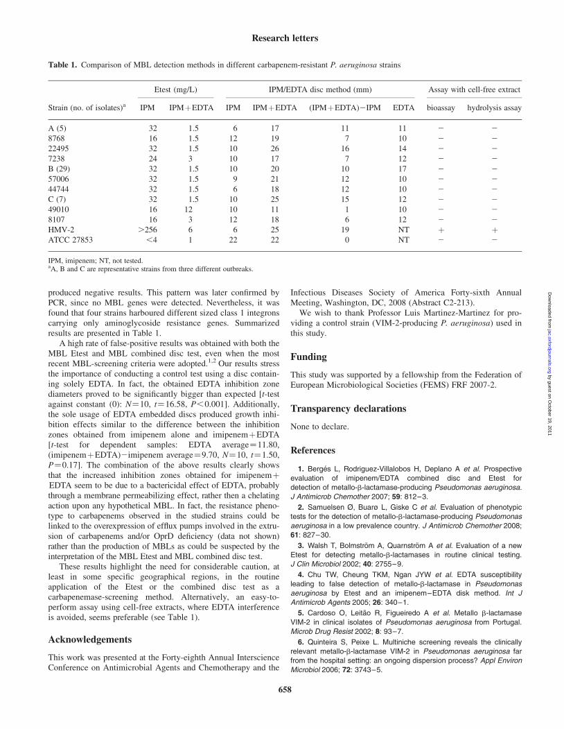
produced negative results. This pattern was later confirmed byPCR, since no MBL genes were detected. Nevertheless, it wasfound that four strains harboured different sized class 1 integronscarrying only aminoglycoside resistance genes. Summarizedresults are presented in Table 1.
A high rate of false-positive results was obtained with both theMBL Etest and MBL combined disc test, even when the mostrecent MBL-screening criteria were adopted.1,2 Our results stressthe importance of conducting a control test using a disc contain-ing solely EDTA. In fact, the obtained EDTA inhibition zonediameters proved to be significantly bigger than expected [t-testagainst constant (0): N¼10, t¼16.58, P,0.001]. Additionally,the sole usage of EDTA embedded discs produced growth inhi-bition effects similar to the difference between the inhibitionzones obtained from imipenem alone and imipenemþEDTA[t-test for dependent samples: EDTA average¼11.80,(imipenemþEDTA)2imipenem average¼9.70, N¼10, t¼1.50,P¼0.17]. The combination of the above results clearly showsthat the increased inhibition zones obtained for imipenemþEDTA seem to be due to a bactericidal effect of EDTA, probablythrough a membrane permeabilizing effect, rather then a chelatingaction upon any hypothetical MBL. In fact, the resistance pheno-type to carbapenems observed in the studied strains could belinked to the overexpression of efflux pumps involved in the extru-sion of carbapenems and/or OprD deficiency (data not shown)rather than the production of MBLs as could be suspected by theinterpretation of the MBL Etest and MBL combined disc test.
These results highlight the need for considerable caution, atleast in some specific geographical regions, in the routineapplication of the Etest or the combined disc test as acarbapenemase-screening method. Alternatively, an easy-to-perform assay using cell-free extracts, where EDTA interferenceis avoided, seems preferable (see Table 1).
Acknowledgements
This work was presented at the Forty-eighth Annual InterscienceConference on Antimicrobial Agents and Chemotherapy and the
Infectious Diseases Society of America Forty-sixth AnnualMeeting, Washington, DC, 2008 (Abstract C2-213).
We wish to thank Professor Luis Martinez-Martinez for pro-viding a control strain (VIM-2-producing P. aeruginosa) used inthis study.
Funding
This study was supported by a fellowship from the Federation ofEuropean Microbiological Societies (FEMS) FRF 2007-2.
Transparency declarations
None to declare.
References
1. Berges L, Rodriguez-Villalobos H, Deplano A et al. Prospective
evaluation of imipenem/EDTA combined disc and Etest for
detection of metallo-b-lactamase-producing Pseudomonas aeruginosa.
J Antimicrob Chemother 2007; 59: 812–3.
2. Samuelsen Ø, Buarø L, Giske C et al. Evaluation of phenotypic
tests for the detection of metallo-b-lactamase-producing Pseudomonas
aeruginosa in a low prevalence country. J Antimicrob Chemother 2008;
61: 827–30.
3. Walsh T, Bolmstrom A, Quarnstrom A et al. Evaluation of a new
Etest for detecting metallo-b-lactamases in routine clinical testing.
J Clin Microbiol 2002; 40: 2755–9.
4. Chu TW, Cheung TKM, Ngan JYW et al. EDTA susceptibility
leading to false detection of metallo-b-lactamase in Pseudomonas
aeruginosa by Etest and an imipenem–EDTA disk method. Int J
Antimicrob Agents 2005; 26: 340–1.
5. Cardoso O, Leitao R, Figueiredo A et al. Metallo b-lactamase
VIM-2 in clinical isolates of Pseudomonas aeruginosa from Portugal.
Microb Drug Resist 2002; 8: 93–7.
6. Quinteira S, Peixe L. Multiniche screening reveals the clinically
relevant metallo-b-lactamase VIM-2 in Pseudomonas aeruginosa far
from the hospital setting: an ongoing dispersion process? Appl Environ
Microbiol 2006; 72: 3743–5.
Table 1. Comparison of MBL detection methods in different carbapenem-resistant P. aeruginosa strains
Strain (no. of isolates)a
Etest (mg/L) IPM/EDTA disc method (mm) Assay with cell-free extract
IPM IPMþEDTA IPM IPMþEDTA (IPMþEDTA)2IPM EDTA bioassay hydrolysis assay
A (5) 32 1.5 6 17 11 11 2 2
8768 16 1.5 12 19 7 10 2 2
22495 32 1.5 10 26 16 14 2 2
7238 24 3 10 17 7 12 2 2
B (29) 32 1.5 10 20 10 17 2 2
57006 32 1.5 9 21 12 10 2 2
44744 32 1.5 6 18 12 10 2 2
C (7) 32 1.5 10 25 15 12 2 2
49010 16 12 10 11 1 10 2 2
8107 16 3 12 18 6 12 2 2
HMV-2 .256 6 6 25 19 NT þ þATCC 27853 ,4 1 22 22 0 NT 2 2
IPM, imipenem; NT, not tested.aA, B and C are representative strains from three different outbreaks.
Research letters
658
by guest on October 19, 2011
jac.oxfordjournals.orgD
ownloaded from

ARTICLE II.

Letters to the Editor 511
Isolation and characterization of an importedextremely-resistant Pseudomonas aeruginosaproducing three different extended-spectrumb-lactamases and hyperproducing twomultidrug-efflux pumps
Dear Editor,
The Journal of Infection has recently published reportson the spread of multi/extremely/pan-resistant Pseudomo-nas aeruginosa strains in clinical settings,1 as well as weface the problem in different hospitals in Hungary.2,3 Wedescribe here the isolation and characterization of an ex-tremely-resistant P. aeruginosa, from a poly-traumatized20-year-old woman transported to our ICU on 28 June2007 from Timisoara, Romania, where she had been treatedfor 2 months after a car accident. Shortly after admission,for reconstruction surgery of her tracheo-esophageal fis-tula, she needed mechanical ventilation because of doublepneumonia, confirmed by chest X-ray. On the day of admis-sion, the endotracheal aspirate and wound sample werepositive for extremely-drug-resistant P. aeruginosa andMRSA, and the blood culture was positive for Candida glab-rata. On 6 July the blood culture, on 18 July the urine sam-ple were positive for P. aeruginosa. She was treated withamphotericin-B (0.5 mg/kg/day intravenously for 10days), colistin (2 million UI intravenously 8-hourly for 7days) and vancomycin (1 g intravenously 12-hourly for 14days). Some laboratory data on days 1, 8, 15 and 20, procal-citonin: 5.44, 4.46, 0.19 and 0.17 ng/l, WBC: 8.72, 7.3, 5.59and 7.01 G/l, and transaminases (aspartate aminotransfer-ase/alanine aminotransferase): 152/117, 111/139, 46/82and 23/59 U/l. By day 17 of therapy her fever decreasedand mechanical ventilation was not needed, but the largetracheo-esophageal fistula necessitated a tracheal tube.On day 21 of therapy, the trachea tube could not be re-placed and, despite 45-min attempted cardio-respiratoryresuscitation, she died.
The Pseudomonas isolates were identified by the VITEK 2system (bioM�erieux, Marcy l’Etoile, France) and subjectedto susceptibility testing by the disk-diffusion method, usingCLSI breakpoints. After isolation, all P. aeruginosa isolateswere stored at �70 �C in CryoBank medium (Copan Diagnos-tics Inc., California, USA) as amember of amulti/extremely-resistant and pan-resistant P. aeruginosa strain collection forfurther investigation. AllP. aeruginosa isolates of this patientproved to be resistant to all anti-pseudomonal antibiotics(piperacillin and tazobactam, cefoperazone, ceftazidime,cefepime, imipenem,meropenem, tobramycin, gentamicin,amikacin, netilmicin, ciprofloxacin, levofloxacin, moxifloxa-cin and aztreonam) except polymyxin-B, which remainedeffective against these isolates. The blood culture isolateof this patient was selected for further molecular investiga-tions of the antibiotic resistance.
The presence of metallo-beta-lactamase genes (blaSPM-1,blaGIM-1, and the most common blaVIM and blaIMP) wassearched for by PCR.4 The lack of carbapenemase genes sug-gested the presence of other mechanisms behind the high-level b-lactam resistance. The genes of the extended-spec-trum b-lactamases [PER-1,5 PER-2, TEM, SHV, GES, VEB-1,
andOXA groups6] were also sought by PCRmethod, using spe-cific primers, and the results were confirmed by sequencingand restriction-fragment-length-polymorphism (RFLP) as-says on the PCR products, as described previously.6 PCR re-vealed the presence of three different extended-spectrumbeta-lactamase genes: blaPER-1, blaOXA-I and blaOXA-II group.Sequencing of the coding region identified the genes ofPER-1 and OXA-74 ESBL, the latter corresponding to theblaOXA-I group. RFLP analysis of the product of blaOXA-II group
PCR revealed the blaOXA-2 gene. No plasmids were isolatedfrom this strain, and it failed to transfer a ceftazidime resis-tance marker by conjugation to other bacterial hosts, sug-gesting that the blaPER-1 and OXA-type genes werechromosomally located.
To investigate the genetic environment of the blaPER-1gene, IS elements characteristic of the composite transpo-son Tn1213 were searched for and found by PCR. Tn1213was first described from P. aeruginosa by Poirel et al. in2005,5 with the ISPa14 and ISPa12 IS elements upstreamfrom the blaPER-1 gene, and the ISPa13, ISPa14 and a gluta-thione-S-transferase gene (gst) downstream from theblaPER-1 gene, which we also found.
To seek further resistance mechanisms behind the multi-ple resistance of the present strain, the expression of thechromosomal genes encoding oprD, mexB, mexX and mexCwas studied in real-time reverse transcriptase PCR assays ac-cording to Quale et al.7 RNA was isolated by the method ofPal�agyi-M�esz�aros et al.8 The expression of mRNA for thegenes of interest was optimized to that of the housekeepinggene rpsL, known to be expressed consistently in P. aerugi-nosa.9 The normalized expression of each gene was cali-brated to the mRNA expression of the reference strainP. aeruginosa PAO1 and the results were given as the expres-sion of the mRNA relative to that of P. aeruginosa PAO1. Thereal-time PCR assays revealed that this strain hyperproducestwo multidrug-efflux pumps, MexAB-OprM and MexXY-OprM(6.68-fold and128-fold increases of expression, respectively,relative to the PAO1 control strain). No change was detectedin the rate of production of the MexCD-OprJ efflux pump, orin the OprD outer membrane protein.
The clonal relatedness of our extremely-resistant isolatewas investigated by MLST analysis as described by Curranet al.10 DNAamplification and sequencing of the sevenhouse-keeping genes (acsA, nuoD, trpE,mutL, guaA, aroE andppsA)were performed. The nucleotide sequences, determined byusing published primers,10,11 and were compared with exist-ing sequences in the MLST database (www.pubmlst.org/paeruginosa). The eBURST algorithm (http://eburst.mlst.net/) was used for phylogenetic analysis.12,13 The MLSTanalyses indicated that this strain (ST235) belongs to aninternationally spreading clonal complex (CC11), previouslyidentified in VIM metallo-b-lactamase and PER-1 ESBL-producing P. aeruginosa strains from patients in Hungary in2008.2,13 A PER-1, OXA-2, OXA-74 and OXA-50-producerstrain with the same ST was isolated from an outbreak inPoland in 2007.14 Although our strain was probablyimported from Romania, the known virulence of the CC11members of MLST types of P. aeruginosa, and thegeographical closeness of Hungary, Romania and Poland,suggest that the same clone, capable of acquiringseveral resistance mechanisms, may be circulating in thesecountries.
512 Letters to the Editor
Acknowledgments
The molecular characterization of the strain was performedin part during a study trip of Cs. Ratkai in Portugal,supported by Federation of European Microbiological Soci-eties (FEMS) FRF 2007-2.
References
1. Suarez C, Pena C, Tubau F, et al. Clinical impact of imipenem-resistant Pseudomonas aeruginosa bloodstream infections.J Infect 2009;58:285e90.
2. Libisch B, Poirel L, Lepsanovic Z, Mirovic V, Balogh B, P�aszti J,et al. Identification of PER-1 extended-spectrum beta-lacta-mase producing Pseudomonas aeruginosa clinical isolates ofthe international clonal complex CC11 from Hungary and Ser-bia. FEMS Immunol Med Microbiol 2008;54:330e8.
3. Szabo D, Szentandrassy J, Juhasz Z, Katona K, Nagy K,Rokusz L. Imported PER-1 producing Pseudomonas aeruginosa,PER-1 producing Acinetobacter baumanii and VIM-2 producingPseudomonas aeruginosa strains in Hungary. Ann Clin MicrobiolAntimicrob 2008;30:7e12.
4. Quinteira S, Peixe L. Multiniche screening reveals the clinicallyrelevant metallo-ß-lactamase VIM-2 in Pseudomonas aerugi-nosa far from the hospital setting: an ongoing dispersion pro-cess? Antimicrobial Agents Chemother 2006;72:3743e5.
5. Poirel L, Cabanne L, Vahaboglu H, Nordmann P. Genetic envi-ronment and expression of the extended-spectrum b-lacta-mase blaPER-1 gene in gram-negative bacteria. AntimicrobAgents Chemother 2005;49:1708e13.
6. Bert F, Branger C, Lambert-Zechovsky N. Identification of PSEand OXA b-lactamase genes in Pseudomonas aeruginosa usingPCR-restriction fragment length polymorphism. J AntimicrobChemother 2002;50:11e8.
7. Quale J, Bratu S, Guptea J, Landman D. Interplay of efflux sys-tem, ampC, and oprD expression in carbapenem resistance ofPseudomonas aeruginosa clinical isolates. Antimicrob AgentsChemother 2006;50:1633e41.
8. Pal�agyi-M�esz�aros SL, Mar�oti J, Latinovics D, Balogh T,Klement E, Medzihradszky KF, et al. Electron-transfer subunitsof the NiFe hydrogenases in Thiocapsa roseopersicina BBS.FEBS J 2009;276:164e74.
9. Fraud S, Campigotto AJ, Chen Z, Poole K. MexCD-OprJ multi-drug efflux system of Pseudomonas aeruginosa: involvementin chlorhexidine resistance and induction by membrane-dam-aging agents dependent upon the AlgU stress response sigmafactor. Antimicrob Agents Chemother 2008;52:4478e82.
10. Curran B, Jonas D, Grundmann H, Pitt T, Dowson CG. Develop-ment of a multi-locus sequence typing scheme for the opportu-nistic pathogen Pseudomonas aeruginosa. J Clin Microbiol2004;42:5644e9.
11. Johnson JK, Arduino SM, Stine OC, Johnson JA, Harris AD.Multi-locus sequence typing compared to pulsed-field gel elec-trophoresis for molecular typing of Pseudomonas aeruginosa.J Clin Microbiol 2007;45:3707e12.
12. Feil EJ, Li BC, Aanensen DM, Hanage WP, Spratt BG. eBURST:inferring patterns of evolutionary descent among clusters ofrelated bacterial genotypes from multi-locus sequence typingdata. J Bacteriol 2004;186:1518e30.
13. Libisch B, Watine J, Balogh B, Gacs M, Muzslay M, Szab�o G,et al. Molecular typing indicates an important role for two in-ternational clonal complexes in dissemination of VIM-producingPseudomonas aeruginosa clinical isolates in Hungary. Res Mi-crobiol 2008;159:162e8.
14. Empel J, Filczak K, Mr�owka A, Hryniewicz W, Livermore DM,Gniadkowski W. Outbreak of Pseudomonas aeruginosa
infections with PER-1 extended-spectrum b-lactamase in War-saw, Poland: further evidence for an international clonal com-plex. J Clin Microbiol 2007;45:2829e34.
Csilla RatkaiElisabeth Nagy
Institute of Clinical Microbiology,University of Szeged, Szeged, Hungary
Lu�ısa PeixeREQUIMTE, Laborat�orio de Microbiologia,
Faculdade de Farm�acia, Universidade do Porto,Porto, Portugal
Vikt�oria BertalanInstitute of Anesthesiology and Intensive Care,
University of Szeged, Szeged, Hungary
Edit Hajd�u*Institute of Clinical Microbiology,
University of Szeged, Szeged, HungaryInfectious Diseases Ward,
First Department of Internal Medicine,University of Szeged,
K�alv�aria sgt. 57, H-6725 Szeged, Hungary*Corresponding author. Infectious Diseases Ward, First De-
partment of Internal Medicine, University of Szeged,K�alv�aria sgt. 57, H-6725 Szeged, Hungary.
Tel./fax: þ36 62 490590/456.E-mail address: [email protected]
Accepted 11 October 2010
Available online 15 October 2010
ª 2010 The British Infection Association. Published by Elsevier Ltd.All rights reserved.
doi:10.1016/j.jinf.2010.10.006
Diagnostic performance of procalcitonin forbacteremia in patients with bacterial infection at theemergency department
Dear Editor,
Recently, Limper et al published a review article andconcluded that procalcitonin (PCT) was helpful in discrimi-nating infectious from non-infectious fever, such as autoim-mune, inflammatory, and malignant disease.1 Fever isa common presentation of patients admitted to the emer-gency department (ED), and bacterial infection is one ofthe most important etiologies. Early and accurate identifica-tion of bacterial infection is of primary importance, becauseearly institution of an appropriate antimicrobial regimenin infected patients is associated with better outcome.Bloodstream infection or bacteremia is one of the mostserious infectious diseases encountered in the ED, buttimely identification of these patients is a great challenge

ARTICLE III.
This article appeared in a journal published by Elsevier. The attachedcopy is furnished to the author for internal non-commercial researchand education use, including for instruction at the authors institution
and sharing with colleagues.
Other uses, including reproduction and distribution, or selling orlicensing copies, or posting to personal, institutional or third party
websites are prohibited.
In most cases authors are permitted to post their version of thearticle (e.g. in Word or Tex form) to their personal website orinstitutional repository. Authors requiring further information
regarding Elsevier’s archiving and manuscript policies areencouraged to visit:
http://www.elsevier.com/copyright
Author's personal copy
Successful application of the DiversiLab repetitive-sequence-based PCRtyping system for confirmation of the circulation of a multiresistant
Pseudomonas aeruginosa clone in different hospital wards☆
Csilla Ratkaia, Luísa V. Peixeb, Filipa Grossob, Ana R. Freitasb, Patricia Antunesb,c,Eleonora Fodora, Edit Hajdúa, Elisabeth Nagya,⁎
aInstitute of Clinical Microbiology, University of Szeged, Semmelweis u. 6., H-6725 Szeged, HungarybREQUIMTE, Laboratório de Microbiologia, Faculdade de Farmácia, Universidade do Porto, Rua Aníbal Cunha 164, 4050-047 Porto, Portugal
cFaculdade de Ciências da Nutrição e Alimentação da Universidade do Porto, Portugal
Received 27 July 2009; accepted 14 January 2010
Abstract
The applicability of the repetitive-sequence-based PCR (rep-PCR)–based DiversiLab system was tested compared with the pulsed fieldgel electrophoresis (PFGE) to type a phenotypically similar subset of a large collection of multiresistant Pseudomonas aeruginosa strainsisolated during a 17-month period from patients treated in different wards including 4 intensive care units (ICUs). Five environmental P.aeruginosa isolates obtained from one of the ICUs were also included. The DiversiLab system and the PFGE demonstrated the geneticrelationship among the isolates with the same efficacy. One of the environmental isolates had the same rep-PCR type as the circulating clone.Multilocus sequence typing of one of the clinical isolates of the circulating clone proved that it is a member of a clonal complex of P.aeruginosa that has not been previously described in clinical samples.© 2010 Elsevier Inc. All rights reserved.
Keywords: Molecular typing methods; Epidemiologic analysis; Multiresistant P. aeruginosa
The surveillance systems of nosocomial infections must berapid, reliable, and capable of differentiating between relatedand unrelated bacterial isolates, especially in highly criticalareas such as intensive care units (ICUs). However, ICUs areoften the scene of the sporadic appearance of multiresistantpathogens such as Pseudomonas aeruginosa, which can bethe origin of nosocomial outbreaks. An outbreak may bedefined as a relatively rapid increase in the incidence ofinfection or colonization by a certain bacterial strain, causedby enhanced patient-to-patient transmission (Belkum et al.,2007). The sporadic isolation of multiresistant strains doesnot often draw attention on the possible nosocomial spread of
1 clone. The collection in routine laboratories of multi-resistant, potential nosocomial pathogens for further typingmay help to discover hidden reservoirs and the circulation ofpossible outbreak clones in different wards. The aim of thepresent study was to use the pulsed field gel electrophoresis(PFGE) and the DiversiLab system to look for genotypicsimilarities among phenotypically similar multiresistantclinical P. aeruginosa isolates. Multilocus sequence typing(MLST) was also used to compare clonal relationship of ourcirculating clone with other known P. aeruginosa clonesfrequently isolated in other geographic areas.
From June 2007 to November 2008, multiresistantP. aeruginosa was isolated from 29 different patients duringtheir stay in different wards, including 4 ICUs at theUniversity Hospital in Szeged, Hungary. Patient's isolatestreated in some other non-ICU wards were also included.Clinical evidences indicated infection in 20 patients, and9 patients displayed simple colonization (Table 1). Theisolates were originally collected for investigation of
Available online at www.sciencedirect.com
Diagnostic Microbiology and Infectious Disease 67 (2010) 202–206www.elsevier.com/locate/diagmicrobio
☆ Part of this study was presented as a poster, “The applicability ofthree different methods for the molecular typing of Pseudomonasaeruginosa,” during the 19th ECCMID in Helsinki 2009 (P519).
⁎ Corresponding author. Institute of Clinical Microbiology, Universityof Szeged, Semmelweis u. 6., H-6725 Szeged, Hungary. Tel.: +36-62-545399; fax: +36-62-545712.
E-mail address: [email protected] (E. Nagy).
0732-8893/$ – see front matter © 2010 Elsevier Inc. All rights reserved.doi:10.1016/j.diagmicrobio.2010.01.010

Author's personal copy
the resistance mechanisms behind the multiresistance of theP. aeruginosa and comprised a subset of a large collection ofcarbapenem-resistant isolates obtained in 2007 and 2008.Twenty-eight of the 29 clinical isolates had the sameantibiotic resistance profile (susceptible only to amikacin,netilmicin, and polymyxin B), and 1 exhibited a slightlydifferent resistance profile (susceptible only to amikacin andpolymyxin B). Environmental sampling was carried out inone of the ICUs during the same period in connection withthe regular hygienic control of the ward. Five P. aeruginosastrains were isolated from different surfaces and included inthis study (Table 2).
PFGE analysis was performed by a slight modification ofa procedure described previously (Deplano et al., 2005). All29 clinical isolates were digested with SpeI, and thefragments were separated by electrophoresis for 12 h in1% agarose gels with a CHEF-DR III System (Bio-RadLaboratories, Hercules, CA). PFGE patterns were analyzedvisually according to criteria described previously (Belkumet al., 2007; Tenover et al., 1995), and 2 unique patterns(designated as pulsotypes, PT-A and PT-B) were found. PT-
A was observed in 28 of the 29 isolates, and PT-Bcorresponded to the isolate with the different antibioticresistance profile (Table 1). Banding patterns are not shown.
The 29 clinical and the 5 environmental P. aeruginosaisolates were fingerprinted by using the automated, repetitive-sequence-based PCR (rep-PCR)–based DiversiLab system,version 3.3. After extraction of the DNA, the isolates werefingerprinted with the Pseudomonas kit (DiversiLab),following the instructions of the rep-PCR worksheet. Afterthe amplification, the amplicons were loaded onto a
Table 2Environmental P. aeruginosa isolates obtained in medical ICU and typed bythe DiversiLab system
Environmental sample Isolates Type with DiversiLab
Right sink drain P. aeruginosa Clone 1Left sink drain P. aeruginosa Clone 3Mechanicalventilation system
P. aeruginosa Clone 3
Wall behind sink P. aeruginosa Clone 4Tap water P. aeruginosa Clone 5
Table 1Data on the 29 multiresistant P. aeruginosa isolates obtained from different patients
Patients Patient's wardat the timeof isolation
First dateof isolation
Wards where thepatient was
I/C Typing
Before after PFGE DiversiLab
isolation of the strain
1 MICU 13/06/2007 SICU3 I PT-A Clone 12 SICU3 20/07/2007 I PT-A Clone 13 MICU 27/11/2007 SICU3 C PT-A Clone 14 MICU 02/01/2008 I PT-A Clone 15 MICU 09/01/2008 I PT-A Clone 16 SICU2 07/07/2008 C PT-A Clone 17 SICU1 15/07/2008 I PT-A Clone 18 SICU2 21/07/2008 NS C PT-A Clone 19 SICU2 23/07/2008 I PT-A Clone 110 SICU2 24/07/2008 MICU MICU I PT-A Clone 111 MICU 26/07/2008 SICU1 I PT-A Clone 112 SICU2 30/07/2008 NE NE I PT-A Clone 113 MICU 02/08/2008 I PT-A Clone 114 SICU2 08/08/2008 NS C PT-A Clone 115 SICU3 25/08/2008 I PT-A Clone 116 MICU 01/09/2008 C PT-A Clone 117 NE 02/09/2008 SICU2 I PT-A Clone 118 SICU1 03/09/2008 I PT-A Clone 119 SICU1 15/09/2008 MICU MICU I PT-A Clone 120 MICU 26/09/2008 I PT-A Clone 121 MICU 30/09/2008 C PT-A Clone 122 SICU1 02/10/2008 I PT-A Clone 123 MICU 09/10/2008 I PT-A Clone 124 SICU1 10/10/2008 C PT-A Clone 125 SICU2 13/10/2008 C PT-A Clone 126 MICU 17/10/2008 I PT-A Clone 127 IM 20/10/2008 SICU2 MICU I PT-A Clone 128 SICU2 20/10/2008 I PT-A Clone 129a SICU2 13/08/2008 C PT-B Clone 2
I/C = infected/colonized; MICU = medical ICU; SICU1 = general surgical ICU; SICU2 = trauma and neurosurgical ICU; SICU3 = cardiac surgical ICU;NS = neurosurgical ward; NE = neurology ward; IM = internal medical ward.
a P. aeruginosa strain with a different antibiotic resistance profile isolated from the same hospital ward.
203C. Ratkai et al. / Diagnostic Microbiology and Infectious Disease 67 (2010) 202–206

Author's personal copy
microfluids chip and the products were detected with theAgilent 2100 Bioanalyzer (Caretto et al., 2008; Healy et al.,2005). Automatically generated reports included dendro-grams, virtual gel images, similarity matrices, scatter plots,and selectable demographic fields. Twenty-eight of the 29clinical isolates were clustered together. The remainingisolate had a different rep-PCR profile together with theslightly different antibiotic susceptibility (Table 1). Fig. 1depicts the similarity matrix and the virtual gel images of 14selected patients isolates clustered together, and the one withthe different rep-PCR pattern. Fig. 2 shows the similarity
matrix and the rep-PCR patterns created by the DiversiLabfor the 5 environmental P. aeruginosa isolates with 4different clones. One of these isolates had the same rep-PCRprofile as that of a representative strain of the circulatingmultiresistant clinical P. aeruginosa (clone 1) (Tables 1 and2). Two environmental P. aeruginosa isolates were alsoclonally related (clone 3), but not related to the strainscolonizing or infecting the 28 patients.
MLST typing was performed on 1 selected clinical isolatecorresponding to PT-A/clone 1. Amplification and sequencingof the 7 housekeeping genes suggested by Curran et al. (2004)
Fig. 1. Dendrogram, virtual gel image, and the similarity matrix of some of the 29 investigated P. aeruginosa isolates typed with the DiversiLab. 1–14: relatedP. aeruginosa isolates (clone 1). 15: unrelated P. aeruginosa isolate with a different antibiotic resistance profile (clone 2).
204 C. Ratkai et al. / Diagnostic Microbiology and Infectious Disease 67 (2010) 202–206

Author's personal copy
were performed. The nucleotide sequences were determinedby using previously published primers (Curran et al., 2004;Johnson et al., 2007) and were compared with existingsequences in the MLST database (http://www.pubmlst.org/paeruginosa). The eBURST algorithm (http://www.eburst.mlst.net/) was used for phylogenetic analysis (Feil et al., 2004;Libisch et al., 2008). The MLST revealed a new sequence type(ST), namely, ST618, belonging to a clonal complex previouslynot described, containing 6 STs. The data on the investigatedstrain are available at http://www.pubmlst.org/paeruginosa.
The DiversiLab system was recently evaluated by severalauthors for confirming different outbreaks caused by Sta-phylococcus aureus, Acinetobacter baumannii, Klebsiellaspp., or P. aeruginosa (Caretto et al., 2008; Doléans-Jordheim et al., 2009; Fontana et al., 2008; Grisold et al.,2009) or to test the discriminative power of it in the case ofknown outbreak strains (Grisold et al., 2010; Tenover et al.,2009) in comparison with the PFGE. In most cases,DiversiLab system proved to be a rapid and reliable method
for molecular analysis of suspected nosocomial outbreakscased by methicillin-resistant S. aureus, A. baumannii, andextended-spectrum β-lactamase–producing Klebsiella spp.It was possible to complete typing and cluster analysisautomatically in a short period (Grisold et al., 2009). In thecase of P. aeruginosa, Doléans-Jordheim et al. (2009)emphasize that rep-PCR provided results similar to thoseobtained by the PFGE-SpeI typing method, and it was mostreliable on nosocomial outbreak isolates.
At the time of the isolation of our strains, epidemiologicrelatedness between the infected or colonized patientstreated in different wards was not suspected. Thephenotypical similarities draw the attention of possibleclonal relatedness of this subset of isolates, which waseasily confirmed first by the DiversiLab system and after byPFGE. When we later followed the routes of the patientsbefore and after isolation of the multiresistant P. aerugi-nosa strain retrospectively, it turned out that some of thepatients visited different wards where patients, infected or
Fig. 2. Dendrogram, virtual gel image and the similarity matrix of the environmental P. aeruginosa isolates, and the representative PT-A and PT-B typed with theDiversiLab system. 1–4, 6: environmental isolates. 5: representative patient isolate (patient 12) (corresponds to PT-A, clone 1). 7: isolate with a differentantibiotic resistance profile (corresponds to PT-B, clone 2) (patient 29).
205C. Ratkai et al. / Diagnostic Microbiology and Infectious Disease 67 (2010) 202–206
Author's personal copy
colonized with the same multiresistant clone, were alsopresent previously (Table 1).
MLST proved that the circulating P. aeruginosa strain is amember of a clonal complex that has previously not beendescribed in clinical samples. P. aeruginosa is known to havenonclonal population structure punctuated by successfulepidemic clones or clonal complexes (Curran et al., 2004).Accordingly, our MLST result may draw the attention ofepidemiologists to the possibility that some clonal lineages aremore likely to be spread among patients and/or causeinfections. It has been shown that only the MLST typingmethod provides information concerning the clonal relation-ship of the bacterial isolates (Curran et al., 2004; Johnson et al.,2007; Turner and Feil, 2007; Urwin andMaiden, 2003). PFGE-and rep-PCR–based methods have higher discriminatorypower than MLST (Johnson et al., 2007); accordingly, theimplication of MLST for population genetics and dynamicsmay be more significant than for everyday molecular typingbecause the polymorphism in the slowly evolving target genesmay not be high enough for useful epidemiologic comparisons.
In conclusion, in our case DiversiLab system and PFGE-SpeI typing of P. aeruginosa strains proved with the sameefficacy the circulation of a successful multiresistant cloneon different wards. According to our experience, thecollection by clinical microbiology laboratories of multi-resistant isolates potentially involved in nosocomial infec-tions even in nonepidemic situation could be important forthe later analysis by molecular typing methods.
Acknowledgments
This study was supported in part by a fellowship awardedby the Federation of European Microbiological Societies(FEMS) FRF 2007-2 to Csilla Ratkai to Porto (Portugal).The study was partly supported by bioMérieux Hungária Kft(Budapest, Hungary).
References
Belkum A, Tassios PT, Dijkshoorn L, Haeggman S, Cookson B, Fry NK,Fussing V, Green J, Feil E, Gerner-Smidt P, Brisse S, Struelens M(2007) Guidelines for the validation and application of typing methodsfor use in bacterial epidemiology. Clin Microbiol Infect 13:1–46.
Caretto E, Barbarini D, Farina C, Grosini A, Nicoletti P, Manso E (2008)Use of the DiversiLab semi-automated repetitive-sequence–basedpolymerase chain reaction for epidemiologic analysis on Acinetobacter
baumannii isolates in different Italian hospitals. Diagn Microbiol InfectDis 60:1–7.
Curran B, Jonas D, Grundmann H, Pitt T, Dowson CG (2004) Developmentof a multi-locus sequence typing scheme for the opportunistic pathogenPseudomonas aeruginosa. J Clin Microbiol 42:5644–5649.
Deplano A, Denis O, Poirel L, Hocquet D, Nonhoff C, Byl B, Nordmann P,Vincent JL, Struelens MJ (2005) Molecular characterization of anepidemic clone of panantibiotic-resistant Pseudomonas aeruginosa.J Clin Microbiol 43:1198–1204.
Doléans-Jordheim A, Cournoyer B, Bergeron E, Croizé J, Salord H, André J,Mazayoer MA, Renaud FNR, Frency J (2009) Reliability of Pseudomonasaeruginosa semi-automated rep-PCR genotyping in various epidemiolog-ical situations. Eur J Clin Microbiol Infect Dis 28:1105–1111.
Feil EJ, Li BC, Aanensen DM, Hanage WP, Spratt BG (2004) eBURST:inferring patterns of evolutionary descent among clusters of relatedbacterial genotypes from multi-locus sequence typing data. J Bacteriol186:1518–1530.
Fontana C, FavaroM, Minelli S, Bossa MC, Testore GP, Leonardis F, NatoliS, Favalli C (2008) Acinetobacter baumannii in intensive care unit: anovel system to study clonal relationship among the isolates. BMC InfectDis 8:79–87.
Grisold AJ, Zarfel G, Strenger V, Feierl G, Leitner E, Masoud L, Hoenigl M,Raggam RB, Dosch V, Marth E (2009) Use of automated repetitive-sequence–based PCR for rapid laboratory conformation of nosocomialoutbreaks. J Infect , doi:10.1016/j.jinf.2009.10.045.
Grisold AJ, Zarfel G, Hoenigl M, Krziwanek K, Feierl G, Masoud L, LeitnerE, Wagner-Eibel U, Badura A, Marth E (2010) Occurrence andgenotyping using automated repetitive-sequence–based PCR of meth-icillin-resistant Staphylococcus aureus ST398 in Southeast Austria.Diagn Microbiol Infect Dis 66:217–221.
Healy M, Huong J, Bittner T, Lising M, Frye S, Raza S, Schrock R, Manry J,Renwick A, Nieto R, Woods C, Versalovic J, Lupski JR (2005)Microbial DNA typing by automated repetitive-sequence–based PCR.J Clin Microbiol 43:199–207.
Johnson JK, Arduino SM, Stine OC, Johnson JA, Harris AD (2007) Multi-locus sequence typing compared to pulsed-field gel electrophoresis formolecular typing of Pseudomonas aeruginosa. J Clin Microbiol 45:3707–3712.
Libisch B, Watine J, Balogh B, Gacs M, Muzslay M, Szabó G, Füzi M(2008) Molecular typing indicates an important role for two internationalclonal complexes in dissemination of VIM-producing Pseudomonasaeruginosa clinical isolates in Hungary. Res Microbiol 159:162–168.
Tenover FC, Arbeit RD, Goering RV, Mickelsen PA, Murray BE, PersingDH, Swaminathan B (1995) Interpreting chromosomal DNA restrictionpatterns produced by pulsed-field gel electrophoresis: criteria forbacterial strain typing. J Clin Microbiol 33:2233–2239.
Tenover FC, Gay EA, Frye S, Eells SJ, Healy M, McGowan Jr JE (2009)Comparison of typing results obtained for methicillin-resistant Staphy-lococcus aureus isolates with the DiversiLab system and pulse-field gelelectrophoresis. J Clin Microbiol 47:2452–2457.
Turner KME, Feil EJ (2007) The secret life of a multi-locus sequence type.Int J Antimicrob Agents 29:129–135.
Urwin R, Maiden MCJ (2003) Multi-locus sequence typing: a tool for globalepidemiology. Trends Microbiol 11:479–487.
206 C. Ratkai et al. / Diagnostic Microbiology and Infectious Disease 67 (2010) 202–206

ARTICLE IV.
Characterisation of Pseudomonas aeruginosa strains isolated from patients suffering
from cystic fibrosis in South-East Hungary
manuscript
Ratkai C.1, Nagy E. 1, Peixe L. 2, Szabó Á. 3, Hajdu E. 4:
1 Institute of Clinical Microbiology, Faculty of Medicine, University of Szeged
Address: Semmelweis u. 6., H-6725 Szeged, Hungary
Phone: +36-62-545399, Fax: +36-62-545712
2 REQUIMTE. Laboratório de Microbiologia. Faculdade de Farmácia. Universidade do Porto
Address: Rua Aníbal Cunha 164, 4050-047 Porto, Portugal
Phone: 351-222078946, Fax: 351-22003977
3:Department of Paediatrics , Faculty of Medicine, University of Szeged
6720 Szeged, Korányi fasor 14-15. Hungary
4. Ward of Infectious Diseases, First Department of Medicine Faculty of medicine, University
of Szeged
Korányi fasor 8-10., H-6720 Szeged,
Corresponding author:
Edit Hajdu
Ward of Infectious Diseases, First Department of Medicine University of Szeged
Korányi fasor 8-10., H-6720 Szeged
E-mail [email protected]
1
Introduction
Cystic fibrosis (CF) is an autosomal recessive disorder that occurs approximately in
1:2000 Caucasian children, in Hungary the rate is 1:4000 [1]. The highest incidence of CF
patients in the world, 1:1353, is found in Ireland [2]. Cystic fibrosis is caused by abnormal
functioning of the CFTR protein, the Cystic Fibrosis Transmembrane Regulator, which is a
cAMP induced chloride channel, and is expressed on the apic membrane of the cells. The
gene encoding the CFTR protein is expressed mainly in epithelial cells in the airway, kidneys,
pancreas, bile ducts, reproductive organs, and the bowel. The main function of CFTR protein
is to transport chloride ions across the cell membrane. CFTR has two nucleotide-binding
domains, both of them is capable to bind and hydrolyze ATP, which helps to open and close
the channel. An alteration of the cftr gene causes the disease. The alteration in cftr gene,
therefore the amino acid residues, can lead to a change in diameter size of the pore, causing
loss in function of the channel. The mutations can occur in different areas of the cftr gene,
each mutation is associated with a different phenotype, and results in a different prognosis for
the patient. In Europe, the most common mutation is the ΔF508, which refers to a deletion of
a phenylalanin at position 508 in the protein. The initial diagnosis of the disease is made via
sweat test, since CF patients produce higher levels of sodium chloride in their sweat (>60
mmol/L). Many newborns are screened for CF if they present with meconium ileus. If the
screening sweat test is positive, additional confirmatory tests are carried out via molecular
methods to detect the exact alteration of the cftr gene.
The pathophysiology of this disease is poorly understood, and symptoms range from
gastrointestinal or nutritional abnormalities, reproductive problems as an adult, chronic
sinusitis, endobronchial disease. In the lungs the loss of the CFTR protein function affects the
volume and viscosity of the airway surface liquid, which cause poor mucociliary clearance.
There is an ongoing debate whether the CF lung becomes first infected by bacteria followed
by inflammation, or vice versa, but it is a fact that bacteria infect the respiratory tract usually
early in the course of cystic fibrosis disease, often fail to be eradicated. The bacterial
infections together with an aggressive host inflammatory response are the key players in the
irreversible airway damage from which most patients die. The contact with these bacteria
usually lead to chronic colonisation of the lung with exacerbations over time. There is a
pattern of the colonisation of the CF lung, which usually involves Staphylococcus aureus in
infancy, followed by Haemophilus influenzae, as a child, P. aeruginosa, Stenotrophomonas
maltophilia, Achromobacter xylosoxidans in teenage years, Burkholderia cepacia complex,
2
non-tuberculous mycobacteria in early twenties, and several newer species, including moulds,
are becoming more common [3].
P. aeruginosa is one of the main organisms responsible for drug-resistant nosocomial
infections, and emerged as the most important bacterial pathogen in lung disease of CF
patients, with a high prevalence in adults, up to 80%. In addition being intrinsically resistant
to several antimicrobial agents, P. aeruginosa can easily develop resistance to all
conventional antipseudomonal antibiotics via different mechanisms. After first infecting the
host, P. aeruginosa has to survive host defences, and repeated courses of antibiotic treatment.
The bacterium has a repertoire of immunoevasive strategies includes the secretion of
exoproducts, antibiotic-resistance proteins, and phenotypic changes, which make them
virtually unrecognizable from the bacteria, that was first isolated, e.g. the siderophores, such
as pyocianin that breaks down intercellular tight junctions, slow the ciliary beat frequency,
therefore affect mucociliary clearance. P. aeruginosa strains found in the CF lung are
unusually hypermutable, they can react promptly to their environment, not only by switching
genes on and off, but by an increased frequency of mutation events within the genome. One
such mutation triggers conversion to a mucoid phenotype. Over-producing mucoid
exopolysaccharide (alginate) surrounds and protects them from external affects, such as
mucociliary clearance, immune response or antibiotics. Another highly successful survival
strategy involves the formation of biofilms. In the CF lung P. aeruginosa and Burkholderia
cepacia isolates exist in biofilms, which protects them from phagocytosis, and prevents
penetration of antibiotics. This phenotypic change plays a major role in the persistence of
pseudomonas infection in the majority of CF patients, despite best medical attempts at
eradication [4].
The aims of this study were to investigate P. aeruginosa isolates infecting/colonising patients
suffering from cystic fibrosis, namely:
- to determine the different resistance mechanisms, especially those causing resistance to
carbapenems
- to study the molecular relatedness between isolates, to find the most reliable typing
method in case of these isolates
3

Methods and materials
Bacterial isolates
From 2003 to 2007 149 P. aeruginosa were isolated from 16 patients suffering from
cystic fibrosis, attending the CF centre in the Children’s Hospital of Szeged, Hungary. In this
period of time all isolates from the 16 patients were stored for further analysis, regardless
their antibiotic susceptibility. 78 isolates (52%) had mucoid colony morphology at the time of
the isolation. Identification of the isolates by VITEK 2 system (bioMérieux) and susceptibility
test by disk-diffusion method and E-test if required were performed, according to CLSI
guidelines. After isolation, the strains were stored at -70 °C in CryoBank medium (Copan
diagnostic Inc., California, USA) for further investigation.
To verify whether strains found in different patients may have been transmitted from
patient-to patient, we tracked possible contacts among the different patients with the MedSol
patients’ data system used in the hospital, and by contacting the patient’s clinicians.
Detection of different antibiotic-resistance mechanisms in P. aeruginosa isolates
Of the 149 isolates 5 were resistant to ceftazidime and to cefepime. ESBL genes were
searched from these isolates. The genes of the extended spectrum β-lactamases (PER-1, PER-
2, TEM, SHV, GES, VEB-1, OXA groups [5] were sought by PCR methods in a
representative P. aeruginosa strain from each genotype, confirmed by sequencing.
Of the 149 isolates 26 were resistant to at least one carbapenem. These imipenem
and/or meropenem resistant isolates were selected for further analyses. MBL presence or
absence was confirmed by PCR, aimed the blaSPM-1, blaGIM-1, and the most common blaVIM
and blaIMP genes [6]. PCR master mix was prepared according to the standard protocol: 1 x
PCR buffer, MgCl2 2,5 mM, dNTP 200 µM, primers 1-1 µM, Taq polymerase 1 U/reaction,
sterile water. Acquired metallo-β-lactamases (MBLs) and some other antiobiotic resistance
markers (especially aminoglycoside-resistance genes) are mostly encoded by integron-borne
genes. Therefore class1 integrons were sought, using PCR.
In order to study the antibiotic resistance due to the efflux-pump overexpression, real-
time PCR was performed, based on the following criteria:
- isolates resistant only to imipenem were tested for OprD deficiency;
4

- isolates resistant to imipenem and/or meropenem and cefepime, but not to ceftazidime
were tested for OprD deficiency and for overexpression of MexXY-OprM;
- isolates resistant imipenem and/or meropenem, cefepime and to ceftazidime were tested
for OprD deficiency and for overexpression of MexXY-OprM, MexAB-OprM, MexCD-
OprJ.
The expression of the chromosomal genes encoding the oprD, mexB, mexX, mexC was
studied with real-time reverse transcriptase PCR assays according to Quale et al. [7]. RNA
was isolated using the method by Palagyi-Meszaros et al [8]. Briefly, the isolate was grown in
60 mL of liquid medium in a hypovial to A600 nm = 1–1.5; 15 mL of culture was centrifuged
at 15 000 g for 2 min, the pellet was suspended in 300 µL of SET buffer [20% sucrose, 50
mm EDTA (pH 8.0) and 50 mm Tris ⁄ HCl (pH 8.0)] and 300 µL of SDS buffer was added
[20% SDS, 1% (NH4)2SO4, pH 4.8]; 500 µL of saturated NaCl was added next, the sample
was centrifuged at 20 000 g for 10 min and the clear supernatant was transferred into a new
tube. 2-Propanol (70% of the total volume of the supernatant) was added to the solution and
the mixture was centrifuged at 20 000 g for 20 min. The pellet was washed twice with 1 mL
of 70% ethanol. The dried pellet was suspended in 20 µL of diethylpyrocarbonate-treated
water.
The expression of mRNA for the genes of interest was optimised to that of the
housekeeping gene rpsL. This gene is known to be expressed consistently in P. aeruginosa
[9]. Normalized expression of each gene was calibrated to the mRNA expression of the
reference strain P. aeruginosa PAO1, results are given as the relative expression of the
mRNA compared to that of P. aeruginosa PAO1. The following values were considered to
represent overexpression compared to the control strain: for mexB ≥3-fold; for mexX≥10-fold;
mexC≥2-fold; and reduced expression of oprD≤0,7-fold [7].
Molecular typing of P. aeruginosa isolates
For bacterial typing with PFGE and DiversiLab methods 44 isolates were selected
from the 149 isolates, based on different antibiotic susceptibility profile.
PFGE analysis was performed as described previously by Deplano et al. with a slight
modification [10]. The DNA of the isolates were digested with SpeI, and the fragments were
separated by electrophoresis in 1% agarose gels with a CHEF- DR III System (Bio-Rad
Laboratories) for 12-12 h, with the switch times ranging from 4 s to 12 s, and from 15 s to 36
s in a Tris-borate-EDTA buffer containing 50 µM thiourea. Gels for PFGE were stained in an
5

aqueous solution containing 0.5 µg ethidium-bromide and photographed under UV
translumination. Patterns of PFGE were visually analysed according to the criteria described
previously [11,12].
The selected P. aeruginosa isolates were fingerprinted using the automated, rep-PCR
based DiversiLab system (bioMériux), version 3.3. DNA was extracted from the bacteria
using the Mo Bio Ultra CleanTM Microbial DNA Isolation Kit (MoBio Laboratories Inc) kit
according to the manufacturer’s instructions. Fingerprinting of the isolates was performed
with the Pseudomonas kit (DiversiLab) following the instructions of the rep-PCR worksheet.
After the amplification, the amplicons were loaded on a microfluids chip and detected, using
the Agilent 2100 Bioanalyzer. The results were sent via internet to the hospital’s secured
DiversiLab website. The analysis of the data was performed using the DiversiLab software
using the Pearson correlation coefficient. Reports were automatically generated, included
dendograms, virtual gel images, similarity matrix, scatter plots and selectable demographic
fields.
Six isolates were randomly selected from different patients for MLST analysis, as
described previously by Curran et al [13]. Briefly, the genomic DNA was purified using the
Mo Bio Ultra CleanTM Microbial DNA Isolation Kit according to the manufacturer’s
instructions. DNA amplification and sequencing of the seven housekeeping genes (acsA,
nuoD, trpE, mutL, guaA, aroE, ppsA) were performed. The nucleotide sequences were
determined by using previously published primers [14,13] and were compared to existing
sequences in the MLST database (www.pubmlst.org/paeruginosa). The eBURST algorithm
(http://eburst.mlst.net/) was used for phylogenetic analysis [15,16].
Results
Antibiotic resistance in P. aeruginosa isolates
From 2003 to 2007 149 P. aeruginosa were collected from 16 different patients
suffering from cystic fibrosis, attending the CF centre in Szeged. The percentage of the
isolates susceptible to the tested agents were as follows: piperacillin/tazobactam: 100 %,
cefoperazone: 87,5 %, ceftazidime: 93,75 %, cefepime: 68,8 %, imipenem: 81,3 %,
meropenem: 87,5 %, gentamycin: 37,5 %, tobramycin: 68,8 %, amikacin: 38,8 %, netilmicin:
6
62,5 %, ciprofloxacin: 93,75 %, levofloxacin: 68,8 %, moxifloxacin: 43,8 %, polymixin B:
100%. The highest resistance was seen in case of gentamycin, amikacin and moxifloxacin.
Of the 149 isolates 26 were resistant to at least one carbapenem. These imipenem
and/or meropenem resistant isolates were selected for MBL detection and PCR detection of
class 1 integrons. No MBL genes were found with PCR. From isolate RN62639 a 689 bp
class 1 integron was amplified, encoding an aacA4 aminoglycoside-acethyltransferase gene
(AAC(6’)II), that confers resistance to tobramycin and amikacin. Of the 149 isolates 5 were
resistant to ceftazidime and to cefepime. ESBL genes were searched, and in isolate RN64944
an OXA-10 ESBL were found. No other β-lactamase or aminoglycoside-resistance genes
have been detected.
Seven isolates were selected for RT-PCR analysis, examining efflux pumps and
porins, based on the criteria detailed in Materials and Methods. One isolate showed
inconclusive results. OprD deficiency was detected in 5 of the 6 cases, hyperproduction of the
MexXY-OprM efflux pump was detected in 3 isolates, overexpression of MexAB-OprM was
detected in one isolate at a relatively high level, ( 148 fold). No isolates hyper-produced the
MexCD-OprJ efflux pump.
Molecular typing of P. aeruginosa isolates recovered from cystic fibrosis patients
Of the 149 P. aeruginosa isolates 44 were selected for bacterial typing by PFGE and
rep-PCR-based DiversiLab methods, 27 (61 %) of them had mucoid colonies. In average ~3
(2,75) isolates per patient were typed. With PFGE 16 different genotypes were determined. In
case of 13 patients one genotype was recovered per patient from their sputum samples, in case
of three patients, two different strains caused the infection/colonisation. With PFGE the same
genotypes, designated as pulso-type K determined typed from two different patients, such as
pulso-type L, and pulso-type F (Table 1).
With DiversiLab 20 different genotypes were determined. The results with the
exception of two patients were contradictory with the PFGE results. With DiversiLab the
same genotype (designated as CloneA, CloneC, CloneD) were determined from more than
one patient in three cases (data not shown).
Six CF isolates were randomly selected from different patients for MLST analysis.
Sequencing the seven alleles resulted six different STs, namely ST343, ST614, ST277,
ST625, ST626 and ST627. The results of the MLST analyses are available at
7

http://pubmlst.org/paeruginosa. With the exception of ST625, which is a member of an
uncharacterized clonal complex, all STs are singletons.
Discussion
Among the 149 P. aeruginosa isolates the highest resistance was seen in case of
gentamycin, amikacin and moxifloxacin (62,5 %, 61,2 %, 56,2 % respectively). Otherwise the
strains had a good in vitro susceptibility, which results do not follow the international
tendency [17,18,19]. Among our CF isolates were no pan- or multidrug resistant P.
aeruginosa, and no developing resistance was observed due to the antimicrobial treatments.
No MBLs were found, only one class 1 integron, and an OXA-10 ESBL was found
among the isolates. Isolate RN64944 producing the OXA-10 ESBL was resistant to cefepime,
but susceptible to all other β-lactams. Isolate RN62639 harbouring the class 1 integron was
resistant to all aminoglycosides. Isolates being only resistant to imipenem were relatively
prevalent, the porin channel analyses proved, that this could be the effect of OprD deficiency.
Isolate BoT83704, which proved to overexpress the MexAB-OprM efflux pump, was resistant
to both carbapenems and all fluoroquinolones. These antibiotic resistance patterns correspond
with the determined resistance mechanisms.
Based on the different antibiotic susceptibility profile 44 isolates were selected for
bacterial typing. With the PFGE method 16 different genotypes were determined. In case of
13 patients one genotype were recovered per patient from their sputum samples, so these
patients were colonised/infected with the same strain from 2003 to 2007. In case of three
patients, two different genotypes could be isolated from their sputum, so they were
colonised/infected with different strains. Overall a high diversity was observed, which
corresponds to the international literature [20]. The isolates were also typed by the DiversiLab
system. The virtual gel image showed too many bands with similar sizes in almost all of the
strains due to an unknown reason (Figure 1). Results did not correspond neither with the
antibiotic susceptibility pattern, nor with the PFGE results, which is considered to be the gold
standard method of bacterial typing.
The same genotype (designated as CloneA, CloneC, CloneD, data not shown) were
determined from more than one patient in three cases. PFGE analysis could not confirm these
results, and also the clinical background studies showed that these patients were not
hospitalised at the same time or did not participate the same CF camp, so the cross-infection
could be excluded. Considering all the above results, we concluded that during the molecular
8

typing of CF isolates the PFGE method is found to be more accurate and reliable than the
DiversiLab system.
Until recently, CF patients were thought to be colonised with their own unique P.
aeruginosa strain acquired from the environment. Cross-infection was considered a relatively
infrequent source of acquisition. With PFGE the same genotype (designated as pulso-type F)
was isolated from patients DM and LA from 2005 (Table 1.), when they participated in the
same CF-camp. Pulso-type L was isolated from patients TZ and KT from 2005. At that time,
they were hospitalised at the same time in the same hospital ward. Pulso-type K recovered
from patients BoT and TZ. The strains were isolated in 2003, and according to their clinical
background the two patients attended the same CF-camp in Budapest, Hungary. Interestingly,
the MLST results suggest that the latter strain, being a member of a clonal complex, and not a
singleton, could be a member of an epidemic clone (Table 1). However the remaining five
strains examined with MLST proved to be singletons with this method, which suggests that
the prevalence of the cluster genotypes is significantly lower among these CF isolates. With
our results we proved, that the transmission of a certain strain from patient to patient in a
hospital ward or in a CF camp is expected, but not inevitable. Nevertheless the incidence of
the cross-infections in the CF centre in Szeged were relatively low due to the infection control
measures applied to prevent patient-to-patient contacts, such as scheduling appointments with
the clinicians at different time points, trying to hospitalize the patients in different hospital
rooms.
Based on the present study we can conclude that the P. aeruginosa isolates obtained
from CF patients have an overall good in vitro susceptibility, which results are in contrast
with the international tendency. Among our isolates there were no MBL producer, no multi-or
pandrug resistant isolate, and no therapy-induced resistance was observed. During the
molecular typing of CF isolates the DiversiLab system is found to be too discriminative and
not as reliable as the PFGE method. These isolates have a high genetic diversity, no clonality
was observed, which corresponds to the international literature. With the exception of a few
sporadic cases of transmission, there were no highly epidemic strain isolated, which is
expected to be a result of the good infection control measures taken by the clinicians, nurses
of the CF centre, and of the good patient compliance.
9

Table 1. Different CF patients colonized/infected with the same pulso-type of P. aeruginosa,
and the result of MLST analysis, if it was performed.
Patient Strain Year of
isolation Mucoid
PFGE
pulso-type MLST
DM 63282 2005 + F NT
LA 61644 2005 + F NT
27013 2003 - K NT BoT
83650 2005 - K ST626
23513 2003 - K NT TZ
23918 2005 - L ST625*
KT 63285 2005 - L NT
NT: not tested, * marks the ST that is a member of a clonal complex.
10

Figure 1. The virtual gel image of some of the examined CF isolates shows the presence of
too many bands in case of almost all of the isolates.
11

References
1. Gilligan P.H.: Microbiology of airway disease in patients with cystic fibrosis. Clin
Microbiol Rev 1991, 4:35-51
2. Farrel P., Joffe S., Foley L., Canny G.J., Mayne P., Rosenberg M.: Diagnosis of cystic
fibrosis in the Republic of Ireland: epidemiology and costs. Ir Med J 2007, 100:557-
560
3. Davies J.C., Bilton D.: Bugs, biofilms, and resistance in cystic fibrosis. Respir Care
2009, 54:628-640
4. Dinesh S.D., Grundmann H., Pitt T.L., Römling U.: Europen-wide distribution of
Pseudomonas aeruginosa clone C. Clin Microbiol Infect 2003, 12:1228-1233
5. Bert F., Branger C., Lambert-Zechovsky N.: Identification of PSE and OXA β-
lactamase genes in Pseudomonas aeruginosa using PCR-restriction fragment-length
polymorphism. J Antimicrob Chemother. 2002, 50:11-18
6. Quinteira S, Peixe L. Multiniche Screening Reveals the Clinically Relevant Metallo-ß-
Lactamase VIM-2 in Pseudomonas aeruginosa Far from the Hospital Setting: an
Ongoing Dispersion Process? Antimicrob Agents Chemother 2006; 72:3743-3745
7. Quale J, Bratu S, Guptea J, Landman D. Interplay of efflux system, ampC, and oprD
expression in carbapenem resistance of Pseudomonas aeruginosa clinical isolates.
Antimicrob Agents Chemother 2006;50(5):1633-1641
8. Palágyi-Mészáros SL, Maróti J, Latinovics D, Balogh T, Klement E,. Medzihradszky
KF. et al. Electron-transfer subunits of the NiFe hydrogenases in Thiocapsa
roseopersicina BBS. FEBS Journal 2009; 276:164–174
9. Fraud S, Campigotto AJ, Chen Z, Poole K. MexCD-OprJ multidrug efflux system of
Pseudomonas aeruginosa: involvement in chlorhexidine resistance and induction by
membrane-damaging agents dependent upon the AlgU stress response sigma factor.
Antimicrob Agents Chemother 2008;52(12):4478–4482
10. Deplano A, Denis O, Poirel L, Hocquet D, Nonhoff C, Byl B, Nordmann P, Vincent
JL, Struelens MJ (2005) Molecular characterization of an epidemic clone of
panantibiotic-resistant Pseudomonas aeruginosa. J Clin Microbiol 43: 1198-1204
11. Belkum A., Tassios P. T., Dijkshoorn L., Haeggman S., Cookson B., Fry N. K.,
Fussing V., Green J., Feil E., Gerner-Smidt P., Brisse S., Struelens M.: Guidelines for
the validation and application of typing methods for use in bacterial epidemiology.
Clin Microbiol Infect. 2007. 13:1-46
12
12. Tenover F. C., Arbeit R. D., Goering R. V., Mickelsen P. A., Murray B. E., Persing D.
H., Swaminathan B.: Interpreting chromosomal DNA restriction patterns produced by
pulsed-field gel electrophoresis: criteria for bacterial strain typing. J Clin Microbiol
1995, 33:2233-2239
13. Curran B., Jonas D., Grundmann H., Pitt T., Dowson C. G.: Development of a
multilocus sequence typing scheme for the opportunistiv pathogen Pseudomonas
aeruginosa. J Clin Microbiol 2004, 42: 5644-5649
14. Johnson J. K., Arduino S. M., Stine O. C., Johnson J. A., Harris A. D. Multilocus
sequence typing compared to pulsed-field gel electrophoresis for molecular typing of
Pseudomonas aeruginosa. J Clin Microbiol 2007, 45: 3707-3712
15. Libisch B, Watine J, Balogh B, Gacs M., Muzslay M., Szabó G., Füzi M.: (2008)
Molecular typing indicates an important role for two international clonal complexes in
dissemination of VIM-producing Pseudomonas aeruginosa clinical isolates in
Hungary. Res Microbiol 159: 162-168
16. Feil EJ, Li BC, Aanensen DM, Hanage WP, Spratt BG. (2004) eBURST: inferring
patterns of evolutionary descent among clusters of related bacterial genotypes from
multilocus sequence typing data. J Bacteriol 186: 1518-1530
17. Wolter DJ, Acquazzino D, Goering RV, Sammut P, Khalaf N, Hanson ND.:
Emergence of carbapenem resistance in Pseudomonas aeruginosa isolates from a
patient with cystic fibrosis in the absence of carbapenem therapy. Clin Infect Dis. 2008
46:137-41
18. Cheng K, Smyth RL, Govan JR, Doherty C, Winstanley C, Denning N, Heaf DP, van
Saene H, Hart CA.: Spread of beta-lactam-resistant Pseudomonas aeruginosa in a
cystic fibrosis clinic. Lancet 1996;348:639-42
19. Pedersen SS, Koch C, Høiby N, Rosendal K.: An epidemic spread of multiresistant
Pseudomonas aeruginosa in a cystic fibrosis centre. J Antimicrob Chemother. 1986
17:505-16
20. Vosahlikova S., Drevinek P., Cinek O., Pohunek P., Maixnerova M., Urbaskova P.,
van den Reijden T.J., Dijkshoorn L., Nemec A.: High genotypic diversity of
Pseudomonas aeruginosa strains isolated from patients with cystic fibrosis in the
Czech Republic. Res Microbiol 2007, 158:324-329
13