Embed Size (px)
Citation preview
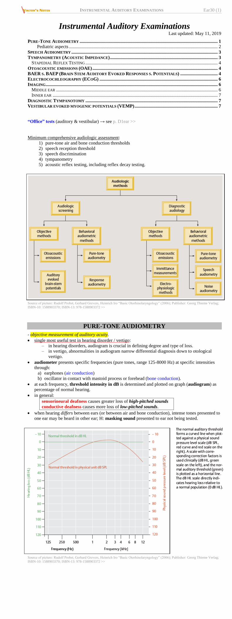
INSTRUMENTAL AUDITORY EXAMINATIONS Ear30 (1)
Instrumental Auditory Examinations Last updated: May 11, 2019
PURE-TONE AUDIOMETRY ...................................................................................................................... 1 Pediatric aspects ............................................................................................................................... 2
SPEECH AUDIOMETRY ............................................................................................................................. 3
TYMPANOMETRY (ACOUSTIC IMPEDANCE) ............................................................................................ 3 STAPEDIAL REFLEX TESTING ................................................................................................................. 4
OTOACOUSTIC EMISSIONS (OAE) ........................................................................................................... 4 BAER S. BAEP (BRAIN STEM AUDITORY EVOKED RESPONSES S. POTENTIALS) ................................ 4
ELECTROCOCHLEOGRAPHY (ECOG) ..................................................................................................... 6
IMAGING ................................................................................................................................................... 6 MIDDLE EAR .......................................................................................................................................... 6
INNER EAR ............................................................................................................................................. 7
DIAGNOSTIC TYMPANOTOMY ................................................................................................................. 7
VESTIBULAR EVOKED MYOGENIC POTENTIALS (VEMP) ....................................................................... 7
“Office” tests (auditory & vestibular) → see p. D1ear >>
Minimum comprehensive audiologic assessment:
1) pure-tone air and bone conduction thresholds
2) speech reception threshold
3) speech discrimination
4) tympanometry
5) acoustic reflex testing, including reflex decay testing.
Source of picture: Rudolf Probst, Gerhard Grevers, Heinrich Iro “Basic Otorhinolaryngology” (2006); Publisher: Georg Thieme Verlag;
ISBN-10: 1588903370; ISBN-13: 978-1588903372 >>
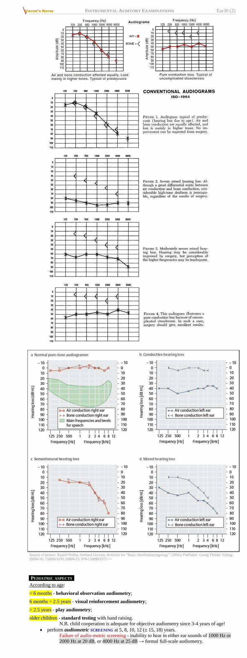
PURE-TONE AUDIOMETRY
- objective measurement of auditory acuity.
single most useful test in hearing disorder / vertigo:
– in hearing disorders, audiogram is crucial in defining degree and type of loss.
– in vertigo, abnormalities in audiogram narrow differential diagnosis down to otological
vertigo.
audiometer presents specific frequencies (pure tones, range 125-8000 Hz) at specific intensities
through:
a) earphones (air conduction)
b) oscillator in contact with mastoid process or forehead (bone conduction).
at each frequency, threshold intensity in dB is determined and plotted on graph (audiogram) as
percentage of normal hearing.
in general:
sensorineural deafness causes greater loss of high-pitched sounds
conductive deafness causes more loss of low-pitched sounds.
when hearing differs between ears (or between air and bone conduction), intense tones presented to
one ear may be heard in other ear; H: masking sound presented to ear not being tested.
Source of picture: Rudolf Probst, Gerhard Grevers, Heinrich Iro “Basic Otorhinolaryngology” (2006); Publisher: Georg Thieme Verlag;
ISBN-10: 1588903370; ISBN-13: 978-1588903372 >>
INSTRUMENTAL AUDITORY EXAMINATIONS Ear30 (2)
Source of picture: Rudolf Probst, Gerhard Grevers, Heinrich Iro “Basic Otorhinolaryngology” (2006); Publisher: Georg Thieme Verlag;
ISBN-10: 1588903370; ISBN-13: 978-1588903372 >>
PEDIATRIC ASPECTS
According to age:
< 6 months - behavioral observation audiometry;
6 months ÷ 2.5 years - visual reinforcement audiometry;
> 2.5 years - play audiometry;
older children - standard testing with hand raising.
N.B. child cooperation is adequate for objective audiometry since 3-4 years of age!
perform audiometric SCREENING at 5, 8, 10, 12 (± 15, 18) years.
Failure of audio-metric screening - inability to hear in either ear sounds of 1000 Hz or
2000 Hz at 20 dB, or 4000 Hz at 25 dB → formal full-scale audiometry.
INSTRUMENTAL AUDITORY EXAMINATIONS Ear30 (3)
FULL-SCALE audiometry should be given to all children prior to beginning school!!!
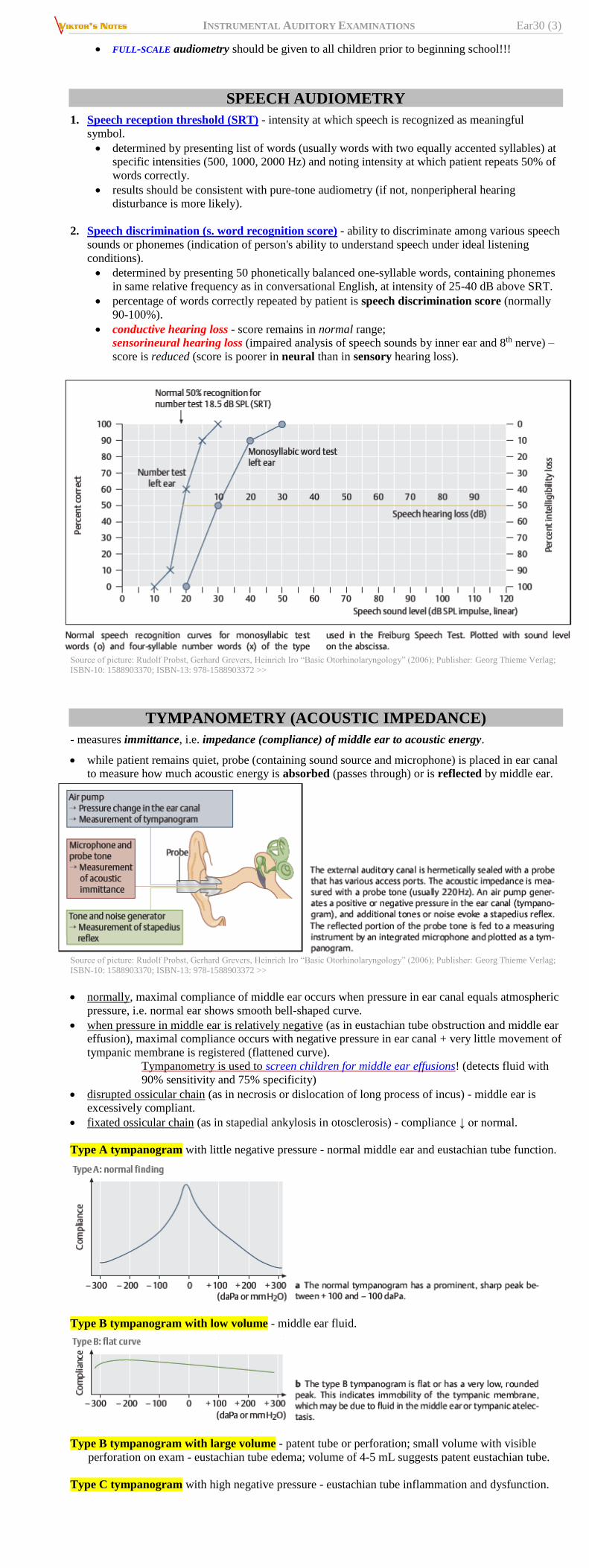
SPEECH AUDIOMETRY
1. Speech reception threshold (SRT) - intensity at which speech is recognized as meaningful
symbol.
determined by presenting list of words (usually words with two equally accented syllables) at
specific intensities (500, 1000, 2000 Hz) and noting intensity at which patient repeats 50% of
words correctly.
results should be consistent with pure-tone audiometry (if not, nonperipheral hearing
disturbance is more likely).
2. Speech discrimination (s. word recognition score) - ability to discriminate among various speech
sounds or phonemes (indication of person's ability to understand speech under ideal listening
conditions).
determined by presenting 50 phonetically balanced one-syllable words, containing phonemes
in same relative frequency as in conversational English, at intensity of 25-40 dB above SRT.
percentage of words correctly repeated by patient is speech discrimination score (normally
90-100%).
conductive hearing loss - score remains in normal range;
sensorineural hearing loss (impaired analysis of speech sounds by inner ear and 8th nerve) –
score is reduced (score is poorer in neural than in sensory hearing loss).
Source of picture: Rudolf Probst, Gerhard Grevers, Heinrich Iro “Basic Otorhinolaryngology” (2006); Publisher: Georg Thieme Verlag;
ISBN-10: 1588903370; ISBN-13: 978-1588903372 >>
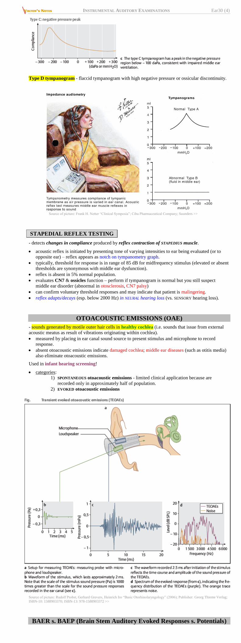
TYMPANOMETRY (ACOUSTIC IMPEDANCE)
- measures immittance, i.e. impedance (compliance) of middle ear to acoustic energy.
while patient remains quiet, probe (containing sound source and microphone) is placed in ear canal
to measure how much acoustic energy is absorbed (passes through) or is reflected by middle ear.
Source of picture: Rudolf Probst, Gerhard Grevers, Heinrich Iro “Basic Otorhinolaryngology” (2006); Publisher: Georg Thieme Verlag;
ISBN-10: 1588903370; ISBN-13: 978-1588903372 >>
normally, maximal compliance of middle ear occurs when pressure in ear canal equals atmospheric
pressure, i.e. normal ear shows smooth bell-shaped curve.
when pressure in middle ear is relatively negative (as in eustachian tube obstruction and middle ear
effusion), maximal compliance occurs with negative pressure in ear canal + very little movement of
tympanic membrane is registered (flattened curve).
Tympanometry is used to screen children for middle ear effusions! (detects fluid with
90% sensitivity and 75% specificity)
disrupted ossicular chain (as in necrosis or dislocation of long process of incus) - middle ear is
excessively compliant.
fixated ossicular chain (as in stapedial ankylosis in otosclerosis) - compliance ↓ or normal.
Type A tympanogram with little negative pressure - normal middle ear and eustachian tube function.
Type B tympanogram with low volume - middle ear fluid.
Type B tympanogram with large volume - patent tube or perforation; small volume with visible
perforation on exam - eustachian tube edema; volume of 4-5 mL suggests patent eustachian tube.
Type C tympanogram with high negative pressure - eustachian tube inflammation and dysfunction.
INSTRUMENTAL AUDITORY EXAMINATIONS Ear30 (4)
Type D tympanogram - flaccid tympanogram with high negative pressure or ossicular discontinuity.
Source of picture: Frank H. Netter “Clinical Symposia”; Ciba Pharmaceutical Company; Saunders >>
STAPEDIAL REFLEX TESTING
- detects changes in compliance produced by reflex contraction of STAPEDIUS muscle.
acoustic reflex is initiated by presenting tone of varying intensities to ear being evaluated (or to
opposite ear) – reflex appears as notch on tympanometry graph.
typically, threshold for response is in range of 85 dB for midfrequency stimulus (elevated or absent
thresholds are synonymous with middle ear dysfunction).
reflex is absent in 5% normal population.
evaluates CN7 & ossicles function – perform if tympanogram is normal but you still suspect
middle ear disorder (abnormal in otosclerosis, CN7 palsy)
can confirm voluntary threshold responses and may indicate that patient is malingering.
reflex adapts/decays (esp. below 2000 Hz) in NEURAL hearing loss (vs. SENSORY hearing loss).
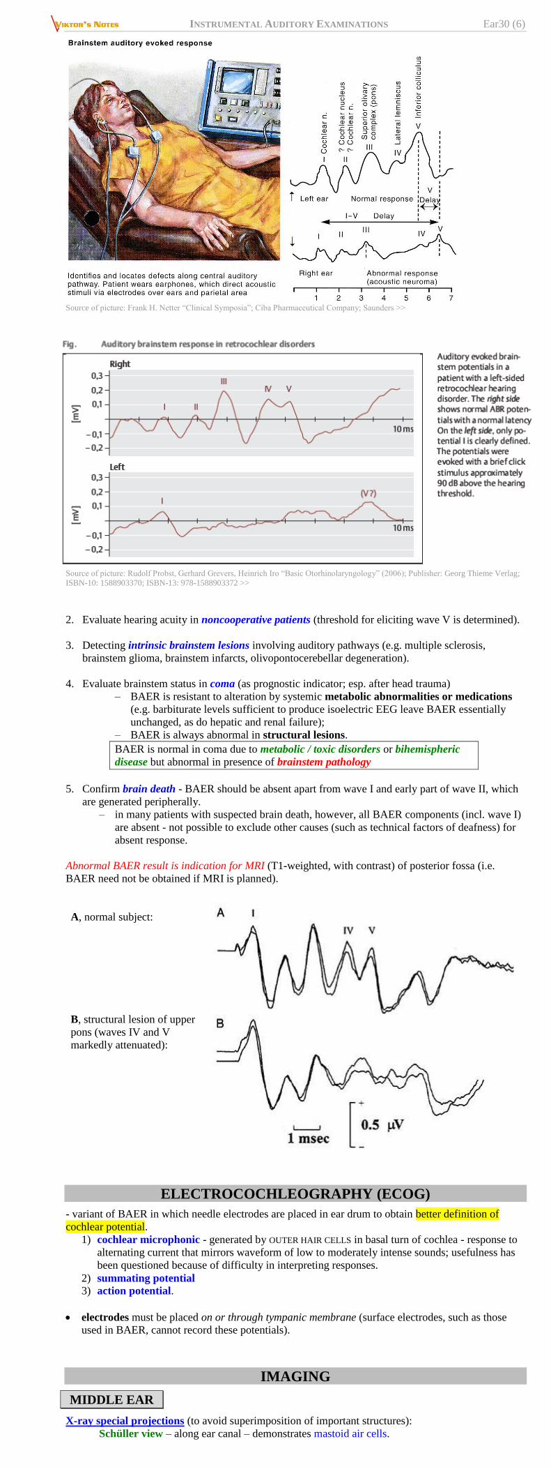
OTOACOUSTIC EMISSIONS (OAE)
- sounds generated by motile outer hair cells in healthy cochlea (i.e. sounds that issue from external
acoustic meatus as result of vibrations originating within cochlea).
measured by placing in ear canal sound source to present stimulus and microphone to record
response.
absent otoacoustic emissions indicate damaged cochlea; middle ear diseases (such as otitis media)
also eliminate otoacoustic emissions.
Used in infant hearing screening!
categories:
1) SPONTANEOUS otoacoustic emissions - limited clinical application because are
recorded only in approximately half of population.
2) EVOKED otoacoustic emissions
Source of picture: Rudolf Probst, Gerhard Grevers, Heinrich Iro “Basic Otorhinolaryngology” (2006); Publisher: Georg Thieme Verlag;
ISBN-10: 1588903370; ISBN-13: 978-1588903372 >>
BAER s. BAEP (Brain Stem Auditory Evoked Responses s. Potentials)

INSTRUMENTAL AUDITORY EXAMINATIONS Ear30 (5)
- registers action potentials along auditory pathways (CN8 ÷ auditory cortex).
stimulus - monaural click 65 dB above patient's hearing threshold.
recorded between vertex of scalp and mastoid process (or earlobe).
attention of subject is not required! – perfect for non-cooperative patients (e.g. children, coma,
malingering).
BAER results may be inaccurate in patients who have no high-frequency hearing (audiometry is
recommended before BAER!).
very low voltage – 1000-2000 responses are recorded so that BAER can be extracted by averaging
from background noise. also see p. D25 >>
Series of up to seven components that occur within 10 msec of click stimulus:
– wave I and early part of wave II - auditory nerve action potential.
– wave II - cochlear nucleus.
– wave III - superior olive.
– wave IV - lateral lemniscus.*
– wave V - inferior colliculus.
– waves VI and VII are inconsistent and of uncertain origin - little clinical utility.
N.B. most consistent are waves I, III, V (CN8, superior olive, inferior colliculus).
*because lateral lemniscus contains second order neurons from cochlea and
third and fourth order neurons from superior olive, it contributes to three waves.
although brain stem auditory pathways decussate at multiple levels, unilateral abnormalities of
waves III and V are most often associated with ipsilateral brain stem disease.
every wave has its own specific shape and latency (bilateral symmetry is also very important) –
lesion localization:
Source of picture: Rudolf Probst, Gerhard Grevers, Heinrich Iro “Basic Otorhinolaryngology” (2006); Publisher: Georg Thieme Verlag;
ISBN-10: 1588903370; ISBN-13: 978-1588903372 >>
BAER is used to:
1. Rule out acoustic neuroma (sensitivity ≈ 90%; higher than CT!) – possible BAER variants:
a) prolongation of I-III interpeak interval (conduction delay between distal eighth
nerve and lower pons).
b) preservation of wave I with loss of subsequent components.
c) loss of all BAER waveforms.
INSTRUMENTAL AUDITORY EXAMINATIONS Ear30 (6)
Source of picture: Frank H. Netter “Clinical Symposia”; Ciba Pharmaceutical Company; Saunders >>
Source of picture: Rudolf Probst, Gerhard Grevers, Heinrich Iro “Basic Otorhinolaryngology” (2006); Publisher: Georg Thieme Verlag;
ISBN-10: 1588903370; ISBN-13: 978-1588903372 >>
2. Evaluate hearing acuity in noncooperative patients (threshold for eliciting wave V is determined).
3. Detecting intrinsic brainstem lesions involving auditory pathways (e.g. multiple sclerosis,
brainstem glioma, brainstem infarcts, olivopontocerebellar degeneration).
4. Evaluate brainstem status in coma (as prognostic indicator; esp. after head trauma)
– BAER is resistant to alteration by systemic metabolic abnormalities or medications
(e.g. barbiturate levels sufficient to produce isoelectric EEG leave BAER essentially
unchanged, as do hepatic and renal failure);
– BAER is always abnormal in structural lesions.
BAER is normal in coma due to metabolic / toxic disorders or bihemispheric
disease but abnormal in presence of brainstem pathology
5. Confirm brain death - BAER should be absent apart from wave I and early part of wave II, which
are generated peripherally.
– in many patients with suspected brain death, however, all BAER components (incl. wave I)
are absent - not possible to exclude other causes (such as technical factors of deafness) for
absent response.
Abnormal BAER result is indication for MRI (T1-weighted, with contrast) of posterior fossa (i.e.
BAER need not be obtained if MRI is planned).
A, normal subject:
B, structural lesion of upper
pons (waves IV and V
markedly attenuated):
ELECTROCOCHLEOGRAPHY (ECOG)
- variant of BAER in which needle electrodes are placed in ear drum to obtain better definition of
cochlear potential.
1) cochlear microphonic - generated by OUTER HAIR CELLS in basal turn of cochlea - response to
alternating current that mirrors waveform of low to moderately intense sounds; usefulness has
been questioned because of difficulty in interpreting responses.
2) summating potential 3) action potential.
electrodes must be placed on or through tympanic membrane (surface electrodes, such as those
used in BAER, cannot record these potentials).
IMAGING
MIDDLE EAR
X-ray special projections (to avoid superimposition of important structures):
Schüller view – along ear canal – demonstrates mastoid air cells.
INSTRUMENTAL AUDITORY EXAMINATIONS Ear30 (7)
Stenvers view – angled 45° forward – demonstrates petrous ridge & apex.
High-resolution thin-slice CT (bone windows) – main imaging modality!!!!
MRI, angiography – only for pulsatile tinnitus, suspected tumors (esp. vascular).
INNER EAR
High-resolution thin-slice CT (bone windows) – for labyrinthine disorders!!!!
MRI – for CN8 (e.g. retrocochlear) disorders.
DIAGNOSTIC TYMPANOTOMY
- under operating microscope, by incising canal skin and reflecting skin and membrane as flap.
most often combined with surgical correction.
VESTIBULAR EVOKED MYOGENIC POTENTIALS (VEMP)
- stimulation of sacculus (by acoustic clicks) and registration of evoked muscle potentials (e.g.
sternocleidomastoideus).
BIBLIOGRAPHY for ch. “Otology” → follow this LINK >>
Viktor’s Notes℠ for the Neurosurgery Resident
Please visit website at www.NeurosurgeryResident.net