Embed Size (px)
Citation preview
STUDIES OF INSULIN CRYSTALS AT LOW TEMPERATURES:EFFECTS ON MOSAIC CHARACTER AND
RADIATION SENSITIVITY*
BY B. W. Low, C. C. H. CHEN, J. E. BERGER, L. SINGMAN, AND J. F. PLETCHER
DEPARTMENT OF BIOCHEMISTRY, COLLEGE OF PHYSICIANS AND SURGEONS, COLUMBIA UNIVERSITY
Communicated by David Shemin, October 12, 1966
The low-temperature X-ray diffraction studies of orthorhombic insulin citratecrystals reported here were initially undertaken because of the rapid deterioration atroom temperature of certain insulin crystals containing heavy-metal cations. Theeffects of cooling on mosaic character and radiation sensitivity were studied. Twotemperature ranges were employed: (a) below -150'C and (b) 00C to -13'C.In normal laboratory practice orthorhombic insulin citrate crystals are grown
and stored at 1 d 10C. Immersion studies in heavy-atom reagents are also made atthis temperature. All X-ray diffraction work is, however, carried out at roomtemperature (.210C).We have found that orthorhombic insulin citrate crystals may be cooled rapidly
to below -150'C without impairing the X-ray diffraction pattern. Rapid coolingdoes, however, enhance the mosaic character of these crystals. X-ray diffractionstudies at 00C and -13'C have provided evidence for marked reduction in radiationsensitivity in certain heavy-atom-containing crystals.
General Background.-Although metal-free insulin crystals are somewhat moresensitive to radiation damage (nickel-filtered CuKa) at room temperature than at00C, the radiation sensitivity at room temperature is not marked enough to preventthe collection of intensity data by counter techniques. ' Furthermore, these crystalsdo not deteriorate on standing at room temperature for several weeks withoutirradiation.However, after immersion (1 ± 1"C) in buffered solutions containing certain
salts with heavy-metal cations (including uranyl) insulin crystals deterioraterapidly at room temperature (sometimes within 1 day or less of exposure) afteronly the minimal radiation exposure (minutes) necessary to monitor the state ofthe crystal. The deterioration has been observed both in the presence and in theabsence of significant changes in X-ray diffraction intensity distribution afterimmersion. It is not markedly accelerated by continuous X-ray irradiation. Thisdeterioration was originally attributed to greatly enhanced radiation damage, aslaboratory practice avoids keeping the crystals at room temperature prior to X-rayphotography. Some apparent enhancement in radiation damage is also observedwith a second group of metal-containing crystals which do not show the strikingand rapid room-temperature deterioration reported above.
Before the deterioration in the first class of crystals was recognized as largely ofthermal origin, studies were made of both classes of metal-containing crystals intowhich radiation protectors had been introduced. These provided marginal evidenceof protection. There was no evidence of protection when the crystals were studiedunder oxygen-free conditions.
In order to collect X-ray data from the crystals which deteriorate rapidly atroom temperature, it would have been necessary to build an apparatus to maintain
1746
VOL. 56, 1966 BIOCHEMISTRY: LOW ET AL. 1747
them at approximately 1VC. The use of data collected at different temperaturesin isomorphous replacement techniques would present serious problems. Coolingmay affect both lattice and thermal parameters and cause structural changes.Therefore, we decided to investigate the general advantages and disadvantages oflow-temperature X-ray diffraction studies with insulin crystals.
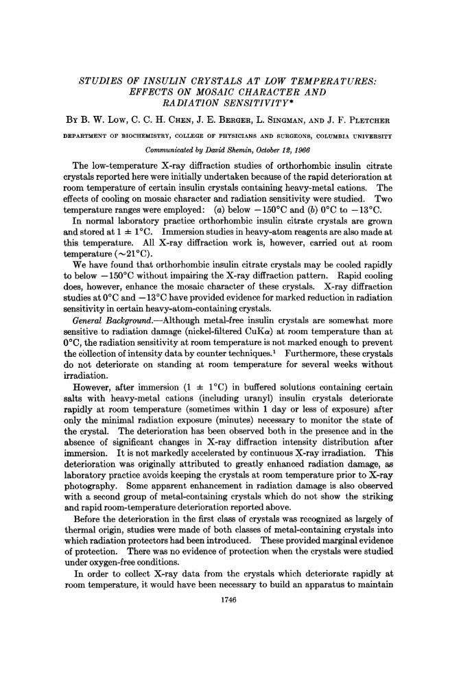
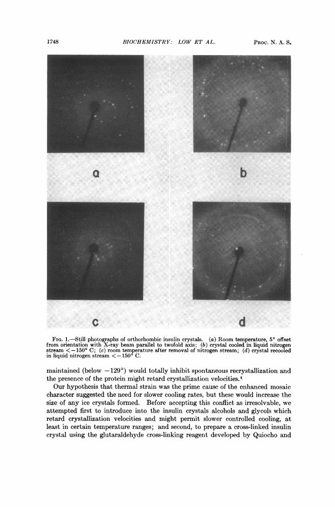
Studies at Temperatures below -150C.-The effects of cooling crystals to tem-peratures below -150'C were first investigated. Such intensive cooling shouldprovide maximum radiation protection effects for all types of crystals. The crys-tals used in this study were metal-free crystals or crystals grown from silver nitrate-containing solution which showed no changes in intensity distribution. The crys-tals, which contain no organic solvent, were wiped free of adhering mother liquorand mounted in a special cell with mylar windows (thickness 0.15 mil). The cellcontained a droplet of mother liquor some distance from the crystal. Still photo-graphs (1-min exposure) were taken of the crystal on a precession camera usingPolaroid ASA 3000 film with an intensifying screen, as described by Smith,2 in amodified film holder.3 After photography of the crystal at room temperature,the cell was dipped into liquid nitrogen and photographed while a stream of liquidnitrogen flowed over it. The temperature within 2 mm of the crystal position wasmeasured with a thermocouple. It was lower than - 150'C, and on cooling reachedthis temperature in less than 1 min. As shown in Figure lb, there was an increasein mosaicity of the crystal on cooling which was enhanced after rapid rewarming toroom temperature, Figure lc, and further enhanced on rapid recooling, Figure ld.Some ice diffraction pattern was always observed which we attribute to condensa-
tion on the mylar film. When it was very heavy, suggesting an accidental wetmounting, the diffraction pattern after warming up was slightly less intense thanbefore cooling. It may be noted in the figure that the crystal orientation changedslightly in the cold, but returned to its original position on warming. This isprobably caused by tension in the mylar film.The mosaic character of these protein crystals after cooling precludes their use
for accurate intensity data collection. To us, it appears probable that the enhance-ment of mosaic character is largely the result of thermal strain on rapid cooling,rather than primarily the disruptive effect of ice crystallite formation between pro-tein crystalline domains. Such enhancement is the well-known consequence ofdipping crystals into liquid air.We do not have sufficient evidence to distinguish between these two possible
causes. It should be noted that the original crystal gave a sharp still. On coolingit showed evidence of a mosaic spread of -0.5-1O and on rewarming a mosaic spreadof about 1.5°, presumably here enhanced by the ice crystallites which must form onrewarming. After two cooling-warming cycles, the diffraction pattern from thesecrystals faded appreciably. In another experiment where a protein crystal en-closed with a film of aqueous solution in a sealed capillary was cooled slowly until thesurrounding droplet froze (-200C), both ice diffraction pattern and a much weak-ened protein diffraction pattern were present in the cold; the protein diffractionpattern weakened further when the crystal was warmed up. Thus, in the certainpresence of external ice crystals, disorder effects in the protein lattice do occur.The rapid cooling procedure employed in these experiments would reduce the
size of ice crystals formed on cooling, the temperature at which the crystals were
1748 BIOCHEMISTRY: LOW ET AL. PROC. N. A. S.
c dFIG. l.--Still photographs of orthorhombic insulin crystals. (a) Room temperature, 50 offset
from orientation with X-ray beam parallel to twofold axis; (b) crystal cooled in liquid nitrogenstream < 15O' C; (c) room temperature after removal of nitrogen stream; (d) crystal recooledin liquid nitrogen stream <-150' C.
maintained (below 129o) would totally inhibit spontaneous recrystallization and
_~~~~~~~
the presence of the protein might retard crystallization velocities.4Our hypothesis that thermal strain was the prime cause of the enhanced mosaic
character suggested the need for slower cooling rates, but these would increase thesize of any ice crystals formed. Before accepting this conflict as irresolvable, weattempted first to introduce into the insulin crystals alcohols and glycols whichretard crystallization velocities and might permit slower controlled cooling, atleast in certain temperature ranges; and second, to prepare a cross-linked insulincrystal using the glutaraldehyde cross-linking reagent developed by Quiocho and
VOL. 5)6 1966 BIOCHEMISTIR: LOW ET AL. 1749
Richards.5 The first experiment failed because these solvents thenmselves increasethe mosaic character of insulin crystals. The second failed because the glutaralde-hyde disordered the structure within the crystalline domains, although it did notincrease the mosaicity and did harden the crystal. With other cross-linked proteincrystals, slow cooling within certain ranges might well permit studies to be madeat these low temperatures.
Efforts to reach very low temperatures as a means of affording maximum protec-tion from both thermal and radiation damage were therefore abandoned and studieswere then made of the effectiveness of relatively minor temperature reductions.
Studies at Roosm Temnperature, 0°, and - 13C.-The crystals used in this studywere heavy-atom-containing crystals which deteriorate rapidly at room tempera-ture. All crystals were mounted in thin-walled glass capillaries by the normalprocedure in the cold room, except that, with adequate insulation for the crystal,one end of the capillary was sealed in a cold flame rather than with wax to avoidturbulence in the cooling gas stream from the protruding drop of wax.The photographs were taken on a Nonius Weissenberg camera with a cooling
attachment, using copper radiation (40 kv 25 ma) and Ilford film. The tempera-ture near the crystal (2 cm) was measured by means of a copper constantan ther-mocouple, the wires of which are led through the Dewar tube. A cylindrical piece ofmylar was used as an extension of the Dewar cylinder to guide the cooling nitrogenstream over the capillary. With this arrangement, condensation was avoided onboth the capillary and mylar tube. The crystals were cooled and irradiated con-tinuously and standard-exposure oscillation photographs were taken at periodicintervals. In order to maintain flow conditions when photographs were not beingrecorded, the layer-line screen was placed close to the crystal and the cassettereplaced by a lead shield.For studies at room temperature, the crystals were allowed 15 min to establish
thermal equilibrium after they were brought out of the cold room. They were thenmounted on the goniometer head and preliminary alignments made with the polariz-ing microscope. For studies at low temperature, the crystals were mounted on thegoniometer head and aligned optically inside the cold room. They were thenrapidly transferred (<10 see) to the precooled camera.The onset of deterioration was defined as evident reduction (visually determined),
however slight, in the over-all intensity of the diffraction pattern. Completedeterioration was defined by absence of the diffraction pattern.Although the lowest temperature investigated ( - 130C) is below the melting
point of the immersion medium (- - 30C) and therefore presumably below themelting point of the liquid of crystallization, it is above the critical nucleation tem-perature (- 250C) of the immersion medium as experimentally determined.
All the crystals studied at 0C which had not deteriorated completely at the end ofthe experiment did so in a few hours when warmed to room temperature withoutfurther X-ray irradiation. The crystal which showed no deterioration after 47hr continuous X-ray exposure at - 13'C gave no diffraction pattern after 2 addi-tional hr at room temperature without irradiation. This crystal, however, showedsome evidence of a normal thermal parameter. It was accidentally permitted towarm up to - 10'C between 40 and 43'/2 hr. The pattern which diminishedslightly at - 10'C was completely restored on recooling to - 13'C.
1750 BIOCHEMISTRY: LOW ET AL. PROC. N. A. S.
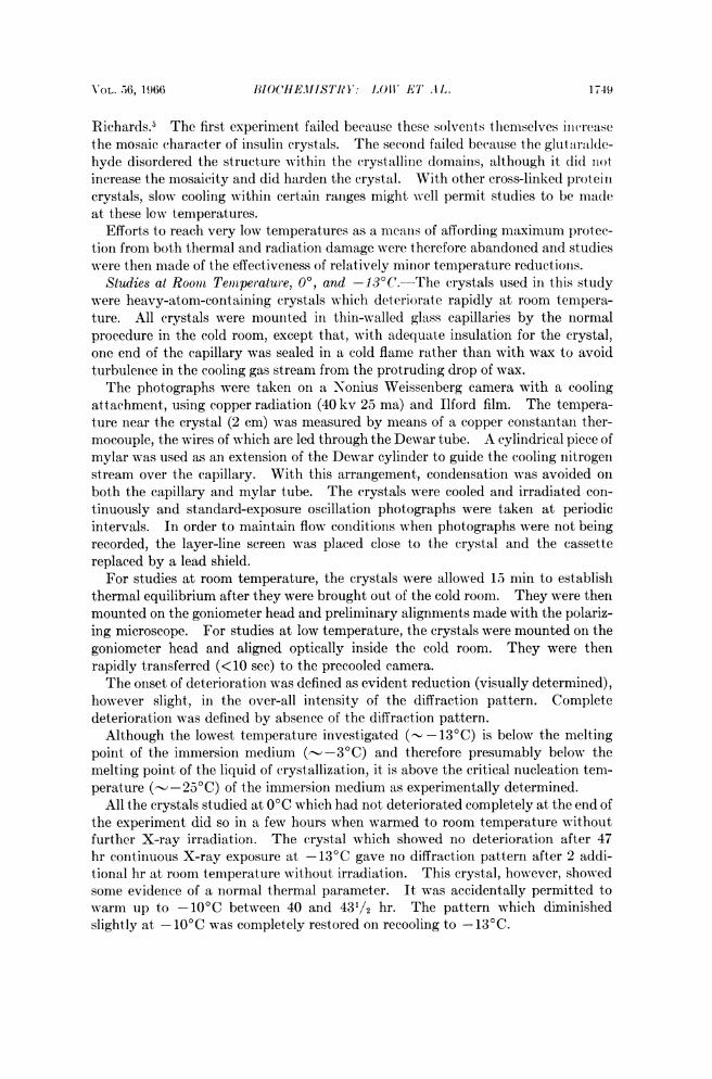
TABLE 1CHANGES IN DIFFRACTION PATTERN OBSERVED FOR CRYSTALS CONTINUOUSLY
IRRADIATED AT DIFFERENT TEMPERATURESExposure Time (hr)
Crystal* Temperature (IC)t Unchanged Diminished DisappearedHg 21 8 ... 21Hg 21 2.5 6 18Hg 0 35 ... ...
Hg 0 25 42 >72UO2a 21 ... 6.5 12UO2a 21 ... ... 16UO2a 21 .. . 5...UO2a 0 29. ...
UO2a 0 27 381/2UO2b 0 81/2 211/2UO2c 21 ... ... isUO2c 0 ... 18-20 ...
UO2c -13 47 ... ...
* The crystals are here identified in terms of the metal cations which were in the diffusion me-dium. U02 with superscripts a, b, and c, represents three different preparations.
t As the thermocouple was 2 cm from the crystal in the capillary, the temperatures cited are notthose at the crystal. The absolute error will therefore be greater at the lower temperature. Thefluctuations in the temperature were rarely greater than +30C.
The results of these studies (Table 1) show that the crystals deteriorate lessrapidly under constant irradiation at 00 than at room temperature. These metal-containing crystals deteriorate at room temperature without irradiation virtuallyas rapidly as under constant irradiation. This contrasts with the normal metal-free crystals, which deteriorate rapidly at room temperature only as the result ofradiation damage. Cooling of the metal-containing crystals to 0° (compare stor-age temperature 1 :i 1VC) must eliminate thermal damage. Moreover, coolingthese crystals to 0° has effectively protected most of them from radiation damage forperiods longer than those observed for metal-free crystals at room temperature.At -130C, the period before onset of radiation damage is considerably longer
than that for normal metal-free crystals at room temperature.These qualitative studies provide clear evidence of enhanced radiation protec-
tion which may be achieved by cooling, even though the absolute temperaturereduction is very small. Quantitative studies have been made of the effects on thediffraction patterns of crystals kept at temperatures in the range of 21'C to- 13'C. These will be reported in detail elsewhere.6
* This investigation was supported in part by U.S. Public Health Service research grant ROI-AMI-01320 from the National Institute of Arthritis and Metabolic Diseases; in part (B. W. L.)by a U.S. Public Health Service Research Career Award, 5-K3-GM-15,246; in part by a U.S.Public Health Service fellowship (J. E. B.) 1-F3-GMI-19,813; and in part by a U.S. Public HealthService fellowship (J. F. P.) 4-FL-GM-13,858.
1 Traub, W., and F. L. Hirshfeld, Acta Cryst., 13, 753 (1960).2 Smith, H. G., Rev. Sci. Instr., 33, 128 (1962).3 Low, B. W., unpublished studies.I Luyet, B., Ann. N.Y. Acad., 85, 549 (1960); Meryman, H. T., Science, 124, 515 (1956).6 Quiocho, F. A., and F. M. Richards, these PROCEEDINGS, 52, 833 (1964).6 Cucka, P., L. Singman, and B. W. Low, in preparation.


![Cement & Concrete Composites - xrm.phys.northwestern.eduxrm.phys.northwestern.edu/research/pdf_papers/2012/... · cement in concrete production [3–5]. The increasing availability](https://img.pdfslide.net/doc/110x75/5ecd0b649698831ef6156275/cement-concrete-composites-xrmphys-cement-in-concrete-production-3a5.jpg)