Embed Size (px)
Citation preview
Unique Research Journal of Medicine and Medical Sciences Vol. 2(3), pp. 019-031, April, 2014 Available online@http://www.uniqueresearchjournals.org/URJMMS ISSN 2333-6935 ©2014 Unique Research Journals
Unique Research Journal of Medicine and Medical Sciences
Review
Inter-mental foramina distance significant and variation among races: Mini review
Mohammed Jasim Al-Juboori1*, Basim K. Atya1, Santhanalaxmi A. Balachandran2, Navenithamaria Eirutharajan2 and Lor Yen Fang2
1Lecturer in of oral surgery department/ Dental Faculty, MAHSA University, Kuala Lumpur, Malaysia.
2Dental Faculty, MAHSA University, level 4, block E, pusatbandardamansara, Kuala Lumpur, Malaysia.
*Correspondence author. E-mail:[email protected]. Tel: (+60)0162417557
Accepted 11 January, 2014
Mental foramen is normally present as a single opening on each side of the mandible. Mental foramen is significant because it plays an essential role during the administration of local anaesthesia and any surgical procedure that may be done in this area. It has been found that the location of the mental foramen varies in different ethnicity. Its position is commonly seen below the second premolar. However, individual variation exists whereby it can be placed anywhere in between the first premolar to the mesial root of the first molar. From this mini review we can conclude that there is a difference in the location of mental foramen which is sometimes related to race, sex or even to the age of the patient. And this may effect on the interforamen distance that may effect on the implant treatment plan. Key words: Mental foramen, races, anatomy, inter-mental foramen distance, inferior dental nerve, dental implant.
INTRODUCTION Trigeminal nerve gives rise to three major branches - the ophthalmic branch [V1], maxillary [V2] and mandibular [V3] nerves. The mandibular nerve is the largest nerve of the three divisions and is both motor and sensory. The mandibular nerve divides into anterior and posterior trunk and the branches of the posterior trunk gives rise to inferior alveolar nerve. The inferior alveolar nerve descends and enters the mandibular canal through the mandibular foramen which is located at the medial surface of the ramus. The inferior alveolar nerve and vessels passes anteriorly within the mandibular canal of the lower jaw. The mandibular canal and its content are inferior to the roots of the molar teeth. Then it divides into its two terminal branches which are the incisive nerve and the mental nerve. The incisive nerve runs
continuously through the mandibular canal while the mental nerve exits the mandible through the mental foramen (Richard et al., 2009; Chummy, 2006; Bernard, 2001; Norton, 2007; Gosling et al., 1985). The mental foramen is an entire funnel-like opening in the lateral surface of the mandible and marks the termination of the mandibular canal. It is positioned entirely within the buccal cortical plate of bone. The mental nerve supplies sensory innervation to the soft tissues of the chin, lower lips and the gingiva on the ipsilateral site as far as the mandibular 2
nd premolar (Ngeow and Yuzawati, 2003;
Mraiwa et al., 2003). Infrequently it innervates the mandibular incisors
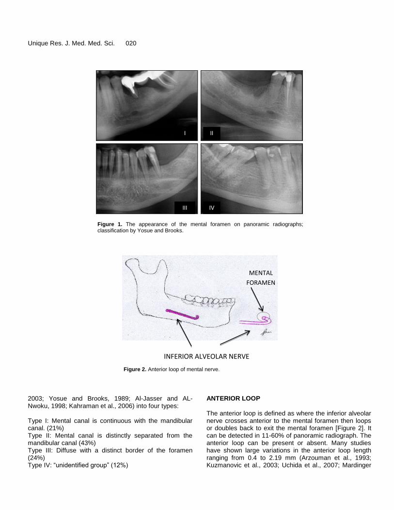
4. Based on the radiographic
appearance [Figure 1], the mental foramen has been classified by Yosue and Brooks (Ngeow and Yuzawati,
Unique Res. J. Med. Med. Sci. 020
I
Type IV: “unidentified group” (12%)
ANTERIOR LOOP
III IV
II
Figure 1⁵: The appearance of the mental foramen on panoramic radiographs; classification
by Yosue and Brooks.
Figure 1. The appearance of the mental foramen on panoramic radiographs; classification by Yosue and Brooks.
INFERIOR ALVEOLAR NERVE
MENTAL
FORAMEN
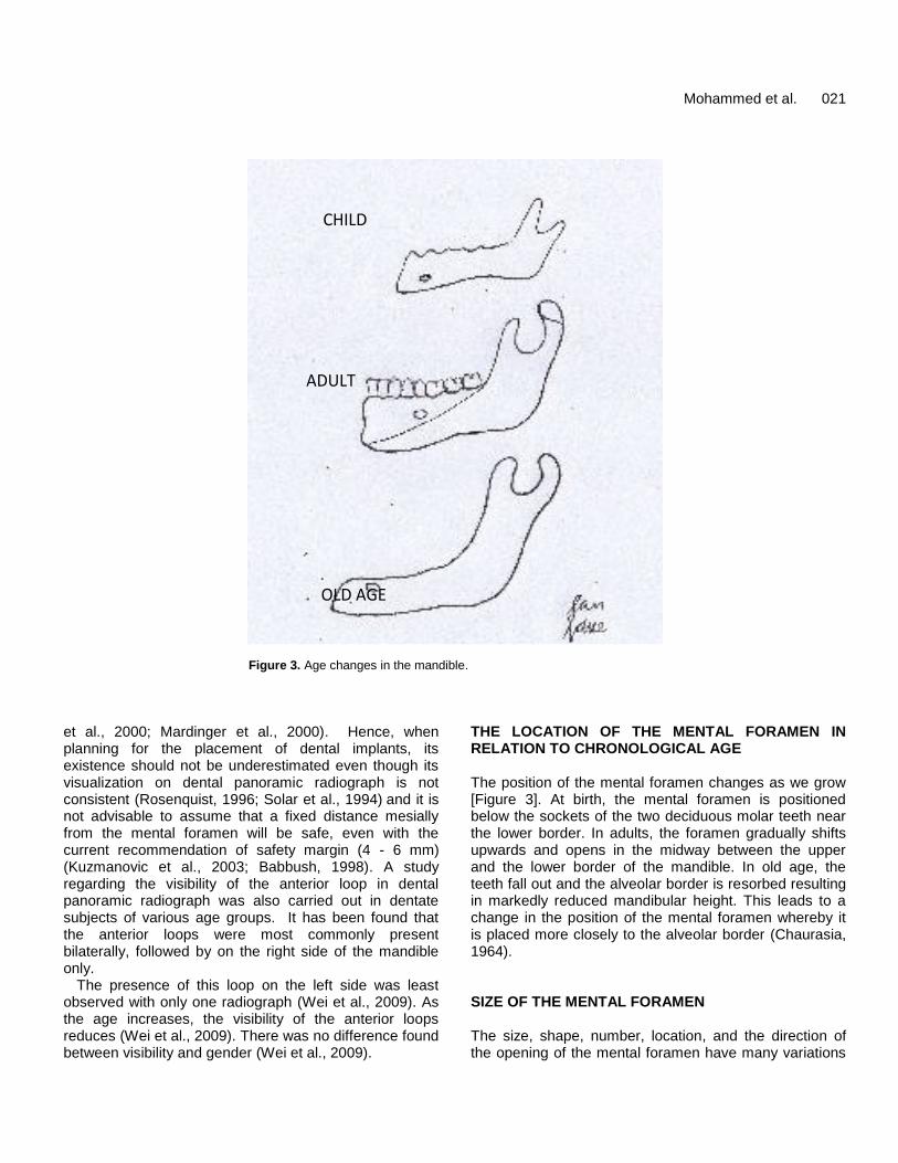
Figure 2. Anterior loop of mental nerve.
2003; Yosue and Brooks, 1989; Al-Jasser and AL-Nwoku, 1998; Kahraman et al., 2006) into four types: Type I: Mental canal is continuous with the mandibular canal. (21%) Type II: Mental canal is distinctly separated from the mandibular canal (43%) Type III: Diffuse with a distinct border of the foramen (24%) Type IV: “unidentified group” (12%)
ANTERIOR LOOP The anterior loop is defined as where the inferior alveolar nerve crosses anterior to the mental foramen then loops or doubles back to exit the mental foramen [Figure 2]. It can be detected in 11-60% of panoramic radiograph. The anterior loop can be present or absent. Many studies have shown large variations in the anterior loop length ranging from 0.4 to 2.19 mm (Arzouman et al., 1993; Kuzmanovic et al., 2003; Uchida et al., 2007; Mardinger
Mohammed et al. 021
CHILD
ADULT
OLD AGE
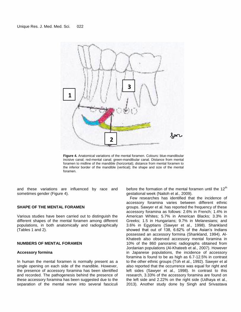
Figure 3. Age changes in the mandible.
et al., 2000; Mardinger et al., 2000). Hence, when planning for the placement of dental implants, its existence should not be underestimated even though its visualization on dental panoramic radiograph is not consistent (Rosenquist, 1996; Solar et al., 1994)
and it is
not advisable to assume that a fixed distance mesially from the mental foramen will be safe, even with the current recommendation of safety margin (4 - 6 mm) (Kuzmanovic et al., 2003; Babbush, 1998). A study regarding the visibility of the anterior loop in dental panoramic radiograph was also carried out in dentate subjects of various age groups. It has been found that the anterior loops were most commonly present bilaterally, followed by on the right side of the mandible only.
The presence of this loop on the left side was least observed with only one radiograph (Wei et al., 2009). As the age increases, the visibility of the anterior loops reduces (Wei et al., 2009). There was no difference found between visibility and gender (Wei et al., 2009).
THE LOCATION OF THE MENTAL FORAMEN IN RELATION TO CHRONOLOGICAL AGE The position of the mental foramen changes as we grow [Figure 3]. At birth, the mental foramen is positioned below the sockets of the two deciduous molar teeth near the lower border. In adults, the foramen gradually shifts upwards and opens in the midway between the upper and the lower border of the mandible. In old age, the teeth fall out and the alveolar border is resorbed resulting in markedly reduced mandibular height. This leads to a change in the position of the mental foramen whereby it is placed more closely to the alveolar border (Chaurasia, 1964). SIZE OF THE MENTAL FORAMEN The size, shape, number, location, and the direction of the opening of the mental foramen have many variations
Unique Res. J. Med. Med. Sci. 022
SIZE OF THE MENTAL FORAMEN
The size, shape, number, location, and the direction
of the opening of the mental foramen have many
variations and these variations are influenced by
race and sometimes gender.
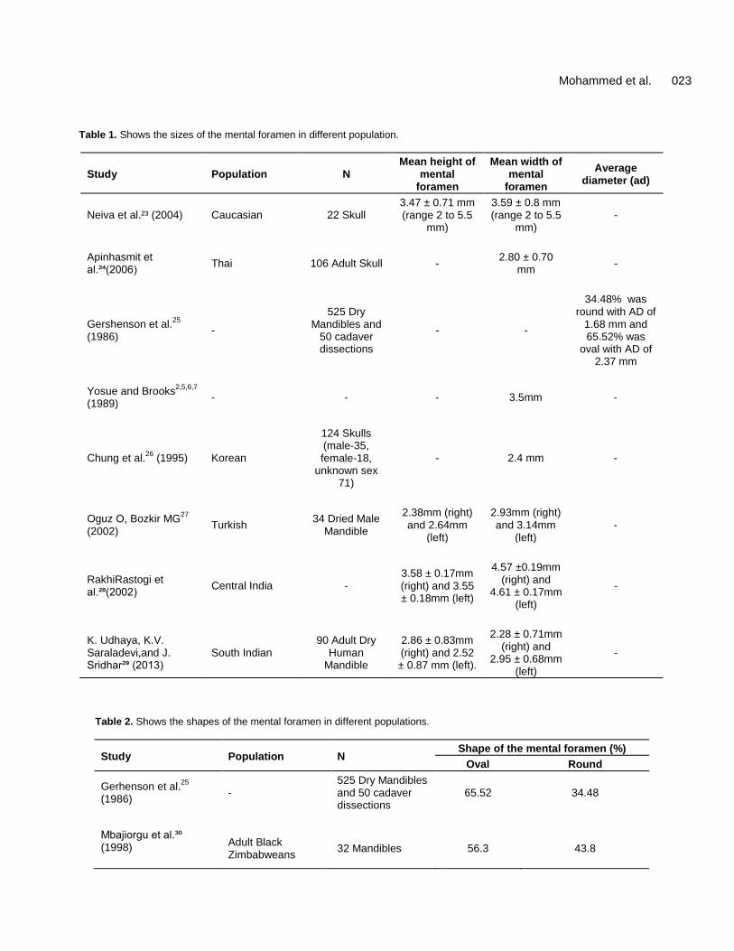
3 Figure 4²²: Anatomical variations of the mental
foramen.
Colours: blue-mandibular incisive canal; red-mental
canal; green-mandibular canal.
1- distance from mental foramen to midline of
the mandible (horizontal)
2- distance from mental foramen to the inferior
border of the mandible (vertical)
3- the shape and size of the mental foramen
1
2
Figure 4. Anatomical variations of the mental foramen. Colours: blue-mandibular incisive canal; red-mental canal; green-mandibular canal. Distance from mental foramen to midline of the mandible (horizontal); distance from mental foramen to the inferior border of the mandible (vertical); the shape and size of the mental foramen.
and these variations are influenced by race and sometimes gender (Figure 4). SHAPE OF THE MENTAL FORAMEN Various studies have been carried out to distinguish the different shapes of the mental foramen among different populations, in both anatomically and radiographically (Tables 1 and 2). NUMBERS OF MENTAL FORAMEN Accessory formina In human the mental foramen is normally present as a single opening on each side of the mandible. However, the presence of accessory foramina has been identified and recorded. The pathogenesis behind the presence of these accessory foramina has been suggested due to the separation of the mental nerve into several fasciculi
before the formation of the mental foramen until the 12th
gestational week (Naitoh et al., 2009). Few researches has identified that the incidence of
accessory foramina varies between different ethnic groups. Sawyer et al. has reported the frequency of these accessory foramina as follows: 2.6% in French; 1.4% in American Whites; 5.7% in American Blacks; 3.3% in Greeks; 1.5 in Hungarians; 9.7% in Melanesians; and 3.6% in Egyptians (Sawyer et al., 1998). Shankland showed that out of 138, 6.62% of the Asian’s Indians possessed an accessory formina (Shankland, 1994). Al-Khateeb also observed accessory mental foramina in 10% of the 860 panoramic radiographs obtained from Jordanian populations (Al-Khateeb et al., 2007). However in Japanese populations, the incidence of accessory foramina is found to be as high as 6.7-12.5% in contrast to the other ethnic groups (Toh et al., 1992). Sawyer et al also reported that the occurrence was equal for right and left sides (Sawyer et al., 1998). In contrast to this research, 3.33% of the accessory foramina are found on the left side and 2.22% on the right side (Udhaya et al., 2013). Another study done by Singh and Srivastava
Mohammed et al. 023
Table 1. Shows the sizes of the mental foramen in different population.
Study Population N Mean height of
mental foramen
Mean width of mental
foramen
Average diameter (ad)
Neiva et al.²³ (2004) Caucasian 22 Skull 3.47 ± 0.71 mm (range 2 to 5.5
mm)
3.59 ± 0.8 mm (range 2 to 5.5
mm) -
Apinhasmit et al.²⁴(2006)
Thai 106 Adult Skull - 2.80 ± 0.70
mm -
Gershenson et al.25
(1986)
-
525 Dry Mandibles and
50 cadaver dissections
- -
34.48% was round with AD of
1.68 mm and 65.52% was
oval with AD of 2.37 mm
Yosue and Brooks2,5,6,7
(1989)
- - - 3.5mm -
Chung et al.26
(1995) Korean
124 Skulls (male-35, female-18,
unknown sex 71)
- 2.4 mm -
Oguz O, Bozkir MG27
(2002)
Turkish 34 Dried Male
Mandible
2.38mm (right) and 2.64mm
(left)
2.93mm (right) and 3.14mm
(left) -
RakhiRastogi et al.²⁸(2002)
Central India - 3.58 ± 0.17mm (right) and 3.55 ± 0.18mm (left)
4.57 ±0.19mm (right) and
4.61 ± 0.17mm (left)
-
K. Udhaya, K.V. Saraladevi,and J. Sridhar²⁹ (2013)
South Indian 90 Adult Dry
Human Mandible
2.86 ± 0.83mm (right) and 2.52 ± 0.87 mm (left).
2.28 ± 0.71mm (right) and
2.95 ± 0.68mm (left)
-
Table 2. Shows the shapes of the mental foramen in different populations.
Study Population N Shape of the mental foramen (%)
Oval Round
Gerhenson et al.25
(1986)
- 525 Dry Mandibles and 50 cadaver dissections
65.52 34.48
Mbajiorgu et al.³⁰ (1998)
Adult Black Zimbabweans
32 Mandibles 56.3 43.8
2
Unique Res. J. Med. Med. Sci. 024
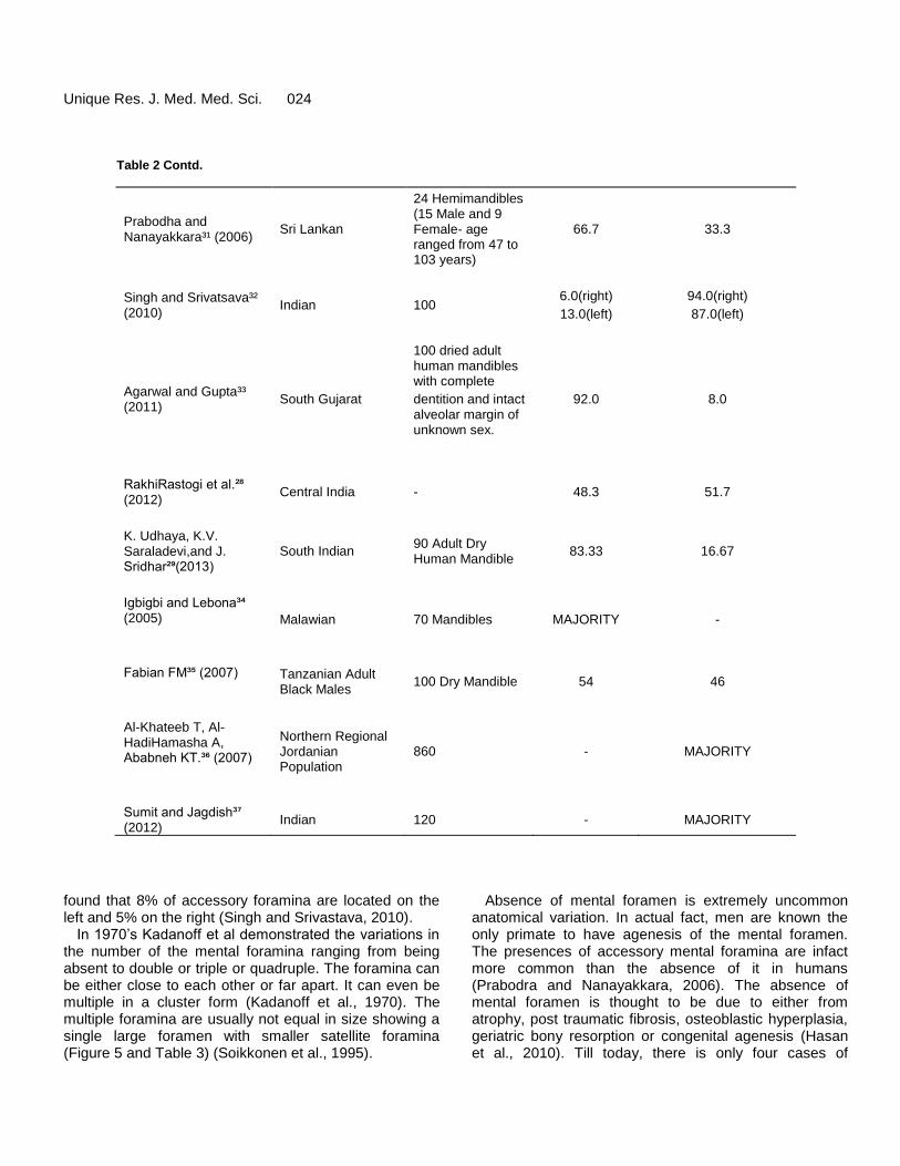
Table 2 Contd.
Prabodha and Nanayakkara³¹ (2006)
Sri Lankan
24 Hemimandibles (15 Male and 9 Female- age ranged from 47 to 103 years)
66.7 33.3
Singh and Srivatsava³² (2010)
Indian 100 6.0(right)
13.0(left)
94.0(right)
87.0(left)
Agarwal and Gupta³³ (2011)
South Gujarat
100 dried adult human mandibles with complete
dentition and intact alveolar margin of unknown sex.
92.0 8.0
RakhiRastogi et al.²⁸ (2012)
Central India - 48.3 51.7
K. Udhaya, K.V. Saraladevi,and J. Sridhar²⁹(2013)
South Indian 90 Adult Dry Human Mandible
83.33 16.67
Igbigbi and Lebona³⁴ (2005)
Malawian 70 Mandibles MAJORITY -
Fabian FM³⁵ (2007)
Tanzanian Adult Black Males
100 Dry Mandible 54 46
Al-Khateeb T, Al-HadiHamasha A, Ababneh KT.³⁶ (2007)
Northern Regional Jordanian Population
860 - MAJORITY
Sumit and Jagdish³⁷ (2012)
Indian 120 - MAJORITY
found that 8% of accessory foramina are located on the left and 5% on the right (Singh and Srivastava, 2010).
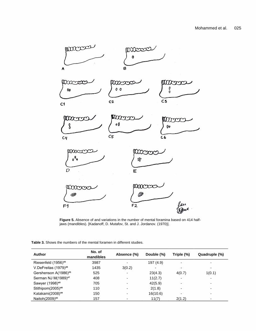
In 1970’s Kadanoff et al demonstrated the variations in the number of the mental foramina ranging from being absent to double or triple or quadruple. The foramina can be either close to each other or far apart. It can even be multiple in a cluster form (Kadanoff et al., 1970). The multiple foramina are usually not equal in size showing a single large foramen with smaller satellite foramina (Figure 5 and Table 3) (Soikkonen et al., 1995).
Absence of mental foramen is extremely uncommon anatomical variation. In actual fact, men are known the only primate to have agenesis of the mental foramen. The presences of accessory mental foramina are infact more common than the absence of it in humans (Prabodra and Nanayakkara, 2006). The absence of mental foramen is thought to be due to either from atrophy, post traumatic fibrosis, osteoblastic hyperplasia, geriatric bony resorption or congenital agenesis (Hasan et al., 2010). Till today, there is only four cases of
Mohammed et al. 025
Figure 5. Absence of and variations in the number of mental foramina based on 414 half-jaws (mandibles). [Kadanoff, D. Mutafov, St. and J. Jordanov. (1970)].
Table 3. Shows the numbers of the mental foramen in different studies.
Author No. of
mandibles Absence (%) Double (%) Triple (%) Quadruple (%)
Riesenfeld (1956)⁴⁵ 3987 - 197 (4.9) - -
V.DeFreitas (1979)⁴⁶ 1435 3(0.2) - - -
Gershenson A(1986)²⁵ 525 - 23(4.3) 4(0.7) 1(0.1)
Serman NJ M(1989)⁴⁷ 408 - 11(2.7) - -
Sawyer (1998)⁴⁰ 705 - 42(5.9) - -
Stithipom(2005)⁴⁸ 110 - 2(1.8) - -
Katakami(2008)⁴⁹ 150 - 16(10.6) - -
Naitoh(2009)⁵⁰ 157 - 11(7) 2(1.2) -
Unique Res. J. Med. Med. Sci. 026 unilateral absence (Azaz and Lustmann, 1973; De Freitas et al., 1979; Freitas et al., 1975; Feritas et al., 1976) and a single case of bilateral absence (Hasan et al., 2010).
The presence and absence of mental foramina is significant because it plays an essential role during the administration of local anaesthesia (Agnieszka and Małgorzata, 2010; Madeira et al., 1978) and the blood vessels present in the accessory foramina can lead to intraosseous haemorrhage during implant procedures (Kaufman et al., 2000; McDonnell et al., 1994). The absence of mental foramen also indicates that there would be probable sensory alterations in the lower lip chin area. Clinical implications would include failure in mental block anaesthesia (Hasan et al., 2010).
Hauser and De Stefano stated that the difference in sizes exist due to the epigenetic traits, as they could be the products of the genetically determined growth processes of other tissues, which had affected the bone formation. Eventually they undergo modifications during morphogenesis and variable degree of expressions. Hence, the differences in the position, shape, number and size of the mental foramen depends on the gene modifications (Hauser and De Stefano, 1989). POSITION OF THE MENTAL FORAMEN In relation to the mandibular teeth It has been found that the location of the mental foramen varies in different ethnicity. Its position is commonly seen below the second premolar. However, individual variation exists whereby it can be placed anywhere in between the first premolar to the mesial root of the first molar.
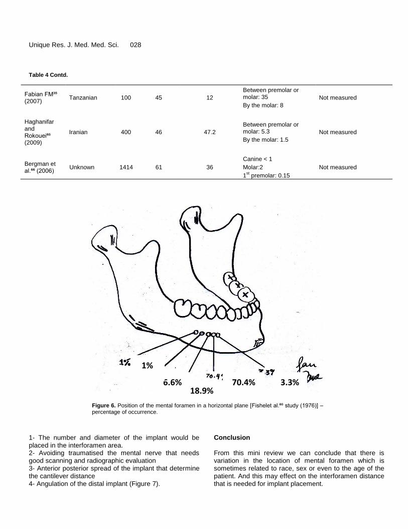
The positioned of the mental foramen can be seen in two different planes which are the horizontal and the vertical plane. Fishel et al. (1976) has proposed the most popular method for the identification of mental foramen. The horizontal position of mental foramen was recorded as either in line with the longitudinal axis of the tooth or lying in between two teeth. Fishel et al. (1976) described the vertical position of the mental foramen to be either situated coronal to the apex, at the apex or apical to the apex (Table 4 and Figure 6) (Fishel et al., 1976). In relation to the symphisismenti, posterior border of the ramus of the mandible, and the lower border of the body of the mandible Some studies measured the location of the mental foramen from various landmark of the mandible. For an instance, the most commonly used landmarks are symphysis menti, posterior border of ramus of mandible
(in horizontal plane) and lower border of body of mandible(in vertical plane).
Various studies have been done by using the midline of the mandible as an anatomical landmark to identify the position of the mental foramen. According to Agthong et al. (2005), the mandibular foramen is located 28 mm from the midline of the mandible and 14 to 15 mm from the inferior border of the mandible. On the other hand, Neiva et al proposed that the distance of this foramen from midline was 27.61±2.29 mm. Neiva also studied about the inter-foramina distance and found that the distance between the left and right foramen was 55.23±5.34 mm (Neiva et al., 2004). Another study which was done by Apinhasmit et al. (2006) founds that the mental foramen was a mean of 28.52±2.15 mm from the midline of the mandible (Table 5).
Therefore, we can conclude that the positions of the mental foramen are different between races and population. The ethnic variations in the position of the mental foramen are also of great significant for estimating its position during the invasive procedures and administration of dental anaesthesia. EMERGENCE PATTERN OF THE MENTAL CANAL AND MENTAL FORAMEN OPENING Solar et al. (1994) has found that the mental canal which is the anterior opening of the mandibular canal, transverses cranially at an angle of inclination ranging from 11 to 77°. They noted that the average gradient among 37 specimens observed was 50°. On the other hand, Kieseret al. (2002) classified the part of emergence into posterior, anterior, right angle or multiple. The most common part of emergence in Caucasoid and Maori was a posterior direction (86.7% of Caucasoid males, 90.2% of Caucasoid females; 85.5% of Maori males, 93.1% of Maori females). In Blacks, the most common pattern is right angle (45.8% of males and 45% of females). Igbigbi and Lebona (2005) and Apinhasmit et al. (2006) recorded that the usual direction of mental foramen opening is posterior superior direction. In addition to this, Fabian (2007) has concluded that the mental foramen opening was superiorly in 44%, posterosuperiorly in 40%, labially in 10%, anteriorly in 3% and posteriorly in 3% of cases. Importance of mental foramen locationid dental implantology Mental foramen location is crucial for dental implant placement in the anterior and premolar area of the mandible. As mental foramen location will determine:
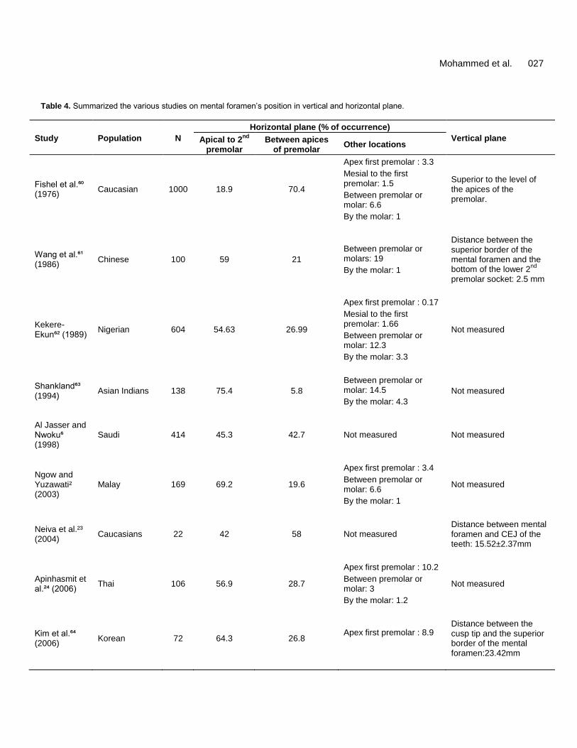
Mohammed et al. 027 Table 4. Summarized the various studies on mental foramen’s position in vertical and horizontal plane.
Study Population N
Horizontal plane (% of occurrence)
Vertical plane Apical to 2nd
premolar
Between apices of premolar
Other locations
Fishel et al.⁶⁰ (1976)
Caucasian 1000 18.9 70.4
Apex first premolar : 3.3
Mesial to the first premolar: 1.5
Between premolar or molar: 6.6
By the molar: 1
Superior to the level of the apices of the premolar.
Wang et al.⁶¹ (1986)
Chinese 100 59 21
Between premolar or molars: 19
By the molar: 1
Distance between the superior border of the mental foramen and the bottom of the lower 2
nd
premolar socket: 2.5 mm
Kekere-Ekun⁶² (1989)
Nigerian 604 54.63 26.99
Apex first premolar : 0.17
Mesial to the first premolar: 1.66
Between premolar or molar: 12.3
By the molar: 3.3
Not measured
Shankland⁶³ (1994)
Asian Indians 138 75.4 5.8
Between premolar or molar: 14.5
By the molar: 4.3
Not measured
Al Jasser and Nwoku⁶ (1998)
Saudi 414 45.3 42.7 Not measured Not measured
Ngow and Yuzawati² (2003)
Malay 169 69.2 19.6
Apex first premolar : 3.4
Between premolar or molar: 6.6
By the molar: 1
Not measured
Neiva et al.²³ (2004)
Caucasians 22 42 58 Not measured Distance between mental foramen and CEJ of the teeth: 15.52±2.37mm
Apinhasmit et al.²⁴ (2006)
Thai 106 56.9 28.7
Apex first premolar : 10.2
Between premolar or molar: 3
By the molar: 1.2
Not measured
Kim et al.⁶⁴ (2006)
Korean 72 64.3 26.8 Apex first premolar : 8.9
Distance between the cusp tip and the superior border of the mental foramen:23.42mm
Unique Res. J. Med. Med. Sci. 028 Table 4 Contd.
Fabian FM³⁵ (2007)
Tanzanian 100 45 12
Between premolar or molar: 35
By the molar: 8
Not measured
Haghanifar and Rokouei⁶⁵ (2009)
Iranian 400 46 47.2
Between premolar or molar: 5.3
By the molar: 1.5
Not measured
Bergman et al.⁶⁶ (2006)
Unknown 1414 61 36
Canine < 1
Molar:2
1st premolar: 0.15
Not measured
18.9% 70.4% 3.3% 6.6%
1%
Figure 6. Position of the mental foramen in a horizontal plane [Fishelet al.⁶⁰ study (1976)] – percentage of occurrence.
1- The number and diameter of the implant would be placed in the interforamen area. 2- Avoiding traumatised the mental nerve that needs good scanning and radiographic evaluation 3- Anterior posterior spread of the implant that determine the cantilever distance 4- Angulation of the distal implant (Figure 7).
Conclusion From this mini review we can conclude that there is variation in the location of mental foramen which is sometimes related to race, sex or even to the age of the patient. And this may effect on the interforamen distance that is needed for implant placement.
Mohammed et al. 029 Table 5. Shows the position of the mental foramen in mean distance from the symphysis menti, posterior border of the ramus and lower border of the body of the mandible.
Study
Mean distance from (mm)
Symphysis menti Posterior border of ramus of mandible
Lower border of body of mandible
Apinhasmit et al.²⁴ (2006) 28.83 68.88 14.88
Prabodha et al.⁶⁸(2006) 26.52 65.38 12.25
Sumit and Jagdish³⁷ (2012) 29.12 74.16 14.45
Agthong et al.⁶⁷ (2005) 28 14.15 -
Neiva et al.²³ (2004) 27.6 - 12mm
Smajilagic A, Dilberovic F⁶⁹ (2004) 25 - -
Figure 7²²: Emergence patterns of the
mental canal and mental foramen opening.
Colours: blue - Mandibular Incisive Canal, red - mental canal (the anterior opening of the mandibular canal), green - mandibular canal. A = superiorly, B = posterosuperiorly; C = labially; D = mesially (anteriorly); E = posteriorly
Figure 7. Emergence patterns of the mental canal and mental foramen opening. Colours: blue - Mandibular Incisive Canal, red - mental canal (the anterior opening of the mandibular canal), green - mandibular canal. A = superiorly, B = posterosuperiorly; C = labially; D = mesially (anteriorly); E = posteriorly.
ACKNOWLEDGMENT Acknowledge goes to Mr. Mohammed Zaki Noor Al-Hashimi/ biostatistics lecturer in MAHSA University, for
his great support and effort REFERENCES Agarwal DR, Gupta SB. Morphometric analysis of mental
foramen in human mandibles of south Gujarat. People’s J Sci Res. 2011;4:15–18.
AgnieszkaP,Małgorzata B. Accessory mandibular foramina: Histological and immunohistochemical studies of their contents .Archives of Oral Biology. 2010; 55: 77-80
Agthong S, Huanmanop T, Chentanez V. Anatomical variation of the supraorbital, infraorbital, and mental foramina related to gender and side. J Oral MaxillofacSurg 2005; 63:800-4
Agthong S, Huanmanop T, Chentanez V. Anatomical variations of the supraorbital, infraorbital, and mental foramina related to gender and side. J Oral MaxillofacSurg2005;63:800-804.
Al-Jasser and AL Nwoku. Radiographic study of the mental foramen in a selected Saudi population. DentomaxillofacRadiol. 1998 Nov;27(6):341-3.
Al-Khateeb T, Al-HadiHamasha A, Ababneh KT. Position of the mental foramen in a northern regional Jordanian population. SurgRadiol Anat. 2007 Apr;29(3):231-7. Epub 2007 Mar 21.
Apinhasmit W, Chompoopong S, Methathrathip D, Sansuk R, Phetphunphiphat W. Supraorbital Notch/Foramen, Infraorbital Foramen and Mental Foramen in Thais: anthropometric measurements and surgical relevance. J. Med Assoc Thai. 2006 May; 89(5):675-82.
Arzouman MJ, Otis L, Kipnis V, Levine D (1993) Observations of the anterior loop of the inferior alveolar canal. Int J Oral Maxillofac Implants 8, 295- 300.
Azaz B, Lustmann J. Anatomical configurations in dry mandibles.Br. J .oral Surg1973 ;2:1-9
Unique Res. J. Med. Med. Sci. 030 Babbush CA (1998) Transpositioning and repositioning
the inferior alveolar and mental nervesin conjunction with endosteal implant reconstruction. Periodontol 2000 17, 183-190
Bergam RA, Afifi AK, Miyauchi R. Mental Foramen. In: Illustrated encyclopedia of Humans Anatomic Variations: Opus V: Skeletal systems. Available at: http://www.anatomyatlases.org/AnatomicVariants/Skeletal-Sysstem/Images/127.shtml. Accessed June 21, 2006.
Bernard Liebgott. The anatomical basis of dentistry. 2nd
ed. Elsevier Mosby, 2001: 311- 313
Chaurasia’s BD.Human Anatomy for Dental Students. 1964: pg154-155
Chummy S. Sinnatamby. Last’s Anatomy: regional and applied. 11
thed. Elsevier Churchill Livingstone, 2006
:366 & 35 Chung MS, Kim HJ, Kang HS, Chung IH. Locational
relationship of the supraorbital notch or foramen and infraorbital and mental foramina in Koreans.Acta Anat. (Basel) 1995;154:162–66.
De Freitas V, Madeira MC, Toledo Filho JL, Chagas CF. Absence of the mental foramen in dry human mandibles.ActaAnat (Basel). 1979; 104(3): 353-355.
De Freitas V, Madeira MC, Toledo Filho JL, Chagas CF. Ab-sence of the mental foramen in dry human mandibles.ActaAnat (Basel) 1979;104:353-5
Fabian FM. Position, shape and direction of opening of the mental foramen in dry mandibles of Tanzanian adult black males. Ital J AnatEmbryol. 2007 Jul-Sep;112(3):169-77
Feritas V ,MadeiraMC,TeixeiraPinto,C, ZorzettoNL.Direction of the mental canal in human mandibles.Austr.dent.J.1976;21:338-340.
Fishel D, Buchner A, Hershkowith A, Kaffe I. Roentgenologic study of the mental foramen. Oral Surg Oral Med Oral Pathol 1976;41:682-686.
Freitas V ,Teixeira Pinto C ,ZorzettoNL ,Madeira,MC ,Pieffer CR .Contribuic,a"o para o estudo da localizac,a"o e das variaco"es do forame mental emmandibulashumanas. Revtabras. Odont. 1975;194:156-160
Gershenson A, Nathan H, Luchansky E. Mental foramen and mental nerve: changes with age. ActaAnat (Basel). 1986;126(1):21-8.
GintarasJuodzbalys, Hom Lay Wang, GintautasSabalys. Anatomical and mandibular vital structure. Part 2: mandibular incisive canal, mental foramen and associated neurovascular bundles in relation with dental implantology. J Oral maxillofac Res. 2010;1:No 1:e3.
Gosling JA, Harris PF, Humpherson JR, Whitmore I &Willan PLT. Head and neck. In: Atlas of human anatomy with integrated text. Edinburgh-London-New
York: Churchill Livingstone; 1985. p. 250-290. Gray’s Anatomy for Students, 2
nd Edition. Richard L.
Drake, A. Wayne Vogl, Adam W.M. Mitchell 2009.pg 931,935
Haghanifa S, and M. Rokouei.Radiographic evaluation of the mental foramen in a selected Iranian population. Indian J. Dent Res. 2009;20:150-152
Hasan T, Fauzi M, Hasan D. Bilateral absence of the mental foramen;a rare variation. International journal of anatomic variations.2010; 3 : 167-169
Hauser G, De Stefano GF. Epigenetic variants of human skull. Ed. Schweizebart. Stuttgart. 1989:230–33
Igbigbi PS, Lebona S. The position and dimensions of the mental foramen in adult Malawian mandibles. West Afr J Med. 2005 Jul-Sep;24(3):184-9.
Kadanoff D, Mutafov ST, Jordanov J, Über die Hauptöffnungen resp. Incisurae des Gesichtsschädels (Incisurafrontalisseu Foramen frontale, Foramen supraorbitaleseuIncisurasupraorbitalis, Foramen infraorbitale, Foramen mentale). GegenbaursMorphol. Jahrbuch ,1970;115:102-118.
KahramanGungor, Mustafa Ozturk Mustafa Semiz and Sharon Lynn Brooks.A Radiographic Study of Location of Mental Foramen in a Selected Turkish Population On Panoramic Radiograph 2006;4 : 801-805
Katakami K, Mishima A, Shiozaki K, Shimoda S, Hamada Y, Kobayashi K. Characteristics of accessory mental foramina observed on limited cone-beam computed tomography images. J Endod 2008;34:1441-5
Kaufman E. N, Serman J , Wang P.D. Bilateral mandibular accessory foramina and canals: a case report and review of the literature, DentomaxillofacRadiol . 2000 ;29 :170–175.
Kekere-Ekun TA. Antero-posterior location of the mental foramen in Nigerians.Afr Dent J 1989;3:2-8.
Kieser J, Kuzmanovic D, Payne A, Dennison J, Herbison P. Patterns of emergence of the human mental nerve. Arch Oral Biol. 2002 Oct;47(10):743-7.
Kim IS, Kim SG, Kim YK, Kim JD. Position of the mental foramen in a Korean population: a clinical and radiographic study. Implant Dent. 2006 Dec;15(4):404-11.
Kuzmanovic DV, Payne AG, Kieser JA, Dias GJ (2003) Anterior loop of the mental nerve: a morphological and radiographic study. Clin Oral Implants Res 14, 464-471.
Madeira C,Percinoto M, Das G, Silva M.. Clinical significance of supplementary innervation of the lower incisor teeth: a dissection study of the mylohyoidnerve.OralSurg Oral Med Oral Pathol. 1978;46: 608–614.
Mardinger O, Chaushu G, Arensburg B, Taicher S, Kaffe I (2000) Anterior loop of the mental canal: an anatomical-radiologic study. Implant Dent 9, 120-125.
Mardinger O, Chaushu G, Arensburg B, Taicher S, Kaffe
I. Anterior loop of the mental canal: an anatomical-
radiologic study. Implant Dent 2000; 9:120-125. Mbajioru EF, Mawera G, Asala SA, Zivanovic S. Position
of the mental foramen in adult Zimbabwean mandibles. A clinical anatomical study.Central Afr J Med. 1988;44:24–30.
McDonnell D, NouriM.R. ,Todd M. The mandibular lingual foramen: a consistent arterial foramen in the middle of the mandible, J Anat .1994; 184:369–371.
Mraiwa N, Jacobs R, van Steenberghe D, Quirynen M. Clinical assessment and surgical implications of anatomic challenges in the anterior mandible. Clin Implant Dent Relat Res 2003;5:219-225.
Naitoh M, Hiraiwa Y, Aimiya H, Gotoh K, Ariji E. Accessory mental foramen assessment using cone-beam computed tomography. Oral Surg Oral Med Oral Pathol Oral RadiolEndod. 2009 Feb;107(2):289-94. Epub 2008 Dec 13.
Naitoh M, Hiraiwa Y, Aimiya H, Gotoh K, Ariji E. Accessory mental foramen assessment using cone-beam computed tomography. Oral Surg Oral Med Oral Pathol Oral RadiolEndod. 2009 Feb;107(2):289-94. Epub 2008 Dec 13.
Neiva RF, Gapski R, Wang HL. Morphometric analysis of implant-related anatomy in Caucasian skulls. J Periodontol 2004;75:1061-1067
Ngeow WC, Yuzawati Y. The location of the mental foramen in a selected Malay population. J Oral Sci 2003; 45: 171-175.
Norton NS. Netter’s Head and Neck Anatomy for Dentistry. Philadelphia: Saunders, 2007:86-96.
Oguz O, Bozkir MG. Evaluation of location of mandibular and mental foramina in dry, young, adult human male, dentulous mandibles. West Indian Med J. 2002;51:14–16
Pogrel MA, Smith R, Ahani R. Innervation of the mandibular incisors by the mental nerve. J Oral MaxillofacSurg 1997;55:961-963.
Prabodha LBL, Nanayakkara BG. The position, dimensions and morphological variations of mental foramen: Galle Medical Journal 2006 Sept, Vol 11;pg 13-15.
Prabodra LBL, Nanayakkara BG. The position, dimension and morphological variations of mental foramen in mandibles.Galle Med J. 2006;11:13–15.
RastogiRakhi, BudhirajaVirendra, Sathpathi DK, Singh Sandeep, GourKranti Kumar, et al. Morphology and morphometry of the mental foramen in dry adult human mandibles from central India and their clinical correlation. Eur J Anat. 2012;16(1):22–26.
Riesenfeld A. Multiple infraorbital, ethmoidal, and mental foramina in the races of man. Am J PhysAnthropol 1956;14:85-100.
Rosenquist B (1996) Is there an anterior loop of the
Mohammed et al. 031 inferior alveolar nerve? Int J Periodontics RestorativeDent
16, 40-45. Sawyer DR, Kiely ML, Pyle MA.The frequency of accessory
mental foramina in four ethnic groups. Arch Oral Biol 1998;43:417-420.
Serman NJ. The mandibular incisive foramen. J Anat 1989;167:195-8
Shankland WE 2nd. The position of the mental foramen in Asian Indians.J Oral Implantol. 1994;20(2):118-123.
Shankland WE 2nd. The position of the mental foramen in Asian Indians.J Oral Implantol. 1994;20(2):118-123.
Singh R, Srivastava AK. Study of position, shape, size and incidence of mental foramen and accessory mental foramen in Indian adult human skulls.Int J Morphol. 2010;28:1141–46.
Smajilagić A, Dilberović F. Clinical and anatomy study of the human mental foramen. 2004 Jul; 4(3):15-23.
Soikkonen K, Wolf J, Ainamo A, Xie Q. "Changes in the position of the mental foramen as a result of alveolar atrophy". J Oral Rehabil. 1995;22: 831–3.
Solar P, Ulm C, Frey G, Matejka M (1994) A comparison of the accuracy of the periapical, panoramic, and computerized tomographic radiographs in locating the mandibular canal. Int J Oral Maxillofac Implants 9, 455-460.
Solar P, Ulm C, Frey G, Matejka M. A classification of the intraosseous paths of the mental nerve.Int J Oral Maxillofac Implants 1994;9:339-344.
Sumit Gupta and Jagdish S. Soni.Study of anatomical variations and incidence of mental foramen and accessory mental foramen in dry human mandibles. National Journal of Health Research 2012;2:28-30.
Toh H, Kodama J, Yanagisako M, Ohmori T. Anatomical study of the accessory mental foramen and the distribution of its nerves. Okajimas Folia AnatJpn. 1992; 69:85-87
Uchida Y, Yamashita Y, Goto M, Hanihara T (2007) Measurement of anterior loop length for the mandibular canal and diameter of the mandibular incisive canal to avoid nerve damage when installingendosseous implants in the interforaminal region. J Oral MaxillofacSurg 65, 1772-1779.
Udhaya K., K.V. Saraladevi, and J. Sridhar. The Morphometric Analysis of the Mental Foramen in Adult Dry Human Mandibles: A Study on the South Indian Population. 2013 : 1547-1551
Wang TM, Shih C, Liu JC, Kuo KJ. A clinical and anatomical study of the location of the mental foramen in adult Chinese mandibles.ActaAnat (Basel) 1986;126:29-33.
Wei Cheong Ngeow, Dionetta D. Dionysius, HayatiIshak and PhrabhakaranNambiar.A radiographic study on the visualization of the anterior loop in dentate subjects of different age groups. Vol. 51, No. 2, 231-237, 2009
Yosue T, Brooks SL. The appearance of mental foramina on panoramic and periapical radiographs. II. Experimental evaluation. Oral Surg Oral Med Oral Pathol 1989;68:488-492.