Embed Size (px)
Citation preview
MOLECULAR AND CELLULAR BIOLOGY,0270-7306/97/$04.0010
Sept. 1997, p. 4957–4966 Vol. 17, No. 9
Copyright © 1997, American Society for Microbiology
Interaction of ATF6 and Serum Response FactorCHENG ZHU, FINN-EIRIK JOHANSEN, AND RON PRYWES*
Department of Biological Sciences, Columbia University, New York, New York 10027
Received 18 April 1997/Returned for modification 23 May 1997/Accepted 2 June 1997
Serum response factor (SRF) is a transcription factor which binds to the serum response element (SRE) inthe c-fos promoter. It is required for regulated expression of the c-fos gene as well as other immediate-earlygenes and some tissue-specific genes. To better understand the regulation of SRF, we used a yeast interactionassay to screen a human HeLa cell cDNA library for SRF-interacting proteins. ATF6, a basic-leucine zipperprotein, was isolated by binding to SRF and in particular to its transcriptional activation domain. The bindingof ATF6 to SRF was also detected in vitro. An ATF6-VP16 chimera activated expression of an SRE reportergene in HeLa cells, suggesting that ATF6 can interact with endogenous SRF. More strikingly, an antisenseATF6 construct reduced serum induction of a c-fos reporter gene, suggesting that ATF6 is involved in activationof transcription by SRF. ATF6 was previously partially cloned as a member of the ATF family. The completecDNA of ATF6 was isolated, and its expression pattern was described.
Expression of the c-fos gene is rapidly transcriptionally ac-tivated in serum-starved cells by serum and many other mito-gens. Mapping of the c-fos promoter suggests that a key se-quence element is the serum response element (SRE) (15).SRE-like elements have also been found to be critical for theexpression of other immediate-early genes, a number of mus-cle-specific genes, and interleukin-2 receptor (15). The mainnuclear protein found to bind the SRE is serum responsefactor (SRF). SRF is a 508-amino-acid protein of the MADSbox family that binds to DNA as a dimer (21). In addition tocentral DNA binding and dimerization domains (spanning theconserved MADS box), it contains a C-terminal transcriptionalactivation domain (13).
One mechanism for serum induction of the SRE is througha family of SRF-associated proteins, ternary complex factors(TCFs) (27). TCFs are encoded by three related genes (Elk1,Sap1, and Sap2/Net/Erp) with ets-related DNA binding do-mains (22, 27). TCFs contact both SRF and a specific sequenceelement and bind to SRF only when a DNA binding site isadjacent to the SRF site. TCFs also contain a transcriptionalactivation domain which is regulated by protein kinases fromthe mitogen-activated protein kinase family (27, 28). Activa-tion of c-fos expression by serum and various growth factors atleast partially works by activating mitogen-activated proteinkinases. A mutation of the TCF site in the c-fos promoter,however, does not have a large effect on serum induction of areporter gene (6, 11). This and other results suggest that thereis a TCF-independent mechanism for serum induction of c-fos(10, 12, 14).
The TCF transcriptional activation domain acts in conjunc-tion with the transcriptional activation domain of SRF to in-crease c-fos transcription (9, 12, 14). The transcriptional acti-vation domain of SRF can also function alone when it is fusedto the DNA binding domain of GAL4, but it is not regulated byserum in this context (13). One mechanism for the activation oftranscription by the SRF transcriptional activation domain ap-pears to be its binding to the general transcription factorTFIIF. We previously found that SRF bound the RAP74 sub-
unit of TFIIF in vitro and that mutations in either SRF orRAP74 that abrogated this binding also reduced the ability ofSRF to activate transcription in vitro (16, 30).
In vivo footprinting suggests that SRF constitutively occu-pies the c-fos SRE in cells (8). However, SRF does not activatetranscription in uninduced cells, suggesting that there is neg-ative regulation of SRF prior to activation. In order to betterunderstand how SRF is regulated by serum and how this reg-ulation controls its ability to activate transcription, we utilizeda yeast interaction screen to identify additional SRF-interact-ing proteins. We describe here the identification of one suchprotein, ATF6, which binds to the SRF transcriptional activa-tion domain and is itself a DNA-binding protein. An antisenseATF6 construct strongly reduced serum induction of a c-fosreporter gene, suggesting that ATF6 is a critical component ofthe SRE-SRF complex.
MATERIALS AND METHODS
Yeast interaction screen. The screening of a human HeLa cell cDNA librarywas performed essentially as previously described (2). The indicator strain, ActL,contains a high-affinity SRF binding site in front of a cyc1-lacZ gene. The SRFexpression vector pSD0.7 encodes full-length SRF under the control of a galac-tose-inducible promoter (2). The HeLa cell cDNA library used was a randomlyprimed cDNA library fused to the C terminus of the VP16 transcriptionalactivation domain under the control of a galactose-inducible promoter as previ-ously described (2). To screen the library, transformed yeast cells were plateddirectly onto nitrocellulose filters and grown for approximately 22 h on selectivemedium containing 2% glucose. The filters were then transferred to plates thatcontained galactose instead of glucose and grown for an additional 20 h. Colorassays for b-galactosidase activity were performed by submerging the filters inliquid nitrogen to permeabilize cells and placing them on Whatman 3MM papersoaked with X-Gal (5-bromo-4-chloro-3-indolyl-b-D-galactopyranoside) sub-strate buffer (2). Positive colonies were retested and purified.
Positive colonies were cured of either the SRF-expressing plasmid (pSD0.7;TRP1 marker) or cDNA-expressing plasmids (URA3 marker) by standard pro-cedures and retested by the color assay. VP16-tagged cDNA library plasmidswere recovered by preparing DNA from a 10-ml culture of pSD0.7-cured yeastand transforming it into Escherichia coli MC1066 by electroporation. The iso-lated plasmids were transformed into the indicator yeast strain containing SRF(pSD0.7) and tested by the color assay for activation of lacZ expression. To testthe interaction with SRF deletion mutants, vectors that express these forms ofSRF were used instead of pSD0.7. For samples without SRF, the control TRP1vector pRS314 was used (25).
For the liquid b-galactosidase assays, yeast cells were grown overnight inselective medium plus 2% glucose, diluted 1:50 in selective medium plus 2%galactose, and grown until the optical density at 600 nm was 0.6 to 0.8. b-Galactosidase activities were determined as previously described (2).
Plasmids. The plasmids constructed for this study are described below. Fur-ther details and maps are available upon request.
* Corresponding author. Mailing address: Department of BiologicalSciences, Columbia University, 1212 Amsterdam Ave. MC2420, NewYork, NY 10027. Phone: (212) 854-8281. Fax: (212) 865-8246. E-mail:[email protected].
4957
Dow
nloa
ded
from
http
s://j
ourn
als.
asm
.org
/jour
nal/m
cb o
n 18
Oct
ober
202
1 by
42.
114.
118.
148.
(i) Yeast expression plasmids. Mutant SRF expression plasmids were derivedfrom pSD0.7, which expresses full-length SRF with a galactose-inducible pro-moter and a TRP1 marker (2). These plasmids included amino acids 114 to 508,114 to 245, or 1 to 412 of SRF.
(ii) Bacterium expression plasmids. A glutathione S-transferase (GST)–c1.12fusion protein was made by using PCR primers to place the c1.12 region of ATF639 of GST between the BamHI and XbaI sites of pGTK, a GST expression vectorprovided by Hua Zhu. A fusion of maltose-binding protein (MBP) and SRF wasmade by placing a fragment that encodes amino acids 266 to 508 of SRF 39 toMBP in plasmid pMalC2 (New England Biolabs). pMBP-c1.12 was obtained byinserting c1.12 into the EcoRI-to-XbaI sites of pMalC2. pSRF(114-508) wasexpressed in pET3a and was described previously (23).
(iii) Mammalian reporter and expression plasmids. The reporter gene plas-mids pO-Fluc, pSRE-Fluc, and pSRE.M-Fluc were described previously (14).They contain a minimal human c-fos reporter gene (253 to 145) fused to aluciferase gene with no site, a single copy of the c-fos SRE, and a mutant SRE,respectively. The reporter gene plasmids Fos-WT and Fos-pm12 contain 2356 to1109 of the mouse c-fos promoter inserted 59 to the luciferase gene in the pGL3vector (Promega). Fos-pm12 contains four base mutations in the SRE thatabolish SRF binding (5).
All of the mammalian expression vectors used were derived from pCGN, inwhich expression is driven by the cytomegalovirus (CMV) promoter and theexpressed proteins are fused to the influenza virus hemagglutinin antigen at theamino terminus at an XbaI site (26). pCFN is identical to pCGN, except for asimian virus 40 nuclear localization signal added after the hemagglutinin epitopetag and before the inserted gene (provided by Hua Zhu). pCFN-VP16c1.12 (seeFig. 3) contains the VP16-c1.12 fusion gene (isolated in the yeast screen) insertedin the XbaI site of pCFN. The full-length ATF6 expression vector, pCGN-ATF6,contains ATF6 from the first start codon to the 39 end at the XbaI site of pCGN.In pCFN-c1.12, the c1.12 fragment of ATF6 was expressed by placing it at theXbaI site of pCFN. The antisense ATF6 vector pAS-ATF6 contains the c1.12region of ATF6 in the antisense orientation at the XbaI site of pCFN.
In vitro binding of SRF and ATF6. GST-c1.12, GST, MBP-c1.12, MBP-SRF(266-508), MBP, and SRF(114-508) proteins were expressed in E. coli BL21.Expression was induced in mid-log phase by the addition of 0.5 mM isopropyl-b-D-thiogalactopyranoside (IPTG) for 3 h. Then cells (1 liter) were harvested,resuspended in 20 ml of BC100 buffer (20 mM Tris [pH 8.0], 100 mM NaCl, 0.2mM EDTA, 100 mM KCl, 20% glycerol, 0.5 mM phenylmethylsulfonyl fluoride,0.5 mM dithiothreitol, 0.05% Nonidet P-40), sonicated, and centrifuged at12,000 3 g for 15 min at 4°C. The supernatant was recentrifuged, and thesubsequent supernatant was used for purification. SRF(114-508) was purified aspreviously described (23). GST and MBP proteins were bound to glutathione-Sepharose 4B (Pharmacia) or amylose agarose (New England Biolabs) by adding0.5 ml of resin for every 5 ml of supernatant for 1 h at 4°C. The resins werewashed with BC100 buffer four times at 4°C. The binding of GST proteins wasconfirmed by eluting three times with 10 mM reduced glutathione in 50 mM Tris(pH 8.0) and pooling the eluates. MBP fusion proteins were eluted from theamylose resin with 10 mM maltose in BC100 buffer. The purities and quantitiesof proteins were analyzed by sodium dodecyl sulfate (SDS)-polyacrylamide gelelectrophoresis and Coomassie blue staining.
Aliquots (20 ml) of glutathione-Sepharose beads bearing equal amounts ofeither GST or the GST-c1.12 fusion protein were incubated with 200 ng ofSRF(114-508) or MBP-SRF(266-508) in 100 ml of BC100 buffer. The mixtureswere gently rocked on a rotating wheel at room temperature for 1 h. The beadswere pelleted at 3,000 3 g for 2 min, and the supernatant was saved as theunbound fraction. The beads were washed four times with 1 ml of BC100 buffer,and bound proteins were eluted three times by incubation with 20 ml of 10 mMreduced glutathione in 50 mM Tris (pH 8.0) at room temperature for 10 min andpooled. Both unbound and eluted proteins (40 ml [each]) were analyzed byimmunoblotting with anti-C-terminal SRF serum (18) and alkaline phosphatase-conjugated goat anti-rabbit immunoglobulin G.
Transfection and luciferase assays. All cells were grown in Dulbecco’s mod-ified Eagle medium containing 10% newborn calf serum. One 60-mm-diameterplate of cells (HeLa or Cos) was transfected by standard calcium phosphatemethods with a total of 7 mg of DNA composed of 2 mg of reporter constructs,3 mg of vector or expression constructs, and 2 mg of pCMV-b-galactosidase as aninternal control. Luciferase and b-galactosidase reporter gene activities wereassayed 42 h later as previously described (14), except that luciferase sampleswere counted for 15 s in a Berthold Lumat luminometer. Luciferase levels werenormalized for transfection efficiency with b-galactosidase activities. In someexperiments at 16 h after the beginning of transfection, HeLa cells were washedand serum starved in Dulbecco’s modified Eagle medium with 0.2% newborn calfserum for 36 h. Starved cells were either left alone or induced with 20% newborncalf serum for 4 h. The results given are the means of three independenttransfection assays done in duplicate, and variations are shown as the standarderrors of the means.
cDNA cloning. ATF6 cDNA was further cloned from the sequence of the c1.12cDNA clone isolated in the yeast screen. The region 39 of c1.12 was cloned byusing a 39 rapid amplification of cDNA ends (RACE) system (Gibco-BRL). Asdescribed by the manufacturer, 5 mg of total HeLa RNA was primed with anoligo(dT)-containing adapter primer (59-GGCCACGCGTCGACTAGTACTTTTTTTTTTTTTTTTT-39) and extended with reverse transcriptase (Super-
Script II RT RNase H2). After first-strand cDNA synthesis, the original mRNAtemplate was digested with RNase H. Then the cDNA was amplified by twostages of PCR with nested ATF6 primers. First, ATF6 primer c1.12-GSP1 (an-nealing to positions 989 to 1012 of ATF6) was used with an abridged universalamplification primer (AUAP; 59-GGCCACGCGTCGACTAGTAC-39) whichoverlaps part of the adapter sequence used to prime first-strand cDNA synthesis.Second, nested ATF6 primer c1.12-AccI (59-ATCGAGAATCCGCTTGTCAGTCTC-39; nucleotides 1033 to 1054) and AUAP were used. The ends weretreated with T4 DNA polymerase, and PCR products were subcloned into theNotI and AccI sites of pBluescript II SK.
The region 59 to the c1.12 clone was isolated by using a 59 RACE system(Gibco-BRL). The first-strand cDNA was primed with ATF6-specific primer GSP-2BT (positions 880 to 856). Terminal transferase (TdT) was used to add poly(dC)tails to the 39 ends of the cDNA. The tailed cDNA was amplified by two-stepPCRs. The first PCR was performed with primer AAP (59-GGCCACGCGTCGACTAGTACGGGIIGGGIIGGGIIG-39) and ATF6-specific primer C1.12BT3(positions 736 to 717). The second amplification was done with this AUAP andnested ATF6 primer GSP-Acc (59-CGAGGGCAGAACTCCAGGTGCTT-39;nucleotides 794 to 773). Three PCR products were obtained and were subclonedinto pBluescript II SK.
The full-length cDNA sequence was obtained by sequencing both strands withvector and gene-specific primers. Sequence comparisons were performed with aBLAST search of the GenBank database.
Northern blot analysis. Total cellular RNA was isolated from an 80% conflu-ent 10-cm-diameter plate with 7 ml of TRIzol reagent (Gibco-BRL). Hybridiza-tion probes were gel purified and labeled with [a-32P]dATP by using a Primer-ItRmT random primer labeling kit (Stratagene) as described by the manufacturer.Total RNA (7 mg) was electrophoresed and transferred to nitrocellulose bystandard procedures. The filters were prehybridized in QuikHyb hybridizationsolution (Stratagene) for 30 min at 68°C and hybridized with 2.5 3 106 cpm ofhybridization solution per ml for 1.5 h at 68°C. The filters were washed twice for15 min at room temperature with 23 SSC (13 SSC is 0.15 M NaCl plus 0.015 Msodium citrate)–0.1% SDS and twice for 30 min at 60°C with 0.13 SSC–0.05%SDS. The mouse multitissue Northern blot (Clontech) contained 2 mg ofpoly(A)1 RNA from the indicated tissues. The human b-actin probe (Clontech)was used to confirm the amount of RNA in each lane.
Immunoblotting of ATF6. MBP-c1.12 fusion protein was affinity purified onamylose agarose as described above and used to raise antiserum against ATF6.MBP-c1.12 (100 mg) was injected subcutaneously into a rabbit, and 50 mg wassubsequently injected at 2, 3, and 7 weeks after the initial injection.
To detect ATF6 in mammalian cells, 2 3 107 Cos or HeLa cells were lysed with300 ml of 13 SDS loading buffer (50 mM Tris [pH 6.8], 0.2% SDS, 0.1%bromophenol blue, 10% glycerol, 0.5 mM dithiothreitol). Lysates (60 ml) wereanalyzed by standard immunoblotting procedures with preimmune or anti-ATF6serum (1:800 dilution) and alkaline phosphatase-conjugated goat anti-rabbitimmunoglobulin G (20 ng/ml).
Cos cells were transfected with 7 mg of full-length ATF6 expression plasmidpCGN-ATF6 or control vector pCGN. Transfected-cell lysates (20 ml) wereanalyzed with anti-ATF6 antiserum as described above or with monoclonalantibody 12CA5 against influenza virus hemagglutinin antigen (1:700) (26).
RESULTS
Screen for SRF-interacting proteins in yeast. To identifyproteins which interact with SRF and which either regulate orexecute its activity, we screened a human HeLa cell cDNAlibrary in the yeast Saccharomyces cerevisiae. We utilized thesystem of Dalton and Treisman (2) that includes a lacZ re-porter gene with an SRF binding site upstream of a cyc1 pro-moter. The SRF binding site used, ActL, binds SRF with highaffinity but does not bind Mcm1, a yeast protein homologous toSRF. SRF and a HeLa cell cDNA library, fused to the Cterminus of the herpesvirus VP16 transcriptional activationdomain, were expressed with galactose-inducible promoters onplasmids with TRP1 and URA3 markers, respectively.
SRF activates transcription only weakly in yeast; therefore,activation of the lacZ gene was dependent upon recruitment ofa VP16 fusion protein to the promoter. This system has anadvantage over the standard two-hybrid method in that it al-lows SRF to bind directly to the promoter and attain its properconformation and potentially allows an interacting protein tobind to both SRF and DNA, thus stabilizing their interaction.This is especially critical since GAL4-SRF chimeras are notserum regulated in HeLa or NIH 3T3 cells (13). This systemwas successfully used to clone one of the TCF family members,Sap1 (2).
4958 ZHU ET AL. MOL. CELL. BIOL.
Dow
nloa
ded
from
http
s://j
ourn
als.
asm
.org
/jour
nal/m
cb o
n 18
Oct
ober
202
1 by
42.
114.
118.
148.
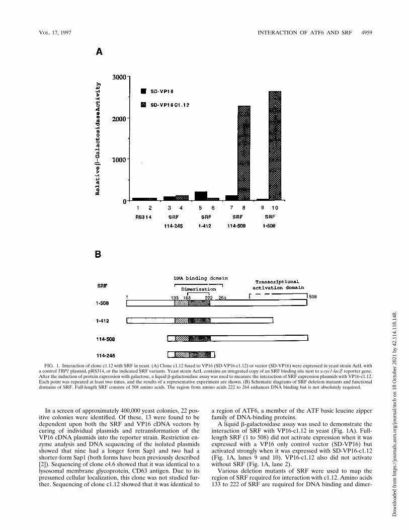
In a screen of approximately 400,000 yeast colonies, 22 pos-itive colonies were identified. Of these, 13 were found to bedependent upon both the SRF and VP16 cDNA vectors bycuring of individual plasmids and retransformation of theVP16 cDNA plasmids into the reporter strain. Restriction en-zyme analysis and DNA sequencing of the isolated plasmidsshowed that nine had a longer form Sap1 and two had ashorter-form Sap1 (both forms have been previously described[2]). Sequencing of clone c4.6 showed that it was identical to alysosomal membrane glycoprotein, CD63 antigen. Due to itspresumed cellular localization, this clone was not studied fur-ther. Sequencing of clone c1.12 showed that it was identical to
a region of ATF6, a member of the ATF basic leucine zipperfamily of DNA-binding proteins.
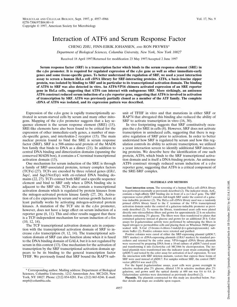
A liquid b-galactosidase assay was used to demonstrate theinteraction of SRF with VP16-c1.12 in yeast (Fig. 1A). Full-length SRF (1 to 508) did not activate expression when it wasexpressed with a VP16 only control vector (SD-VP16) butactivated strongly when it was expressed with SD-VP16-c1.12(Fig. 1A, lanes 9 and 10). VP16-c1.12 also did not activatewithout SRF (Fig. 1A, lane 2).
Various deletion mutants of SRF were used to map theregion of SRF required for interaction with c1.12. Amino acids133 to 222 of SRF are required for DNA binding and dimer-
FIG. 1. Interaction of clone c1.12 with SRF in yeast. (A) Clone c1.12 fused to VP16 (SD-VP16-c1.12) or vector (SD-VP16) were expressed in yeast strain ActL witha control TRP1 plasmid, pRS314, or the indicated SRF variants. Yeast strain ActL contains an integrated copy of an SRF binding site next to a cyc1-lacZ reporter gene.After the induction of protein expression with galactose, a liquid b-galactosidase assay was used to measure the interaction of SRF expression plasmids with VP16-c1.12.Each point was repeated at least two times, and the results of a representative experiment are shown. (B) Schematic diagrams of SRF deletion mutants and functionaldomains of SRF. Full-length SRF consists of 508 amino acids. The region from amino acids 222 to 264 enhances DNA binding but is not absolutely required.
VOL. 17, 1997 INTERACTION OF ATF6 AND SRF 4959
Dow
nloa
ded
from
http
s://j
ourn
als.
asm
.org
/jour
nal/m
cb o
n 18
Oct
ober
202
1 by
42.
114.
118.
148.
ization as well as interaction with TCFs (21, 27). The C-termi-nal region of SRF contains a transcriptional activation domain(13). Only deletions that retained the DNA binding domainwere used since this assay requires the binding of SRF to theSRE-containing reporter gene. As shown in Fig. 1A, deletionof the SRF amino terminus [SRF(114-508)] did not abolish thisinteraction (lane 8). However, deletion of the SRF C terminus[SRF(1-412)] abolished binding to c1.12 (Fig. 1A, lane 6). Inaddition, the SRF DNA binding region alone [SRF(114-245)]was not sufficient for binding to c1.12 (Fig. 1A, lane 4). Similarresults were obtained by b-galactosidase filter assay (data notshown). Both SRF(1-412) and SRF(114-245) were expressedsince they interacted well with VP16-Sap1 (16) (data notshown). Besides affecting the binding of SRF to c1.12, a dele-tion to amino acid 412 of SRF abolishes transcriptional acti-vation by a GAL4-SRF chimera (13). These results suggestthat the c1.12 protein product interacts with the SRF transcrip-tional activation domain.
As described below (see Fig. 5), the predicted amino acidsequence from the 570-bp sequence of c1.12 shows that it isidentical to the ATF6 sequence in a region that spans the basicregion but includes only half of the leucine zipper dimerizationdomain. As such, it would not be predicted to bind DNAdirectly. This suggests that c1.12 (and therefore ATF6) canbind SRF without making additional DNA contacts.
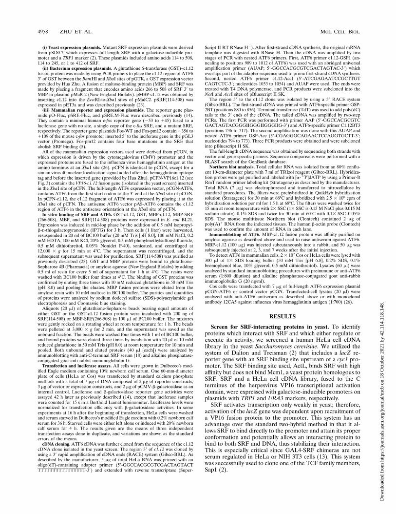
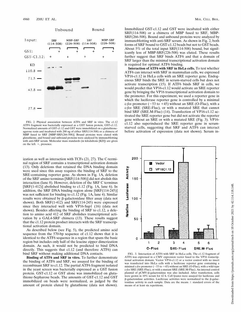
Binding of ATF6 and SRF in vitro. To further demonstratethe binding of ATF6 and SRF, we assayed for the binding ofrecombinant SRF to c1.12. The partial ATF6 fragment isolatedin the yeast screen was bacterially expressed as a GST fusionprotein. GST-c1.12 or GST alone was immobilized on gluta-thione-Sepharose beads. The amounts of GST-c1.12 and GSTimmobilized on beads were normalized, as judged by theamount of protein eluted by glutathione (data not shown).
Immobilized GST-c1.12 and GST were incubated with eitherSRF(114-508) or a chimera of MBP fused to SRF, MBP-SRF(266-508). Bound and unbound proteins were analyzed byimmunoblotting with anti-SRF serum. As shown in Fig. 2, bothforms of SRF bound to GST-c1.12 beads but not to GST beads.About 5% of the total input SRF(114-508) bound, but signif-icantly less of MBP-SRF(226-508) was eluted. These resultsfurther suggest that SRF binds ATF6 and that a domain ofSRF larger than the minimal transcriptional activation domainis required for optimal ATF6 binding.
Interaction of ATF6 with SRF in HeLa cells. To test whetherATF6 can interact with SRF in mammalian cells, we expressedVP16-c1.12 in HeLa cells with an SRE reporter gene. Endog-enous SRF binds the SRE in serum-starved cells but does notactivate transcription (15). If ATF6 binds SRF in cells, wewould predict that VP16-c1.12 would activate an SRE reportergene by bringing the VP16 transcriptional activation domain tothe promoter. For this experiment, we used a reporter gene inwhich the luciferase reporter gene is controlled by a minimalc-fos promoter (253 to 145) without an SRE (O-Fluc), with ac-fos SRE (SRE-Fluc), or with a mutated SRE that cannotbind SRF (SRE.M-Fluc) (14). Transfection of VP16-c1.12 ac-tivated the SRE reporter gene but did not activate the reportergene without an SRE or with a mutated SRE (Fig. 3). VP16-c1.12 also superinduced the SRE reporter gene in serum-starved cells, suggesting that SRF and ATF6 can interactbefore activation of expression (data not shown). Serum in-
FIG. 2. Physical association between ATF6 and SRF in vitro. The c1.12ATF6 fragment was bacterially expressed as a GST fusion protein, GST-c1.12.Normalized amounts of GST-c1.12 and GST were immobilized on a glutathione-agarose resin and incubated with 200 ng of either SRF(114-508) or a chimera ofMBP fused to SRF [MBP-SRF(266-508)]. Bound proteins were eluted withglutathione, and bound and unbound proteins were analyzed by immunoblottingwith anti-SRF serum. Molecular mass standards (in kilodaltons [KD]) are givenon the left. 1, presence.
FIG. 3. Interaction of ATF6 with SRF in HeLa cells. The c1.12 fragment ofATF6 was expressed in a CMV expression vector fused to the VP16 transcrip-tional activation domain. Vector VP16-c1.12 or a vector control with no insertwas transfected into HeLa cells with a luciferase reporter gene containing aminimal c-fos promoter (253 to 145) without an SRE (O-Fluc), with a wild-typec-fos SRE (SRE-Fluc), or with a mutant SRE (SRE.M-Fluc). An internal controlplasmid of pCMV-b-galactosidase was also included. After transfection, cellswere grown in 10% serum for 42 h. Cell lysates were assayed for luciferase andb-galactosidase activities. Luciferase activities were normalized to the b-galac-tosidase activity in each sample. Data are the means 6 standard errors of themeans of at least six repetitions.
4960 ZHU ET AL. MOL. CELL. BIOL.
Dow
nloa
ded
from
http
s://j
ourn
als.
asm
.org
/jour
nal/m
cb o
n 18
Oct
ober
202
1 by
42.
114.
118.
148.
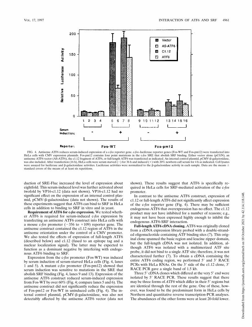
duction of SRE-Fluc increased the level of expression abouteightfold. This serum-induced level was further activated abouttwofold by VP16-c1.12 (data not shown). VP16-c1.12 had nosignificant effect on the expression of an internal control plas-mid, pCMV-b-galactosidase (data not shown). The results ofthese experiments suggest that ATF6 can bind to SRF in HeLacells in addition to binding to SRF in vitro and in yeast.
Requirement of ATF6 for c-fos expression. We tested wheth-er ATF6 is required for serum-induced c-fos expression bytransfecting an antisense ATF6 construct into HeLa cells witha mouse c-fos promoter (2356 to 1109) reporter gene. Theantisense construct contained the c1.12 region of ATF6 in theantisense orientation under the control of a CMV promoter.We also tested the effects of expression of full-length ATF6(described below) and c1.12 (fused to an epitope tag and anuclear localization signal). The latter may be expected tofunction as a dominant negative by interfering with endoge-nous ATF6 binding to SRF.
Expression from the c-fos promoter (Fos-WT) was inducedby serum induction of serum-starved HeLa cells (Fig. 4, lanes1 and 5). A mutant c-fos promoter (Fos-pm12) showed thatserum induction was sensitive to mutations in the SRE thatabolish SRF binding (Fig. 4, lanes 9 and 13). Expression of theantisense ATF6 construct reduced serum-induced expressionfrom Fos-WT by over 60% (Fig. 4; compare lanes 5 and 6). Theantisense construct did not significantly reduce the expressionof Fos-pm12 or Fos-WT in uninduced cells (Fig. 4). The in-ternal control plasmid, pCMV-b-galactosidase, was also notdetectably affected by the antisense ATF6 vector (data not
shown). These results suggest that ATF6 is specifically re-quired in HeLa cells for SRF-mediated activation of the c-fospromoter.
In contrast to the antisense ATF6 construct, expression ofc1.12 or full-length ATF6 did not significantly affect expressionof the c-fos reporter gene (Fig. 4). There may be sufficientendogenous ATF6 that overexpression has no effect. The c1.12product may not have inhibited for a number of reasons; e.g.,it may not have been expressed highly enough to inhibit theendogenous ATF6-SRF interaction.
Full-length ATF6 cDNA cloning. ATF6 was originally clonedfrom a cDNA expression library probed with a double-strand-ed oligonucleotide containing ATF binding sites (7). This orig-inal clone spanned the basic region and leucine zipper domain,but the full-length cDNA was not isolated. In addition, al-though ATF6 was isolated with a multimerized ATF siteprobe, it did not bind to a single ATF site; therefore, it was notcharacterized further (7). To obtain a cDNA containing theentire ATF6 coding region, we performed 59 and 39 RACEPCRs with HeLa RNAs. On the 39 side of the c1.12 clone, 39RACE PCR gave a single band of 1.5 kb.
Three 59 cDNA clones which differed at the very 59 end wereisolated by 59 RACE PCR. These results suggest that theremay be three forms of ATF6 which differ in their 59 regions butare identical through the rest of the gene. One of these, how-ever, was found to be the predominant form in HeLa cells byNorthern and quantitative reverse transcription-PCR analyses.The abundances of the other forms were at least 20-fold lower.
FIG. 4. Antisense ATF6 reduces serum-induced expression of a c-fos reporter gene. c-fos–luciferase reporter genes (Fos-WT and Fos-pm12) were transfected intoHeLa cells with CMV expression plasmids. Fos-pm12 contains four point mutations in the c-fos SRE that abolish SRF binding. Either vector alone (pCGN), anantisense ATF6 vector (AS-ATF6), the c1.12 fragment of ATF6, or full-length ATF6 was transfected as indicated. An internal control plasmid, pCMV-b-galactosidase,was also included. After transfection (16 h), HeLa cells were serum starved (2) for 36 h and induced (1) with 20% newborn calf serum for 4 h as indicated. Cell lysateswere assayed for luciferase and b-galactosidase activities. Luciferase activities were normalized to the b-galactosidase activity in each sample. Data are the means 6standard errors of the means of at least six repetitions.
VOL. 17, 1997 INTERACTION OF ATF6 AND SRF 4961
Dow
nloa
ded
from
http
s://j
ourn
als.
asm
.org
/jour
nal/m
cb o
n 18
Oct
ober
202
1 by
42.
114.
118.
148.
The predominant form extended 507 bases 59 of the c1.12clone.
cDNA sequence. Sequencing of the major ATF6 cDNA formrevealed that the length of the complete ATF6 cDNA was2,474 bp. A diagram of the structure of the predicted aminoacid sequence is shown in Fig. 5A. The length of the cDNA isconsistent with the size of the major mRNA band detected onNorthern blots of HeLa RNA (Fig. 6A). The cDNA sequencepredicts an open reading frame from nucleotides 43 to 2052 fora protein product of 670 amino acids with a calculated molec-ular mass of 74,576 Da. The 39 end of the cDNA contains 422nucleotides of untranslated sequence which contains two po-tential polyadenylation signals (AAATAA) followed by apoly(A) tail.
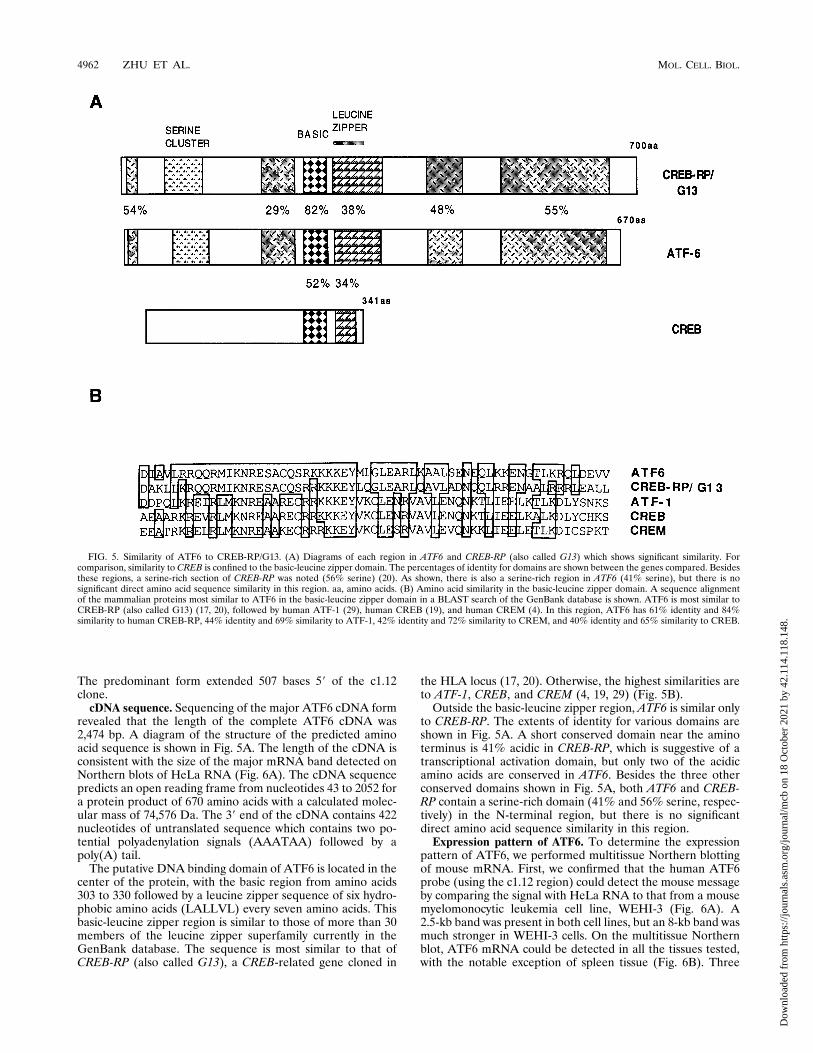
The putative DNA binding domain of ATF6 is located in thecenter of the protein, with the basic region from amino acids303 to 330 followed by a leucine zipper sequence of six hydro-phobic amino acids (LALLVL) every seven amino acids. Thisbasic-leucine zipper region is similar to those of more than 30members of the leucine zipper superfamily currently in theGenBank database. The sequence is most similar to that ofCREB-RP (also called G13), a CREB-related gene cloned in
the HLA locus (17, 20). Otherwise, the highest similarities areto ATF-1, CREB, and CREM (4, 19, 29) (Fig. 5B).
Outside the basic-leucine zipper region, ATF6 is similar onlyto CREB-RP. The extents of identity for various domains areshown in Fig. 5A. A short conserved domain near the aminoterminus is 41% acidic in CREB-RP, which is suggestive of atranscriptional activation domain, but only two of the acidicamino acids are conserved in ATF6. Besides the three otherconserved domains shown in Fig. 5A, both ATF6 and CREB-RP contain a serine-rich domain (41% and 56% serine, respec-tively) in the N-terminal region, but there is no significantdirect amino acid sequence similarity in this region.
Expression pattern of ATF6. To determine the expressionpattern of ATF6, we performed multitissue Northern blottingof mouse mRNA. First, we confirmed that the human ATF6probe (using the c1.12 region) could detect the mouse messageby comparing the signal with HeLa RNA to that from a mousemyelomonocytic leukemia cell line, WEHI-3 (Fig. 6A). A2.5-kb band was present in both cell lines, but an 8-kb band wasmuch stronger in WEHI-3 cells. On the multitissue Northernblot, ATF6 mRNA could be detected in all the tissues tested,with the notable exception of spleen tissue (Fig. 6B). Three
FIG. 5. Similarity of ATF6 to CREB-RP/G13. (A) Diagrams of each region in ATF6 and CREB-RP (also called G13) which shows significant similarity. Forcomparison, similarity to CREB is confined to the basic-leucine zipper domain. The percentages of identity for domains are shown between the genes compared. Besidesthese regions, a serine-rich section of CREB-RP was noted (56% serine) (20). As shown, there is also a serine-rich region in ATF6 (41% serine), but there is nosignificant direct amino acid sequence similarity in this region. aa, amino acids. (B) Amino acid similarity in the basic-leucine zipper domain. A sequence alignmentof the mammalian proteins most similar to ATF6 in the basic-leucine zipper domain in a BLAST search of the GenBank database is shown. ATF6 is most similar toCREB-RP (also called G13) (17, 20), followed by human ATF-1 (29), human CREB (19), and human CREM (4). In this region, ATF6 has 61% identity and 84%similarity to human CREB-RP, 44% identity and 69% similarity to ATF-1, 42% identity and 72% similarity to CREM, and 40% identity and 65% similarity to CREB.
4962 ZHU ET AL. MOL. CELL. BIOL.
Dow
nloa
ded
from
http
s://j
ourn
als.
asm
.org
/jour
nal/m
cb o
n 18
Oct
ober
202
1 by
42.
114.
118.
148.

bands, of 2.5, 4.5, and 8 kb, were detected. The distribution ofthese bands varied. Each of these bands was also detected byprobes from the 59 and 39 ends of ATF6 (data not shown). Wecannot account for the different-sized messages at this time.Since the three ATF6 probes detected the same bands and weused fairly stringent hybridization conditions, it does not ap-pear that we detected cross-reactive genes.
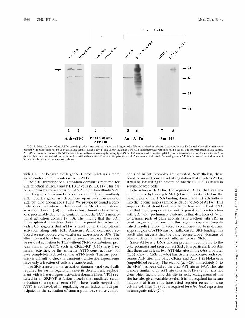
ATF6 protein product. We raised antiserum to ATF6 toshow that its protein product was made in cells. The c1.12region of ATF6 was expressed in E. coli as an MBP fusionprotein, purified, and injected into rabbits. First, the antiserumwas found to bind a GST-c1.12 fusion protein on an immuno-blot, showing that it is reactive to ATF6 (data not shown). Italso detected in vitro-translated ATF6 but not in vitro-trans-lated ATF-1 or ATF-2 (data not shown). To determine themolecular weight of ATF6 in mammalian cells, we used theantiserum to probe an immunoblot of HeLa and Cos celllysates. In both lysates, a 90-kDa band was detected. This wasnot detected with preimmune serum, although a 65-kDa bandwas detected by both sera (Fig. 7, lanes 1 to 4). Transfectioninto Cos cells of ATF6 in a CMV expression vector, fused to aninfluenza virus epitope tag, showed overexpression of a comi-grating 90-kDa band. This band was detected by anti-ATF6and anti-epitope (HA) sera (Fig. 7, lanes 6 and 8). The migra-tion of the ATF6 band was somewhat higher than the expectedmolecular mass (75 kDa). This may be due to conformationaleffects on ATF6 migration or to posttranslational modifica-tions. In argument somewhat against posttranslational modifi-
cations, in vitro-translated ATF6 comigrated with HeLa cellATF6 at 90 kDa (data not shown). The 90-kDa ATF6 band wasalso detected in nuclear extracts of HeLa cells, suggesting thatit is at least partially localized to the nucleus (data not shown).
Two pieces of evidence suggest that we have cloned theentire ATF6 coding region. First, the ATF6 cDNA is similar insize to the RNA band detected on Northern blots (Fig. 6).Second, the transfected ATF6 protein product (90 kDa) comi-grated with an endogenous band reactive with the anti-ATF6serum (Fig. 7). Nevertheless, although we did not obtain anycDNA clones with further 59 sequence by 59 RACE, it remainspossible that the mRNA 59 sequence extends further.
DISCUSSION
We have identified ATF6 as a protein which interacts withSRF in yeast and in vitro. The results of transfection experi-ments showed that ATF6 can also interact with SRF in HeLacells and is required for serum-induced expression of c-fos.
Interaction with SRF. Deletion mutants of SRF showed thatthe SRF transcriptional activation domain is required for itsinteraction with ATF6 in yeast. Deletion of the C terminus ofSRF (amino acids 413 to 508) abolished transcriptional acti-vation by SRF in HeLa cells (13) and the interaction of SRFwith ATF6 in yeast. The C-terminal half of SRF was sufficientfor binding to ATF6 in vitro, but a larger fragment bound moreeffectively. This may be because there are additional contacts
FIG. 6. Tissue distribution of ATF6 expression. (A) HeLa (human cervical carcinoma) and WEHI-3 (mouse promyelocytic leukemia) cell RNAs were probed withthe ATF6-specific c1.12 probe. (B) Northern blot of mRNAs from the indicated mouse tissues hybridized with the ATF6-specific c1.12 probe. To control for mRNAamounts, the blot was reprobed with a human b-actin cDNA probe. An alternatively spliced form of b-actin is detected in spleen mRNA.
VOL. 17, 1997 INTERACTION OF ATF6 AND SRF 4963
Dow
nloa
ded
from
http
s://j
ourn
als.
asm
.org
/jour
nal/m
cb o
n 18
Oct
ober
202
1 by
42.
114.
118.
148.
with ATF6 or because the larger SRF protein attains a morestable conformation to interact with ATF6.
The SRF transcriptional activation domain is required forSRF function in HeLa and NIH 3T3 cells (9, 10, 14). This hasbeen shown by overexpression of SRF with low-affinity SREreporter genes. Serum-induced expression of these low-affinitySRE reporter genes are dependent upon overexpression ofSRF but bind endogenous TCFs. We previously found a com-plete loss of activity with deletion of the SRF transcriptionalactivation domain (14), but others have found only a partialloss, presumably due to the contribution of the TCF transcrip-tional activation domain (9, 10). The finding that the SRFtranscriptional activation domain is required for activationwith TCF suggests that ATF6 is involved in transcriptionalactivation along with TCF. Antisense ATF6 expression re-duced serum-induced c-fos–luciferase expression by 60%. Theeffect may not have been larger for several reasons. There maybe residual activation by TCF without SRF’s contribution; pro-teins similar to ATF6, such as CREB-RP (G13), may havesimilar activities; or the antisense ATF6 construct may nothave completely reduced cellular ATF6 levels. This last possi-bility is difficult to check in transient-transfection experimentssince only a fraction of cells are transfected.
The SRF transcriptional activation domain is not specificallyrequired for serum regulation since its deletion and replace-ment with a heterologous activation domain (from VP16) re-sulted in an SRF-VP16 fusion protein that mediated seruminduction of a reporter gene (14). These results suggest thatATF6 is not involved in regulating serum induction but par-ticipates in the activation of transcription once other compo-
nents of an SRF complex are activated. Nevertheless, therecould be an additional level of regulation that involves ATF6.It will be interesting to determine whether ATF6 is altered inserum-induced cells.
Interaction with ATF6. The region of ATF6 that was iso-lated in yeast by binding to SRF (clone c1.12) starts before thebasic region of the DNA binding domain and extends halfwayinto the leucine zipper (amino acids 155 to 345 of ATF6). Thissuggests that it should not be able to dimerize or bind DNAand that these properties are not required for its interactionwith SRF. Our preliminary evidence is that deletions of N- orC-terminal parts of c1.12 abolish its interaction with SRF inyeast, suggesting that much of this region is required (unpub-lished results). Since in these experiments the basic-leucinezipper region of ATF6 was not sufficient for SRF binding, thisresult also suggests that the basic-leucine zipper domains ofother such proteins are not sufficient to bind SRF.
Since ATF6 is a DNA-binding protein, it could bind to thec-fos promoter and then contact SRF. It is particularly notablethat there are at least two ATF-like sites in the c-fos promoter(1, 3). One (a CRE at 260) has strong homologies with con-sensus ATF sites and binds CREB and ATF-1 in HeLa cells(unpublished results). The second (at 2290 immediately 39 tothe SRE) has been called the c-fos AP1 site or FAP. This siteis more similar to an AP1 site than an ATF site, but it is notclear which factors bind this site in cells. Mutagenesis of thissite has also given variable results. It is not required for seruminduction of transiently transfected reporter genes in tissueculture cell lines (1, 3) but is required for c-fos–lacZ expressionin transgenic mice (24).
FIG. 7. Identification of an ATF6 protein product. Antiserum to the c1.12 region of ATF6 was raised in rabbits. Immunoblots of HeLa and Cos cell lysates wereprobed with either anti-ATF6 or preimmune serum (lanes 1 to 4). The arrow indicates a 90-kDa band detected with anti-ATF6 serum but not with preimmune serum.A CMV expression vector with ATF6 fused to an influenza virus epitope tag (pCGN-ATF6) and a control vector (pCGN) were transfected into Cos cells (lanes 5 to8). Cell lysates were probed on immunoblots with either anti-ATF6 or anti-epitope (anti-HA) serum as indicated. An endogenous ATF6 band was detected in lane 5but cannot be seen in the exposure shown.
4964 ZHU ET AL. MOL. CELL. BIOL.
Dow
nloa
ded
from
http
s://j
ourn
als.
asm
.org
/jour
nal/m
cb o
n 18
Oct
ober
202
1 by
42.
114.
118.
148.
ATF6 was originally isolated in a cDNA expression libraryprobed with a multimerized ATF site but was not able to binda single ATF site (7). We have not yet been able to demon-strate specific DNA binding with ATF6. This may requireidentifying a preferred binding site for ATF6 and/or finding aheterodimerization partner for ATF6 that is required for it tobind DNA.
Models for ATF6’s role in c-fos regulation. Two attractivemodels are suggested by our results. One is that ATF6 func-tions as a coactivator for SRF. Since ATF6 binds to the SRFtranscriptional activation domain, it may bridge an interactionto the general transcription machinery. We previously foundthat SRF binds the RAP74 subunit of the general transcriptionfactor TFIIF in vitro and suggested that this is the mechanismfor transcriptional activation by SRF (16, 30). There may bealternative means for activation by SRF; it is also possible thatSRF binds both ATF6 and TFIIF at the same time, eventhough their binding domains overlap.
A second model, not necessarily exclusive to the first, is thatATF6 binds a sequence element in the c-fos promoter and thenbinds SRF. The binding of ATF6 to the promoter could en-hance and stabilize its interaction with SRF. This would alsoallow the formation of a larger promoter complex that could beimportant in cells for the activation of transcription. Such acomplex was proposed from the results with mutations of thec-fos promoter in transgenic mice. Mutations in multiple ele-ments had a deleterious effect in transgenic mice, even thoughthey did not affect the expression of transiently transfectedreporter genes in tissue culture (24).
ATF6 sequence. ATF6 was originally partially cloned (7), butwe have cloned the complete cDNA sequence here. The fullsequence reveals that ATF6 is most similar to CREB-RP (G13)both in the basic-leucine zipper domain and in a number ofother domains (Fig. 5A). While ATF6 is slightly less similar toATF-1, CREB, and CREM in the basic-leucine zipper domain(Fig. 5B), it contains no similarity to those genes outside of thisregion. The CREB-RP (G13) gene was identified during exten-sive cloning and sequencing of the human HLA locus (17, 20).Its expression was detected in all the cell lines and tissuestested (20). The extended similarity of CREB-RP (G13) andATF6 suggests that they have similar functions, such as acti-vating transcription with SRF.
It will be interesting to determine the DNA binding anddimerization properties of ATF6 and CREB-RP (G13) andwhether CREB-RP (G13) can also bind SRF. This will help tocharacterize the SRF transcriptional activation complex anddetermine whether ATF6 or CREB-RP (G13) binds to a spe-cific site in the c-fos promoter. Since these leucine zipperproteins must dimerize to bind DNA, it will be critical todetermine with which proteins they dimerize. They may ho-modimerize, but they may also require a distinct partner toheterodimerize and bind DNA.
The functional domains of ATF6 also need to be deter-mined. It may have a transcriptional activation domain as wellas regulatory domains. Since the SRF complex does not acti-vate transcription in uninduced cells, an important mystery tobe solved is what changes upon serum induction. One answeris phosphorylation of TCF, but TCF is not required for seruminduction (15). One piece of evidence, the transfection ofVP16-c1.12 (Fig. 3), suggests that ATF6 can bind SRF inuninduced cells. This suggests that there must be a change inthe SRF complex and/or ATF6 that leads to transcriptionalactivation in serum-induced cells.
ACKNOWLEDGMENTS
We thank Stephen Dalton and Richard Treisman for generouslyproviding the yeast SRF interaction system and cDNA library. We alsothank Hua Zhu for the pGTK and pCFN plasmids, Michael Gilmanfor mouse c-fos promoter plasmids, and Wolfgang Fecke for the mousec-fos–luciferase plasmids.
This work was supported by grant CA 50329 from the NationalCancer Institute to R.P.
REFERENCES
1. Berkowitz, L. A., K. T. Riabowol, and M. Z. Gilman. 1989. Multiple sequenceelements of a single functional class are required for cyclic AMP respon-siveness of the mouse c-fos promoter. Mol. Cell. Biol. 9:4272–4281.
2. Dalton, S., and R. Treisman. 1992. Characterization of SAP, a proteinrecruited by serum response factor to the c-fos serum response element. Cell68:597–612.
3. Fisch, T., R. Prywes, and R. G. Roeder. 1987. c-fos sequences necessary forbasal expression and induction by epidermal growth factor, 12-O-tetrade-canoyl phorbol-13-acetate, and the calcium ionophore. Mol. Cell. Biol. 7:3490–3502.
4. Fujimoto, T., J. Fujisawa, and M. Yoshida. 1994. Novel isoforms of humancyclic AMP-responsive element modulator (hCREM) mRNA. J. Biochem.115:298–303.
5. Gilman, M. Z. 1988. The c-fos serum response element responds to proteinkinase C-dependent and independent signals but not to cyclic AMP. GenesDev. 2:394–402.
6. Graham, R., and M. Gilman. 1991. Distinct protein targets for signals actingat the c-fos serum response element. Science 251:189–192.
7. Hai, T., F. F. Liu, W. Coukos, and M. Green. 1989. Transcription factor ATFcDNA clones: an extensive family of leucine zipper proteins able to selec-tively form DNA-binding heterodimers. Genes Dev. 3:2083–2090.
8. Herrera, R. E., P. E. Shaw, and A. Nordheim. 1989. Occupation of the c-fosserum response element in vivo by a multi-protein complex is unaltered bygrowth factor induction. Nature 340:68–70.
9. Hill, C. S., R. Marais, S. John, J. Wynne, S. Dalton, and R. Treisman. 1993.Functional analysis of a growth factor-responsive transcription factor com-plex. Cell 73:395–406.
10. Hill, C. S., J. Wynne, and R. Treisman. 1994. Serum-regulated transcriptionby serum response factor (SRF): a novel role for the DNA binding domain.EMBO J. 13:5421–5432.
11. Hill, C. S., and R. Treisman. 1995. Differential activation of c-fos promoterelements by serum, lysophosphatidic acid, G proteins and polypeptidegrowth factors. EMBO J. 14:5037–5047.
12. Hill, C. S., J. Wynne, and R. Treisman. 1995. The Rho family GTPasesRhoA, Rac1, and CDC42Hs regulate transcriptional activation by SRF. Cell81:1159–1170.
13. Johansen, F.-E., and R. Prywes. 1993. Identification of transcriptional acti-vation and inhibitory domains in serum response factor (SRF) by usingGAL4-SRF constructs. Mol. Cell. Biol. 13:4640–4647.
14. Johansen, F.-E., and R. Prywes. 1994. Two pathways for serum regulation ofthe c-fos serum response element require specific sequence elements and aminimal domain of serum response factor. Mol. Cell. Biol. 14:5920–5928.
15. Johansen, F.-E., and R. Prywes. 1995. Serum response factor: transcriptionalregulation of genes induced by growth factors and differentiation. Biochim.Biophys. Acta 1242:1–10.
16. Joliot, V., M. Demma, and R. Prywes. 1995. Interaction with RAP74 subunitof TFIIF is required for transcriptional activation by serum response factor.Nature 373:632–635.
17. Khanna, A., and R. D. Campbell. 1996. The gene G13 in the class III regionof the human MHC encodes a potential DNA-binding protein. Biochem. J.319:81–89.
18. Manak, J. R., N. de Bisschop, R. M. Kris, and R. Prywes. 1990. Casein kinaseII enhances the DNA binding activity of SRF. Genes Dev. 4:955–967.
19. Meyer, T. E., and J. F. Habener. 1992. Cyclic AMP response element bindingprotein CREB and modulator protein CREM are products of distinct genes.Nucleic Acids Res. 20:6106.
20. Min, J., H. Shukla, H. Kozono, S. Bronson, S. Weissman, and D. Chaplin.1995. A novel Creb family gene telomeric of HLA-DRA in the HLA com-plex. Genomics 30:149–156.
21. Norman, C., M. Runswick, R. Pollock, and R. Treisman. 1988. Isolation andproperties of cDNA clones encoding SRF, a transcription factor that bindsto the c-fos serum response element. Cell 55:989–1003.
22. Price, M. A., A. E. Rogers, and R. Treisman. 1995. Comparative analysis ofthe ternary complex factors Elk-1, Sap-1a and Sap-2 (ERP/NET). EMBO J.14:2589–2610.
23. Prywes, R., and H. Zhu. 1992. In vitro squelching of activated transcription byserum response factor: evidence for a common coactivator used by multipletranscriptional activators. Nucleic Acids Res. 20:513–520.
24. Robertson, L. M., T. K. Kerppola, M. Vendrell, D. Luk, R. J. Smeyne, C.Bocchiaro, J. I. Morgan, and T. Curran. 1995. Regulation of c-fos expression
VOL. 17, 1997 INTERACTION OF ATF6 AND SRF 4965
Dow
nloa
ded
from
http
s://j
ourn
als.
asm
.org
/jour
nal/m
cb o
n 18
Oct
ober
202
1 by
42.
114.
118.
148.
in transgenic mice requires multiple interdependent transcription controlelements. Neuron 14:241–252.
25. Sikorski, R. S., and P. Hieter. 1989. A system of shuttle vectors and yeasthost strains designed for efficient manipulation of DNA in Saccharomycescerevisiae. Genetics 122:19–27.
26. Tanaka, M., and W. Herr. 1990. Differential transcriptional activation byOct-1 and Oct-2: interdependent activation domains induce Oct-2 phosphor-ylation. Cell 60:375–386.
27. Treisman, R. 1994. Ternary complex factors: growth factor regulated tran-scriptional activators. Curr. Opin. Genet. Dev. 4:96–101.
28. Whitmarsh, A. J., P. Shore, A. D. Sharrocks, and R. J. Davis. 1995. Inte-gration of MAP kinase signal transduction pathways at the serum responseelement. Science 269:403–407.
29. Yoshimura, T., J. Fujisawa, and M. Yoshida. 1990. Multiple cDNA clonesencoding nuclear proteins that bind to the tax-dependent enhancer ofHTLV-1: all contain a leucine zipper structure and basic amino acid domain.EMBO J. 9:2537–2542.
30. Zhu, H., V. Joliot, and R. Prywes. 1994. Role of transcription factor TFIIFin serum response factor-activated transcription. J. Biol. Chem. 269:3489–3497.
4966 ZHU ET AL. MOL. CELL. BIOL.
Dow
nloa
ded
from
http
s://j
ourn
als.
asm
.org
/jour
nal/m
cb o
n 18
Oct
ober
202
1 by
42.
114.
118.
148.