Embed Size (px)
Citation preview

Cancer Immunol Immunother (2008) 57:1091–1104
DOI 10.1007/s00262-007-0446-5ORIGINAL ARTICLE
Interactions of host IL-6 and IFN-� and cancer-derived TGF-�1 on MHC molecule expression during tumor spontaneous regression
Ya-Wen Hsiao · Kuang-Wen Liao · Tien-Fu Chung · Chen-Hsuan Liu · Chia-Da Hsu · Rea-Min Chu
Received: 7 June 2007 / Accepted: 18 December 2007 / Published online: 8 February 2008© Springer-Verlag 2008
Abstract Many tumors down-regulate major histocom-patibility complex (MHC) antigen expression to evade hostimmune surveillance. However, there are very few in vivomodels to study MHC antigen expression during tumorspontaneous regression. In addition, the roles of trans-forming growth factor beta1 (TGF-�1), interferon gamma(IFN-�), and interleukin (IL)-6 in modulating MHC antigenexpression are ill understood. We previously reported thattumor inWltrating lymphocyte (TIL)-derived IL-6 inhibitsTGF-�1 and restores natural killing (NK) activity. Using anin vivo canine-transmissible venereal tumor (CTVT) tumormodel, we presently assessed IL-6 and TGF-� involvementassociated with the MHC antigen expression that is com-monly suppressed in cancers. IL-6, IFN-�, and TGF-�1,closely interacted with each other and modulated MHCantigen expression. In the presence of tumor-derived TGF-�1, host IFN-� from TIL was not active and, therefore,there was low expression of MHC antigen during tumorprogression. TGF-�1-neutralizing antibody restored IFN-�-activated MHC antigen expression on tumor cells. The
addition of exogenous IL-6 that has potent anti-TGF-�1activity restored IFN-� activity and promoted MHC antigenexpression. IFN-� and IL-6 in combination acted synergisti-cally to enhance the expression of MHC antigen. Thus, thethree cytokines, IL-6, TGF-�1, and IFN-�, closely inter-acted to modulate the MHC antigen expression. Further-more, transcription factors, including STAT-1, STAT-3,IRF-1, NF-�B, and CREB, were signiWcantly elevated afterIL-6 and IFN-� treatment. We conclude that the host IL-6derived from TIL works in combination with host IFN-� toenhance MHC molecule expression formerly inhibited byTGF-�1, driving the tumor toward regression. It is sug-gested that the treatment of cancer cells that constitutivelysecrete TGF-�1 should incorporate anti-TGF-� activity.The Wndings in this in vivo tumor regression model havepotential applications in cancer immunotherapy.
Keywords MHC · IFN-� · IL-6 · TGF-�1 · Transcription factors
Introduction
Major histocompatibility complex (MHC) antigen expres-sion is pivotal to the initiation of immune responses. MHC-dependent activities are important for dendritic cells topresent self or foreign antigens including those of cancercells, and for antigen-speciWc cytotoxicity of T cells.Tumor cells often suppress the expression of MHC antigenthrough a variety of mechanisms to evade immune surveil-lance [1], facilitating their progressive growth. Antagonismof MHC expression activates anti-tumor immune responsesin vitro [2-4]. However, in vivo models are presently lack-ing, so it is unclear how host interaction with tumors regu-lates MHC antigen expression.
Y.-W. Hsiao · T.-F. Chung · C.-H. Liu · C.-D. Hsu · R.-M. ChuDepartment of Veterinary Medicine, National Taiwan University, Taipei, Taiwan, ROC
Y.-W. HsiaoInstitute of Biomedical Sciences, Academia Sinica, Taipei, Taiwan, ROC
K.-W. LiaoDepartment of the Biological Science and Technology, National Chiao Tung University, Hsin-Chu, Taiwan, ROC
R.-M. Chu (&)Department of Veterinary Medicine, Animal Cancer Research Center, 1 Roosevelt Road, Section 4, Taipei 106, Taiwan, ROCe-mail: [email protected]
123
1092 Cancer Immunol Immunother (2008) 57:1091–1104
Canine-transmissible venereal tumor (CTVT) is a uniquetumor caused by transfer of the cancer cell itself [5]. One ofthe characteristic features of the tumor is spontaneousregression following progressive growth with signiWcantlyelevated MHC antigen expression [6]. CTVT is the solemodel to study the mechanisms of host-cancer cell inter-actions during tumor growth and spontaneous regression.That CTVT has an aneuploid karyotype and a long inter-spersed nuclear element insertion near c-myc is globallyevident in tumors [7,8]. During CTVT progression, MHCclass I and class II antigens are scarcely expressed [6],while transforming growth factor-beta1 (TGF-�1) pro-duced by the tumor cells is elevated [9]. After progressivegrowth for 3–4 months, the tumor usually spontaneouslyregresses, with MHC antigen being expressed in up to 40%of the regressing cells [6].
Cytokines produced by tumor cells and tumor-inWltrat-ing lymphocyte (TIL) play signiWcant roles in regulatingtumor cell growth and in the cytotoxic activity of TIL.Interleukin (IL)-6 produced by TIL antagonizes TGF-� andrestores natural-killer (NK) activity [9]. TGF-�1 has beendetected in tissue specimens from a variety of tumor typesand is a pleiotropic growth factor with wide-ranging eVectson proliferation, diVerentiation, migration, apoptosis, andextra-cellular matrix remodeling [10]. In addition, TGF-�1exerts strong immunosuppressive eVects by inhibiting theproliferation of B and T cells, allowing tumors to escapeimmune surveillance [11,12]. This cytokine directly targetsCD8+ cytotoxic T lymphocytes (CTL) by inhibiting theexpression of perforin, granzymes, and other gene productsresponsible for CTL-mediated tumor cytotoxicity duringtumor immune evasion [12]. Finally, TGF-�1 has an inhibi-tory eVect on interferon gamma (IFN-�)-induced MHCclass I and class II antigen expression [13,14] by attenuat-ing IFN-�-induced expression of MHC genes. IFN-� is oneof the major cytokines responsible for up-regulating MHCclass I antigen expression and also for inducing MHC classII antigens on a variety of leukocytes and epithelial cells[15].
IL-6 is another pleiotropic cytokine produced by avariety of cells that act on a wide range of tissues. Thefunctions of IL-6 depend on sources and/or types of tumors.IL-6 can enhance tumor growth or assist host immunityagainst tumor cells, and promote cervical cancer growth byvascular endothelial growth factor-mediated angiogenesis[16]. We have previously demonstrated that IL-6 producedby TIL in CTVT increases the host immune responses [9].During CTVT regression, high concentrations of IL-6secreted by TIL block TGF-�1 inhibition of LAK natural-killing activity [9]. In addition, IL-6 signiWcantly antago-nizes the immunosuppressive eVects of TGF-�1 on T cellproliferation in the eyes with endotoxin-induced uveitis[17]. IL-6 also induces T-cell-mediated anti-tumor eVects
in animal models and directly activates human NK cells[18, 19]. Combined IL-2 and IL-6 gene therapy involvingliposome-mediated intra-tumoral transfer of the genes tomice bearing B16F10 melanoma signiWcantly enhancesCTL and NK activities of splenocytes and TIL [20].
Using the CTVT model, we presently studied the host/cancer interactions in vivo, focusing on cytokine-medi-ated modulation of MHC antigen expression. We demon-strate that the anti-TGF-�1 eVect of IL-6 restores theability of IFN-� to promote MHC class I and II antigenexpressions. IFN-� alone does little in promoting MHCexpression during tumor progression. However, treatmentwith both IL-6 and IFN-� (IL-6/IFN-�) markedly increasesthe expression of MHC antigens on tumor cells. The tran-scription factors (TF) associated with these activities werealso studied.
Materials and methods
In vivo tumor growth
Six beagles (three males, three females) were used for thetransplantation of tumors at eight sites subcutaneously inthe back. Tumor introduction and excision have beendescribed previously [6]. The size of each tumor was mea-sured weekly with calipers, and tumor volume (cm3) wascalculated as � £ length £ width £ thickness/4. Animalswere maintained in the Veterinary Central Animal Facilityof National Taiwan University, Taipei, Taiwan, in accor-dance with the guidelines of the Institutional Animal Careand Use Committee.
PuriWcation of CTVT cells and TIL
One tumor mass was surgically removed from each experi-mental dog every 2–3 weeks post-implantation. Thosecollected after 6–8 weeks were used as the progressionphase samples, and those collected 1–2 weeks after tumorsdecreased in size represented regression phase samples.Tumor cells and TIL were isolated as described previously[6]. BrieXy, aseptic tumor tissue was minced in Hank’sbuVered salt solution (HBSS: Invitrogen, Carlsbad, CA).Samples were mechanically crushed using a stainless steelmesh, and the resulting single cells were overlaid on agradient of 42% Percoll (Amersham Pharmacia Biotech,Piscataway, NJ) and centrifuged (820g, 4°C, 25 min). CTVTcells and TIL located in the liquid–air interface and the tubebottom, respectively, were harvested and washed threetimes with Dulbecco’s ModiWed Eagle medium (DMEM;Invitrogen) supplemented with 10% fetal calf serum. Thepurities were determined by staining with Hemacolor(Merck, Whitehouse Station, NJ).
123
Cancer Immunol Immunother (2008) 57:1091–1104 1093
Antibodies and reagents
TGF-� and �-actin antibodies were purchased from Sigma–Aldrich (St. Louis, MO). Mouse anti-canine MHC class IH58A and MHC class II H42A monoclonal antibody(mAb) and appropriate isotype controls were purchasedfrom VMRD (Pullman, WA). Goat anti-canine IFN-�, anti-canine IL-6 mAb, recombinant canine IFN-� (rcIFN-�), andrecombinant canine IL-6 (rcIL-6) were purchased fromR&D systems (Minneapolis, MN). The phosphor-STAT1(Try701) and phosphor-STAT3 (Try705) rabbit mAbs werefrom Cell Signaling Technology (Beverly, MA). STAT1and STAT3 antibodies were purchased from Santa CruzBiotechnology (Santa Cruz, CA).
Cell lysis and Western immunoblotting
Progression or regression phase CTVT cells and TILs werelysed in lysis buVer containing protease inhibitors. Equalamounts of protein were fractionated by sodium dodecylsulfate-polyacrylamide gel electrophoresis and transferredto a polyvinylidenediXuoride membrane. Each membranewas blocked, probed with primary antibody, and thenwith the appropriate horseradish peroxidase-conjugatedsecondary antibody. The results were visualized usingthe Supersignal West™ Pico detection system (PierceBiotechnology, Rockford, IL), according to the manufac-turer’s instructions. Intensities of immunoreactive bands(target gene normalized to actin) were quantiWed usingMulti-Gauge Version 2.2 software (Fuji Film, Tokyo,Japan).
Immunohistochemistry staining
Super sensitive non-biotin horseradish peroxidase detec-tion system (BioGenex Laboratories, San Ramon, CA)was used to detect TGF-�1 in the deparaYned tumor sec-tions that were adhered to glass slides and stained follow-ing the procedures described previously [21]. Antigenunmasking was performed by the immersion of sections inTrilogyTM (CellMarque, Hot Springs, AR) and heating at121°C for 15 min in a SA-252F autoclave (Sturdy Indus-trial, Taipei, Taiwan). Sections were immediately trans-ferred to fresh hot TrilogyTM solution for 10 min at 80°C.Antibody against TGF-�1 (T 9429; Sigma–Aldrich)diluted 50£ was applied for 24 h at 4°C followed bySuper EnhancerTM (BioGenex, San Ramon, CA) treatmentfor another hour at room temperature. Tris-buVered saline(TBS; DakoCytomation, Carpinteria, CA) was used towash the slides following every staining step. The slideswere treated with the substrate, diaminobenzidine tetrahy-drochloride (BioGenex), for 1–2 min and counterstainedwith hematoxylin for 1–2 min.
Tumor-speciWc T cell cytotoxicity
CTVT cells at P and R phase (8 £ 106 cells/ml) wereincubated with mitomycin (15 �g/ml, Sigma–Aldrich) for1 h. After three washes, CTVT cells (0.8 £ 106 cells/ml)were incubated with peripheral blood monomuclear cells(3.2 £ 106 cells/ml) for 6 days to prepare CTVT-speciWcCTLs (eVector cells; E). Following washing, the freshlyprepared target cells (T) were incubated at 37°C overnightin U-bottom microtiter plates (4 £ 103 cells/well) with theeVector cells at E:T ratios of 100:1, 50:1, 25:1, 12.5:1, or6.25:1. After incubation, supernatants were collected andthe CTL cytotoxic activity was measured by the CytoTox96® Non-Radioactive Cytotoxicity Assay (Promega, Madi-son, WI) following manufacturer’s instructions.
EVect of TGF-�, IFN-�, and IL-6 interactions on MHC antigen expression
PuriWed progression phase CTVT cells (1 £ 106) were treatedwith rcIFN-� (250, 500, or 1,000 U/ml) or rcIL-6 (5, 10, 20,40 ng/ml) for 24, 48, or 72 h. In another experiment, CTVTcells (1 £ 106) were pre-incubated with anti-TGF-� antibodyfor 4 h and then treated with IFN-� (1,000 U/ml), IL-6 (10 ng/ml), or IFN-�/IL-6 (1,000 U/ml and 10 ng/ml, respectively)for another 72 h. Finally, the combined eVects of IFN-� andIL-6 were investigated by adding IFN-� (1,000 U/ml) and 2.5,5, 10, 20, 40, or 60 ng IL-6/ml, or 10 ng IL-6/ml and variousamounts of IFN-� (250, 500, or 1,000 U/ml) to progressionphase CTVT cells and incubating for 72 h. MHC class I andclass II antigen expressions were determined as described pre-viously [9] by measuring the intensity of the positive surfaceimmunoXuorescence with a FACScaliber™ Xow cytometer(BD Biosciences, Franklin Lakes, NJ).
Reverse transcription polymerase chain reaction
Total RNA from the P and R phase TIL were preparedusing TRIzol™ (Invitrogen). �The IFN-� sense primer was5�-CCAGATGTATCGGACGGTGG-3 and the anti-senseprimer was 5�-TTATCGCCTTGCGCTGGACC-3�; the IL-6sense primer was 5�-AACAAGTGTGAAGACAGCAAAGAGGCACTG-3� and the anti-sense primer was 5�-CATTATCCGAACAGCCCTCA-3�. AmpliWcation of �-actincDNA served as an internal standard. Cycle conditionswere 94°C for 30 s, 56°C for 30 s, and 72°C for 1 min, for atotal of 34 cycles. The RT-PCR products were separated on2% agarose gels.
Enzyme-linked immunosorbant assay
The concentration of TGF-�1 in the supernatant from the Pand R phase tumor cells was measured with an ELISA
123

1094 Cancer Immunol Immunother (2008) 57:1091–1104
using the TGF-�1 Emax ImmunoAssay system (Promega)according to the manufacturer’s instructions.
TranSignal™ protein/DNA array
A nuclear extraction kit (Panomics, Fremont, CA) and theprocedures following the manufacture’s instructions wereused to isolate nuclear extracts from progression phaseCTVT cells that had been stimulated by IFN-� (1,000 U/ml) or IL-6 (10 ng/ml), or IFN-� (1,000 U/ml) plus IL-6(10 ng/ml) for 72 h. TF levels were determined using Tran-SignalTM protein/DNA arrays (Panomics). DNA/proteinhybridization was carried out according to the manufac-turer’s instructions. The array membrane was spotted withbiotinylated DNA in the right columns and bottom rowsand was used as positive controls for the assay. Weadjusted the exposure time such that the majority of thepositive control spots displayed equal signal intensity atthe same exposure time, ensuring that control spots had asimilar intensity. QuantiWcation of the data was done byspot-densitometry using NIH Image 1.61 software. Anyspots with a twofold increase were considered signiWcant.
Detection of transcription factors
The sequence of the single-stranded oligodeoxynucleotide(ODN) corresponding to the selected transcription factorswas designed according to previous publications (under-lined letters denote phosphorothioate-bonded bases)[22–24]. Double-stranded decoy ODN were prepared fromcomplementary, single-stranded phosphorothioate-bondedODN (MWG-Biotech, Ebersberg, Germany) by melting at95°C for 5 min, followed by a 3-h cool-down at roomtemperature. The decoy ODN were: 5�-CATGTTATGCATATTCCTGTAAGTG-3�, signal transducer and activatorof transcription-1 (STAT-1); 5�-AGTTGAGGGGACTTTCCCAGGC-3�, nuclear factor-�B (NF-�B); 5�-AGTTGAGGTGAGTTTCACAGGC-3�, NF-�B mutant control,NF-�B mut; 5�-GGAAGCGAAA ATGAAATTGAC-3�,interferon regulatory factor-1 (IRF-1) [22]; 5�-CCTGCATTCTGGGAACTGTAG-3�, signal transducer and activator oftranscription-3 (STAT-3); 5�-TGACGTCATGACGTCATGACGTCA-3�, cyclic AMP response element (CRE); and5�–CTAGCTAGCTAGCTAGCTAGCTAG-3�, nonsensesequence palindrome control ODN (CREC) [23,24]. CRECODN does not bind to other TF DNA binding sites [23].The eVective concentrations for STAT-1, STAT-3, NF-�B,NF-�Bmut, and IRF-1 decoy ODN were 7–10 �M. TheeVective concentrations for CRE and control CRECdecoy ODN were 150–200 nM. The cationic lipid N-(2,3-dioleoyloxy-1-propyl) trimethylammonium methyl sulfate(DOTAP) (Sigma–Aldrich) was used to increase the deliveryof CRE-decoy ODN and control CREC-decoy ODN into
cells [23]. However, the uptake of other decoy ODN wasachieved without using cationic lipid. IFN-� (1,000 U/ml)and IL-6 (10 ng/ml) were added to the P phase CTVT cellspretreated with decoy ODN.
Statistical analysis
Experiments were performed in triplicate and repeated atleast six times. Results are expressed as mean § SE. Thestatistical signiWcance of diVerences between mean valueswas estimated using Student’s t-test (Microsoft Excel). Val-ues of P < 0.05 were considered signiWcant.
Results
MHC class I and II antigen expression during CTVT progression and regression
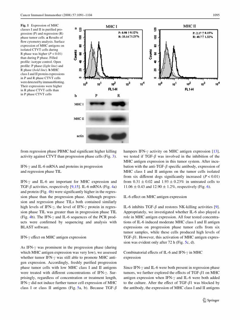
Flow cytometry determined MHC classe I and II antigenexpressions on tumor cells during growth. These analysesconWrmed the previous Wndings [6] that a much higher pro-portion of tumor cells (33.14 § 1.21% for class I and40.7 § 1.32% for class II) expressed MHC antigen in theregression phase than those during the progression phase(0.98 § 0.12% for class I and 2.17 § 0.15% for class II)(P < 0.01) (Fig. 1a). Western blotting results analyzed byactin-normalized densitometry also revealed markedlyhigher levels of MHC class I (0.82 § 0.1) and class II anti-gens (0.76 § 0.1) in regression phase tumor cells comparedto progression phase tumor cells (0.3 § 0.08 for MHC I and0.12 § 0.05 for MHC II) (Fig. 1b).
TGF-�1 expression in CTVT progression and regression
Similarly performed Western blotting established thatthe TGF-�1 protein was expressed at a high level bothin the progression (0.93 § 0.08) and regression phases(0.84 § 0.11) (Fig. 2a). ELISA veriWed that the active formof TGF-�1 was highly expressed both in cultured progres-sion phase cells (472.16 § 17.85 pg/ml) and regressionphase cells (499.96 § 12.45 pg/ml). Expressions of activeTGF-�1 were not signiWcantly diVerent (Fig. 2b). Immuno-histochemistry was positive for TGF-�1 in the cytoplasmof most progression and regression phase tumor cells(Fig. 2c–e).
Tumor-speciWc T cell cytotoxicity
To further prove that the regression phase T cells weremore potent in cytotoxicity against the tumor cells, regres-sion and progression phase PBMC were collected toperform tumor-speciWc cytotoxicity assay. The cytotoxicity
123
Cancer Immunol Immunother (2008) 57:1091–1104 1095
from regression phase PBMC had signiWcant higher killingactivity against CTVT than progression phase cells (Fig. 3).
IFN-� and IL-6 mRNA and proteins in progression and regression phase TIL
IFN-� and IL-6 are important for MHC expression andTGF-� activities, respectively [9,15]. IL-6 mRNA (Fig. 4a)and protein (Fig. 4b) were signiWcantly higher in the regres-sion phase than the progression phase. Although progres-sion and regression phase TILs both contained similarlyhigh levels of IFN-�, the level of IFN-� protein in regres-sion phase TIL was greater than in progression phase TIL(Fig. 4b). The IFN-� and IL-6 sequences of the PCR prod-ucts were conWrmed by sequencing and analysis withBLAST software.
IFN-� eVect on MHC antigen expression
As IFN-� was prominent in the progression phase (duringwhich MHC antigen expression was very low), we assessedwhether tumor IFN-� was still able to promote MHC anti-gen expression. Accordingly, freshly puriWed progressionphase tumor cells with low MHC class I and II antigenswere treated with diVerent concentrations of IFN-�. Sur-prisingly, regardless of concentration or treatment length,IFN-� did not induce further tumor cell expression of MHCclass I or class II antigens (Fig. 5a, b). Because TGF-�
hampers IFN-� activity on MHC antigen expression [13],we tested if TGF-� was involved in the inhibition of theMHC antigen expression in this tumor system. After incu-bation with the anti-TGF-� speciWc antibody, expression ofMHC class I and II antigens on the tumor cells isolatedfrom six diVerent dogs signiWcantly increased (P < 0.01)from 0.31 § 0.02 and 1.93 § 0.23% in untreated cells to11.06 § 0.43 and 12.90 § 1.2%, respectively (Fig. 6).
IL-6 eVect on MHC antigen expression
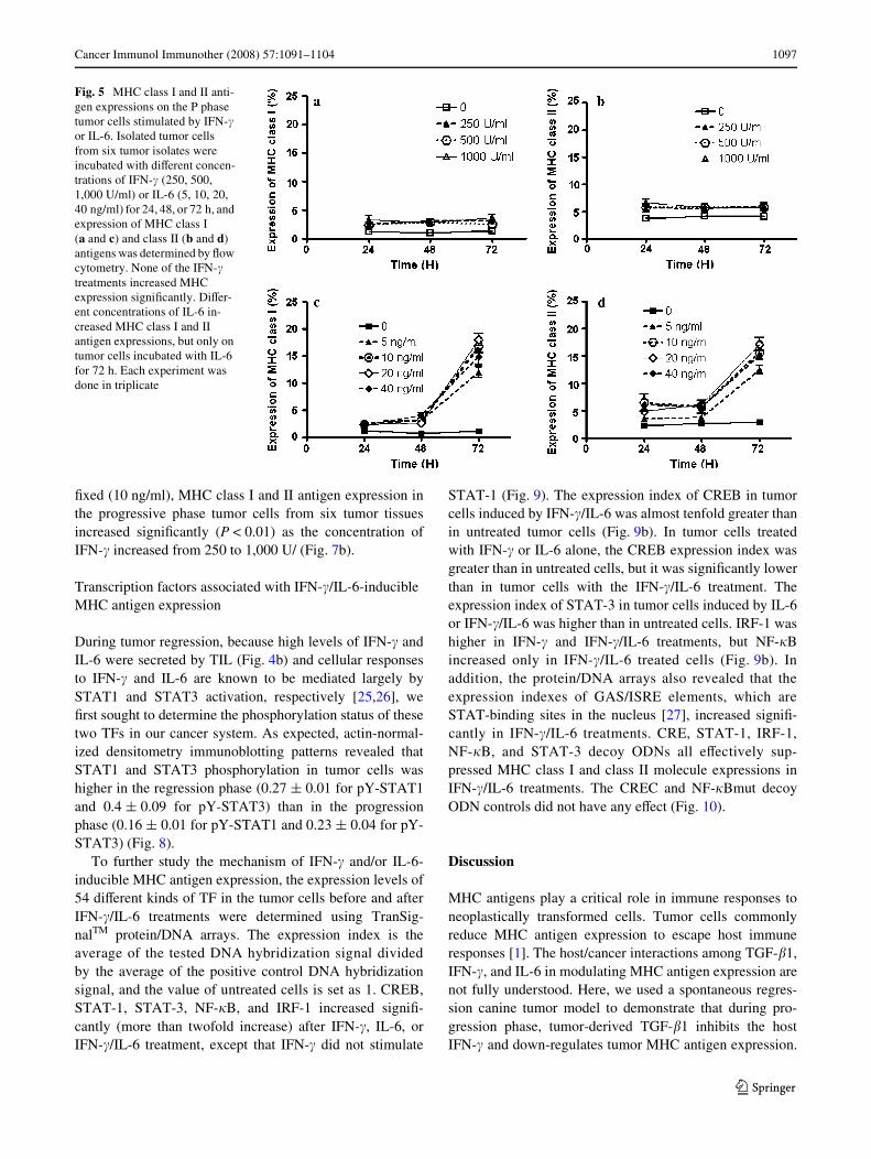
IL-6 inhibits TGF-� and restores NK-killing activities [9].Appropriately, we investigated whether IL-6 also played arole in MHC antigen expression. All four tested concentra-tions of IL-6 induced moderate MHC class I and II antigenexpressions on progression phase tumor cells from sixtumor samples, while these cells produced high levels ofTGF-�1. However, this activation of MHC antigen expres-sion was evident only after 72 h (Fig. 5c, d).
Combinatorial eVects of IL-6 and IFN-� in MHC expression
Since IFN-� and IL-6 were both present in regression phasetumors, we further explored the eVects of TGF-�1 on MHCantigen expression when IFN-� and IL-6 were both addedto the culture. After the eVect of TGF-�1 was blocked bythe antibody, the expression of MHC class I and II antigens
Fig. 1 Expression of MHC classes I and II in puriWed pro-gression (P) and regression (R) phase tumor cells. a Results of Xow cytometry analysis. Surface expression of MHC antigens on isolated CTVT cells during R phase was higher (P < 0.01) than during P phase. Filled proWle: isotype control. Open proWle: P phase (light line) and R phase (bold line). b MHC class I and II protein expressions in P and R phase CTVT cells were detected by immunoblotting. Their expressions were higher in R phase CTVT cells than in P phase CTVT cells
123

1096 Cancer Immunol Immunother (2008) 57:1091–1104
were both increased from 0.31 § 0.02 to 11.06 § 0.43%and 1.93 § 0.23 to 12.90 § 1.2%, respectively (P < 0.01).The addition of IFN-� or IL-6 further increased the expres-
sion to 16.93 § 0.31% (class I) and 20.06 § 1.02% (classII) for IFN-� (P < 0.01), and 16.6 § 1.28% (class I) to23.49 § 1.47% (class II) for IL-6 (P < 0.01). In addition,IFN-�/IL-6 was the more potent inducer for both MHCantigen (24.42 § 0.89% for class I and 40.04 § 1.11% forclass II) (P < 0.01) (Fig. 6). These synergistic eVects ofIFN-�/IL-6 were further investigated using diVerent cyto-kine dosages, alone or in combination. When progressivephase tumor cells were co-cultured with a Wxed amount ofIFN-� (1,000 U/ml) and various amounts of IL-6, IFN-�/IL-6 signiWcantly increased the expression of MHC class I andII antigens. Peak expression was achieved with 1,000 U/mlIFN-� and 10 ng/ml IL-6 (P < 0.05) (Fig. 7a). Nevertheless,IL-6 concentration > 10 ng/ml resulted in lower levels ofMHC antigen expression. When the IL-6 concentration was
Fig. 2 TGF-�1 expression in progression (P) and regression (R) phaseCTVT cells. a Western blotting revealed high levels of the 12.5 kDaTGF-�1 protein in both P and the R phase cells. b Expression of theactive form of TGF-�1 in P and R phase tumor cell supernatants wasdetected by ELISA. The amounts of active TGF-�1 in P and the R phaseCTVT supernatants were not signiWcantly diVerent. Immunohistochem-ical analyses of deparaVinized formalin-Wxed sections revealed that themajority of P phase (D) and R phase (E) tumor cells were positive forTGF-�1 (arrows). c Negative control without TGF-�1 antibody staining
Fig. 3 Tumor speciWc T cell cytotoxicity. P phase and R phase CTVTcells (8 £ 106 cells/ml) were incubated with mitomycin C and R phaseperipheral blood mononuclear cells (PBMC; 3.2 £ 106 cells/ml) for6 days. Freshly prepared target (T) cells (CTVT; 4 £ 103/well) wereincubated at 37°C overnight with the eVector (E) cells (R phasePBMC) at various E:T ratios (100:1, 50:1, 25:1, 12.5:1, 6.25:1). Thesupernatants were collected and the CTL cytotoxic activity wasmeasured by the CytoTox 96® Non-Radioactive Cytotoxicity Assay(Promega, USA)
Fig. 4 IFN-� and IL-6 mRNA, and protein in TIL during the P and theR phases. a P and the R phase TIL expressed both IFN-� and IL-6 mR-NA. The levels of IFN-� mRNA in the P and the R phase TIL were notsigniWcantly diVerent. However, the levels of IL-6 mRNA were higherin R phase than in P phase. b A similar result for the IL-6 protein wasobtained by Western blotting, but the level of IFN-� protein was higherin R phase than in P phase
123
Cancer Immunol Immunother (2008) 57:1091–1104 1097
Wxed (10 ng/ml), MHC class I and II antigen expression inthe progressive phase tumor cells from six tumor tissuesincreased signiWcantly (P < 0.01) as the concentration ofIFN-� increased from 250 to 1,000 U/ (Fig. 7b).
Transcription factors associated with IFN-�/IL-6-inducible MHC antigen expression
During tumor regression, because high levels of IFN-� andIL-6 were secreted by TIL (Fig. 4b) and cellular responsesto IFN-� and IL-6 are known to be mediated largely bySTAT1 and STAT3 activation, respectively [25,26], weWrst sought to determine the phosphorylation status of thesetwo TFs in our cancer system. As expected, actin-normal-ized densitometry immunoblotting patterns revealed thatSTAT1 and STAT3 phosphorylation in tumor cells washigher in the regression phase (0.27 § 0.01 for pY-STAT1and 0.4 § 0.09 for pY-STAT3) than in the progressionphase (0.16 § 0.01 for pY-STAT1 and 0.23 § 0.04 for pY-STAT3) (Fig. 8).
To further study the mechanism of IFN-� and/or IL-6-inducible MHC antigen expression, the expression levels of54 diVerent kinds of TF in the tumor cells before and afterIFN-�/IL-6 treatments were determined using TranSig-nalTM protein/DNA arrays. The expression index is theaverage of the tested DNA hybridization signal dividedby the average of the positive control DNA hybridizationsignal, and the value of untreated cells is set as 1. CREB,STAT-1, STAT-3, NF-�B, and IRF-1 increased signiW-cantly (more than twofold increase) after IFN-�, IL-6, orIFN-�/IL-6 treatment, except that IFN-� did not stimulate
STAT-1 (Fig. 9). The expression index of CREB in tumorcells induced by IFN-�/IL-6 was almost tenfold greater thanin untreated tumor cells (Fig. 9b). In tumor cells treatedwith IFN-� or IL-6 alone, the CREB expression index wasgreater than in untreated cells, but it was signiWcantly lowerthan in tumor cells with the IFN-�/IL-6 treatment. Theexpression index of STAT-3 in tumor cells induced by IL-6or IFN-�/IL-6 was higher than in untreated cells. IRF-1 washigher in IFN-� and IFN-�/IL-6 treatments, but NF-�Bincreased only in IFN-�/IL-6 treated cells (Fig. 9b). Inaddition, the protein/DNA arrays also revealed that theexpression indexes of GAS/ISRE elements, which areSTAT-binding sites in the nucleus [27], increased signiW-cantly in IFN-�/IL-6 treatments. CRE, STAT-1, IRF-1,NF-�B, and STAT-3 decoy ODNs all eVectively sup-pressed MHC class I and class II molecule expressions inIFN-�/IL-6 treatments. The CREC and NF-�Bmut decoyODN controls did not have any eVect (Fig. 10).
Discussion
MHC antigens play a critical role in immune responses toneoplastically transformed cells. Tumor cells commonlyreduce MHC antigen expression to escape host immuneresponses [1]. The host/cancer interactions among TGF-�1,IFN-�, and IL-6 in modulating MHC antigen expression arenot fully understood. Here, we used a spontaneous regres-sion canine tumor model to demonstrate that during pro-gression phase, tumor-derived TGF-�1 inhibits the hostIFN-� and down-regulates tumor MHC antigen expression.
Fig. 5 MHC class I and II anti-gen expressions on the P phase tumor cells stimulated by IFN-� or IL-6. Isolated tumor cells from six tumor isolates were incubated with diVerent concen-trations of IFN-� (250, 500, 1,000 U/ml) or IL-6 (5, 10, 20, 40 ng/ml) for 24, 48, or 72 h, and expression of MHC class I (a and c) and class II (b and d) antigens was determined by Xow cytometry. None of the IFN-� treatments increased MHC expression signiWcantly. DiVer-ent concentrations of IL-6 in-creased MHC class I and II antigen expressions, but only on tumor cells incubated with IL-6 for 72 h. Each experiment was done in triplicate
123

1098 Cancer Immunol Immunother (2008) 57:1091–1104
However, during the regression phase, TIL produce highlevel of IL-6 that antagonizes TGF-� activities and restoresIFN-�-mediated MHC antigen expression.
We and other investigators [5,6] have found that thisspontaneously transmissible tumor expresses extremelylow levels of MHC class I and class II antigens. At thesame time, a large amount of activated TGF-�1 is secretedduring the growth of the tumor [9]. This was presentlyreconWrmed by RT-PCR, Western blotting, and immuno-histochemical staining. Although Treg cells might be onepossible source, CD4 positive cells comprised only around10% of total the TIL; considering Foxp-3 reduces the per-centage further, such that the numbers of Treg cells is non-proportional to tumor cells. Thus, the total contribution of
TGF-� originating from cells other than the tumor cellscould be minimal.
TGF-� inhibits MHC antigen expression throughthe inhibition of type III and type IV-CIITA mRNA inmicroglial cells [28] and can down-regulate MHC I antigenexpression in ocular melanoma cells [29], the high concen-trations of TGF-� secreted by the tumor cells could be oneof the factors causing low MHC antigen expression of thistumor. Thus, when the activity of TGF-� is neutralized byanti-TGF-� polyclonal antibody, tumor cell expression ofMHC class I and II antigens increases signiWcantly. In addi-tion, IFN-� is one of the most potent cytokines modulatingthe expression of MHC antigens [15]. The high level ofIFN-� expression in TIL during the tumor growth was areason to suspect that TIL-secreted IFN-� might lose itscapability to induce MHC antigen expression during tumorprogression. This was conWrmed by the failure to promoteMHC antigen expression on the tumor cells by additionof up to 1000 U/ml of exogenous IFN-� to the culture.Unresponsiveness of MHC antigen expression to IFN-� intumors that secrete TGF-�1 is not common. However, inTGF-�¡/¡ mice, IFN-� mRNA and circulating levels ofIFN-� are increased [30] as are IRF-1 and STAT1a [31],and expression of MHC class I and class II antigens are alsoincreased in TGF-� deWcient mice [32]. Presently, theobservation that addition of IFN-� in the anti-TGF-� poly-clonal Ab-treated tumor cells further promoted the MHCantigen expression provides additional supportive evidence.Although tumor-derived TGF-� eYciently inhibited thefunction of IFN-� secreted by TIL during the tumor’s pro-gression phase, we are not able to rule out the possibilitythat other unknown factors might also be involved in thesuppression of MHC antigen expression.
The observation that exogenous IL-6 alone can inducethe expression of MHC antigens on tumor cells supports theview that in the presence of TGF-�1, TIL-derived IL-6could also promote MHC antigen expression of the tumorcells through its anti-TGF-�1 activity. Further supportcomes from our observation that STAT3, a major TF asso-ciated with IL-6 activity [26], was signiWcantly activatedonly when IL-6 was used. In addition, as described previ-ously when the tumor cells were pre-treated with anti-TGF-�antibody, exogenous IL-6 further enhanced the expressionof MHC class I and II antigens. Thus, IL-6 secreted by TILin the regression phase of the tumor might be a key elementin promoting tumor MHC antigen expression.
IL-6 is multivalent cytokine. In some cases, IL-6enhances tumor growth [33], whereas in others, it assistshost immune activity against tumor cells [33,34], and IL-6gene has also been used to treat cancers such as melanoma[34] and Lewis mouse lung cancer [35]. In addition, IL-6function may depend, in part, on the type of cells that pro-duces it. IL-6 secreted by a tumor cell may protect the
Fig. 6 EVect of IFN-�, IL-6, or IFN-�/IL-6 on MHC class I and II anti-gen expressions in tumor cells pretreated with anti-TGF-� Ab. Pro-gression phase CTVT cells were incubated with anti-TGF-� Ab for 4 hand then treated with IFN-� (1,000 U/ml) or IL-6 (10 ng/ml) or IFN-�/IL-6 for 24 h. Expression of MHC molecules was measured by Xowcytometry. Data for each experimental group is shown with a bold line;data for the isotype control is shown with a light line. Numbers (topright) represent the percentage of cells showing signiWcant inductioncompared to the control. Data are representative of least three separateexperiments
123

Cancer Immunol Immunother (2008) 57:1091–1104 1099
tumor from host immune attack [36]. In other cases, asshown previously [9] and presently, IL-6 produced by TILstimulates host immune responses. Thus, IL-6 activitiesrely on the situations that it encounters.
Furthermore, co-application of IFN-� and IL-6 promotedthe highest MHC antigen expression among all of the testedgroups. Therefore, TIL-derived IL-6 may play a dual role inregression phase tumor cells. First, IL-6 overcomes TGF-�-
Fig. 7 Expression of MHC class I and II antigens on tumor cells stimulated by diVerent con-centrations of IFN-�/IL-6 when anti-TGF-� Ab is not present. EVect of 72 h exposures a IFN-� (1,000 U/ml) and IL-6 (2.5, 5, 10, 20, 40, or 60 ng/ml), and b IFN-� (250, 500, or 1,000 U/ml) and IL-6 (10 ng/ml) on the expression of MHC class I and II antigens in six tumor isolates were determined by Xow cytometry. The upper portions of a, b displayed the expression curves of MHC I and II antigens stimulated by diVerent concentrations of IL-6 or IFN-�, respectively. Peak expression was achieved with 1,000 U/ml IFN-� and 10 ng/ml IL-6 (P < 0.05). Nevertheless, IL-6 concentration > 10 ng/ml resulted in lower levels of MHC antigen expression. The lower portions of a and b displayed the histograms corresponding to a and b
123

1100 Cancer Immunol Immunother (2008) 57:1091–1104
mediated suppression, which likely constitutes the mostimportant activity in promoting or initiating the MHCantigen expression, abrogating IFN-� inhibition. Second,because when the tumor cells were pre-treated with anti-TGF-� antibody, exogenous IFN-� were then able toenhance the expression of MHC class I and II antigens andIL-6 might act in combination with the functionallyrestored IFN-� in increasing MHC antigen expression. Onthe other hand, TGF-�1 is the major obstacle for IFN-� toovercome to promote MHC antigen expression. Thus, IL-6,IFN-�, and TGF-�1 work alone and/or together to modulateMHC antigen expression on the tumor cells. These threecytokines should be important in controlling the tumorMHC antigen expression. As far as we know, this is the Wrstexperimental in vivo system that has demonstrated thecombinatorial eVect of host IFN-�/IL-6 in the promotion ofMHC antigen expression in TGF-�-producing tumor duringtumor regression. This appears to be an important mecha-nism used by the host to inhibit tumor-progressive growthand eventually to cause its regression. Knowing that as longas the TGF-�1 activities are present IFN-� will not workeYciently, the blocking of TGF-�1 eVect may allow for thetreatment of cancer cells that constitutionally produce thiscytokine and suppress immune responses.
Regulatory T cell is an important cell type in regulatingimmune homeostasis [37]. Treg cells increase in numbersin the tumor microenvironment and induce tumor-speciWcimmune tolerance [38]. TGF-�1 actively promotes theexpansion and diVerentiation of Treg cells [39]. TGF-�1converts CD4+CD8¡ cells into CD4+CD8+ Treg cells byinducing transcription factor Foxp3 [40]. Treg cells were
not presently examined. While IL-6 is a potent anti-TGF-�1cytokine, the high concentration of IL-6 secreted by TILduring regression phase might block the expansion anddiVerentiation of Treg cells, which could possibly explainthe tumor regression.
During regression phase, up to 60% of the tumor cellsstill lack MHC antigen expression. However, in this phase,cytotoxic T cells increased in numbers with enhancedtumor-speciWc T cell cytotoxicity, and the inhibited NKcytotoxicity by TGF-�1 was also restored [9]. Therefore,
Fig. 8 Phosphorylation of STAT1 and STAT3 in tumor cells. Westernblotting results showed that STAT1 and STAT3 were activated byphosphorylation at Try 701 and Try 705, respectively. Their phosphor-ylated levels were higher in R phase than in P phase. pY-STAT1 andpY-STAT3 were antibodies against the STAT1 and STAT3 whenphosphorylated accordingly at tyrosine 701 and tyrosine 705
Fig. 9 Transcription factor expression levels in tumor cells exposed toIFN-�, IL-6, or IFN-�/IL-6. a Transcription factor expression levels innuclear extracts from P phase CTVT cells, which had been stimulatedfor 72 h by IFN-� (1,000 U/ml), IL-6 (10 ng/ml), or IFN-� (1,000U/ml)and IL-6 (10 ng/ml), were determined using TranSignalTM protein/DNA. On each array, genes were spotted in duplicate. Expressionsof CREB, STAT-1, STAT-3, IRF-1, NF-�B, and GAS/ISRE wereincreased signiWcantly in nuclei by IFN-�/IL-6 treatment. b All spotswere quantiWed by scanning densitometry. The expression index wasthe average of the tested DNA hybridization signal divided by the aver-age of the positive control DNA hybridization signal and the value ofuntreated cells was set as 1. Data are representative of two experi-ments. Any spots with a twofold increase were considered signiWcant
123

Cancer Immunol Immunother (2008) 57:1091–1104 1101
the remaining 60% of regression phase CTVT cells that stilllacked suYcient MHC antigen expression were presumablytargeted by the reactivated NK cells during the regressionphase.
The proper regulation of the cytokine-activated JAK/STAT pathway is critical because abnormal JAK/STATsignaling is closely associated with some cancers [41] andimmune disorders [42]. The cytokine-activated JAK tyro-sine kinases recruit STATs that are latent TFs in the cyto-plasm, facilitating phosphorylation of the STATs. Tyrosinephosphorylation of STATs is required for their dimeriza-tion, nuclear translocation, and DNA binding [25]. The sig-niWcant increase of phosphorylation of STAT1 at Try701and STAT3 at Try705 in the regression phase tumor cellssuggests the involvement of STAT1- and STAT3-associ-ated activities. Cellular responses to IFN-� are largely med-iated by STAT1 activation and are rarely associated withSTAT3 [25,43]. In contrast, IL-6 strongly activates STAT3and only weakly activates STAT1 [26]. Similar relation-ships between these cytokines and STATs were found exvivo in this tumor and the combinatorial eVect of IFN-�/IL-6that activated both STAT1 and STAT3 in the tumor cellsalso highlights these activities. The coincident low expres-sion of STAT1 in the presence of IFN-� and inability ofIFN-� to stimulate MHC expression suggest that TGF-�1inhibits IFN-�-activated MHC antigen expression duringthe progression phase of a tumor at the transcription levelvia STAT1. Also noteworthy is our Wnding of the decreasedprotein level during this phase compared to regressionphase TIL, but that the mRNA level of IFN-� was similarlyhigh during both phases. Thus, in addition to the inhibitoryeVect to the IFN-� activities through the STAT1 activation,this tumor might also inhibit IFN-� protein expression.STATs cooperate with the histone acetyltransferase CREB-binding protein (CBP)/p300 for gene activation [44] andactivate numerous genes including the MHC class II andthe CIITA by directly binding to the promoters [45, 46].We found that both IL-6 alone and IFN-�/IL-6 treatmentgroups, strongly activated CREB in the tumor cells, sug-gesting that CREB activity is also associated with the MHC
Fig. 10 EVect of decoy ODN on cytokine-mediated MHC class I andII antigen expressions. Double-stranded, decoy ODN were preparedfrom complementary single-stranded, phosphorothioate-bonded bases.Decoy ODN were added 1 day after seeding) to the wells for 24 h. IFN-� (1,000U/ml) and IL-6 (10 ng/ml) were added to the CTVT cells pre-treated with decoy ODN. After incubation at 37°C for 72 h, MHC classI and II antigen expressions were d ODN (CRE, STAT-1, STAT-3,IRF-1, NF-�B) signiWcantly decreased the expression of MHC class Iand II molecules on the surface of tumor cells exposed to IFN�/IL-6 for72 h. Filled proWle: isotype control. Open proWle: with ODN (bold line)and without ODN (light line). Data are representative of three experi-ments
�
123

1102 Cancer Immunol Immunother (2008) 57:1091–1104
antigen expression in this tumor under the inXuence of thecytokines. This might explain the present results in cellsexposed solely to IFN-�, where there was no promotion inMHC antigen expression and much less of an eVect onCREB.
The biological functions of STAT3 are diversiWed.STAT3 is constitutively expressed in a number of primaryhuman tumors [47] and is critical in maintaining cancer cellproliferation [48] and immunosuppression [49]. STAT3 isinducible by many cytokines and growth factors includingTGF-alpha, EGF, hepatocyte growth factor, vascular endo-thelial growth factor, and IL-6, and oncogenic kinases aswell [47,50]. Therefore, the activities also depend on sub-stance(s) that initiate STAT3 activation or induction. IL-6production increases signiWcantly by TILs isolated fromregression rather than progression phase, and functions asan anti-TGF beta agent [9]. Signaling of IL-6 follows theSTAT3 pathway [51]. Thus, it is reasonable to suggest thatthe increased pSTAT3 presently demonstrated was a resultof increased concentration of IL-6, which preludes anti-TGF beta activity. Our evidence also indicates that IL-6acts indirectly in promoting MHC expression and the decoyODN of STAT3 inhibits the expression of MHC expres-sion. Therefore, IL-6 is activated during regression phase,by a yet unknown mechanism, to transduce a STAT3-medi-ated signal, antagonize the TGF-beta activities, and pro-mote MHC expression. In addition, IL-6 displayed aconcentration threshold phenomenon. When IL-6 concen-tration exceeded 10 ng/ml, MHC expression was sup-pressed. Thus, it is also possible that in CTVT, IL-6concentration and pSTAT3 are at relatively low levels thatfavor the activation of MHC expression of CTVT, while, inother cancers, IL-6 and pSTAT3 are relatively more abun-dant and mediate cancer cell proliferation.
IRF-1 is expressed at low levels in unstimulated cellsand can be activated by many cytokines including type Iand II IFN, TNF-�, IL-1, and IL-6 [52,53]. IRF-1 is medi-ated through STAT1 and NF-�B, whose binding sites are inthe IRF-1 promoter [54,55]. The interaction of NF-�B withIRF-1 is critical for promoter activation [55]. Presently,both STAT1 and NF-�B were activated in the IFN-�/IL-6treatment. Gamma-activated sites (GAS) are associatedwith the induction of transcription by IFN-� [56] and as abinding site for STATs [57]. IFN-stimulated response ele-ment (ISRE) is also important in MHC I gene promoters.Upon treatment of IFN-�, IRF-1 serves as an adapter pro-tein for the binding of STATs to the ISRE or GAS, whichin turn induces MHC I transactivation [56,58]. Presently,IFN-�/IL-6 treatment activated GAS/ISRE most eVectively.Thus, IFN-�/IL-6 treatment activates STAT1, which bindsto GAS/ISRE to activate the promoter for IRF-1, ultimatelypromoting MHC I antigen expression. In the present IRF-1decoy ODN study, MHC expression was almost totally
suppressed. Thus, IRF-1 should play a more important rolein IFN-�- and IL-6-stimulated MHC antigen expression.However, exogenous IFN-� alone signiWcantly activatedthe interferon-inducible genes GAS/ISRE, but did not acti-vate STATs. IRF dimers that do not form a complex withSTATs can also regulate interferon-stimulating genes bydirectly binding the ISRE and bypassing STATs [56]. Theactivation of the GAS/ISRE observed in the presence ofonly IFN-� likely occurred via this STATs-independentpathway. In addition, IFN-� alone can activate IRF-1 evenin the presence of TGF-� eVect, but does not promote MHCantigen expression on the progression phase tumor cells,even when a high concentration of exogenous IFN-� isadded to the culture. This dichotomy can be at least par-tially explained by the Wndings that TGF-� inhibits IFN-�-induced MHC antigen expression, but is incapable of actingglobally to inhibit IFN-�-induced gene expression includ-ing IRF-1[13]. Taken together, the presence of high level ofIRF-1 does not necessarily relate to STATs or promote theMHC expression.
In conclusion, CTVT, a TGF-�-producing transmissibletumor, can markedly suppress the expression levels ofMHC. In the presence of TGF-�, IFN-� is not active. How-ever, the host TIL produces high concentrations of IL-6,which show powerful anti-TGF-� activity and activateMHC antigen expression. Furthermore, IFN-� and IL-6 actsynergistically in combination to enhance the expression ofMHC antigens through mechanisms associated with TFssuch as STAT-1, STAT-3, CREB, NF-�B, and IRF-1. Rela-tionships between the TFs are closely interactive and com-plicated. It is suggested that the regimen for treating cancercells that constitutively secrete TGF-� should considerincorporating a strategy of anti-TGF-� activity. Finally, thesigniWcance of this research is that the results and the mech-anisms depicted were obtained from an in vivo system ofthe tumor presented with spontaneous regression. This Wnd-ing should prove useful for designing eVective immuno-therapies for cancer patients.
Acknowledgments This project was supported by National ScienceCouncil of Taiwan (NSC95-2313-B-002-375) and Council of Agricul-ture of Taiwan (96AS-1.2.1-AD.U1(20)).
References
1. Garrido F, Ruiz-Cabello F, Cabrera T, Perez-Villar JJ, Lopez-Botet M, Duggan-Keen M, Stern PL (1997) Implications forimmunosurveillance of altered HLA class I phenotypes in humantumours. Immunol Today 18:89–95
2. HoVmann TK, Meidenbauer N, Muller-Berghaus J, Storkus WJ,Whiteside TL (2001) ProinXammatory cytokines and CD40 ligandenhance cross-presentation and cross-priming capability of humandendritic cells internalizing apoptotic cancer cells. J Immunother24:162–171
123

Cancer Immunol Immunother (2008) 57:1091–1104 1103
3. Overwijk WW (2005) Breaking tolerance in cancer immunother-apy: time to ACT. Curr Opin Immunol 17:187–194
4. Yu Z, Restifo NP (2002) Cancer vaccines: progress reveals newcomplexities. J Clin Invest 110:289–294
5. Murgia C, Pritchard JK, Kim SY, Fassati A, Weiss RA (2006)Clonal origin and evolution of a transmissible cancer. Cell126:477–487
6. Hsiao YW, Liao KW, Hung SW, Chu RM (2002) EVect of tumorinWltrating lymphocytes on the expression of MHC molecules incanine transmissible venereal tumor cells. Vet Immunol Immuno-pathol 87:19–27
7. Katzir N, Arman E, Cohen D, Givol D, Rechavi G (1987) Com-mon origin of transmissible venereal tumors (TVT) in dogs. Onco-gene 1:445–448
8. Liao KW, Lin ZY, Pao HN, Kam SY, Wang FI, Chu RM (2003)IdentiWcation of canine transmissible venereal tumor cells using insitu polymerase chain reaction and the stable sequence of the longinterspersed nuclear element. J Vet Diagn Invest 15:399–406
9. Hsiao YW, Liao KW, Hung SW, Chu RM (2004) Tumor-inWltrat-ing lymphocyte secretion of IL-6 antagonizes tumor-derivedTGF-beta 1 and restores the lymphokine-activated killing activity.J Immunol 172:1508–1514
10. Li MO, Wan YY, Sanjabi S, Robertson AK, Flavell RA (2006)Transforming growth factor-beta regulation of immune responses.Annu Rev Immunol 24:99–146
11. Cross D, Cambier JC (1990) Transforming growth factor beta 1has diVerential eVects on B cell proliferation and activation anti-gen expression. J Immunol 144:432–439
12. Thomas DA, Massague J (2005) TGF-beta directly targets cyto-toxic T cell functions during tumor evasion of immune surveil-lance. Cancer Cell 8:369–380
13. Panek RB, Lee YJ, Benveniste EN (1995) TGF-beta suppressionof IFN-gamma-induced class II MHC gene expression does notinvolve inhibition of phosphorylation of JAK1, JAK2, or signaltransducers and activators of transcription, or modiWcation of IFN-gamma enhanced factor X expression. J Immunol 154:610–619
14. Ljunggren G, Anderson DJ (1998) Cytokine induced modulationof MHC class I and class II molecules on human cervical epithelialcells. J Reprod Immunol 38:123–138
15. Ting JP, Baldwin AS (1993) Regulation of MHC gene expression.Curr Opin Immunol 5:8–16
16. Wei L-H, Kuo M-L, Chen C-A, Chou C-H, Lai K-B, Lee C-N,Hsieh C-Y (2003) Interleukin-6 promotes cervical tumor growthby VEGF-dependent angiogenesis via a STAT3 pathway. Onco-gene 22:1517
17. Ohta K, Yamagami S, Taylor AW, Streilein JW (2000) IL-6 antag-onizes TGF-beta and abolishes immune privilege in eyes with endo-toxin-induced uveitis. Invest Ophthalmol Vis Sci 41:2591–2599
18. Rabinowich H, Sedlmayr P, Herberman RB, Whiteside TL (1993)Response of human NK cells to IL-6 alterations of the cell surfacephenotype, adhesion to Wbronectin and laminin, and tumor necro-sis factor-alpha/beta secretion. J Immunol 150:4844–4855
19. Scheid C, Young R, McDermott R, Fitzsimmons L, ScarVe JH,Stern PL (1994) Immune function of patients receiving recombi-nant human interleukin-6 (IL-6) in a phase I clinical study: induc-tion of C-reactive protein and IgE and inhibition of natural killerand lymphokine-activated killer cell activity. Cancer ImmunolImmunother 38:119–126
20. Cao X, Wang Q, Ju DW, Tao Q, Wang J (1999) EYcient induca-tion of local and systemic antitumor immune response by lipo-some-mediated intratumoral co-transfer of interleukin-2 gene andinterleukin-6 gene. J Exp Clin Cancer Res 18:191–200
21. Liang CT, Chueh LL, Pang VF, Zhuo YX, Liang SC, Yu CK,Chiang H, Lee CC, Liu CH (2007) A non-biotin polymerizedhorseradish-peroxidase method for the immunohistochemicaldiagnosis of canine distemper. J Comp Pathol 136:57–64
22. Wagner AH, Gebauer M, Pollok-Kopp B, Hecker M (2002) Cyto-kine-inducible CD40 expression in human endothelial cells ismediated by interferon regulatory factor-1. Blood 99:520–525
23. Park YG, Nesterova M, Agrawal S, Cho-Chung YS (1999) Dualblockade of cyclic AMP response element- (CRE) and AP-1-di-rected transcription by CRE-transcription factor decoy oligonu-cleotide. gene-speciWc inhibition of tumor growth. J Biol Chem274:1573–1580
24. Krzesz R, Wagner AH, Cattaruzza M, Hecker M (1999) Cytokine-inducible CD40 gene expression in vascular smooth muscle cellsis mediated by nuclear factor kappaB and signal transducer andactivation of transcription-1. FEBS Lett 453:191–196
25. Darnell JE, Kerr I, Stark G (1994) Jak-STAT pathways and tran-scriptional activation in response to interferons and other extracel-lular signaling proteins. Science 264:1415–1420
26. Heinrich PC, Behrmann I, Muller-Newen G, Schaper F, Graeve L(1998) Interleukin-6-type cytokine signalling through the gp130/Jak/STAT pathway. Biochem J 334(Pt 2):297–314
27. Ehret GB, Reichenbach P, Schindler U, Horvath CM, Fritz S,Nabholz M, Bucher P (2001) DNA binding speciWcity of diVerentSTAT proteins. Comparison of in vitro speciWcity with naturaltarget sites. J Biol Chem 276:6675–6688
28. Pazmany T, Tomasi TB (2006) The major histocompatibility com-plex class II transactivator is diVerentially regulated by interferon-gamma and transforming growth factor-beta in microglial cells.J Neuroimmunol 172:18–26
29. Ma D, Niederkorn JY (1995) Transforming growth factor-betadown-regulates major histocompatibility complex class I antigenexpression and increases the susceptibility of uveal melanoma cellsto natural killer cell-mediated cytolysis. Immunology 86:263–269
30. Shull MM, Ormsby I, Kier AB, Pawlowski S, Diebold RJ, Yin M,Allen R, Sidman C, Proetzel G, Calvin D et al (1992) Targeteddisruption of the mouse transforming growth factor-beta 1 generesults in multifocal inXammatory disease. Nature 359:693–699
31. McCartney-Francis NL, Wahl SM (2002) Dysregulation of IFN-gamma signaling pathways in the absence of TGF-beta 1. J Immu-nol 169:5941–5947
32. Geiser AG, Letterio JJ, Kulkarni AB, Karlsson S, Roberts AB,Sporn MB (1993) Transforming growth factor beta 1 (TGF-beta 1)controls expression of major histocompatibility genes in the post-natal mouse: aberrant histocompatibility antigen expression in thepathogenesis of the TGF-beta 1 null mouse phenotype. Proc NatlAcad Sci USA 90:9944–9948
33. Shouda T, Hiraoka K, Komiya S, Hamada T, Zenmyo M, IwasakiH, Isayama T, Fukushima N, Nagata K, Yoshimura A (2006) Sup-pression of IL-6 production and proliferation by blocking STAT3activation in malignant soft tissue tumor cells. Cancer Lett231:176–184
34. Mule JJ, Custer MC, Travis WD, Rosenberg SA (1992) Cellularmechanisms of the antitumor activity of recombinant IL-6 in mice.J Immunol 148:2622–2629
35. Usuda J, Okunaka T, Furukawa K, Tsuchida T, Kuroiwa Y, OheY, Saijo N, Nishio K, Konaka C, Kato H (2001) Increased cyto-toxic eVects of photodynamic therapy in IL-6 gene transfectedcells via enhanced apoptosis. Int J Cancer 93:475–480
36. Code S, Simard C, Lemieux R (2002) Regulation of growth-relat-ed genes by interleukin-6 in muring myeloma cells. Cytokine20:113–120
37. Sakaguichi S (2005) Naturally arising Foxp3-expressingCD25+CD4+ regulatory T cells in immunological tolerance to selfand non-self. Nat Immunol 6:345–352
38. Wang HY, Wang RF (2007) Regulatory T cells and cancer. CurrOpin Immunol 19:217–223
39. Zhang L, Yi H, Xia XP, Zhao Y (2006) Transforming growth fac-tor-beta: an important role in CD4+CD25+ regulatory T cells andimmune tolerance. Autoimmunity 39:269–276
123

1104 Cancer Immunol Immunother (2008) 57:1091–1104
40. Chen W, Jin W, Hardegen N, Lei KJ, Li L, Marinos N, McGradyG, Wahl SM (2003) Conversion of peripheral CD4+CD25- naiveT cells to CD4+CD25+ regulatory T cells by TGF-beta inductionof transcription factor Foxp3. J Exp Med 198:1875–1886
41. Baxter EJ, Scott LM, Campbell PJ, East C, Fourouclas N, Swan-ton S, Vassiliou GS, Bench AJ, Boyd EM, Curtin N, Scott MA,Erber WN, Green AR (2005) Acquired mutation of the tyrosinekinase JAK2 in human myeloproliferative disorders. Lancet365:1054–1061
42. Shuai K, Liu B (2003) Regulation of JAK-STAT signalling in theimmune system. Nat Rev 3:900–911
43. BalhoV JP, Stephens JM (1998) Highly speciWc and quantitativeactivation of STATs in 3T3-L1 adipocytes. Biochem Biophys ResCommun 247:894–900
44. Christian M, Marangos P, Mak I, McVey J, Barker F, White J,Brosens JJ (2001) Interferon-gamma modulates prolactin and tis-sue factor expression in diVerentiating human endometrial stromalcells. Endocrinology 142:3142–3151
45. Moreno CS, Beresford GW, Louis-Plence P, Morris AC, Boss JM(1999) CREB regulates MHC class II expression in a CIITA-dependent manner. Immunity 10:143–151
46. van der Stoep N, Quinten E, van den Elsen PJ (2002) Transcrip-tional regulation of the MHC class II trans-activator (CIITA) pro-moter III: identiWcation of a novel regulatory region in the 5’-untranslated region and an important role for cAMP-responsiveelement binding protein 1 and activating transcription factor-1 inCIITA-promoter III transcriptional activation in B lymphocytes.J Immunol 169:5061–5071
47. Huang S (2007) Regulation of metastases by signal transducer andactivator of transcription 3 signaling pathway: clinical implica-tions. Clin Cancer Res 13:1362–1366
48. Bowman T, Garcia R, Turkson J, Jove R (2000) STATs in onco-genesis. Oncogene 19:2474–2488
49. Kortylewski M, Kujawski M, Wang TH, Wei S, Zhang SM, Guil-ian STP, Kay NHK, William JM, Richard GK, Pardoll JW, Yu H(2005) Inhibiting Stat3 signaling in the hematopoietic system elic-its multicomponent antitumor immunity. Nat Med 11:1314–1321
50. Aggarwal B, Sethi G, Ahn KS, Sandur SK, Pandey MK, Kunnu-makkara AB, Sung B, Ichikawa H (2006) Targeting signal-trans-ducer-and-activator-of-transcription-3 for prevention and therapyof cancer: modern target but ancient solution. Ann N Y Acad Sci1091:151–169
51. Wagenka UM, Buschmann L, Lutticken C, Heinrich PC, Horn F(1993) Acute-phase response factor, a muclear factor binding toacute-phase response elements, is rapidly activated by interleukin-6 at the posttranslational level. Mol Cell Biol 13:2762–2788
52. Revel M, Katz A, Eisenbach L, Feldman M, Haran-Ghera N,Harroch S, Chebath J (1995) Interleukin-6: eVects on tumormodels in mice and on the cellular regulation of transcriptionfactor IRF-1. Ann N Y Acad Sci 762:342–355; Discussion355–346
53. Agresti C, Bernardo A, Del Russo N, Marziali G, Battistini A, Alo-isi F, Levi G, Coccia EM (1998) Synergistic stimulation of MHCclass I and IRF-1 gene expression by IFN-gamma and TNF-alphain oligodendrocytes. Eur J Neurosci 10:2975–2983
54. Meraz MA, White JM, Sheehan KC, Bach EA, Rodig SJ, DigheAS, Kaplan DH, Riley JK, Greenlund AC, Campbell D, Carver-Moore K, DuBois RN, Clark R, Aguet M, Schreiber RD (1996)Targeted disruption of the Stat1 gene in mice reveals unexpectedphysiologic speciWcity in the JAK-STAT signaling pathway. Cell84:431–442
55. Chelbi-alix MK, Bobe P, Benoit G, Canova A, Pine R (2003) Ar-senic enhances the activation of Stat1 by interferon gamma leadingto synergistic expression of IRF-1. Oncogene 22:9121–9130
56. Wesoly J, Szweykowska-Kulinska Z, Bluyssen HA (2007) STATactivation and diVerential complex formation dictate selectivity ofinterferon responses. Acta Biochim Pol 54:27–38
57. Kim OS, Park EJ, Joe EH, Jou I (2002) JAK-STAT signalingmediates gangliosides-induced inXammatory responses in brainmicroglial cells. J Biol Chem 277:40594–40601
58. Gobin SJ, van Zutphen M, Woltman AM, van den Elsen PJ (1999)Transactivation of classical and nonclassical HLA class I genesthrough the IFN-stimulated response element. J Immunol163:1428–1434
123


![Thérapeutiques ciblées dans o v µ ]vW o[ vîìíò · 2017-10-11 · IFN Rini ASCO 2009 17.4 BEV/IFN 18.3 Gore ASCO 2008 18.5 18.7 IFN 19.8 NR 22.9 Bevacizumab + IFN 20.5 Sternberg](https://img.pdfslide.net/doc/110x75/5f0257867e708231d403cbc1/thrapeutiques-cibles-dans-o-v-vw-o-v-2017-10-11-ifn-rini-asco.jpg)