Embed Size (px)
Citation preview
JOURNAL OF VIROLOGY, Feb. 2007, p. 1479–1491 Vol. 81, No. 30022-538X/07/$08.00�0 doi:10.1128/JVI.01861-06Copyright © 2007, American Society for Microbiology. All Rights Reserved.
Variable Deficiencies in the Interferon Response Enhance Susceptibilityto Vesicular Stomatitis Virus Oncolytic Actions in Glioblastoma
Cells but Not in Normal Human Glial Cells�
Guido Wollmann,1 Michael D. Robek,2 and Anthony N. van den Pol1*Departments of Neurosurgery1 and Pathology,2 Yale University School of Medicine, New Haven, Connecticut 06520
Received 25 August 2006/Accepted 5 November 2006
With little improvement in the poor prognosis for humans with high-grade glioma brain tumors, alternativetherapeutic strategies are needed. As such, selective replication-competent oncolytic viruses may be useful as apotential treatment modality. Here we test the hypothesis that defects in the interferon (IFN) pathway could beexploited to enhance the selective oncolytic profile of vesicular stomatitis virus (VSV) in glioblastoma cells. Twogreen fluorescent protein-expressing VSV strains, recombinant VSV and the glioma-adapted recombinant VSV-rp30a, were used to study infection of a variety of human glioblastoma cell lines compared to a panel of control cells,including normal human astrocytes, oligodendrocyte precursor cells, and primary explant cultures from humanbrain tissue. Infection rate, cell viability, viral replication, and IFN-�/�-related gene expression were compared inthe absence and presence of IFN-� or polyriboinosinic polyribocytidylic acid [poly(I:C)], a synthetic inducer of theIFN-�/� pathway. Both VSV strains caused rapid and total infection and death of all tumor cell lines tested. To alesser degree, normal cells were also subject to VSV infection. In contrast, IFN-� or poly(I:C) completely attenuatedthe infection of all primary control brain cells, whereas most glioblastoma cell lines treated with IFN-� or poly(I:C)showed little or no sign of protection and were killed by VSV. Together, our results demonstrate that activation ofthe interferon pathway protects normal human brain cells from VSV infection while maintaining the vulnerabilityof human glioblastoma cells to viral destruction.
The majority of primary brain tumors display a malignantclinical course, with glioblastoma multiforme being the mostfrequent and most aggressive form. The prognosis is dismal,with a mean survival of 1 year despite advances in multidisci-plinary treatment strategies, including surgery, irradiation, andchemotherapy. New investigative treatment modalities such asreplication-competent oncolytic viruses may be particularly ef-fective due to their ability to selectively proliferate within thetumor, leading to a self-amplification of the antitumor effectand intratumoral spread.
In a previous study, we found vesicular stomatitis virus(VSV) to have the strongest selective oncolytic action on hu-man glioblastoma cells out of a panel of nine different virusesthat were compared (42). VSV is an enveloped, negative-strand RNA virus belonging to the Rhabdoviridae family. Its11.2-kb genome consists of five protein-encoding genes (N, P,M, G, and L). VSV causes mild disease in livestock. Infectionin humans is rare and is usually asymptomatic, with sporadiccases of mild flu-like symptoms (28). We adapted VSV throughmultiple passages on glioblastoma cells to generate a modifiedVSV phenotype, VSV-rp30a (42).
Glioblastoma brain tumors are susceptible to VSV onco-lysis (2, 7, 11), though the mechanisms or distinct charac-teristics that contribute to this effect remain largely elusive.Glioblastomas are extremely heterogeneous in their geno-typic and phenotypic makeups. Common aberrations in-
clude epidermal growth factor receptor amplification, p53and PTEN mutations, p16 deletions, and chromosomal ab-errations (22). Links of viral oncolytic actions to certaintumor-associated mutations have been established for a va-riety of potential antitumor agents. Here we sought to ad-dress what facilitates selective VSV targeting and oncolysisof glioblastoma cells. Several hypotheses for selective VSVtargeting have been suggested. A defective interferon (IFN)system has been postulated (21, 36) for VSV oncolytic ac-tion for a variety of peripheral tumors, including colon (36),breast (12), prostate (1), and liver (34), or mesenchymaltumors such as leukemia (18). The innate cellular immuneresponse to VSV infection has not been tested for braintumors. Another hypothesis argues that there is an affinity ofVSV for hypoxic tumor environments (7). A third hypoth-esis argues that changes in tumor cell translational regula-tion (3) may be a critical mechanism of VSV oncolysis.
Compared to many other organs, the interferon system ap-pears to be quite distinct in the brain (4). The blood-brainbarrier limits the access of circulating IFNs and many IFN-responsive cells to the brain (8), making an effective local IFNsystem even more important. Systemic IFN may penetrate intothe brain at restricted leaky areas, could find access throughspecific carriers, or could bind to brain endothelial cells andinitiate the generation of central modulators (41). IFN-� and-�, also known as type I IFN, are expressed in many cells as anearly response to viral infection. In the brain, expression ofIFN-�/� could be shown in astrocytes and microglial cells (38,43). Effects of IFN-�/� on microglial cells, astrocytes, andneurons have been described (6, 19, 30, 39). In contrast, littleis known about the antiviral repertoire of glioblastoma cells.
The fatal nature of glioblastoma and the lack of successful
* Corresponding author. Mailing address: Department of Neurosur-gery, Yale University School of Medicine, 333 Cedar St., New Haven,CT 06520. Phone: (203) 785-5823. Fax: (203) 737-2159. E-mail:[email protected].
� Published ahead of print on 15 November 2006.
1479
on August 15, 2017 by guest
http://jvi.asm.org/
Dow
nloaded from
treatments suggest alternative approaches are needed. To spe-cifically target tumors in general and glioblastomas in partic-ular, it is important to characterize tumor-specific defects thatcould be exploited with appropriate means. Here we showinterferon deficiencies at different levels that appear to pro-mote effective VSV oncolysis of human glioblastoma cells.Furthermore, we show that boosting the innate antiviral de-fense with IFN-� and the IFN-�/� pathway inducer polyribo-inosinic polyribocytidylic acid [poly(I:C)] provides protectionfor human astroglial cells, the normal cell type most closelyrelated to glioblastoma cells, but does not block the ability ofeither VSV or VSV-rp30a to kill glioblastoma cells.
MATERIALS AND METHODS
Glioma cell lines. The human glioblastoma cell lines U-87MG and MO59Jwere obtained from the ATCC (Manassas, VA), and cell lines U-373, U-118, andA-172 were kindly provided by R. Matthews and propagated in minimal essentialmedium supplemented with 10% fetal bovine serum. Medium for U-87 andU-373 cells additionally contained 1% sodium pyruvate and 1% nonessentialamino acids.
Normal human control cells. Normal human astrocytes and oligodendrocyteprecursor cells were purchased from Sciencell (San Diego, CA) and kept inculture using astrocyte growth medium or oligodendrocyte medium. In addition,primary astrocyte cultures were established from human brain tissue obtainedfrom epilepsy surgery from three patients different in age and gender and des-ignated A, B, and C. Patient welfare was the sole basis for resection of braintissue. Tissue blocks were cut into small cubes and plated onto Millipore filterinsets or poly-L-lysine coated coverslips for explant cultures. Cells were harvestedand propagated in minimal essential medium with 10% fetal bovine serum.
Red fluorescent protein (RFP)-expressing U-87 cells. To generate humanglioblastoma cells that expressed a colored reporter gene, U-87 cells were platedin 25-cm2 tissue culture flasks. Confluent cultures were transfected with plasmidpDsRed-Monomer-C1 (Clontech), containing an expression cassette with dsReddriven by a cytomegalovirus promoter and a neomycin resistance gene, by usingLipofectamin2000 (Invitrogen). G418 (Sigma) was used to select and maintainstably transfected U-87 cells.
All cells were propagated in a humidified atmosphere containing 5% CO2 at37°C.
Viruses. Green fluorescent protein (GFP)-expressing VSV (9) and VSV-rp30a, derived from VSV-rp30 (42), were generated as previously described.Titers for VSV and VSV-rp30a were determined through plaque assay on BHKcells.
Quantitative real-time PCR. Glioblastoma cells or control cells were grown in25-cm2 flasks in triplicates for each treatment condition. Ninety percent conflu-ent cultures were treated with either IFN-� (100 u/ml), poly(I:C) (25 �g/ml), orVSV-rp30a (multiplicity of infection [MOI] of 2), or mock treated, for 6 h. Cellswere lysed, and RNA was extracted using TRIzol reagent (Invitrogen) accordingto the manufacturer’s instructions. The TaqMan reverse transcription kit (Ap-plied Biosystems, Foster City, CA) was used to reverse transcribe 1 �g of totalRNA, using random hexamers. Quantitative PCR was performed on duplicate25-�l PCR mixtures containing 100 ng reverse-transcribed RNA, 12.5 �l SYBRgreen reaction mix (Applied Biosystems), and 200 nM sense and antisenseprimers. Unless otherwise specified, all tests were run on at least three indepen-dent cell samples per group. Reactions were run using an Applied Biosystems7500 real-time PCR system. After an initial incubation at 95°C for 5 min, PCRamplification was performed by cycling 50 times for 1 min at 95°C followed by 1min at 60°C. Gene expression was quantified using the 7500 system sequencedetection software (Applied Biosystems) after normalization to GAPDH (glyc-eraldehyde-3-phosphate dehydrogenase) expression.
Cell growth and viability. Cells were plated at a density of 5,000 per well in96-well dishes and incubated overnight before medium (0.1 ml per well) wasreplaced and virus was added. Viability was assessed using an MTT (MolecularProbes) assay according to the manufacturer’s instructions. Optical density wasread at 570 nm using a Dynatech MR500 enzyme-linked immunosorbent assayplate reader (Dynatech Lab Inc., Alexandria, VA) and corrected from a back-ground control. Each condition was tested in triplicate.
Cytotoxicity assay. Cells were seeded in 24-well plates at a density of 50,000cells per dish. After 12 h, 1 ml serum-free medium containing 5 � 105 PFU(MOI, 5) of VSV, VSV-rp30, or no virus (control) was added, and 6 h later, fetal
bovine serum was added to each dish to make a final concentration of 10%. GFPexpression was recorded at the indicated time points. Ethidium homodimer(EthD-1; Molecular Probes, Eugene, OR) was used to label dead cells; 20 �l ofEthD-1 stock was dissolved in 10 ml PBS (with Ca2� and Mg2�). Medium wascarefully removed and replaced by 250 �l EthD-1 solution per dish. After 40 minof incubation at 37°C, dead cells were detected based on red fluorescence ofnuclei.
Coculture time-lapse recordings. Normal human astrocytes or explant culturesfrom normal human brain tissue were plated onto 35-mm culture dishes withglass bottoms (MatTek Co., Ashland, MA) at medium density. After 1 day,50,000 to 100,000 U-87 cells expressing dsRed were added to the dish to formcocultures. Pretreatment was started 12 h later and involved a medium change toLeibovitz’s L-15 medium (Gibco) containing 10% fetal bovine serum and pre-treatment drugs consisting of human IFN-� (Sigma-Aldrich; catalog no. I4401)or poly(I:C) (Amersham) at the indicated concentrations. At 6 to 12 h later, viruswas added to reach an MOI of 5. The dish was sealed and placed on a heated(37°C) stage of an Olympus IX-81 fluorescence microscope. The focus, fieldcoordinates, fluorescence shutter, filter revolver, and Hamamatsu digital camerawere all computer controlled using Slidebook software (Intelligent Imaging In-novations Inc., Denver, CO). Images were recorded for GFP (virus), dsRed(human glioblastoma cells), and phase contrast (normal, non-red human astro-cytes) at 6-min intervals. The total recording periods varied between 50 and 80 h.
RESULTS
We and others have previously shown that cell lines derivedfrom human glioblastomas are highly susceptible to VSV-me-diated oncolysis (2, 7, 11, 42). To determine whether this is dueto specific deficits in antiviral defense exhibited by the tumorcells, we compared a panel of five different glioblastoma celllines with a number of control brain-derived cells for VSV-induced cytolysis in the presence or absence of interferon path-way activation. Consistent with previous findings (42), all glio-blastoma cell lines were killed by VSV and the glioma-adaptedstrain VSV-rp30a (data not shown). VSV-rp30a was substan-tially faster in its infection rate and oncolytic effects than VSV,but this was seen primarily at early time intervals. At 6 hpostinfection, the relative ratios of infected cells (denoted byexpression of the GFP reporter) to uninfected cells (mean of10 random microscope fields) for VSV-rp30a and VSV werequantified. The respective ratios for each type of glioblastomawere 94.1% � 1.2% and 56.5% � 6.1% for U-87, 87.8% �1.3% and 44.1% � 4.5% for U-118, 65.4% � 4.2% and 12.7% �1.8% for U-373, and 68.6% � 2.9% and 8.8% � 1.1% for A-172.Thus, in all cases, VSV-rp30a infected glioblastoma cells morerobustly at this time point than the other VSV strain.
In the absence of IFN-�, all tumor cells were dead orshowed pronounced cytopathic effects within 48 h. Preincuba-tion with IFN-� (100 U/ml) or the double-stranded RNApoly(I:C) (25 �g/ml) had only a slight protective effect ontumor cells against VSV and VSV-rp30a at an early time point.Figure 1 displays representative images of four glioblastomacell types pretreated and infected with VSV-rp30a. U-87 andU-118 were completely infected within 24 h, with most of thecells dead or dying, as indicated by the red fluorescence of theEthD-1 nuclear staining. The course of VSV-rp30a infection ofU-373 cells was unaffected by poly(I:C) pretreatment but wasmoderately attenuated in IFN-�-pretreated cultures, leadingto delayed onset of GFP expression and cell death. A-172glioblastoma cells showed signs of protection from VSV-rp30ainfection after IFN-� and poly(I:C) treatment, with fewer thanhalf of the cells expressing GFP or displaying cytopathic ef-fects. Together, these results indicate that three out of fourglioblastoma cell lines tested remain highly susceptible to VSV
1480 WOLLMANN ET AL. J. VIROL.
on August 15, 2017 by guest
http://jvi.asm.org/
Dow
nloaded from
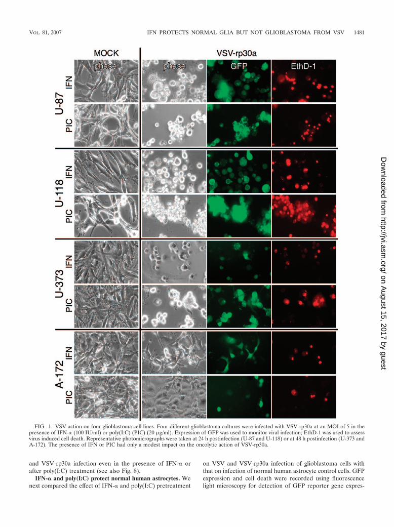
and VSV-rp30a infection even in the presence of IFN-� orafter poly(I:C) treatment (see also Fig. 8).
IFN-� and poly(I:C) protect normal human astrocytes. Wenext compared the effect of IFN-� and poly(I:C) pretreatment
on VSV and VSV-rp30a infection of glioblastoma cells withthat on infection of normal human astrocyte control cells. GFPexpression and cell death were recorded using fluorescencelight microscopy for detection of GFP reporter gene expres-
FIG. 1. VSV action on four glioblastoma cell lines. Four different glioblastoma cultures were infected with VSV-rp30a at an MOI of 5 in thepresence of IFN-� (100 IU/ml) or poly(I:C) (PIC) (20 �g/ml). Expression of GFP was used to monitor viral infection; EthD-1 was used to assessvirus induced cell death. Representative photomicrographs were taken at 24 h postinfection (U-87 and U-118) or at 48 h postinfection (U-373 andA-172). The presence of IFN or PIC had only a modest impact on the oncolytic action of VSV-rp30a.
VOL. 81, 2007 IFN PROTECTS NORMAL GLIA BUT NOT GLIOBLASTOMA FROM VSV 1481
on August 15, 2017 by guest
http://jvi.asm.org/
Dow
nloaded from
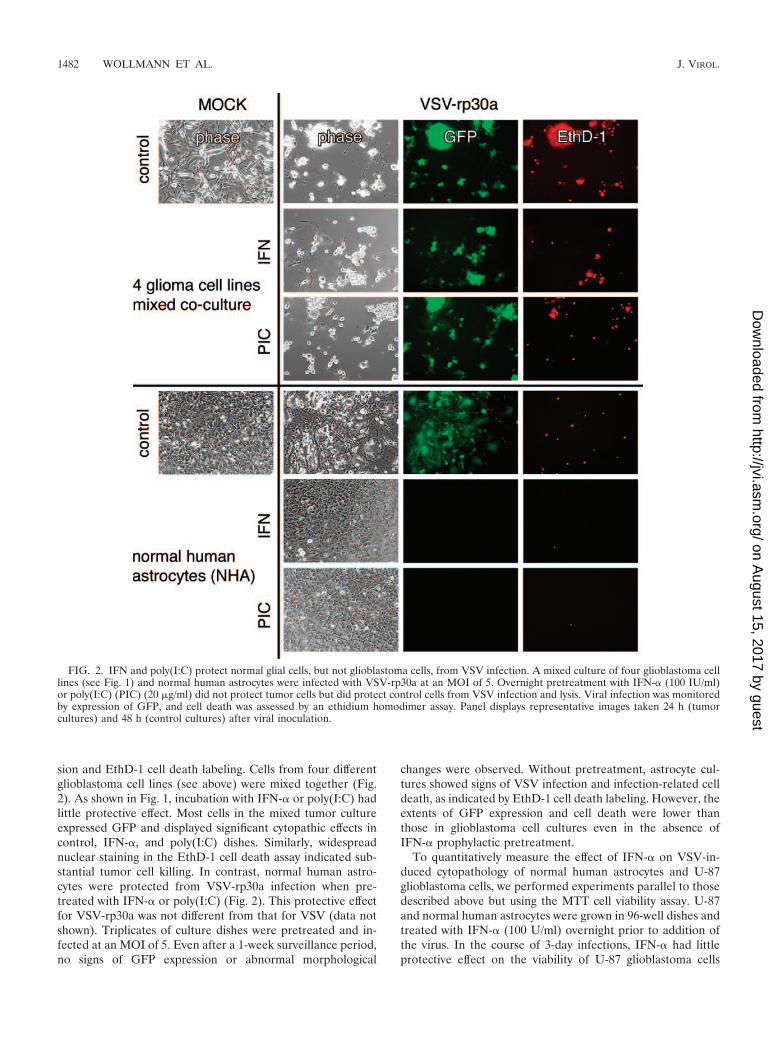
sion and EthD-1 cell death labeling. Cells from four differentglioblastoma cell lines (see above) were mixed together (Fig.2). As shown in Fig. 1, incubation with IFN-� or poly(I:C) hadlittle protective effect. Most cells in the mixed tumor cultureexpressed GFP and displayed significant cytopathic effects incontrol, IFN-�, and poly(I:C) dishes. Similarly, widespreadnuclear staining in the EthD-1 cell death assay indicated sub-stantial tumor cell killing. In contrast, normal human astro-cytes were protected from VSV-rp30a infection when pre-treated with IFN-� or poly(I:C) (Fig. 2). This protective effectfor VSV-rp30a was not different from that for VSV (data notshown). Triplicates of culture dishes were pretreated and in-fected at an MOI of 5. Even after a 1-week surveillance period,no signs of GFP expression or abnormal morphological
changes were observed. Without pretreatment, astrocyte cul-tures showed signs of VSV infection and infection-related celldeath, as indicated by EthD-1 cell death labeling. However, theextents of GFP expression and cell death were lower thanthose in glioblastoma cell cultures even in the absence ofIFN-� prophylactic pretreatment.
To quantitatively measure the effect of IFN-� on VSV-in-duced cytopathology of normal human astrocytes and U-87glioblastoma cells, we performed experiments parallel to thosedescribed above but using the MTT cell viability assay. U-87and normal human astrocytes were grown in 96-well dishes andtreated with IFN-� (100 U/ml) overnight prior to addition ofthe virus. In the course of 3-day infections, IFN-� had littleprotective effect on the viability of U-87 glioblastoma cells
FIG. 2. IFN and poly(I:C) protect normal glial cells, but not glioblastoma cells, from VSV infection. A mixed culture of four glioblastoma celllines (see Fig. 1) and normal human astrocytes were infected with VSV-rp30a at an MOI of 5. Overnight pretreatment with IFN-� (100 IU/ml)or poly(I:C) (PIC) (20 �g/ml) did not protect tumor cells but did protect control cells from VSV infection and lysis. Viral infection was monitoredby expression of GFP, and cell death was assessed by an ethidium homodimer assay. Panel displays representative images taken 24 h (tumorcultures) and 48 h (control cultures) after viral inoculation.
1482 WOLLMANN ET AL. J. VIROL.
on August 15, 2017 by guest
http://jvi.asm.org/
Dow
nloaded from

(Fig. 3). Under untreated conditions, both VSV and VSV-rp30a effectively killed nearly all U-87 cells. The action ofVSV-rp30a was slightly stronger than that of VSV (P � 0.05 atan MOI of 5; P � 0.3 at an MOI of 0.1). Pretreatment withIFN-� was ineffective at protecting glioblastoma cells (P �0.001 by analysis of variance). On the other hand, pretreatmentwith IFN-� completely protected normal human astrocytesfrom VSV cytotoxicity (Fig. 3). Even in the absence of IFN-�,the effect of VSV infection on viability of astrocytes was sig-nificantly less pronounced than in U-87 cells (P � 0.01 bypaired t test). Interestingly, viral load appeared to have littleeffect. The unaffected viability of human astrocytes in the pres-ence of IFN-� is consistent with the lack of GFP expressionupon VSV and VSV-rp30a infection mentioned above.
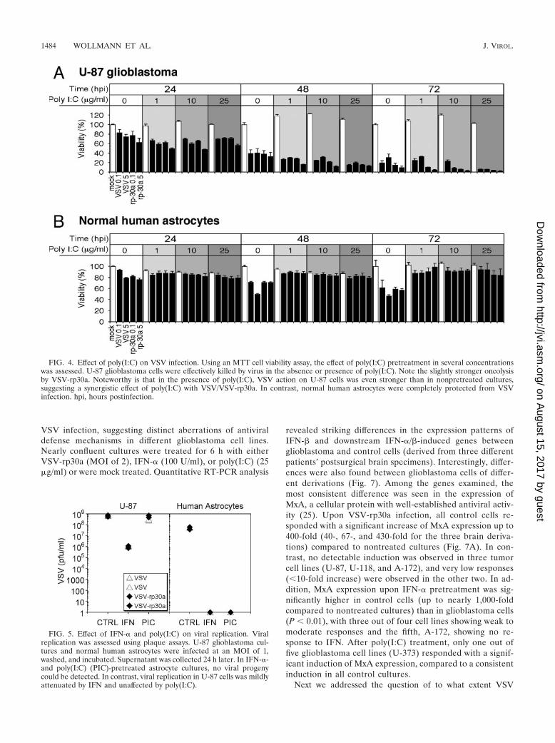
Synthetic double-stranded RNA polymers are potent induc-ers of the endogenous IFN-�/� pathway. If preincubation withpoly(I:C) leads to endogenous IFN production in human as-trocytes, then a protection from VSV-induced cytopathologywould be expected. U-87 glioblastoma cells and normal humanastrocytes were treated with poly(I:C) at three different con-centrations (1, 10, and 25 �g/ml) for 24 h prior to addition ofVSV and VSV-rp30a at MOIs of 0.1 and 5. Preincubation withPoly(I:C) had no protective effect on viability of U-87 cells; infact, poly(I:C) reduced the viability of glioblastoma cells inde-pendent of viral inoculation (P � 0.05) and enhanced theantitumor potency of VSV and VSV-rp30a in a dose-depen-dent manner (Fig. 4A). In these experiments, VSV-rp30a dis-played a moderately stronger antitumor effect than VSV-GFP.In contrast, poly(I:C) preincubation at all concentrations ef-
fectively protected normal human astrocytes (Fig. 4B). The cellviability of normal human astrocytes was not impaired by VSVand VSV-rp30a infection even at the low poly(I:C) concentra-tion. Poly(I:C) alone had no effect on cell viability (Fig. 4).
Together, these data indicate that normal human astrocytesare protected from VSV infection by administration of bothexogenous IFN-� and the IFN-�/� pathway inducer poly(I:C).
Effects of IFN and poly(I:C) on viral replication. Viral rep-lication was assessed by plaque assay of serial dilutions ofsupernatants 24 h after infection of U-87 cultures or normalhuman astrocyte cultures with VSV or VSV-rp30a at an MOIof 1. Experiments were performed in duplicate (Fig. 5). Undercontrol conditions, supernatants of U-87 glioblastoma culturesinfected with VSV and VSV-rp30a contained nearly 109
PFU/ml (7.5 � 108 and 6.1 � 108 PFU/ml for VSV and 7.7 �108 and 5.6 � 108 for VSV-rp30a [values for duplicates ofexperiments]). Though pretreatment with IFN-� resulted infewer progeny, indicating moderately attenuated viral replica-tion, glioblastoma cell supernatants still contained about 106
PFU/ml of both VSV and VSV-rp30a. Pretreatment withpoly(I:C), on the other hand, had nearly no effect on viralreplication in U-87 cells, resulting in PFU concentrations sim-ilar to those under nontreated control conditions (3.2 and 2.6 �108 PFU/ml for VSV and 7.1 and 5.0 � 108 PFU/ml for VSV-rp30a). In striking contrast, there was virtually no viral replicationin IFN-�- and poly(I:C)-treated normal human astrocyte controlcells, as indicated by the absence of any detectable plaques. Evenunder nontreated control conditions, viral replication in normalhuman astrocytes was lower than that in nontreated U-87 cells(6.0 and 5.5 � 107 PFU/ml for VSV and 6.3 and 3.9 � 107
PFU/ml for VSV-rp30a). Together these data indicate that IFN-�and poly(I:C) dramatically reduce viral proliferation in normalcells but not in glioblastoma cells.
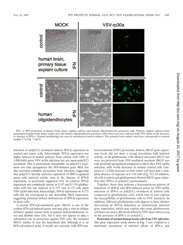
Protective effect of IFN-� on other human central nervoussystem (CNS) cells. In addition to normal human astrocytes,we tested whether IFN-� could protect primary human braintissue derived from epilepsy surgery from VSV infection. Pri-mary explant cultures were established from brain specimensfrom three patients of different ages and genders, resulting insemiconfluent cell layers with glial morphology. We also testedcommercially available human oligodendrocytes (Sciencell,San Diego, CA). In the absence of IFN treatment, both humanoligodendrocytes and, to a lesser extent, primary human braintissue explant cultures showed some GFP expression and cy-topathic morphology changes upon VSV infection. In contrast,IFN-� pretreatment (100 U/ml overnight) resulted in completeprotection of the respective cells from both VSV and VSV-rp30a infection, and neither cytopathic effects nor GFP expres-sion was detected for up to 6 days postinfection, when theexperiment was stopped. Representative photomicrographsfrom these experiments are displayed in Fig. 6.
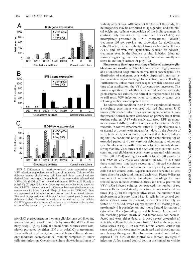
Different expressional responses to VSV-rp30a, IFN-�, orpoly(I:C). To address further underlying mechanisms thatmay mediate VSV oncolysis of glioblastoma cells, we usedquantitative reverse transcription-PCR (RT-PCR) to exam-ine the expression of IFN-� and two representative well-characterized IFN-�/�-stimulated genes, MxA and ISG15.All five glioblastoma cell lines tested were exquisitely sus-ceptible to oncolytic action of VSV. However, pretreatmentwith IFN-� or poly(I:C) resulted in various outcomes of
FIG. 3. Effect of IFN on VSV infection. Using an MTT cell viabilityassay, the effect of IFN pretreatment on the action of VSV on U-87cells and normal human astrocytes was assessed. IFN had little effecton glioblastoma cell destruction by VSV but completely protectednormal astrocytes. Low (MOI, 0.1) and high (MOI, 5) concentrationsof VSV and VSV-rp30a were used. Note the slightly stronger oncolysisby VSV-rp30a. Error bars indicate standard errors of the means fromtriplicate experiments. hpi, hours postinfection.
VOL. 81, 2007 IFN PROTECTS NORMAL GLIA BUT NOT GLIOBLASTOMA FROM VSV 1483
on August 15, 2017 by guest
http://jvi.asm.org/
Dow
nloaded from
VSV infection, suggesting distinct aberrations of antiviraldefense mechanisms in different glioblastoma cell lines.Nearly confluent cultures were treated for 6 h with eitherVSV-rp30a (MOI of 2), IFN-� (100 U/ml), or poly(I:C) (25�g/ml) or were mock treated. Quantitative RT-PCR analysis
revealed striking differences in the expression patterns ofIFN-� and downstream IFN-�/�-induced genes betweenglioblastoma and control cells (derived from three differentpatients’ postsurgical brain specimens). Interestingly, differ-ences were also found between glioblastoma cells of differ-ent derivations (Fig. 7). Among the genes examined, themost consistent difference was seen in the expression ofMxA, a cellular protein with well-established antiviral activ-ity (25). Upon VSV-rp30a infection, all control cells re-sponded with a significant increase of MxA expression up to400-fold (40-, 67-, and 430-fold for the three brain deriva-tions) compared to nontreated cultures (Fig. 7A). In con-trast, no detectable induction was observed in three tumorcell lines (U-87, U-118, and A-172), and very low responses(�10-fold increase) were observed in the other two. In ad-dition, MxA expression upon IFN-� pretreatment was sig-nificantly higher in control cells (up to nearly 1,000-foldcompared to nontreated cultures) than in glioblastoma cells(P � 0.01), with three out of four cell lines showing weak tomoderate responses and the fifth, A-172, showing no re-sponse to IFN. After poly(I:C) treatment, only one out offive glioblastoma cell lines (U-373) responded with a signif-icant induction of MxA expression, compared to a consistentinduction in all control cultures.
Next we addressed the question of to what extent VSV
FIG. 4. Effect of poly(I:C) on VSV infection. Using an MTT cell viability assay, the effect of poly(I:C) pretreatment in several concentrationswas assessed. U-87 glioblastoma cells were effectively killed by virus in the absence or presence of poly(I:C). Note the slightly stronger oncolysisby VSV-rp30a. Noteworthy is that in the presence of poly(I:C), VSV action on U-87 cells was even stronger than in nonpretreated cultures,suggesting a synergistic effect of poly(I:C) with VSV/VSV-rp30a. In contrast, normal human astrocytes were completely protected from VSVinfection. hpi, hours postinfection.
FIG. 5. Effect of IFN-� and poly(I:C) on viral replication. Viralreplication was assessed using plaque assays. U-87 glioblastoma cul-tures and normal human astrocytes were infected at an MOI of 1,washed, and incubated. Supernatant was collected 24 h later. In IFN-�-and poly(I:C) (PIC)-pretreated astrocyte cultures, no viral progenycould be detected. In contrast, viral replication in U-87 cells was mildlyattenuated by IFN and unaffected by poly(I:C).
1484 WOLLMANN ET AL. J. VIROL.
on August 15, 2017 by guest
http://jvi.asm.org/
Dow
nloaded from
infection or poly(I:C) treatment induces IFN-� expression incontrol and tumor cells. Interestingly, IFN-� expression washighly induced in human primary brain culture cells (400- to1,000-fold) upon VSV-rp30a infection but not upon poly(I:C)treatment. This is particularly remarkable, as poly(I:C) treat-ment not only upregulated the IFN-induced gene MxA butalso provided complete protection from infection, suggestingthat poly(I:C) directly activates expression of IRF3-regulatedgenes with antiviral activity even in the absence of IFN-�expression, as previously suggested (10). In contrast IFN-�expression was completely absent in U-87 and U-118 glioblas-toma cells but was induced in U-373 and A-172 cells uponVSV-rp30a infection. Interestingly, IFN-� expression in A-172cells did not correspond to any detectable MxA expression,suggesting potential defects downstream of IFN-� expressionin these cells.
A second IFN-�/�-stimulated gene, ISG15, is one of theearliest IFN-�/�-induced genes and may play a role in cellulardefenses against viruses such as human immunodeficiency vi-rus and Sindbis virus (16), but it does not appear to play asubstantial role in protection against VSV (24). We includedISG15 studies to test the hypothesis that although it is anIFN-�/�-induced gene, it would not correlate with IFN-con-
ferred antiviral (VSV) protection. Indeed, ISG15 gene expres-sion levels did not show a strong correlation with antiviralactivity, as all glioblastoma cells showed increased ISG15 butwere not protected from VSV-mediated oncolysis. ISG15 wasonly modestly upregulated compared to MxA after VSV-rp30ainfection, with 8-fold increases in normal control cells com-pared to 12-fold increases in four tumor cell lines and a com-plete absence of response in U-118 cells (Fig. 7C). In addition,all cells (control and glioblastoma) showed ISG15 gene induc-tion after IFN-� or poly(I:C) pretreatment.
Together, these data indicate a heterogeneous pattern ofinduction of IFN-� and IFN-induced genes by VSV-rp30ainfection or IFN-� or poly(I:C) treatment in control cellscompared to glioblastoma cells, which may in part explainthe susceptibility of glioblastoma cells to VSV oncolysis. Inaddition, different glioblastoma cells appear to have distinctaberrations in IFN-� induction or downstream antiviralgene expression, which may explain why some glioblastomacell lines are more effectively killed by VSV than others evenin the presence of IFN-� or poly(I:C).
Protection of normal human brain cells from VSV infection.The gene expression study shown in Fig. 7 was coupled to afunctional assessment of antiviral effects of IFN-� and
FIG. 6. IFN protection of human brain tissue explant cultures and human oligodendrocyte precursor cells. Primary explant cultures frompostsurgery human brain tissue (upper set) and human oligodendrocyte precursor cells (lower set) were infected with VSV-rp30a in the presenceor absence of IFN-�. Normal morphology was seen in noninfected control cultures. The primary brain culture used here corresponds to controlsample C in Fig. 7 and 8.
VOL. 81, 2007 IFN PROTECTS NORMAL GLIA BUT NOT GLIOBLASTOMA FROM VSV 1485
on August 15, 2017 by guest
http://jvi.asm.org/
Dow
nloaded from
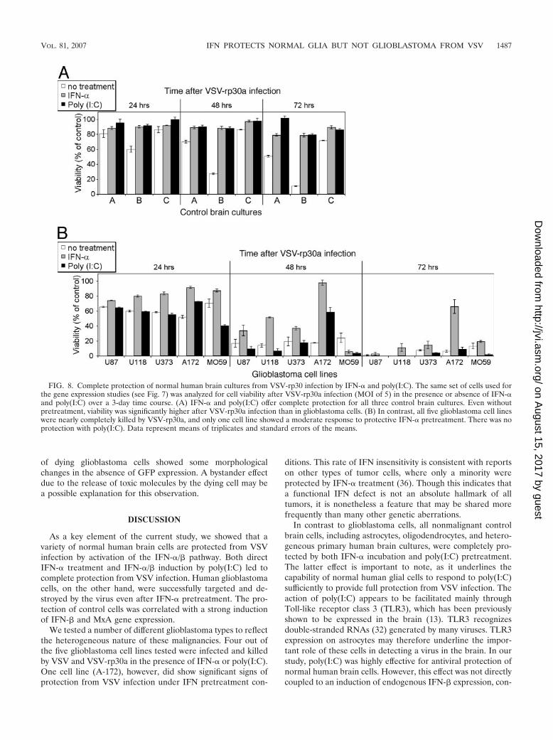
poly(I:C) pretreatment on the same glioblastoma cell lines andnormal human control brain cells by using the MTT cell via-bility assay (Fig. 8). Normal human brain cultures were com-pletely protected by either IFN-� or poly(I:C) pretreatment.Even without treatment, two normal brain cultures showedonly moderate decreases in cell viability compared to tumorcells after infection. One normal culture showed impairment of
viability after 3 days. Although not the focus of this study, thisheterogeneity may be attributed to age, gender, and anatomi-cal origin and cellular composition of the brain specimen. Incontrast, only one out of five tumor cell lines (A-172) wasincompletely protected by IFN-� pretreatment. Poly(I:C)treatment did not provide any protection for glioblastomacells. Of note, the cell viability of two glioblastoma cell lines,A-172 and MO59J, was significantly reduced by poly(I:C)treatment even in the absence of viral infection (data notshown), suggesting that these two cell lines were directly sen-sitive to antitumor actions of poly(I:C).
Fluorescence time-lapse recording of infected astrocyte-glio-blastoma cell cocultures. Glioblastoma cells are highly invasiveand often spread deep into the normal brain parenchyma. Thisdistribution of malignant cells widely dispersed in normal tis-sue presents a major challenge for selective tumor cell killing.Furthermore, unlike most inert reagents, which decrease withtime after application, the VSV concentration increases. Thisraises a question of whether in a mixed normal astrocyte/glioblastoma cell culture, the normal astrocytes would be ableto still resist VSV infection when surrounded by tumor cellsreleasing replication-competent virus.
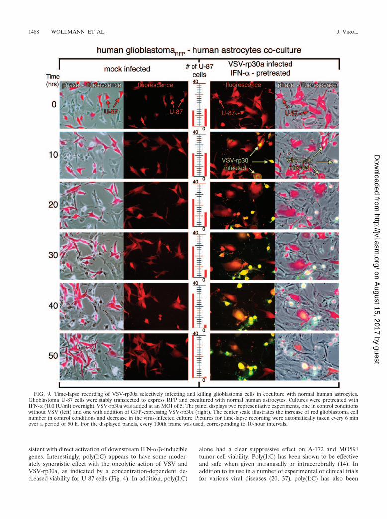
To address this condition in an in vitro experimental model,a coculture experiment was used with red fluorescent U-87tumor cells seeded into dishes containing subconfluent non-fluorescent normal human astrocytes or primary brain tissueexplant cultures. U-87 cells stably expressed RFP (a mono-meric form of dsRed); cultures of these cells contained �99%red cells. In control experiments, either RFP glioblastoma cellsor normal astrocytes were imaged for 4 days. In the absence ofvirus, both cell types continued to grow and replicate, indicat-ing that the conditions of digital imaging continuously for anextended period of 4 days were not debilitating to either celltype. Similar controls with IFN-� or poly(I:C) similarly showedstrong viability. Cocultures of the two cell types (normal astro-cytes and red glioblastoma cells) were pretreated with humanIFN-� (200 U/ml) overnight or with poly(I:C) (20 �g/ml) for6 h. VSV or VSV-rp30a was added at an MOI of 5. Underthese conditions, time-lapse recording of infected coculturesconfirmed the selective infection and cell lysis of glioblastomacells but not control cells. Experiments were repeated at leastthree times for each condition and each virus. Figure 9 displaystwo sets of representative time-lapse recordings for non-treated, mock-infected control cultures and IFN-�-pretreated,VSV-rp30a-infected cultures. As expected, the number of redtumor cells increased steadily over time in mock-infected cul-tures (Fig. 9). In this representative series, the number of redglioblastoma cells rose from 14 to 41 cells in the control con-dition without virus. In contrast, VSV-rp30a selectively in-fected U-87-dsRed, which expressed viral GFP starting at ap-proximately 6 h postinfection and showed rapid formation ofcytopathic effects (rounding up and blebbing). At the end ofthe recording period, nearly all red tumor cells had been in-fected and were either dead or showed severe cytopathic ef-fects (the cell number decreased from 16 to 1 in the represen-tative series in Fig. 9). In contrast, normal control cells in thesame culture dish were mostly unaffected and showed normalmorphology throughout the observation period and did notexpress GFP; �2% of the control cells showed any signs ofinfection. A few normal control cells in the immediate vicinity
FIG. 7. Differences in interferon-related gene expression uponVSV infection in glioblastoma and control brain cells. Cultures of fivedifferent human glioblastoma cell lines and three control culturesderived from postsurgery human brain tissue were either infected withVSV-rp30a (MOI of 2) or treated with human IFN-� (100 IU/ml) orpoly(I:C) (25 �g/ml) for 6 h. Gene expression studies using quantita-tive RT-PCR revealed marked differences between glioblastoma andcontrol cells for MxA (A) and IFN-� (B) but not for ISG15 (C). Dataare expressed as fold induction relative to control untreated cultures.The level of expression was different for each tested gene (y axes havedifferent scales). Expression levels are normalized to the cellularGAPDH gene and are presented as means of triplicates with standarderrors of the means. n.d., none detected.
1486 WOLLMANN ET AL. J. VIROL.
on August 15, 2017 by guest
http://jvi.asm.org/
Dow
nloaded from
of dying glioblastoma cells showed some morphologicalchanges in the absence of GFP expression. A bystander effectdue to the release of toxic molecules by the dying cell may bea possible explanation for this observation.
DISCUSSION
As a key element of the current study, we showed that avariety of normal human brain cells are protected from VSVinfection by activation of the IFN-�/� pathway. Both directIFN-� treatment and IFN-�/� induction by poly(I:C) led tocomplete protection from VSV infection. Human glioblastomacells, on the other hand, were successfully targeted and de-stroyed by the virus even after IFN-� pretreatment. The pro-tection of control cells was correlated with a strong inductionof IFN-� and MxA gene expression.
We tested a number of different glioblastoma types to reflectthe heterogeneous nature of these malignancies. Four out ofthe five glioblastoma cell lines tested were infected and killedby VSV and VSV-rp30a in the presence of IFN-� or poly(I:C).One cell line (A-172), however, did show significant signs ofprotection from VSV infection under IFN pretreatment con-
ditions. This rate of IFN insensitivity is consistent with reportson other types of tumor cells, where only a minority wereprotected by IFN-� treatment (36). Though this indicates thata functional IFN defect is not an absolute hallmark of alltumors, it is nonetheless a feature that may be shared morefrequently than many other genetic aberrations.
In contrast to glioblastoma cells, all nonmalignant controlbrain cells, including astrocytes, oligodendrocytes, and hetero-geneous primary human brain cultures, were completely pro-tected by both IFN-� incubation and poly(I:C) pretreatment.The latter effect is important to note, as it underlines thecapability of normal human glial cells to respond to poly(I:C)sufficiently to provide full protection from VSV infection. Theaction of poly(I:C) appears to be facilitated mainly throughToll-like receptor class 3 (TLR3), which has been previouslyshown to be expressed in the brain (13). TLR3 recognizesdouble-stranded RNAs (32) generated by many viruses. TLR3expression on astrocytes may therefore underline the impor-tant role of these cells in detecting a virus in the brain. In ourstudy, poly(I:C) was highly effective for antiviral protection ofnormal human brain cells. However, this effect was not directlycoupled to an induction of endogenous IFN-� expression, con-
FIG. 8. Complete protection of normal human brain cultures from VSV-rp30 infection by IFN-� and poly(I:C). The same set of cells used forthe gene expression studies (see Fig. 7) was analyzed for cell viability after VSV-rp30a infection (MOI of 5) in the presence or absence of IFN-�and poly(I:C) over a 3-day time course. (A) IFN-� and poly(I:C) offer complete protection for all three control brain cultures. Even withoutpretreatment, viability was significantly higher after VSV-rp30a infection than in glioblastoma cells. (B) In contrast, all five glioblastoma cell lineswere nearly completely killed by VSV-rp30a, and only one cell line showed a moderate response to protective IFN-� pretreatment. There was noprotection with poly(I:C). Data represent means of triplicates and standard errors of the means.
VOL. 81, 2007 IFN PROTECTS NORMAL GLIA BUT NOT GLIOBLASTOMA FROM VSV 1487
on August 15, 2017 by guest
http://jvi.asm.org/
Dow
nloaded from
sistent with direct activation of downstream IFN-�/�-induciblegenes. Interestingly, poly(I:C) appears to have some moder-ately synergistic effect with the oncolytic action of VSV andVSV-rp30a, as indicated by a concentration-dependent de-creased viability for U-87 cells (Fig. 4). In addition, poly(I:C)
alone had a clear suppressive effect on A-172 and MO59Jtumor cell viability. Poly(I:C) has been shown to be effectiveand safe when given intranasally or intracerebrally (14). Inaddition to its use in a number of experimental or clinical trialsfor various viral diseases (20, 37), poly(I:C) has also been
FIG. 9. Time-lapse recording of VSV-rp30a selectively infecting and killing glioblastoma cells in coculture with normal human astrocytes.Glioblastoma U-87 cells were stably transfected to express RFP and cocultured with normal human astrocytes. Cultures were pretreated withIFN-� (100 IU/ml) overnight. VSV-rp30a was added at an MOI of 5. The panel displays two representative experiments, one in control conditionswithout VSV (left) and one with addition of GFP-expressing VSV-rp30a (right). The center scale illustrates the increase of red glioblastoma cellnumber in control conditions and decrease in the virus-infected culture. Pictures for time-lapse recording were automatically taken every 6 minover a period of 50 h. For the displayed panels, every 100th frame was used, corresponding to 10-hour intervals.
1488 WOLLMANN ET AL. J. VIROL.
on August 15, 2017 by guest
http://jvi.asm.org/
Dow
nloaded from
tested in a clinical trial for direct treatment of glioblastoma(29). Both IFN and poly(I:C) protected normal brain cellsfrom recombinant VSVs. It is important to note that cellswithin the normal brain would benefit from many additionalantiviral defenses of the systemic immune system, includingactivated B and T cells and macrophages/microglia, which wereminimal or absent in our culture conditions.
Conditional viral replication is a central feature of oncolyticviral use in comparison to vector-based gene therapy strategiesthat utilize replication-deficient virions. Replication of VSVand VSV-rp30a was only moderately reduced by IFN-� andwas not affected by poly(I:C) in U-87 cells; in contrast, littleviral replication was detected in nontumor control CNS cellspretreated with IFN-� or poly(I:C). This is particularly impor-tant, as it would theoretically prevent further viral spread be-yond the interface of tumor mass and normal brain paren-chyma. For our replication studies, we used normal humanastrocytes, the cell type most closely related to glioblastomatumors, as control cells. Of note, a similar inhibitory effect ofIFN on VSV replication was shown with mouse neuron cul-tures in a recent report (39). Previous work (42) showed thatVSV-rp30 was more effective in killing glioblastoma cells thanVSV-GFP; this was particularly noticeable at shorter time in-tervals. For this reason we used VSV-rp30a for the experi-ments here. In some experiments we also used VSV-GFP, andwe found that VSV-GFP and VSV-rp30 showed similar actionsand responses at the longer time intervals in which both viruseshad sufficient time to replicate during multiple cycles, produc-ing a final high MOI. A faster-acting virus such as VSV-rp30amay provide some advantage in brain tumors, in which hun-dreds of millions of tumor cells would ideally be destroyedbefore a substantive immune response would eliminate thevirus.
Our gene expression studies revealed a number of interest-ing correlations. First, MxA was highly induced after viralinfection in control cells but was hardly detectable in any of thefive glioblastoma cell lines. When pretreated with IFN-� orpoly(I:C), four out of five glioblastoma cell lines respondedwith a small induction, compared to control cells that had asignificantly greater response. It is of interest to note thatalthough the complete protection of control cells is associatedwith an MxA gene induction of up to 1,000-fold, the onlyglioblastoma cell type that was somewhat protected by IFN-�pretreatment, A-172, did not express MxA to any significantlevels. Although MxA has been primarily associated with myxo-virus resistance, evidence exists for a role for MxA in theIFN-mediated inhibition of VSV (31, 35). However, the re-quirement for MxA in the antiviral defense against VSV maydiffer in tumor and normal brain cells. IFN-� expression wasvery strong in control cells upon VSV-rp30a infection but waslacking after poly(I:C) treatment. In tumor cells, on the otherhand, IFN-� mRNA was completely absent in two tumor celltypes, U-87 and U-118. Interestingly, these tumor cells havethe greatest sensitivity for VSV oncolysis but also display thefastest growth pattern in our experimental conditions. In thatregard, it is of importance to note that though they wereoriginally described as antiviral elements of the innate immunesystem, IFN-�/� also may play a role in control of cell differ-entiation and proliferation and in prevention of malignanttransformation (8). Functional loss of IFN-� and -� appears to
be common in the mutation repertoire of gliomas, especially inhighly malignant glioblastoma multiforme (33), and the extentof IFN-�/� defects increases with malignant progression. Aprevious study revealed an intact IFN status in low-grade gli-omas (WHO grade 2), hemizygous IFN deletions in anaplasticastrocytomas (grade 3), and a nullizygous IFN status in glio-blastoma multiforme (grade 4) (15). However, expression ofcertain IFN-�/�-stimulated genes does not necessarily corre-late with antiviral potential in glioblastoma cells against VSV.In fact, our studies on ISG15 gene expression revealed aslightly greater induction in tumor cells than in normal cells.ISG15 is a ubiquitin-like protein suggested to be involved incellular protein modification. However, a role for ISG15 inantiviral defense is complex, as ISG15 appears to inhibit thereplication of some viruses (human immunodeficiency virustype 1 and Sindbis virus) (16, 23) but has been shown to bedispensable for defense against lymphocytic choriomeningitisvirus and VSV (24).
This study was designed to address the important issue ofenhancing the safety of a potential application of VSV as anoncolytic agent in the brain. VSV has only recently been rec-ognized as a potential tool to attack malignancies. A number ofreports have demonstrated the oncolytic effects of VSV inseveral in vitro and in vivo tumor models (reviewed in refer-ences 3 and 17), and the importance of the IFN-�/� system asthe first line of defense for normal cells outside the brain hasbeen well established. Application of replication-competentoncolytic viruses in the brain, however, represents a particularchallenge considering the special immune status of the CNSparenchyma. Glioblastoma brain tumors may be an attractivetarget for VSV-based oncolytic approaches. The molecularmakeup of those tumors can be heterogeneous, making it dif-ficult for viral vectors that are limited in targeting only specificmutations. For example, reovirus requires activated Ras sig-naling pathways, and oncolytic adenoviruses are based uponp53 defects (5). VSV on the other hand, selectively killed anumber of glioblastoma types with different genetic aberra-tions but in the presence of IFN-� did not kill normal humanastrocytes or mixed primary cultures from human brain. Im-portantly, as the time-lapse video experiments showed, evenwith a potentially increasing virus titer during infection andVSV replication in glioblastoma cells, the normal astrocytes inthe same culture dish were continuously protected by a singleapplication of IFN or poly(I:C) at the beginning of the 4-daytime course of the experiment.
Another consideration is the size and invasive nature ofglioblastomas, which are two of the main factors why the out-come of previous clinical trials with replication-incompetentvectors has been disappointing (reviewed in reference 27).VSV rapidly replicates and has been shown in a previous studyfrom our lab to spread through the whole tumor mass of U-87xenografts with minimal infection of surrounding host tissue(42), indicating potential for application in a widespread andinvasive glioblastoma mass. However, uncontrolled spread ofthe virus, particularly in the brain, would result in deleteriousconsequences. We show here that IFN-�/� as well as an in-ducer of the IFN-�/� pathway can protect normal human braincells from VSV infection in vitro. It is important to note thatintravenous IFN-� is an approved treatment modality for se-vere multiple sclerosis cases, indicating effective and safe ac-
VOL. 81, 2007 IFN PROTECTS NORMAL GLIA BUT NOT GLIOBLASTOMA FROM VSV 1489
on August 15, 2017 by guest
http://jvi.asm.org/
Dow
nloaded from
tion of this group of drugs within the brain. Moreover, IFN-�/�has also been shown to be effective against glioma directlythrough apoptosis induction (44) and indirectly through inhi-bition of angiogenesis (26). In our experimental study, wesuccessfully combined an oncolytic VSV with IFN, which notonly may result in improved safety but also may act synergis-tically against glioma.
As the protective effects of IFN on normal brain cells butnot on glioblastoma cells were found with both VSV variantsused here, it is likely that these results may also generalizeto other VSV strains that also may possess oncolytic poten-tial (17, 18, 21). In addition, IFN can also protect brain cellsagainst other unrelated viruses. For example, IFN-�, -�, and- protect the developing brain from cytomegalovirus infec-tion (40). The antiviral effect was significantly strongeragainst VSV than against cytomegalovirus, further support-ing the view that IFN sensitivity of VSV can serve as apotential safety measure. Additionally, the data here mayalso prove useful in devising strategies for protecting normalbrain, but not brain tumors, during treatment with oncolyticstrains of herpesvirus, adenovirus, reovirus, and other rep-lication-competent viruses (5, 27).
ACKNOWLEDGMENTS
We thank Prabhat Ghosh and Koray Ozduman for construction andgeneration of red fluorescent U-87 cells and Prasanthi Bandi for tech-nical assistance.
Support for this work was provided by NIH grants K22 AI064757 (toM.D.R.) and AI48854 (to A.N.V.D.P.).
REFERENCES
1. Ahmed, M., S. D. Cramer, and D. S. Lyles. 2004. Sensitivity of prostatetumors to wild type and M protein mutant vesicular stomatitis viruses.Virology 330:34–49.
2. Balachandran, S., M. Porosnicu, and G. N. Barber. 2001. Oncolytic activityof vesicular stomatitis virus is effective against tumors exhibiting aberrantp53, Ras, or myc function and involves the induction of apoptosis. J. Virol.75:3474–3479.
3. Barber, G. N. 2005. VSV-tumor selective replication and protein translation.Oncogene 24:7710–7719.
4. Chesler, D. A., and C. S. Reiss. 2002. The role of IFN-gamma in immuneresponses to viral infections of the central nervous system. Cytokine GrowthFactor Rev. 13:441–454.
5. Chiocca, E. A. 2002. Oncolytic viruses. Nat. Rev. Cancer 2:938–950.6. Colton, C. A., J. Yao, J. E. Keri, and D. Gilbert. 1992. Regulation of micro-
glial function by interferons. J. Neuroimmunol. 40:89–98.7. Connor, J. H., C. Naczki, C. Koumenis, and D. S. Lyles. 2004. Replication
and cytopathic effect of oncolytic vesicular stomatitis virus in hypoxic tumorcells in vitro and in vivo. J. Virol. 78:8960–8970.
8. Dafny, N., and P. B. Yang. 2005. Interferon and the central nervous system.Eur. J. Pharmacol. 523:1–15.
9. Dalton, K. P., and J. K. Rose. 2001. Vesicular stomatitis virus glycoproteincontaining the entire green fluorescent protein on its cytoplasmic domain isincorporated efficiently into virus particles. Virology 279:414–421.
10. Doyle, S., S. Vaidya, R. O’Connell, H. Dadgostar, P. Dempsey, T. Wu, G.Rao, R. Sun, M. Haberland, R. Modlin, and G. Cheng. 2002. IRF3 mediatesa TLR3/TLR4-specific antiviral gene program. Immunity 17:251–263.
11. Duntsch, C. D., Q. Zhou, H. R. Jayakar, J. D. Weimar, J. H. Robertson,L. M. Pfeffer, L. Wang, Z. Xiang, and M. A. Whitt. 2004. Recombinantvesicular stomatitis virus vectors as oncolytic agents in the treatment ofhigh-grade gliomas in an organotypic brain tissue slice-glioma coculturemodel. J. Neurosurg. 100:1049–1059.
12. Ebert, O., S. Harbaran, K. Shinozaki, and S. L. Woo. 2005. Systemic therapyof experimental breast cancer metastases by mutant vesicular stomatitis virusin immune-competent mice. Cancer Gene Ther. 12:350–358.
13. Farina, C., M. Krumbholz, T. Giese, G. Hartmann, F. Aloisi, and E. Meinl.2005. Preferential expression and function of Toll-like receptor 3 in humanastrocytes. J. Neuroimmunol. 159:12–19.
14. Ichinohe, T., I. Watanabe, S. Ito, H. Fujii, M. Moriyama, S. Tamura, H.Takahashi, H. Sawa, J. Chiba, T. Kurata, T. Sata, and H. Hasegawa. 2005.Synthetic double-stranded RNA poly(I:C) combined with mucosal vaccineprotects against influenza virus infection. J. Virol. 79:2910–2919.
15. James, C. D., J. He, E. Carlbom, M. Nordenskjold, W. K. Cavenee, and V. P.Collins. 1991. Chromosome 9 deletion mapping reveals interferon alpha andinterferon beta-1 gene deletions in human glial tumors. Cancer Res. 51:1684–1688.
16. Lenschow, D. J., N. V. Giannakopoulos, L. J. Gunn, C. Johnston, A. K.O’Guin, R. E. Schmidt, B. Levine, and H. W. Virgin IV. 2005. Identificationof interferon-stimulated gene 15 as an antiviral molecule during Sindbis virusinfection in vivo. J. Virol. 79:13974–13983.
17. Lichty, B. D., A. T. Power, D. F. Stojdl, and J. C. Bell. 2004. Vesicularstomatitis virus: re-inventing the bullet. Trends Mol. Med. 10:210–216.
18. Lichty, B. D., D. F. Stojdl, R. A. Taylor, L. Miller, I. Frenkel, H. Atkins, andJ. C. Bell. 2004. Vesicular stomatitis virus: a potential therapeutic virus forthe treatment of hematologic malignancy. Hum. Gene Ther. 15:821–831.
19. Mendoza-Fernandez, V., R. D. Andrew, and C. Barajas-Lopez. 2000. Inter-feron-alpha inhibits long-term potentiation and unmasks a long-term depres-sion in the rat hippocampus. Brain Res. 885:14–24.
20. Morrey, J. D., C. W. Day, J. G. Julander, L. M. Blatt, D. F. Smee, and R. W.Sidwell. 2004. Effect of interferon-alpha and interferon-inducers on WestNile virus in mouse and hamster animal models. Antivir. Chem. Chemother.15:101–109.
21. Obuchi, M., M. Fernandez, and G. N. Barber. 2003. Development of recom-binant vesicular stomatitis viruses that exploit defects in host defense toaugment specific oncolytic activity. J. Virol. 77:8843–8856.
22. Ohgaki, H. 2005. Genetic pathways to glioblastomas. Neuropathology 25:1–7.
23. Okumura, A., G. Lu, I. Pitha-Rowe, and P. M. Pitha. 2006. Innate antiviralresponse targets HIV-1 release by the induction of ubiquitin-like proteinISG15. Proc. Natl. Acad. Sci. USA 103:1440–1445.
24. Osiak, A., O. Utermohlen, S. Niendorf, I. Horak, and K. P. Knobeloch. 2005.ISG15, an interferon-stimulated ubiquitin-like protein, is not essential forSTAT1 signaling and responses against vesicular stomatitis and lymphocyticchoriomeningitis virus. Mol. Cell. Biol. 25:6338–6345.
25. Pavlovic, J., H. A. Arzet, H. P. Hefti, M. Frese, D. Rost, B. Ernst, E. Kolb, P.Staeheli, and O. Haller. 1995. Enhanced virus resistance of transgenic miceexpressing the human MxA protein. J. Virol. 69:4506–4510.
26. Puduvalli, V. K., and R. Sawaya. 2000. Antiangiogenesis—therapeutic strat-egies and clinical implications for brain tumors. J. Neurooncol. 50:189–200.
27. Pulkkanen, K. J., and S. Yla-Herttuala. 2005. Gene therapy for malignantglioma: current clinical status. Mol. Ther. 12:585–598.
28. Rose, J. K., and M. A. Whitt. 2001. Rhabdoviridae: the viruses and their repli-cation, p. 1221–1244. In D. M. Knipe and P. M. Howley (ed.), Fields virology,4th ed., vol. 1. Lippincott Williams and Wilkins, Philadelphia, PA.
29. Salazar, A. M., H. B. Levy, S. Ondra, M. Kende, B. Scherokman, D. Brown,H. Mena, N. Martin, K. Schwab, D. Donovan, D. Dougherty, M. Pulliam, M.Ippolito, M. Graves, H. Brown, and A. Ommaya. 1996. Long-term treatmentof malignant gliomas with intramuscularly administered polyinosinic-poly-cytidylic acid stabilized with polylysine and carboxymethylcellulose: an openpilot study. Neurosurgery 38:1096–1103.
30. Satoh, J., and Y. Kuroda. 2001. Differing effects of IFN beta vs IFN gammain MS: gene expression in cultured astrocytes. Neurology 57:681–685.
31. Schanen, C., V. Chieux, P. E. Lobert, J. Harvey, and D. Hober. 2006. Correlationbetween the anti-virus-induced cytopathic effect activity of interferon-alpha sub-types and induction of MxA protein in vitro. Microbiol. Immunol. 50:19–24.
32. Scumpia, P. O., K. M. Kelly, W. H. Reeves, and B. R. Stevens. 2005. Double-stranded RNA signals antiviral and inflammatory programs and dysfunc-tional glutamate transport in TLR3-expressing astrocytes. Glia 52:153–162.
33. Sehgal, A. 1998. Molecular changes during the genesis of human gliomas.Semin. Surg. Oncol. 14:3–12.
34. Shinozaki, K., O. Ebert, and S. L. Woo. 2005. Eradication of advancedhepatocellular carcinoma in rats via repeated hepatic arterial infusions ofrecombinant VSV. Hepatology 41:196–203.
35. Staeheli, P., and J. Pavlovic. 1991. Inhibition of vesicular stomatitis virusmRNA synthesis by human MxA protein. J. Virol. 65:4498–4501.
36. Stojdl, D. F., B. D. Lichty, B. R. tenOever, J. M. Paterson, A. T. Power, S.Knowles, R. Marius, J. Reynard, L. Poliquin, H. Atkins, E. G. Brown, R. K.Durbin, J. E. Durbin, J. Hiscott, and J. C. Bell. 2003. VSV strains withdefects in their ability to shutdown innate immunity are potent systemicanti-cancer agents. Cancer Cell 4:263–275.
37. Tazulakhova, E. B., O. V. Parshina, T. S. Guseva, and F. I. Ershov. 2001.Russian experience in screening, analysis, and clinical application of novelinterferon inducers. J. Interferon Cytokine Res. 21:65–73.
38. Tedeschi, B., J. N. Barrett, and R. W. Keane. 1986. Astrocytes produceinterferon that enhances the expression of H-2 antigens on a subpopulationof brain cells. Cell Biol. 102:2244–2253.
39. Trottier, M. D., Jr., B. M. Palian, and C. Shoshkes Reiss. 2005. VSVreplication in neurons is inhibited by type I IFN at multiple stages of infec-tion. Virology 333:215–225.
40. van den Pol, A. N., M. D. Robek, P. K. Ghosh, K. Ozduman, P. Bandi, M. D.Whim, and G. Wollmann. 2007. Cytomegalovirus induces interferon-stimu-lated gene expression and is attenuated by interferon in the developing brain.J. Virol. 81:332–348.
41. Watkins, L. R., S. F. Maier, and L. E. Goehler. 1995. Cytokine-to-brain
1490 WOLLMANN ET AL. J. VIROL.
on August 15, 2017 by guest
http://jvi.asm.org/
Dow
nloaded from
communication: a review & analysis of alternative mechanisms. Life Sci.57:1011–1026.
42. Wollmann, G., P. Tattersall, and A. N. van den Pol. 2005. Targeting humanglioblastoma cells: comparison of nine viruses with oncolytic potential. J. Vi-rol. 79:6005–6022.
43. Yamada, T., M. A. Horisberger, N. Kawaguchi, I. Moroo, and T. Toyoda.
1994. Immunohistochemistry using antibodies to alpha-interferon and itsinduced protein, MxA, in Alzheimer’s and Parkinson’s disease brain tissues.Neurosci. Lett. 181:61–64.
44. Yoshida, J., M. Mizuno, and T. Wakabayashi. 2004. Interferon-beta genetherapy for cancer: basic research to clinical application. Cancer Sci.95:858–865.
VOL. 81, 2007 IFN PROTECTS NORMAL GLIA BUT NOT GLIOBLASTOMA FROM VSV 1491
on August 15, 2017 by guest
http://jvi.asm.org/
Dow
nloaded from