Embed Size (px)
Citation preview
Page 1 of 66
The Royal College of Ophthalmologists
Interim Guidelines for Management of Retinal Vein Occlusion
December 2010
Scientific Department 17 Cornwall Terrace London NW1 4QW
Telephone: 020 7935 0702 Facsimile: 020 7487 4674
www.rcophth.ac.uk
© The Royal College of Ophthalmologists 2010 All rights reserved For permission to reproduce any of the content contained herein please contact [email protected]
Page 2 of 66
1 CONTENTS
1 Contents.......................................................................................................................3
2 List of Tables……………………………………………………………………………… 5
3 Introduction ................................................................................................................10
3.1 Background ............................................................................................................10
3.2 Remit of the guidelines...........................................................................................12
4 Methods .....................................................................................................................14
4.1 The Guideline Development Group........................................................................14
4.2 Gathering the evidence ..........................................................................................14
4.3 Assessing the evidence and forming recommendations .......................................16
4.4 Consultation process..............................................................................................18
5 Aetiology and risk factors...........................................................................................19
5.1 Strength of evidence ..............................................................................................19
5.2 Other Important Observations................................................................................21
6 Natural history of retinal vein occlusions ...................................................................23
6.1 CRVO .....................................................................................................................23
6.2 BRVO .....................................................................................................................24
6.3 Low Vision and Living with RVO…………………………………………… …... …13
7 Management ..............................................................................................................26
7.1 OPHTHALMOLOGICAL MANAGEMENT..............................................................26
7.1.1 Central retinal vein occlusion (CRVO)…………………………………………...26
7.1.1.1 Management of ischaemic central retinal vein occlusion and anterior
segment neovascularisation......................................................................28
7.1.1.2 Posterior segment neovascularisation ......................................................30
7.1.1.3 Management of established neovascular glaucoma.................................32
7.1.1.4 Macular oedema........................................................................................33
7.1.1.5 Recommendations for Further Follow-up..................................................42
Page 3 of 66
7.1.1.6 Experimental treatments ...........................................................................43
7.1.2 Branch Retinal Vein Occlusion ........................................................................44
7.1.2.1 Treatment of neovascularisation...............................................................45
7.1.2.2 Laser treatments for macular oedema ......................................................46
7.1.2.3 Pharmacologic Treatments ......................................................................48
7.1.2.4 Other Treatments ......................................................................................56
7.1.3 Hemisphere vein occlusion..............................................................................56
7.2 MEDICAL MANAGEMENT............................................................................... …. 58
7.2.1 Referral for medical investigation and treatment .............................................58
7.2.2 Medical Management.......................................................................................60
7.2.2.1 Restoring venous patency.........................................................................60
7.2.2.2 Ameliorate cardiovascular morbidity and mortality associated with retinal
vein occlusion............................................................................................62
7.2.2.3 To prevent the recurrence of retinal vein occlusion ..................................63
7.2.3 Management of younger patients (less than 50 years of age) ........................65
7.3 TREATMENT ALGORITHMS.................................................................................67
7.3.1 Minimum Service Specifications ......................................................................68
7.3.2 Treatment of Risk Factors................................................................................69
7.3.3 Treatment Algorithm for CRVO........................................................................70
7.3.3.1 Baseline Assessments ..............................................................................70
7.3.3.2 Management at baseline...........................................................................71
7.3.3.3 Non-Ischaemic CRVO...............................................................................72
7.3.3.4 Ischaemic CRVO.......................................................................................75
7.3.4 Treatment Algorithm for BRVO........................................................................77
7.3.4.1 NON –ISCHAEMIC BRVO........................................................................77
7.3.4.2 Unlicensed and Contraindicated pharmacological agents - Considerations
…………………………………………………………………………………..80
7.3.4.3 Ischaemic BRVO.......................................................................................80
Page 4 of 66
7.3.5 Hemispheric Vein Occlusion Algorithm ...........................................................80
7.4 RVO SERVICE PROVISION..................................................................................80
7.4.1 Burden of disease due to RVO........................................................................80
7.4.2 Existing service provision and referral pathways.............................................80
7.4.3 Anticipated workload........................................................................................80
7.4.4 RVO Service Specifications.............................................................................80
7.4.4.1 Early access ..............................................................................................80
7.4.4.2 Geographical equity of access to all regions within the UK......................80
7.4.4.3 Minimum clinical services required for effective management .................80
7.4.5 RVO Referral Pathways...................................................................................80
7.4.6 Resources........................................................................................................48
7.4.7 Low Vision and Living with RVO......................................................................48
8 Tables.................................................................................................................... ….80
8.1 Table 1: Predominant associations for vein occlusions .........................................80
8.2 Table 2: Initial Medical Investigations for Patients Presenting with Retinal Vein
Occlusion ...................................................................................................................51
8.3 Table 3: Guide to diagnosis and targets for cardiovascular risk factors ................52
9 Cited References .......................................................................................................80
10 Working Group Membership 2010.............................................................................80
Page 5 of 66
2. Tables
8.1 Table 1. Predominant associations for retinal vein occlusions.................................51
8.2 Table 2. Initial medical investigations for patients presenting
with retinal vein occlusion……………………………………………………….52
8.3 Table 3. Guide to diagnosis and targets for cardiovascular risk factors……………….53
Appendices
Appendix A. Search Methodology
Appendix B. Search Strategy
Page 6 of 66
3 INTRODUCTION
3.1 Background
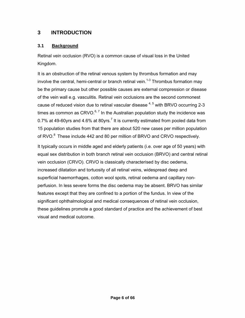
Retinal vein occlusion (RVO) is a common cause of visual loss in the United
Kingdom.
It is an obstruction of the retinal venous system by thrombus formation and may
involve the central, hemi-central or branch retinal vein.1-3 Thrombus formation may
be the primary cause but other possible causes are external compression or disease
of the vein wall e.g. vasculitis. Retinal vein occlusions are the second commonest
cause of reduced vision due to retinal vascular disease 4, 5 with BRVO occurring 2-3
times as common as CRVO.6, 7 In the Australian population study the incidence was
0.7% at 49-60yrs and 4.6% at 80yrs.7 It is currently estimated from pooled data from
15 population studies from that there are about 520 new cases per million population
of RVO.8 These include 442 and 80 per million of BRVO and CRVO respectively.
It typically occurs in middle aged and elderly patients (i.e. over age of 50 years) with
equal sex distribution in both branch retinal vein occlusion (BRVO) and central retinal
vein occlusion (CRVO). CRVO is classically characterised by disc oedema,
increased dilatation and tortuosity of all retinal veins, widespread deep and
superficial haemorrhages, cotton wool spots, retinal oedema and capillary non-
perfusion. In less severe forms the disc oedema may be absent. BRVO has similar
features except that they are confined to a portion of the fundus. In view of the
significant ophthalmological and medical consequences of retinal vein occlusion,
these guidelines promote a good standard of practice and the achievement of best
visual and medical outcome.
Page 7 of 66
3.2 Remit of the guidelines
The document aims to provide updated recommendations on the management of
RVO in the light of recent developments in both diagnostic tools and treatment
options that supersede those in the previous RVO guidelines. It has also reviewed
the risk factors for RVOs and included recommendations for investigations and
indications for medical management. These guidelines are intended for the use of
ophthalmologists, but will also be useful to physicians, general practitioners, and
commissioners.
The guidelines are considered interim and will be reviewed in a year (or earlier, as
necessary) as new evidence continues to emerge.
The recommendations in this document are based on scientific and medical
evidence. The guidelines do not address the NHS funding of its recommendations,
which are in the remit of NICE and the NHS.
Page 8 of 66
4 METHODS
4.1 The Guideline Development Group
Three ophthalmologists with expertise in medical retinal diseases, and a medical
ophthalmologist constituted the RVO Guidelines Development Group.
4.2 Gathering the evidence
4.2.1 Search methodology
The searches for the evidence base were conducted by the Management Team at
NHS Evidence-eyes and vision. Full details of the search methodology and the
search strategy are provided in Appendix A and Appendix B respectively.
(The search output was also used to inform the NHS Evidence Update on Retinal Vein Occlusion March 2010, http://www.library.nhs.uk/eyes/viewResource.aspx?resid=345418).
Period of Search: January 2002 to 15th February 2010
This time interval covered the period since the searches for the 2004 Retinal Vein
Occlusion Guidelines had been undertaken.
Databases searched: NHS Evidence - eyes and vision; PubMed; Medline; EMBASE; CINAHL; AMED; BNI; and PsycINFO
Inclusion criteria:
a) Publication type – Secondary publications (including Cochrane systematic reviews, systematic
reviews, reviews, meta or cost analysis)
Interventional studies (randomised controlled trials and controlled clinical trials).
Observational studies (cohort, case control, validation studies, observational or comparative studies, case reports/series, population based cross-sectional and cohort studies and qualitative surveys).
b) Relevancy to the scope of the Guideline
Page 9 of 66
In addition references from the Central Vein Occlusion Study and the Branch Vein
Occlusion Study (trials that reported between 15 and 25 years ago) were identified
as seminal research underpinning the evidence base for the current management of
retinal vein occlusion in the NHS, and were also included.
4.2.2 Supplemental searches
a) These were conducted by the Guideline Development Group and covered the period from February to August 2010.
b) Citations from the 2004 Guideline were selected by the Guideline
Development Group for their relevance to the scope of the guideline and the
updated evidence base (see 4.2.)
4.3 Assessing the evidence and forming recommendations
Relevant literature was identified and the level of evidence graded.
Recommendations for a good standard of practice were formed using the following
categories (i.e. strength of the evidence) and included in the text of the guidelines.
At least one meta-analysis, systematic review, or good quality randomised
control trial (RCT) directly applicable to the target population; or a body of
evidence consisting principally of RCTs, directly applicable to the target
population, and demonstrating overall consistency of results.
A body of evidence including high quality systematic reviews of case-control
or cohort studies, directly applicable to the target population and
demonstrating overall consistency of results or extrapolated evidence from
RCTs.
A body of evidence including studies rated as well conducted case control or
cohort studies with a low risk of confounding or bias and a moderate
probability that the relationship is causal, directly applicable to the target
population and demonstrating overall consistency of results; or extrapolated
A
B
C
Page 10 of 66
evidence from studies rated as high quality systematic reviews of case-control
or cohort studies.
Evidence from non-analytic studies, e.g. case reports, case series or expert
opinion
4.4 Consultation process
The Guideline Development Group invited comments on the draft guideline from all
UK consultant ophthalmologists prior to publication over a month consultation period.
Two external experts from outside the UK were also invited to evaluate the
guidelines. The comments were evaluated, and where appropriate, incorporated into
the final version of the guideline.
D
Page 11 of 66
5 AETIOLOGY AND RISK FACTORS
Retinal vein occlusion is due to thrombosis within retinal veins (central, hemi or
branch)1-3 although it remains unclear whether it is a primary or secondary effect.
Established cardiovascular risk factors are the predominant medical associations for
both central and branch vein occlusions and are summarised below and include
differentiation by age and ethnic groups. (See table 1) 9,10
5.1 Strength of evidence
Hypertension
This is the predominant risk factor with up to 64% of patients having hypertension
(Table 1) in the older age group (more than 50 years).11 This is more prevalent in
BRVO than CRVO. A new diagnosis or uncontrolled hypertension is a common
finding. Inadequately controlled hypertension is associated with recurrence of RVO
in the same eye or fellow eye involvement.
Hyperlipidaemia
Hyperlipidaemia (cholesterol > 6.5 mmol/l) is the predominant association in the
younger age group (< 50 years) of patients with retinal vein occlusion and is
associated in up to 50% of older patients.12
Diabetes mellitus
Diabetes mellitus (table 1) is associated with retinal vein occlusion. This may be due
to an increase of other cardiovascular risk factors (e.g. 70% of type II diabetics are
hypertensive).11,13,14
Glaucoma
Current evidence suggests an association between central retinal vein occlusion and
glaucoma.7, 15 One study suggests that BRVO is associated with glaucoma. 13
B
C
B
C
Page 12 of 66
Thrombophilia
Antiphospholipid antibody syndrome and hyperhomocysteinaemia are the two
haematological factors with the strongest evidence for association with CRVO,
although this is not proven. Factor V Leiden, protein S,C, and anti-thrombin 3
deficiency have also been reported.16 Thombophilia and the other rarer associations
e.g. oral contraceptive pill, and optic disc vasculitis assume more importance in
younger patients (<50 years).17, 18
5.2 Other Important Observations
Myeloproliferative disorders occur in 1% of patients presenting with retinal
vein occlusion.10
Other rare associations with retinal vein occlusion include:
Inflammatory diseases that cause or are associated with retinal vasculitis –
Behçets disease, polyarteritis nodosa, sarcoidoisis, Wegener’s
Granulomatosis and Goodpasture’s Syndrome.
Chronic renal failure and other secondary causes of hypertension and
diabetes e.g. acromegaly, Cushing’s syndrome.
Secondary causes of hypercholesterolaemia eg hypothyroidism.
C D
D
Page 13 of 66
6 NATURAL HISTORY OF RETINAL VEIN OCCLUSIONS, AND
LIVING WITH RETINAL VEIN OCCLUSIONS
6.1 CRVO
Natural history data from the CVOS study 19,20, and a systematic literature review8
demonstrated that visual outcome of CRVO depends on the visual acuity at
presentation. Eyes with initial visual acuity of 20/40 (6/12) or better have a better
prognosis for retaining good vision than those with worse vision. Only 20% of eyes
with initial visual acuity of 20/50-20/200 (6/15 -6/60) improve spontaneously to 20/50
(6/15) while 80% of patients with baseline vision worse than 20/200 (6/60) remain at
this level or worsen. Furthermore, the longer the duration of macular oedema, the
more the structural damage at the fovea so it is justifiable that early treatment be
initiated.
6.2 BRVO
Natural history data from an evidence based systematic review of 24 studies by
Rogers et al (2010) 21 indicated that VA was moderately poor (worse than 6/12) at
presentation, and that although there may be some improvement in the follow-up
period, such improvement was limited such that the average improvement did not
result in VA better than 6/12. Macular oedema may develop in 5 to 15% of eyes over
a 1 year period; however, of the eyes that had macular oedema at presentation, 18
to 40% may show some resolution. Approximately 20% of untreated eyes
experienced significant vision deterioration over time. In the BVOS, approximately
50% of untreated eyes with BRVO retain vision of 6/12 or better whilst 25% will have
vision of <6/60.5 Fellow eye involvement by BRVO may occur in 10% of cases over
time.
6.3 Low Vision and Living with RVO
It is known that the sudden onset of visual loss whether unilateral or bilateral results
in significant distress. CRVO is reported to be associated with a decreased vision-
Page 14 of 66
related quality of life as measured by the VFQ-25. The decrease in VFQ-25 scores is
related to the degree of visual loss in the better-seeing eye and the overall systemic
health of the patient. 22 Another study has shown that BRVO is associated with a
decrease in vision-related quality of life as determined by the VFQ-25 and that the
decrease in VFQ-25 score correlated well with the visual acuity of the involved eye,
even when good visual acuity is maintained in the uninvolved eye.23 Patients with
either central or branch retinal vein occlusion with macular oedema have significant
impact on their quality of life, and were willing to undergo potentially invasive
treatment. 24,25
Page 15 of 66
7 MANAGEMENT
There are two aims in the management of retinal vein occlusion: the identification of
modifiable risk factors and their medical management and the recognition and
management of sight-threatening complications.
Although the systemic investigation and treatment in all types of vein occlusion is
similar, the ophthalmological management of central (CRVO) and branch retinal vein
occlusion (BRVO) differs. These will therefore be considered separately.
7.1 OPHTHALMOLOGICAL MANAGEMENT
7.1.1 Central retinal vein occlusion (CRVO)
The main management problem is to differentiate ischaemic from non-ischaemic
central retinal vein occlusion. Patients with ischaemic CRVO are at risk of
neovascular glaucoma. This risk of iris neovascularisation is higher if the area of
retinal ischaemia (retinal non-perfusion as determined by FFA) is >10 disc
diameters.19 Ischaemic central retinal vein occlusion is associated with one or more
of the following characteristics:-
1. Poor visual acuity (44% of eyes with vision of <6/60 develop rubeosis 19
2. Relative afferent pupillary defect
3. Presence of multiple dark deep intra-retinal haemorrhage
4. Presence of multiple cotton wool spots
5. Fluorescein angiography showing greater than 10 disc areas of retinal capillary
non-perfusion (CVOS) 19
6. Electrodiagnostic tests (ERG): reduced b wave amplitude, reduced b:a ratio and
prolonged b-wave implicit time 26-30
7. Degree of retinal vein dilatation and tortuosity

Page 16 of 66
There is no evidence as to which combination of the above characteristics best
defines ischaemic CRVO. It is important to note that up to 30% of patients with
initially non-ischaemic central retinal vein occlusion will develop ischaemic
transformation.20,31-33 This is usually heralded by further rapid visual deterioration
and requires further assessment. CRVO especially of the non-ischaemic type needs
to be differentiated from the ocular ischaemic syndrome and other simulating
retinopathies.
7.1.1.1 Management of ischaemic central retinal vein occlusion and anterior
segment neovascularisation
An initial evaluation of risk factors and the appropriate treatment of the present risks
must proceed alongside management of the ocular findings.
The evidence supports the use of laser pan-retinal photocoagulation (PRP)
when iris new vessels (INV) or angle new vessels (ANV) are visible. 19
Recent evidence indicates that intravitreal anti-VEGF agents in combination
with PRP results in dramatic regression of the INV/ANV. 34-37 iCRVO should
be monitored monthly for new vessels iris and/ or angle. Repeat anti-VEGF
and PRP are advocated in case of recurrence of new vessels. In some
patients, it may not be logistically possible to review these patients monthly, 2-
3 monthly reviews may be sufficient, unless there are particular risk factors.
Particular individualized arrangements need to be made for these patients.
In circumstances when regular follow-up is impractical, prophylactic treatment
with PRP and anti-VEGF agent may be appropriate.38 However, none of the
available or commonly used anti-VEGF agents (bevacizumab, ranibizumab,
pegaptanib) currently have regulatory approval for such an indication.
A
C
C

Page 17 of 66
There is no proven protective effect of intravitreal triamcinolone acetonide on
anterior segment neovascularisation and it may exacerbate any pre-existing
neovascular glaucoma.This treatment option is not recommended.
7.1.1.2 Posterior segment neovascularisation
This is an uncommon complication following ischaemic central retinal vein occlusion
in eyes which have not developed neovascular glaucoma or who have been
successfully treated for rubeosis by laser.39 There is anecdotal evidence that new
vessels may be managed with a combination of anti-VEGF and PRP.
Pan-retinal photocoagulation for CRVO with INV or ANV requires 1500 – 2000
of 500-micron burns at the retina. This is best applied with 0.05-0.1 second
applications one burn width apart with sufficient energy to produce a pale burn
in the retina. Treatment is usually placed in the periphery avoiding areas of
retinal haemorrhage. Some cases require further treatment if the iris
neovascularisation fails to regress.19
The pan-VEGF A blockers, ranibizumab and bevacizumab have been shown
to cause regression of new vessels of the iris, angle and retina when given
intravitreally at the dose of 0.5mg/0.05ml and 1.25mg/0.05ml respectively.34-36
However, the effect is transient and recurrence of new vessels is common so
repeated treatment, typically every six weeks with these agents supplemented
with PRP may be required.
No anti-VEGF agent (bevacizumab, ranibizumab, pegaptanib) currently has a
licensed indication for posterior segment neovascularisation following ischaemic
CRVO. As such, GMC Guidelines on “Good Medical Practice” as it relates to the use
of both off-label and unlicensed medications and the manufacturer’s advice should
guide any physician directed potential intraocular use.
D
D
C
Page 18 of 66
7.1.1.3 Management of established neovascular glaucoma
The aim of management of this condition in a blind eye is to keep the eye pain
free. This is usually achieved by topical steroids and atropine. However, if the
eye has any visual potential intraocular pressure should be controlled with
topical pressure-lowering agents,cyclo-ablative procedures or filtering surgery
Intravitreal and intracameral bevacizumab has been shown to cause
regression of iris new vessels and decrease angle obstruction.40,41
Comparative case series indicate that iris new vessels regress faster after
intravitreal bevacizumab with PRP than with PRP alone.36,42 The reports also
suggest that bevacizumab may reduce the need for surgical interventions and
serve as a useful adjunct to filtering surgery. 37,44
7.1.1.4 Macular oedema
Macular oedema following central retinal vein occlusion results from leakage
of perifoveal capillaries. It results in visual loss. Randomised controlled trials
have failed to indicate benefit with grid laser photocoagulation, although a
trend in favour of treatment has been observed in younger patients. 45
Although there was significant reduction in the severity of macular oedema in
treated eyes compared to controls there was no visual acuity benefit.45
Triamcinolone acetonide (TRIVARIS): The rationale for the use of
intravitreal triamcinolone acetonide (IVTA) to treat macular oedema is that
corticosteroids reduce retinal capillary permeability and inhibit the expression
of the VEGF gene and the metabolic pathway of VEGF.
Evidence for the use of a specific preparation of triamcinolone in CRVO is
from the SCORE-CRVO Study (SCORE Study Report 5). 46 In this study, a
preservative-free form of triamcinolone (TRIVARIS, Allergan) given at different
doses, 1mg and 4mg, at four monthly intervals and with pre-defined re-
treatment criteria, was compared to observation. Results showed that both
D
C
A
A
Page 19 of 66
doses of TRIVARIS produced both anatomical and functional improvement of
macular oedema due to CRVO, compared to observation. However, at month
12, the 1mg dose had a better safety profile compared to the 4mg dose in
terms of a lower incidence of raised intraocular pressure (IOP) >35mmHg (5%
vs. 8%), incidence of cataract formation or progression (26% vs. 33%, cf. 18%
for observation) and need for cataract surgery (0% vs. 4%). 46
However, although FDA approved, TRIVARIS is not available for use in
clinical practice anywhere in the world and there are significant differences
between TRIVARIS and other currently available triamcinolone preparations.
Specifically, TRIVARIS is a single-use, pre-filled, preservative free
preparation, containing an injectable suspension of triamcinolone acetonide at
a concentration of 80mg/mL. This formulation contains hyaluronic acid and a
uniform and narrow distribution of triamcinolone particles and is buffered such
that the pH is in a narrow range of 7.0-7.4.
In contrast, the triamcinolone preparation that is commonly used in the UK, is
4mg from the KENALOG formulation (Squibb) which is indicated for intra-
articular joint use and has a contraindication for ocular use although it has
been used widely in Europe and the USA in the last few years. KENALOG is
typically presented in 1mL glass vials containing triamcinolone at a
concentration of 40mg/mL with a preservative, Benzyl Alcohol at 0.99% w/v,
which contains a wide variation in triamcinolone particle size. In addition to the
known risks of cataract and raised IOP seen with TRIVARIS, the presence of
a preservative may also lead to an increased risk of sterile endophthalmitis.
A preservative-free preparation of triamcinolone TRIESENCE (Alcon) has
been produced for use in the USA, but is currently unavailable in the UK, has
no ocular license for use in the UK and has no randomised controlled, clinical
trial data to support its use.
Therefore, there is no Grade A evidence to suggest that the visual and
anatomical responses seen with TRIVARIS in SCORE-CRVO would be
replicated with off-label IVTA preparations such as KENALOG or
TRIESENCE. 47 As such, GMC Guidelines on “Good Medical Practice” as it
Page 20 of 66
relates to the use of both off-label and unlicensed medications and the
manufacturer’s advice should guide physician directed intraocular use.
Dexamethasone Biodegradeable Implant: The rationale for the use of
intravitreal dexamethasone to treat macular oedema is similar to that of IVTA,
although dexamethasone has been show to be a more potent corticosteroid
that IVTA but also is able to reduce retinal capillary permeability and inhibit
the expression of the VEGF gene and the metabolic pathway of VEGF.
However, dexamethasone when injected intravitreally in its free form, has a
short half-life that limits its clinical utility as an injectable suspension. 48
A pre-filled applicator single-use, sustained release biodegradeable implant
containing 0.7mg of dexamethasone (OZURDEX, Allergan) has been studied
in the GENEVA study programme.49 In this study, OZURDEX and an
alternative dose of dexamethasone implant (0.35mg) were compared to a
sham injection, in patients with CRVO and BRVO in 2 parallel multicentre
studies and published together as the GENEVA study. Re-treatment was
possible 6 months after the first injection under pre-specified re-treatment
criteria. The first trial did not meet its original primary end-point , namely
proportion of eyes gaining 15 letters. The two trials were analysed together
and the primary outcome measure for all patients was time to achieve a ≥ 15
letter gain. The percentage of eyes with ≥ 15 letter gain in BCVA was
significantly higher in both implant groups compared with sham at days 30 to
90 with a peak effect at 60 days. Subgroup analyses of the BRVO and CRVO
subjects showed a significantly greater number achieved ≥ 15 letter gain from
30 to 90 days than sham treated eyes, and that sham treated eyes in the
BRVO subgroup were more likely to improve spontaneously than similar
managed CRVO eyes
Anatomically, improvements in macular oedema as seen by OCT were also
seen. In terms of safety, raised IOP peaked again at month 2 (3.2% of
patients had an IOP>35 mmHg), but declined significantly by month 3 and
was close to 0% by month 6, with 19% of patients requiring an IOP lowering
agent at month 6 and 0.7% of patients requiring any IOP lowering surgical
A

Page 21 of 66
procedures. Similarly, rates of cataract progression were low with 7%
progression at month 6, compared to 4% in the sham group. 49
Based on the GENEVA study programme, OZURDEX has received FDA and EU
approval for the 0.7 mg preparation, and is licensed in the UK for the treatment of
adult patients with macular oedema following either BRVO or CRVO.50 A post hoc
analysis suggested that eyes treated within 90 days of CMO being present were
more likely to improve than eyes commencing treatment after this time point..
Ranibizumab: The pan-VEGF blocker, ranibizumab (LUCENTIS, Novartis)
when given in 2 doses (0.3mg and 0.5mg) every month for 6 months, in the
CRUISE Trial, was shown to produce a 3-line gain of visual acuity and
corresponding anatomical response.51 The mean gain in VA was 12.7 and
14.9 letters respectively with the 0.3 and 0.5 mg compared to the sham
treated group at 6 months. Following the first 6 months, all patients were
enrolled into an open-label extension for an additional 6 months and the
overall 12 months results suggest that the visual gain established in the first 6
months can be retained with a slightly less intensive pro re nata (PRN)
therapy with ranibizumab (an average of 5.6 injections in 1st 6 months, vs. 3.3
injections in 2nd PRN 6 month phase). These results also show that patients in
the usual care group who were subsequently treated with ranibizumab 0.5mg
benefited from such treatment. Early treatment may be preferable as
confirmed from the earlier smaller observational studies 53-54, and a sham
controlled study.55
Ranibizumab 0.5mg (LUCENTIS) has subsequently received a license for the
treatment of macular edema following retinal vein occlusion (RVO) in the
USA, although in the EU, regulatory authorisation is not expected till 2011.
Bevacizumab: The pan-VEGF blocker, bevacizumab is unlicensed for
intraocular use. Several case series (without controls) indicate that
approximately 50% of subjects with non-ischaemic CRVO improve 2 or more
lines with intravitreal bevacizumab, whilst 90% of eyes showed vision
A
D
Page 22 of 66
stabilization by 12 months.56-60 However, the dosing schedule is unclear and
the long-term outcomes remain unclear. The SmPC for bevacizumab has
recently been altered to include cases of severe intraocular inflammation
following intravitreal administration of the drug.
(http://www.medicines.org.uk/EMC/medicine/15748/SPC/Avastin)
GMC Guidelines on “Good Medical Practice” as it relates to the use of both
off-label and unlicensed medications and the manufacturer’s advice should
guide physician directed intraocular use.
Pegaptanib: A phase II trial, and prospective case series indicate that
intravitreal 0.3mg pegaptanib sodium when given every 6 weekly for 6 months
improved the visual acuity by approximately 7 letters at 6 months. 61 The
reported follow-up periods are short and so the treatment regimen and the
response to treatment in the long-run remain unclear.
7.1.1.5 Recommendations for Further Follow-up
Follow-up after the initial 6 months of treatment will depend upon initiation of
anti-VEGF agent or steroid treatment for macular oedema but will normally be
required for up to 2 years in uncomplicated cases. The eyes should be
monitored for ischaemia (> 10DD non-perfusion) and for
occurrence/recurrence of macular oedema. The development of disc
collaterals +/- resolution of the macular oedema should lead to discharge from
clinical supervision. Detailed treatment and follow-up algorithms are provided
in subsequent sections of this guideline.
7.1.1.6 Experimental treatments
Chorio-retinal anastomosis (C-RA) was recently evaluated in a small (n=113)
randomised clinical trial. 62 Of patients in whom the C-RA was patent (76%), VA
improved by a mean of 11.7 letters compared to controls. Side effects included
C
D
Page 23 of 66
neovascularisation at the site of the anastomosis in18% and vitrectomy was required
in 9%, due to macular traction or non-resolving vitreous haemorrhage. The
procedure requires a special high power laser and significant operator experience. It
is only recommended in the context of prospective data collection by an
ophthalmologist specifically trained in its use. An Australian review of the technique
concluded that there was only level IV evidence available.63 The procedure was
therefore classified as experimental, with potential to cause serious side effects.
Other studies have reported significant complications associated with the procedure
e.g. choroidal neovascularisation64, retinal and subretinal fibrosis or traction65, and
vitreous haemorrhage.66
Trials of other treatments such as radial optic neurotomy (RON) with pars plana
vitrectomy, and thrombolytic therapies are under way.67, 68 RON is essentially a
procedure in which a radial incision is made in the nasal segment of the scleral ring
in order to decompress the presumed pressure within this compartment so as to
relieve pressure on the CRV. These, however, are only experimental at present and
are, therefore, not recommended except as part of clinical trials.
7.1.2 Branch Retinal Vein Occlusion
The diagnosis of branch retinal vein occlusion is clinical, as described before. In
doubtful cases, especially small BRVO, fluorescein angiography may be indicated to
confirm the diagnosis. Fluorescein angiography is particularly useful in determining
the extent of macular oedema and ischaemia. In the BVOS, approximately 50% of
untreated eyes with BRVO retain vision of 6/12 or better whilst 25% will have vision
of <6/60. Macular oedema and neovascularisation of the retina or disc are the two
major complications which may require therapy. Retinal neovascularisation occurs in
36% of eyes with >5 DD, and 62% with >4DD area of non-perfusion, as reported in 2
independent studies.6, 69

Page 24 of 66
7.1.2.1 Treatment of neovascularisation
Disc or retinal neovascularisation is an indication for photocoagulation to the
ischaemic retina (sector photocoagulation), although available evidence suggests
that waiting until vitreous haemorrhage occurs before laser treatment does not
adversely affect the visual prognosis. 6 ,69 New vessels occur only when there is at
least a quadrant of capillary closure and commonly after six months following the
occlusion.
Follow up visits at 3- 4 monthly intervals are recommended in patients with one
quadrant or more retinal ischaemia. It is recommended that sector laser
photocoagulation is applied once retinal or optic disc neovascularisation occur.
Fluorescein angiography is not usually necessary prior to laser because the area of
ischaemia is visible clinically.
Photocoagulation for neovascularisation is applied to the sector of retinal capillary
closure.6 500-micron burns at the retina are used and are applied in a scatter pattern
to the affected sector, one burn width apart are appropriate with sufficient energy to
create a gentle burn. A quadrant usually requires 400-500 burns.
7.1.2.2 Laser treatments for macular oedema
Laser Photocoagulation: Randomised clinical studies in the laser treatment
of macular oedema have demonstrated that a grid pattern of photocoagulation
in the distribution of leaking capillaries is beneficial but it is recommended only
after a period of three to six months following the initial event and following
absorption of the majority of haemorrhage.5, 70.
Fluorescein angiography should be carried out prior to this therapy usually at
> 3 months if visual acuity is 6/12 or less. This has two functions. Firstly it
identifies the leaking capillaries and secondly will indicate the degree of
macula ischaemia, which may limit the value of photocoagulation.70 It will also
help to avoid laser to collaterals.
A
Page 25 of 66
Laser Photocoagulation: Those with severe visual loss (less than 6/60
vision) and those in whom symptoms have been present for more than one
year are unlikely to benefit from photocoagulation. 70
Laser Photocoagulation: The optimal technique to administer laser
photocoagulation for macular oedema requires gentle burns of 50 to 100um.
The power depends on the individual patient. An average of between 20 to
100 applications (depending on the area of vascular leakage) are required in
a grid pattern to the areas of vascular leakage but avoiding the foveal
avascular zone (i.e. the burns must not approach the foveal centre by less
than 1/2 DD). Collaterals should be avoided. 5,70
Initial follow-up in all patients treated with laser photocoagulation should be at
three months following the occlusion. Subsequent follow-up at three to six
monthly intervals will depend on complications and laser treatment, and will
not normally be required after two years in uncomplicated cases
7.1.2.3 Pharmacologic Treatments
Triamcinolone acetonide (TRIVARIS): Evidence for the use of a specific
preparation of triamcinolone in BRVO is from the SCORE-BRVO Study
(SCORE Study Report 6).71,72 In this study, a preservative-free form of
triamcinolone (TRIVARIS, Allergan) given at different doses, 1mg and 4mg, at
four monthly intervals and with pre-defined re-treatment criteria, was
compared to laser photocoagulation. Results showed that both doses of
TRIVARIS produced both anatomical and functional improvement of macular
oedema due to BRVO, but this was similar in magnitude to laser. In addition,
at month 12, both the 1mg and 4mg doses had an inferior safety profile
compared to laser in terms of a higher incidence of raised intraocular pressure
>35mmHg (IOP) (2% and 14%, vs. 1%), incidence of cataract formation or
progression (25% and 35%, vs. 13%) and need for cataract surgery (0% and
D
D
A
Page 26 of 66
4%, vs. 3%). As such, laser is considered to have a more favourable
benefit:risk profile to TRIVARIS in BRVO.
Similar to the case in CRVO, there is no Grade A evidence to suggest that
the visual and anatomical responses seen with TRIVARIS in SCORE-BRVO
would be replicated with off-label IVTA preparations such as KENALOG or
TRIESENCE.73-75 As such, GMC Guidelines on “Good Medical Practice” as it
relates to the use of both off-label and unlicensed medications and the
manufacturer’s advice should guide physician directed intraocular use.
Dexamethasone Biodegradeable Implant: In the GENEVA study
programme48 (Haller, 2010), OZURDEX and an alternative dose of
dexamethasone in an implant (0.35mg) was compared to a sham injection, in
patients with CRVO and BRVO in 2 parallel multicentre studies. Re-treatment
was possible 6 months after the first injection under pre-specified re-treatment
criteria. . The first trial did not meet its original primary end-point , namely
proportion of eyes gaining 15 letters. The two trials were analysed together
and the primary outcome measure for all patients was time to achieve a ≥ 15
letter gain. The percentage of eyes with ≥ 15 letter gain in BCVA was
significantly higher in both implant groups compared with sham at days 30 to
90 with a peak effect at 60 days. Subgroup analyses of the BRVO and CRVO
subjects showed a significantly greater number achieved ≥ 15 letter gain from
30 to 90 days than sham treated eyes, and that sham treated eyes in the
BRVO subgroup were more likely to improve spontaneously than similar
managed CRVO eyes
Anatomically, improvements in macular oedema as seen by OCT were also
seen. In terms of safety, raised IOP peaked again at month 2 (3.2% of
patients had an IOP>35 mmHg), but declined significantly by month 3 and
was close to 0% by month 6, with 19% of patients requiring an IOP lowering
agent at month 6 and 0.7% of patients requiring any IOP lowering surgical
procedures. Similarly, rates of cataract progression were low with 7%
progression at month 6, compared to 4% in the sham group. 49
A
Page 27 of 66
Based on the GENEVA study programme, OZURDEX has received FDA and EU
approval for the 0.7 mg preparation, and is licensed in the UK for the treatment of
adult patients with macular oedema following either BRVO or CRVO.50 A post hoc
analysis suggested that eyes treated within 90 days of CMO being present were
more likely to improve than eyes commencing treatment after this time point..
Ranibizumab: The pan-VEGF blocker, ranibizumab (LUCENTIS, Novartis)
given in 2 doses (0.3mg and 0.5mg) every month for 6 months, was
compared with sham, in the BRAVO study. 76 At 6 months, the mean gain in
VA was +16.6 and +18.3 letters (0.3 and 0.5 mg respectively) compared to
+7.3 letters in the sham injection group. Sixty-one percent of the ranibizumab
0.5mg group achieved a 15 letter gain vrs 29% in the sham treated group.
However from months 3-5, a single application of rescue laser
photocoagulation was also allowed in all study arms if hemorrhages had
cleared sufficiently to allow safe application of laser and the following criteria
were met: Snellen equivalent BCVA ≤20/40 or mean central subfield thickness
250 m, and compared with the visit 3 months before the current visit,
patient had a gain of <5 letters in BCVA or a decrease of <50 m in mean
central subfield thickness. Based on these criteria, approximately 20% of
patients in both ranibizumab arms received adjunctive laser, versus 55% in
the sham injection arm. Following the first 6 months, all patients were enrolled
into an open-label extension for an additional 6 months and the overall 12
months results suggest that the visual gain established in the first 6 months
can be retained with a slightly less intensive pro re nata (PRN) therapy with
ranibizumab (an average of 5.7 injections in 1st 6 months, vs. 2.7 injections in
2nd PRN 6 month phase).76 These results also show that patients in the sham
injection group who were subsequently treated with ranibizumab 0.5mg
benefited from such treatment. However, as seen with the results of GENEVA
& CRUISE studies, the visual acuity outcome never caught up in this delayed
treated group compared to eyes treated earlier.
Ranibizumab 0.5mg (LUCENTIS) has subsequently received a license for the
treatment of macular oedema following retinal vein occlusion (RVO) in the
A

Page 28 of 66
USA, although in the EU, it has yet to receive regulatory approval. As such,
GMC Guidelines on “Good Medical Practice” as it relates to the use of both
off-label and unlicensed medications and the manufacturer’s advice should
guide physician directed intraocular use.
Bevacizumab: Currently, increasing short-term data support the fact that
multiple intravitreal bevacizumab injections reduce macular oedema
secondary to branch retinal vein occlusion including those that had failed
previous laser treatment.57,59,60,77-79 The most common treatment regimen is
two to three injections over the first 5-6 months.
However, further randomized, controlled trials are required to assess long-
term safety and efficacy of intravitreal bevacizumab. No recommendations on
the use of intravitreal bevacizumab can be made at this time. Due to the
unlicensed nature of bevaciumab when compounded and distributed to third
parties, GMC Guidelines on “Good Medical Practice” as it relates to the use of
both off-label and unlicensed medications and the manufacturer’s advice
should guide physician directed intraocular use.
Periocular triamcinolone: Periocular (orbital floor or retrobulbar)
triamcinolone has been administered as treatment of macular oedema in
BRVO.80, 81 Although both routes of administration demonstrated efficacy, the
results are short-lived. 81
7.1.2.4 Other Treatments
The evidence on the efficacy of surgical interventions in BRVO are limited to
case reports and case series.82
NICE has reviewed the evidence of arteriovenous sheathotomy for this
condition and recommended that this procedure be done only as part of a
research study. 83
C
C
A
Page 29 of 66
7.1.3 Hemisphere vein occlusion
The risk of rubeosis in ischaemic hemi-central vein occlusion is greater than that of
BRVO but less than that of CRVO. The risk of disc neovascularisation appears
greater for hemispheric vein occlusion than either ischaemic CRVO or BRVO.
The management of hemispheric vein occlusion is similar to that described for
branch retinal vein occlusion, the guidelines for treatment options being those
described above for retinal branch vein occlusion.
Page 30 of 66
7.2 MEDICAL MANAGEMENT
7.2.1 Referral for medical investigation and treatment
IT IS THE RESPONSIBILITY OF THE OPHTHALMOLOGICAL TEAM TO ENSURE
MEDICAL INVESTIGATION AND TREATMENT IS INITIATED ON DIAGNOSIS OF
RETINAL VEIN OCCLUSION.
Recommended investigations for patients with retinal vein occlusion are listed in
Table 2. It is the responsibility of the diagnosing physician or ophthalmologist to:
1. Investigate and interpret results.
2. Refer the patient for appropriate medical advice with urgency according to the
severity of underlying risk factor(s).
3. Ensure that specialists in the relevant field should manage the rarer causes of
retinal vein occlusion.
4. Ensure that initiation of medical management occurs within 2 months of
diagnosis.
The importance of detecting and treating underlying medical conditions lies in the
need to prevent further non-ocular target organ damage, as well as to prevent
recurrence of venous occlusion particularly in the fellow eye.68 Two long-term follow-
up studies of patients with retinal vascular disease (retinal vein occlusion and retinal
arterial occlusion) demonstrate excess cardiovascular morbidity, mortality from
stroke, 69, 70 and myocardial infarction over a ten-year period.
Page 31 of 66
7.2.2 Medical Management
Medical management should be targeted at three areas:
7.2.2.1 Restoring venous patency
Clinical & Diagnostic Work-up: This is applicable in a limited number of
cases. Patients with ‘incipient’ retinal vein occlusion (consisting of the
presence of dilated retinal veins and few widely scattered haemorrhages
without any macular oedema in patients who are either asymptomatic or have
transient episodes of blurring in the affected eye and may have slight increase
in retinal circulation time on fluorescein angiography 88 should have medical
investigation for underlying systemic risk factors and treatment urgently as
there is the potential to prevent progression, or to reverse the existing
occlusion.
The medical therapies explored to improve retinal venous flow include: -
Anti-coagulants: heparin
Fibrinolytic agents: streptokinase, tissue plasminogen activator (intravitreal
or systemic)
Anti-platelet drugs: aspirin, prostacyclin, ticlopidine
These would seem to be logical treatments, but results from trials using heparin,
streptokinase and warfarin have been disappointing with limited evidence of benefit
owing to adverse effects of retinal and vitreous haemorrhage. Aspirin is not
recommended for primary prevention of cardiovascular events. If aspirin is used in
primary prevention, the balance of benefits and risks should be considered for each
individual, particularly the presence of risk factors .89 Given that there is insufficient
evidence to suggest that RVO is a risk factor for stroke or vascular mortality, the role
of aspirin in RVO remains equivocal.
D
C
Page 32 of 66
Haemodilution: The effects of haemodilution have been inconsistent in
completed control trials in RVO and the treatment may have adverse affects
on the patients’ general well-being.
7.2.2.2 Ameliorate cardiovascular morbidity and mortality associated with
retinal vein occlusion
Manage underlying risk factors: Although reports on the association of RVO
with cardiovascular morbidity and mortality are conflicting, it is crucial that all
cardiovascular risk factors be identified and treated in patients with RVO. 86, 87
Cardiovascular risk factors identified in patients with retinal vein occlusion
should be managed according to the Joint British recommendations on
prevention of coronary heart disease and the recent updates on the
management of hypertension and the use of statins.91- 93
Patients with rarer underlying conditions such as myeloma and inflammatory
disorders should be referred and managed by appropriate specialists.
7.2.2.3 To prevent the recurrence of retinal vein occlusion
Several series have demonstrated that recurrence of retinal vein occlusion
may occur in the affected eye or in the fellow eye in up to 15% of patients
over a five year follow up period.85 Rates vary according to studies in differing
countries from 9 to 15%. In view of the poor potential visual outcome of
patients with recurrent retinal vein occlusion, this aspect has been studied, but
not in controlled trials. Available data supports the concept that recurrence of
retinal vein occlusion may be reduced by medical treatments of underlying
cardiovascular risk factors.
Hormone Replacement Therapy: Although estrogen-containing HRT should
not be commenced in those women with retinal vein occlusion, continued use
does not appear to be associated with a higher rate of recurrence.94
C
C
C
D
Page 33 of 66
Historically, HRT was contraindicated and discontinued following central vein
thrombosis.13 Following the work of the Eye Disease Case-Control Study
Group and Kirwan and associates17, medical practice showed a trend to
continue HRT following retinal vein occlusion due to the epidemiological
evidence supporting HRT in the prevention of cardiovascular disease.
This policy has not lead to the potentially disastrous visual outcome of
recurrence of retinal vein occlusion in the fellow eye. Currently, the decision
about whether to continue HRT in a woman with retinal vein occlusion should
be made on a case by case basis. The decision should be based on the
woman’s individual case history, including the indication for HRT use.
The degree of residual visual impairment may influence the decision as a
recurrence in the fellow eye may have a potentially devastating visual
outcome. Further guidance may be obtained from the results of thrombophilia
screening, as this may provide an indicator of future risk. The current
uncertainty about the effects of HRT on cardiovascular risk and recent
guidelines for the use of HRT should also be considered.
7.2.3 Management of younger patients (less than 50 years of age)
Central retinal vein occlusion in this age group has been thought to have a more
benign outcome in a greater proportion of patients, with spontaneous regression of
the central retinal venous occlusive event being more common. However, at least
20% of patients develop poor visual outcome with severe neovascular
complications.95 Some authorities advocate the use of steroid therapy but this has
not been tested in controlled trials.
Patients in this age group with BRVO usually have underlying systemic conditions
such as hypertension or hyperlipidaemia which should be managed appropriately.95
Those with CRVO present a particular problem in investigation and management.
Many of these patients will have no identifiable underlying cause despite extensive
investigation including the specialised investigations listed in Table 2.

Page 34 of 66
In females the contraceptive pill is the most common underlying association, and
caution is advised in patients with retinal vein occlusion. There is debate as to the
exact prevalence of thrombophilic disorders in this patient group as well as
appropriate therapy. Identified inflammatory disease should be treated as
appropriate to the condition and referred for specialist medical advice.
Page 35 of 66
7.3 TREATMENT ALGORITHMS
Dexamethasone intravitreal implant (Ozurdex) has received its FDA and EU licenses
for treatment of retinal vein occlusions. These were based on the GENEVA Study
results.
Clinical trial evidence for ranibizumab’s effects in retinal vein occlusion are available
from two phase III clinical studies in branch occulsions (BRAVO Study) and central
occlusions (CRUISE study). Ranibizumab has recently received FDA approval for
the treatment of RVO in the US. It is assumed that the EU licence for ranibizumab in
the treatment of retinal vein occlusion will be available shortly.
The GMC Good Medical Practice Guidelines, and the manufacturer’s advice should
guide the intraocular use of ranibizumab in conditions outside its current indications.
7.3.1 Minimum Service Specifications for retinal vein occlusions
The minimum service specifications include personnel and equipment and are
similar to those for neovascular age-related macular degeneration (nAMD). A
consultant ophthalmologist with expertise in the management of medical retinal
diseases is expected to lead the team. Support would be provided by other
ophthalmologists at consultant, middle grade as well as trainees.
It is expected that there will be adequate support from the nurses, ophthalmic
photographers/technicians. A clinic coordinator and data entry personnel equipped
with an electronic patient record (EPR) system are essential to running an efficient
service delivery. LogMAR visual acuity systems, and an OCT (Stratus or higher
specification) are required as part of the minimum service requirements. (See RVO
Service Provision, below).
7.3.2 Treatment of Risk Factors
It is essential to treat risks known to be associated with all types of RVO. It is the
responsibility of the ophthalmological team to ensure that medical investigations and
Page 36 of 66
treatment is initiated on diagnosis of RVO. This ensures that the risk of recurrence of
RVO, or the occurrence of new occlusions are reduced. It also improves the chance
of reversing the retinal vein occlusion, as well as ameliorate cardiovascular morbidity
and mortality associated with RVO.
It is expected that the ophthalmic team will evaluate, or arrange for such evaluation,
of the patient for common risk factors of systemic hypertension, diabetes,
hyperlipidemia, and glaucoma/ocular hypertension. Referral would be expected to
the appropriate physician for optimal management.
Patients should also be referred to the appropriate specialists in the relevant field for
investigation and management of the rarer risk factors.
7.3.3 Treatment Algorithm for CRVO
7.3.3.1 Baseline Assessments
The minimum assessments required before commencing treatments for CRVO
include:
1. Clinical examination including
a. Best corrected visual acuity (BCVA)
b. Pupillary reactions- the presence of a brisk afferent papillary defect
(APD)
c. IOP
d. Gonioscopy
e. Slit lamp biomicroscopy of the anterior segment and fundus
2. Retinal Imaging
a. Colour fundus photographs in all cases
b. Optical coherent tomography (OCT) with Zeiss Stratus or higher
specification OCT
Page 37 of 66
c. Fundus fluorescein angiography (FFA) where the interpretation is not
confounded by the presence of marked intraretinal haemorrhage or can
be based on clinical judgement.
7.3.3.2 Management at baseline
This depends on whether the CRVO is ischaemic or non-ischaemic. There is no
evidence as to which combination of characteristics best defines ischaemic CRVO. It
is important to note that up to 30% of patients with initially non-ischaemic central
retinal vein occlusion will develop ischaemic transformation. This is usually heralded
by further rapid visual deterioration and requires further assessment. CRVO
especially of the non-ischaemic type needs to be differentiated from the ocular
ischaemic syndrome and other simulating retinopathies.
7.3.3.3 Non-Ischaemic CRVO
By definition, there will be no iris or angle NV
1. If VA is 6/12 or worse +OCT ≥250 microns (Stratus, or equivalent) consider
pharmacotherapy with Ozurdex which is licensed or Ranibizumab which is
unlicensed but has robust evidence.
2. However, the presence of a brisk APD associated with VA<6/96 indicates
potentially poor treatment outcomes.
a. As such no treatment would be recommended for such cases. Watch
for NVI/NVA, and treat as ischaemic CRVO below.
7.3.3.3.1 Management – Subsequent Follow-Up
1. Depending on baseline VA, OCT & FFA findings, and initial treatment options,
monitoring will be required at varying frequencies during the first 6 months.
Page 38 of 66
a. Assessments at each visit include VA, IOP, gonioscopy, fundoscopy,
and OCT
b. From month 6 to 18 months, monitoring at monthly or 3 monthly,
depending on the particular treatment of choice
2. Re-treatment as per the criteria below
7.3.3.3.2 Re-treatment Criteria
1. Based on the results of the clinical trials, treatment may be repeated unless
a. VA>6/7.5 (84 letters on LogMAR) OR
b. Central Retina Thickness (CRT) on OCT<250 microns OR
c. Treatment is discontinued at the clinician’s discretion (See below)
2. Re-treatment with dexamethasone implant (OZURDEX) should take place at
4 to 6 month intervals. There is only limited case report data to support dosing
intervals less than 6 monthly.
3. Based on the CRUISE study, consider following the monthly injection
schedule for the first 6-12 months, and the PRN re-treatment criteria from the
study should be used as the basis for a PRN dosing regimen.
7.3.3.3.3 Treatment discontinuation
1. Treatment may be discontinued in the presence of continuing deterioration of
vision or morphology of the macula.
2. Criteria for stopping treatment include
a. No evidence of benefit from treatment, e.g. continued worsening / lack
of stabilisation of vision despite an adequate trial of therapy.
b. Rise in IOP uncontrolled by effective IOP lowering agents when
dexamethasone implant (OZURDEX) has been the treatment
Page 39 of 66
c. When using ranibizumab, or off-label agents such as other anti-VEGF
agent e.g. bevacizumab, if in the clinician’s opinion, the benefit: risk
profile of further treatment is unfavourable, e.g. new MI or CVA.
7.3.3.4 Ischaemic CRVO
7.3.3.4.1 Management at baseline
In the presence of significant retinal ischaemia at baseline, regular monitoring is
advised.
7.3.3.4.2 Subsequent Management
1. Monitoring should be at monthly intervals wherever possible. Where this is
impossible, two monthly monitoring may be acceptable.
a. If Iris or angle NV present and anterior chamber angle is open
i. There is limited anecdotal evidence for the use of intravitreal
bevacizumab in such cases and its use would be considered
unlicensed, e.g. Panretinal photocoagulation (PRP), in combination
with intravitreal bevacizumab and review 6-weekly.
ii. Repeat PRP +/- intravitreal bevacizumab if NVI/NVA still persists at
follow-up
iii. Follow-up 3 monthly to up to 12 months. Subsequent follow-up will be
guided by the clinical findings and on-going other treatment
b. If iris or angle NV and anterior chamber angle is closed
i. There is limited anecdotal evidence for the use of intravitreal
bevacizumab in such cases and its use would be considered
unlicensed, e.g. Advise PRP +/- intravitreal bevacizumab.
Page 40 of 66
ii. Consider specialist glaucoma input and the options of cyclodiode laser
therapy or tube/ shunt surgery
c. Where ischaemic CRVO occurs but there is no anterior segment
vascularisation (NVI/NVG) as yet, and regular follow-up is impractical, it is
reasonable to provide prophylactic treatment with PRP
7.3.4 Treatment Algorithm for BRVO
7.3.4.1 NON –ISCHAEMIC BRVO
7.3.4.1.1 BASELINE ASSESSMENTS
The minimum assessments required before commencing treatments for BRVO
include:
3. Clinical examination including
a. Best corrected visual acuity (BCVA)
b. Pupillary reactions- the presence of a brisk afferent papillary defect
(APD)
c. IOP
d. Gonioscopy if clinically indicated
e. Slit lamp biomicroscopy of the anterior segment and fundus
4. Retinal Imaging
a. Colour fundus photographs in all cases
b. Optical coherent tomography (OCT) with Zeiss Stratus or higher
specification OCT
c. Fundus fluorescein angiography (FFA) where the interpretation is not
confounded by the presence of marked intraretinal haemorrhage or as
per clinical judgement.
Page 41 of 66
7.3.4.1.2 Management of macular oedema secondary to BRVO with no or minimal
evidence of macular ischaemia
1. 1. If patients with macular oedema secondary to BRVO are seen within 3
months of onset of BRVO, consider pharmacotherapy with Ozurdex which is
licensed or Ranibizumab which is unlicensed but has robust clinical evidence
of efficacy.
2. If patients are seen after 3 months from onset of BRVO, consider laser
photocoagulation or pharmacotherapy with Ozurdex which is licensed or
Ranibizumab which is unlicensed but has robust clinical evidence of efficacy.
7.3.4.1.3 Management in eyes with evidence of marked macular ischaemia No
immediate treatment is recommended. Watch for conversion of the RVO to
ischaemic type and subsequent neovascularisation
7.3.4.1.4 Re-treatment criteria
1. Based on the results of the clinical trials, treatment may be repeated unless.
a. VA>6/7.5 (84 letters on LogMAR) OR
b. Central Retina Thickness (CRT) on OCT<250 microns
c. Treatment should be discontinued (See below)
2. Re-treatment with dexamethasone implant (OZURDEX) should take place
with 4-6 months after first treatment.
3. Re-treatment with ranibizumab injections should occur monthly for the first 6
months followed by a PRN schedule based on re-treatment criteria from the
BRAVO study
4. Re-treatment with modified Grid Laser Photocoagulation should be
considered at 4 monthly intervals
Page 42 of 66
7.3.4.1.5 Discontinuation of treatment
1. Treatment may be discontinued in the presence of continuing deterioration of
vision or morphology of the macular
2. Criteria for stopping treatment include
a. No evidence of benefit from treatment, e.g. Continued worsening / lack
of stabilisation of vision despite treatment on 2 consecutive treatment
visits
b. Rise in IOP uncontrolled by effective IOP lowering agents when
dexamethasone implant (OZURDEX) has been the treatment
c. When using ranibizumab, or off-label agents such as other anti-VEGF
agent e.g. bevacizumab, if in the clinician’s opinion, the benefit: risk
profile of further treatment is unfavourable, e.g. New MI or CVA.
7.3.4.2 Unlicensed and Contraindicated pharmacological agents -
Considerations
1. Triamcinolone
There are no randomised controlled trials for any clinically available
triamcinolone preparation, in retinal vein occlusion.
Clinical trial evidence for the use of triamcinolone in retinal vein
occlusion comes from the SCORE Study and involves a single-use,
preservative free preparation, using a triamcinolone concentration of
80mg/mL and of a narrow particle size distribution (TRIVARIS).
This TRIVARIS preparation is not currently available for clinical use
anywhere in the world and is different from the commonly available
Kenalog which is formulated in large vials with a preservative (BAK),
has a triamcinolone concentration of 40mg/mL and has a wide variation
in triamcinolone particle size.
The manufacturer of Kenalog (Bristol Myers Squibb) has specifically
advised against its intraocular use and the product license in the UK
specifically states that it is contraindicated for use intraocularly.
Page 43 of 66
The GMC Good Medical Practice Guidelines, and the manufacturer’s
advice should guide the intraocular use of Kenalog.
2. Anti-VEGF - Bevacizumab
Bevacizumab received its initial UK product license for the
management of metastatic colorectal cancer in combination with 5-FU,
a chemotherapeutic agent
Bevacizumab does not have a license for the management of any
ocular conditions.
However, it has been used extensively in clinical practice with some
success, for the management of many retinal conditions that have a
VEGF driven pathophysiology, despite a lack of randomised controlled,
clinical trial evidence.
The MHRA has recently confirmed that the license status in the UK for
bevacizumab for the management of any retinal disease including
retinal vein occlusion when compounded in a pharmacy and distributed
to a third party in single dose pre-filled syringes, is “unlicensed”, since
its formulation is different from that used in the oncology setting.
The GMC Good Medical Practice Guidelines, and the manufacturer’s
advice should guide the intraocular use of bevacizumab.
Page 44 of 66
7.3.4.3 Ischaemic BRVO
7.3.4.3.1 Management
1. Watch carefully for NV
2. Perform 3 Monthly follow, especially if the area of retinal ischaemia is >
4DD, and treatment is not required for macular oedema.
3. If NVE occurs, there is limited anecdotal evidence for the use of
intravitreal bevacizumab in such cases and its use would be considered
unlicensed, e.g.
a. PRP+/- intravitreal bevacizumab 4-6 weekly until quiescent.
b. The use of bevacizumab must be guided by the GMC Good
Medical Practice Guidelines on the use of unlicensed products.
4. Follow-up 3 monthly to up to 12 months. Subsequent follow-up will be
guided by the clinical findings and on-going treatment.
7.3.4.3.2 Other Options
All other treatments for BRVO, including A-V sheathotomy currently remain
investigative, and as such are not recommended as part of routine clinical
practice.
7.3.5 Hemispheric Vein Occlusion Algorithm
1. The management of hemispheric vein occlusion is similar to that
described for branch retinal vein occlusion. Particularly, macular oedema
secondary to hemi-vein occlusion is managed similarly to than in BRVO.
2. The risk of rubeosis in ischaemic hemi-central vein occlusion is greater
than that of BRVO but less than that of CRVO. Assessment for anterior
segment neovascularisation, including gonioscopy is therefore indicated.
The management of NVI/NVG is the same as that secondary to CRVO.
Page 45 of 66
7.4 RVO SERVICE PROVISION
Patients with retinal vein occlusion have previously been evaluated and followed
up in eye clinics. Essentially, these clinic visits were aimed at identifying
modifiable risk factors and managing the sight threatening complications of the
vein occlusion. A few patients benefited from laser treatment. Recent large
controlled clinical trials have unequivocally demonstrated the clinical efficacy for
intravitreal injections of ranibizumab (Lucentis) and dexamethasone implants
(Ozurdex) in preventing visual loss, and improving vision in all types of RVO.
7.4.1 Burden of disease due to RVO
There are currently no UK based studies on the prevalence of RVO. It is currently
estimated from pooled data from 15 population studies from that there are about
520 new cases per million population of RVO.8 These include 442 and 80 per
million of BRVO and CRVO respectively. However, only 200-260/million will
require treatment as some patients with RVO retain good vision and do not
require any treatment.8,19,20,21 BRVO occurs 2-3 times as common as CRVO.
7.4.2 Existing service provision and referral pathways
The management of an individual patient depends on the type of RVO and
complications. Until recently, the management of retinal vein occlusion has been
retinal laser photocoagulation for macular oedema (in BRVO), retinal or iris
neovascularisation. Some cases of iris neovascularisation require cyclodiode
laser or cyclocryotherapy. With the introduction of intravitreal delivery of
dexamethasone and anti-VEGF treatments, the management of RVO is
undergoing significant change as indicated in other parts of this guideline.
Page 46 of 66
7.4.3 Anticipated workload
Given the effectiveness of intravitreal steroid injections and anti-VEGF therapies
in all types of RVO, the number of patients eligible for treatment, and treatment
frequency will increase significantly.
Patients receiving anti-VEGF therapy will require 4 - 6 weekly visits whilst those
receiving dexamethasone may require injections every 4-6 months, but require
monitoring visits as well at 4-6 weeks intervals. It will not be feasible to ask these
patients to travel long distances for repeat treatments at these intervals. It is
therefore essential to provide comprehensive treatment in the local hospital eye
unit.
As these patients already attend the different local eye clinics for diagnosis and
investigation, it is only the frequency of attendances and provision of injections
that will alter. It is expected that clinic attendances will increase, probably to as
much as 4-6 times the current attendances for RVO in the first 12 months
following diagnosis. In addition, the times required for administering treatments
have to be allowed for.
7.4.4 RVO Service Specifications
7.4.4.1 Early access
It is recommended that the time from referral from the primary source to initial
evaluation and treatment by the retinal specialist at the eye clinic is not more
than 2-4 weeks from presentation. This recommendation is based on reports
from the CVOS that reported that the final visual acuity depends on the visual
acuity at presentation.19,20 More recently, the GENEVA Study 49 also suggested
that visual recovery is better for eyes that are treated early after the onset of
RVO.
Page 47 of 66
7.4.4.2 Geographical equity of access to all regions within the UK
There needs to be immediate access to retinal specialists with expertise in the
management of RVO for all patients, irrespective of geographic location. Referral
pathways of RVO to treating specialists may vary but must be appropriate for
different regions, as there may be several variations in geographic population
distribution, logistics, expertise, and physician workload. The guiding principle is
that no particular patient or region should be disadvantaged.
7.4.4.3 Minimum clinical services required for effective management
These include
1. Best corrected visual acuity assessments by optometrist or certified VA
examiners
2. Colour Fundus photographs and Fundus Fluorescein angiography (FFA)
by trained technical staff
3. Optical coherence tomography (OCT) with the Stratus or higher
specification equipment by trained technical staff
4. Treatment initiated within 1-2 weeks of assessment
5. Appropriate facilities for IVT injection
6. Appropriate capacity for follow up, monitoring and re-treatment
7.4.5 RVO Referral Pathways
All patients suspected to have RVO by the optometrist, general practitioner, or
other health workers should be referred directly to the nearest Eye Casualty, or
Eye Clinic. Optometrists may be used for ‘screening’ or first examination of
patients suspected of having RVO. Referrals from the optometrist should be sent
directly to an ophthalmology department, and should not necessarily pass
through the general practitioner as such a route introduces unnecessary delays.
Page 48 of 66
Self referral or presentation to the Eye Casualty/Clinic should be encouraged,
especially in patients who have second eye involvement.
7.4.6 Resources
The contemporary management of RVO requires collaboration between the
ophthalmology multidisciplinary team and physicians. 96 The multidisciplinary
ophthalmic team is similar to that required for the management of wet age-
related macular degeneration (wAMD).
(http://www.rcophth.ac.uk/docs/publications/AMD_GUIDELINES_FINAL_VERSI
ON_Feb_09.pdf /). Intravitreal injection facilities will exist and be shared with
AMD services.
It is expected that all patients with RVO will require refraction LogMAR visual
acuities, FFA and OCT at the commencement of treatment. Subsequent follow
up will require OCTs, and FFA thereafter only when indicated.
7.4.7 Low Vision and Living with RVO
It is known that the sudden onset of visual loss whether unilateral or bilateral
results in significant distress. CRVO is reported to be associated with a
decreased vision-related quality of life as measured by the VFQ-25.22,23
Patients with reduced BCVA secondary to RVO or other causes are offered the
opportunity of accessing low vision support and advice at an early stage. Advice
and use of task lighting and magnifiers reduce the early impact of sight loss and
the risk of falls. It is important not to wait until all treatment options have been
explored or until an individual’s vision deteriorates to a level that merits
registration as visually impaired/severely visually impaired before considering
referring an individual to low vision and rehabilitation services.

Page 49 of 66
It is easier to introduce the patient to low vision services at an earlier than latter
stage of the disease. An individual can learn how to use their remaining vision
more effectively, retaining independence and confidence.
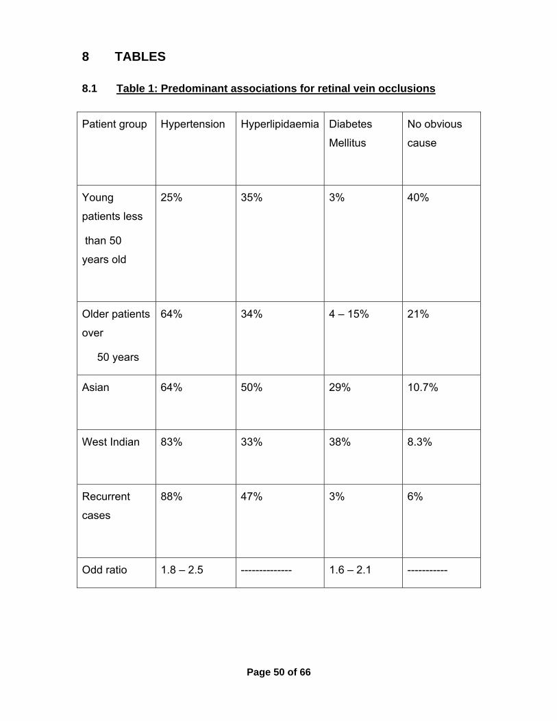
Page 50 of 66
8 TABLES
8.1 Table 1: Predominant associations for retinal vein occlusions
Patient group
Hypertension
Hyperlipidaemia
Diabetes
Mellitus
No obvious
cause
Young
patients less
than 50
years old
25%
35% 3% 40%
Older patients
over
50 years
64% 34% 4 – 15% 21%
Asian
64% 50% 29% 10.7%
West Indian
83% 33% 38% 8.3%
Recurrent
cases
88% 47% 3% 6%
Odd ratio 1.8 – 2.5 -------------- 1.6 – 2.1 -----------
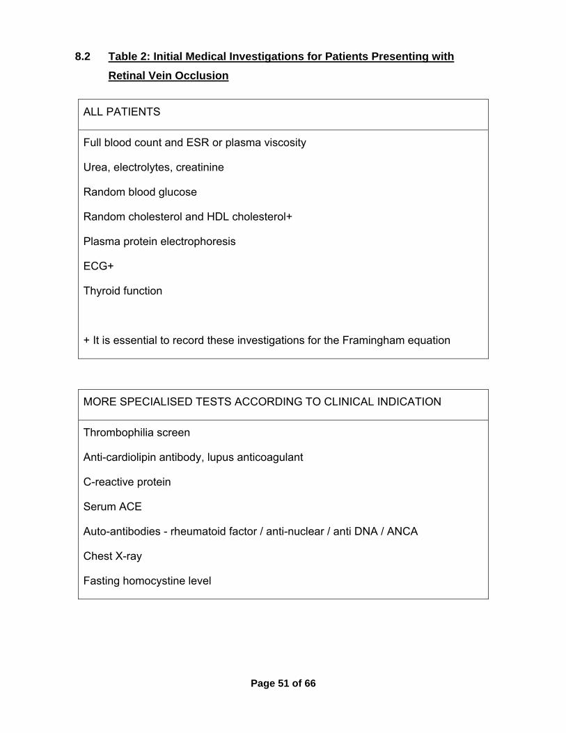
Page 51 of 66
8.2 Table 2: Initial Medical Investigations for Patients Presenting with
Retinal Vein Occlusion
ALL PATIENTS
Full blood count and ESR or plasma viscosity
Urea, electrolytes, creatinine
Random blood glucose
Random cholesterol and HDL cholesterol+
Plasma protein electrophoresis
ECG+
Thyroid function
+ It is essential to record these investigations for the Framingham equation
MORE SPECIALISED TESTS ACCORDING TO CLINICAL INDICATION
Thrombophilia screen
Anti-cardiolipin antibody, lupus anticoagulant
C-reactive protein
Serum ACE
Auto-antibodies - rheumatoid factor / anti-nuclear / anti DNA / ANCA
Chest X-ray
Fasting homocystine level
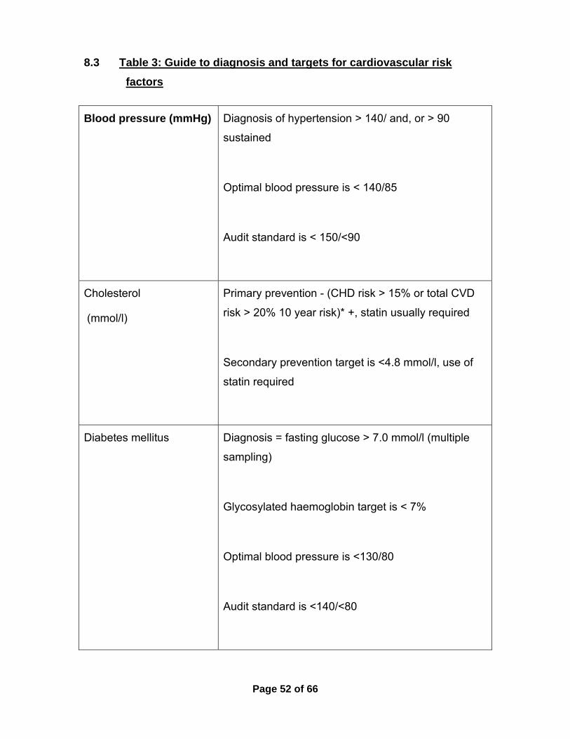
Page 52 of 66
8.3 Table 3: Guide to diagnosis and targets for cardiovascular risk
factors
Blood pressure (mmHg) Diagnosis of hypertension > 140/ and, or > 90
sustained
Optimal blood pressure is < 140/85
Audit standard is < 150/<90
Cholesterol
(mmol/l)
Primary prevention - (CHD risk > 15% or total CVD
risk > 20% 10 year risk)* +, statin usually required
Secondary prevention target is <4.8 mmol/l, use of
statin required
Diabetes mellitus
Diagnosis = fasting glucose > 7.0 mmol/l (multiple
sampling)
Glycosylated haemoglobin target is < 7%
Optimal blood pressure is <130/80
Audit standard is <140/<80
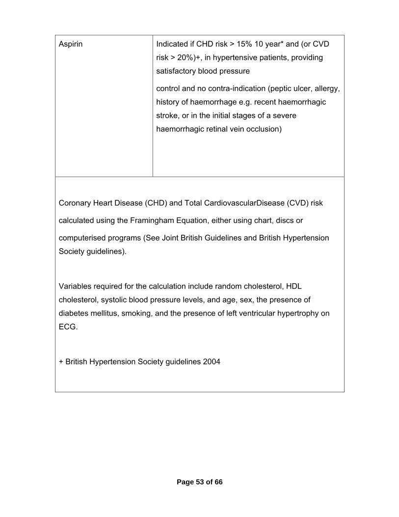
Page 53 of 66
Aspirin
Indicated if CHD risk > 15% 10 year* and (or CVD
risk > 20%)+, in hypertensive patients, providing
satisfactory blood pressure
control and no contra-indication (peptic ulcer, allergy,
history of haemorrhage e.g. recent haemorrhagic
stroke, or in the initial stages of a severe
haemorrhagic retinal vein occlusion)
Coronary Heart Disease (CHD) and Total CardiovascularDisease (CVD) risk
calculated using the Framingham Equation, either using chart, discs or
computerised programs (See Joint British Guidelines and British Hypertension
Society guidelines).
Variables required for the calculation include random cholesterol, HDL
cholesterol, systolic blood pressure levels, and age, sex, the presence of
diabetes mellitus, smoking, and the presence of left ventricular hypertrophy on
ECG.
+ British Hypertension Society guidelines 2004
Page 54 of 66
9 CITED REFERENCES
1. Green WR, Chan CC, Hutchins GM, Terry JM et al. Central retinal vein
occlusions: A prospective histopathologic study of 29 eyes in 28 cases. Retina
1981;1: 27-55.
2. Green WR . Retina. In Spencer WH (Ed): Ophthalmic pathology. An Atlas
and Textbook. 3rd Ed. Philadephia. WB Saunders. 1985; p589.
3. Frangieh GT, Green WR, Barraquer-Soers E et al. Histopathologic study of
nine branch retinal vein occlusions. Arch Ophthalmol 1982;100: 1132-1140.
4. Orth DH, Patz A. Retinal branch vein occlusion. Surv Ophthalmol 1978; 22:
357-376.
5. The Branch Vein Occlusion Study Group. Argon laser photocoagulation for
macular edema in branch vein occlusion. Am J Ophthalmol 1984; 98(3): 271-
82.
6. Branch Vein Occlusion Study Group Argon laser scatter photocoagulation
for prevention of neovascularization and vitreous hemorrhage in branch vein
occlusion. A randomized clinical trial. Arch Ophthalmol 1986;104: 34-41.
7. Mitchell, P., Smith, W., and Chang, A. Prevalence and associations of
retinal vein occlusion in Australia. The Blue Mountains Eye Study. Arch
Ophthalmol 1996; 114:1243-7.
8. Rogers S, McIntosh RL, Cheung N, Lim L et al. The prevalence of retinal
vein occlusion: Pooled data from population studies from the United States,
Europe, Asia, and Australia. Ophthalmology 2010; 117:313-319.
9. Dodson P M, Kritzinger E E. Medical cardiovascular treatment trials:
relevant to medical ophthalmology in 1997? Eye 1997;11: 3-11.
10. Dodson PM, Kritzinger E E, Clough C G, Diabetes mellitus and retinal vein
occlusion in patients of Asian, West Indian and White European origin. Eye
1992; 6: 66-68.
Page 55 of 66
11. The Eye Disorders Case-Control Study Group Risk Factors for Central
Retinal Vein Occlusion. Arch Ophthalmol 1996;114:545-54.
12. Dodson PM, Galton DJ, Hamilton A M et al. Retinal Vein Occlusion and
the Prevalence of Lipoprotein Abnormalities. Br J Ophthalmol 1982;66:161-
164.
13. The Eye Disease Case-Control Study Group Risk Factors for Branch
Retinal Vein Occlusion. Am J Ophthalmol 1993;116: 286-96.
14. Elman MJ, Bhatt AK, Quinlan PM, Enger C. The risk for systemic vascular
diseases and mortality in patients with central retinal vein occlusion.
Ophthalmology 1990; 97:1543-8.
15. Hirota A, Mishima HK, Kiuchi Y. Incidence of retinal vein occlusion at the
Glaucoma Clinic of Hiroshima University. Ophthalmologica 1997; 211: 288-91.
16. Rehak M, Rehak J, Muller M, Faude S, Faude F, Siegemund A, et al. The
prevalence of activated protein C (APC) resistance and factor V Leiden is
significantly higher in patients with retinal vein occlusion without general risk
factors. Case-control study and meta-analysis. Thrombosis and Haemostasis
2008 May;99(5):925-9.
17. Kirwan,JF, Tsaloumas MD, Vinall H, Prior P et al. Sex hormone
preparations and retinal vein occlusion. Eye1997; 11:53-56.
18. Dodson PM, Kritzinger EE. Underlying Medical Conditions in Young
Patients and Ethnic Differences in Retinal Vein Occlusion. Trans Ophthalmol
Soc UK 1985; 104:114-119.
19. The Central Vein Occlusion Study Group A randomized clinical trial of
early panretinal photocoagulation for ischemic central vein occlusion: The
Central Retinal Vein Occlusion Study Group N Report. Ophthalmology1995;
102: 1434-44.
20.Central Retinal Vein Occlusion Study Group. Natural history and clinical
management of central retinal vein occlusion. Arch Ophthalmol 1997; 115:
486-491.
Page 56 of 66
21. Rogers SL, McIntosh RL, Lim L, Mitchell P et al. Natural history of branch
retinal vein occlusion: an evidence-based systematic review. Ophthalmology
2010; 117:1094-1101.
22. Deramo VA, Cox TA, Syed AB et al. Vision-related quality of life in people
with central retinal vein occlusion using the 25-item NIE Visual Function
Questionnaire. Arch Ophthalmol 2003; 121:1297-1302.
23. Awdeh RM, Elsing SH, Deramo VA et al. Vision-related quality of life in
persons with branch retinal vein occlusion (BRVO) using the 25-item NIE
Visual Function Questionairre. Br J Ophthalmol 2010; 94:319-323.
24. Chang MA, Fine HF, Bass E, Bressler SB et al. Patients preferences in
choosing therapy for retinal vein occlusions. Retina 2007;27:789-797.
25. Sakuma H, Azukzawa M, Higuchi R. Patients’ satisfaction after trans-
Tenon injection of retrobulbar triamcinolone for macular edema. Jap J Clin
Ophthalmol 2005; 59:1337-1340.
26. Sabates R, Hirose T, McMeel JW. Electrophysiology in the prognosis and
classification of central retinal vein occlusion. Arch Ophthalmol 1983; 101:
232-235.
27. Hayreh SS, Klugman MR, Podhajsky P et al. Electroretinography in
central retinal vein occlusion. Correlation of electro-retinographic changes
with pupillary abnormalities. Graefe Arch Clin Exp Ophthalmol 1989;227: 549-
561.
28. Breton ME, Quinn GE, Keene SS et al. Electroretinogram parameters at
presentation as predictors of rubeosis in CRVO patients. Ophthalmology
1989: 96: 1343-1352.
29. Kay SB, Harding SP. Early electroretinography in unilateral central retinal
vein occlusion as a predictor of rubeosis iridis. Arch Ophthalmol 1988; 106:
353- 6.
30. Bresnick GH. Following up patients with central retinal vein occlusion.
Arch Ophthalmol 1988; 106:324- 6.
Page 57 of 66
31. Hayreh SS, Rojas P, Podhajsky P et al: Ocular neovascularisation with
retinal vascular occlusion. III. Incidence of ocular neovascularisation with
retinal vein occlusion. Ophthalmology 1983; 90: 488-506.
32. Quinlan PM, Elman MJ, Kaur Bhatt A et al. The natural course of central
retinal vein occlusion. Am J Ophthalmol 1990; 110: 118-123.
33. Miturn J, Brown GC. Progression of non-ischemic central retinal vein
obstruction to the ischemic variant. Ophthalmology 1986; 93:1158-1162.
34. Davidorf FH, Mouser JG, Derick RJ. Rapid improvement of rubeosis iridis
from a single bevacizumab (Avastin) injection. Retina 2006; 26(3):354-6.
35. Beutel J, Peters S, Lüke M, Aisenbrey S, Szurman P, Spitzer MS,
Yoeruek E; the Bevacizumab Study Group, Grisanti S. Bevacizumab as
adjuvant for neovascular glaucoma. Acta Ophthalmol 2010;88:103-9. [Epub
2008 Sep 20 ahead of publication ahead of print] PMID:18811641[Pubmed as
supplied by publisher].
36. Moraczewski A, Lee RK, Palmberg PF et al. Outcomes of treatment of
neovascular glaucoma with intravitreal bevacizumab. Br J Ophthalmol 2009;
93:589-593.
37. Yazdani S, Hendi K, Pakravan M, Mahdavi M, Yaseri M. Intravitreal
bevacizumab for neovascular glaucoma: a randomized controlled trial. J
Glaucoma 2009;18:632-7.
38. Laatikainen, L. A prospective follow-up study of panretinal photocagulation
in preventing neovascular glaucoma following ischaemic central retinal vein
occlusion. Graefe Arch Clin Exp Ophthalmol 1983; 220: 236-239.
39. Murdoch I E, Rosen PH, Shilling JS. Neovascular response in ischaemic
CRVO after panretinal photocoagulation. Br J Ophthalmol 1991; 75:459-61.
40. Chalam KV, Gupta SK, Grover S, Brar VS, Agarwal S. Intracameral
Avastin dramatically resolves iris neovascularization and reverses
neovascular glaucoma. Eur J Ophthalmol 2008;18:255-62.
Page 58 of 66
41. Batioğlu F, Astam N, Ozmert E. Rapid improvement of retinal and iris
neovascularization after a single intravitreal bevacizumab injection in a patient
with central retinal vein occlusion and neovascular glaucoma. Int Ophthalmol
2008;28:59-61.
42. Gheith ME, Siam GA, de Barros DS et al. Role of intravitreal bavacizumab
in neovascular glaucoma J Ocul Pharmacol Ther 2007; 23:487-491.
43. Alkawas AA, Shahien EA, Hussein AM. Management of neovascular
glaucoma with panretinal photocoagulation, intravitreal bevacizumab and
subsequent trabeculectomy with Mitomycin C. J Glaucoma 2010. Epub ahead
of print PMID:20179624.
44. Chen CH, Lai IC, Wu PC, Chen YJ, Chen YH, Lee JJ, Liu YC, Kuo HK.
Adjunctive intravitreal bevacizumab-combined trabeculectomy versus
trabeculectomy alone in the treatment of neovascular glaucoma. J Ocul
Pharmacol Ther 2010;26:111-8.
45. The Central Vein Occlusion Study Group. Evaluation of grid pattern
photocoagulation for macular edema in central vein occlusion. Ophthalmology
1995;102:1425-33.
46. Ip MS, Scott IU, VanVeldhuisen PC, Oden NL et al. A randomized trial
compaqring the efficacy and safety of intravitreal triamcinolone with standard
care to treat vision loss associated with macular edema secondary to central
retinal vein occlusion: the Standard Care vrs Corticosteroid for Retinal Vein
Occlusion (SCORE) study report 5. Arch Ophthalmol 2009; 127:1101-14.
47. Gewaily D, Greenberg PB. Intravitreal steroids versus observation for
macular edema secondary to central retinal vein occlusion. Cochrane
Database Syst Rev 2009 Jan 21; 1:CD007324.
48. Kwak HW and D’Amico DJ. Evaluation of the retinal toxicity and
pharmacokinetics of dexamethasone after intravitreal injection. Arch
Ophthalmol 1992;110:259–266.
Page 59 of 66
49. Haller JA, Bandello F, Belfort R Jr, Blumenkranz MS et al, OZURDEX
GENEVA Study Group. Randomised, sham-controlled trial of dexamethasone
intravitreal implant in patients with macular edema due to retinal vein
occlusion. Ophthalmology 2010; 117:1134-1146.e3.
50. Electronic Medicines Compendium (2010) OZURDEX Summary of
Product Characteristics. Available from:
http://www.medicines.org.uk/EMC/medicine/23422/SPC/Ozurdex/ [Accessed
1st September 2010]
51. Brown DM, Campochiaro PA, Singh RP,Li Z et al for CRUISE
Investigators. Ranibizumab for macular edema following central retinal vein
occlusion: 6-month primary endpoint results of a phase III study.
Ophthalmology 2010; 117:1124-1133.
52. Spaide RF, Chang LK, Klancnik JM, Yannuzzi LA, Sorenson J, Slakter JS,
Freund KB, Klein R. Prospective study of intravitreal ranibizumab as a
treatment for decreased visual acuity secondary to central retinal vein
occlusion. Am J Ophthalmol 2009;147(2):298-306.
53. Campochiaro PA, Hafiz G, Shah SM, Nguyen QD, Ying H, Do DV,
Quinlan E,Zimmer-Galler I, Haller JA, Solomon SD, Sung JU, Hadi Y, Janjua
KA, Jawed N, Choy DF, Arron JR. Ranibizumab for macular edema due to
retinal vein occlusions: implication of VEGF as a critical stimulator. Mol Ther
2008;16(4):791-9.
54. Pieramici DJ, Rabena M, Castellarin AA, Nasir M, See R, Norton T,
Sanchez A,Risard S, Avery RL. Ranibizumab for the treatment of macular
edema associated with perfused central retinal vein occlusions.
Ophthalmology 2008; 115(10):e47-54.
55. Kinge B, Stordahl PB, Forsaa V et al. Efficacy of ranibizumab in patients
with macular edema secondary to central retinal vein occlusion: results from
the sham-controlled ROCC Study. Am J Ophthalmol 2010; 150:310-314.
56. Beutel J, Ziemssen F, Lüke M, Partsch M, Bartz-Schmidt KU; The
Bevacizumab Study Group, Gelisken F. Intravitreal bevacizumab treatment of
macular edema in central retinal vein occlusion: one-year results. Int
Page 60 of 66
Ophthalmol 2010:30:15-22. 2008 Dec 20. [Epub ahead of publication ahead
of print] PMID: 19099203 [Pubmed as supplied by publisher].
57. Prager F, Michels S, Kriechbaum K, Georgopoulos M, Funk M,
Geitzenauer W,Polak K, Schmidt-Erfurth UM. Intravitreal bevacizumab
(Avastin(R)) for macular edema secondary to retinal vein occlusion - twelve-
month results of a prospective clinical trial. Br J Ophthalmol 2009; 93:452-
456.
58. Ferrara DC, Koizumi H, Spaide RF. Early bevacizumab treatment of
central retinal vein occlusion. Am J Ophthalmol 2007;144(6):864-71.
59. Hoeh AE, Ach T, Schaal KB, Scheuerle AF, Dithmar S. Long-term follow-
up of OCT guided bevacizumab treatment of macular edema due to retinal
vein occlusion. Graefes Arch Clin Exp Ophthalmol 2009; 47:1635-1641.
60. Figueroa MS, Contreras I, Noval S, Arruabarrena C. Results of
bevacizumab as the primary treatment for retinal vein occlusion. Br J
Ophthalmol 2010; 94:1052-1056.
61. Wroblewski JJ, Wells JA, III, Adamis AP, Buggage RR et al. Pegaptanib
sodium for macular edema secondary to central retinal vein occlusion. Arch
Ophthalmol 2009; 127:374-380.
62. McAllister IL, Gillies ME, Smithies LA, Rochtchina E et al. The Central
Retinal Vein Occlusion Bypass Study: a trial of laser-induced chorioretinal
anastomosis for central retinal vein occlusion. Ophthalmology 2010; 17:594-
565.
63. Centre for Reviews and Dissemination Health Technology Assessment
Database. Mundy L, Merlin T, Hodgkinson B, Parella A. Yag laser for blocked
retinal venous circulation to prevent or restore visual loss in patients suffering
non-ischemic central retinal vein occlusion. Horizon Scanning Prioritisatising
Summary – Vol 3. Adelaide Health Technology Assessment (AHTA). 2004.
64. Eccarius SG, Moran MJ, Slingby JG. Choroidal neovascular membrane
after laser-induced chorioretinal anastomosis. Am J Ophthalmol 1996;
122:590-1.
Page 61 of 66
65. Luttrull JK. Epiretinal membrane and traction retinal detachment
complicating laser-induced chorioretinal venous anastomosis. Am J
Ophthalmol 1997; 123: 698-9.
66. Browning DJ, Rotberg MH. Vitreous hemorrhage complicating laser-
induced chorioretinal anastomosis for central retinal vein occlusion. Am J
Ophthalmol 1996; 122:588-9.
67. Arevalo JF, Garcia RA, Wu L, Rodriguez FJ, Dalma-Weiszhausz J,
Quiroz-Mercado H, Morales-Canton V, Roca JA, Berrocal MH, Graue-
Wiechers F, Robledo V;Pan-American Collaborative Retina Study Group.
Radial optic neurotomy for central retinal vein occlusion: results of the Pan-
American Collaborative Retina Study Group (PACORES). Retina 2008;
28(8):1044-52.
68. Murakami T, Takagi H, Ohashi H, Kita M, Nishiwaki H, Miyamoto K,
Watanabe D,Sakamoto A, Yamaike N, Yoshimura N. Role of posterior
vitreous detachment induced by intravitreal tissue plasminogen activator in
macular edema with central retinal vein occlusion. Retina 2007; 27(8):1031-7.
69. Shilling JS, Kohner EM. New vessel formation in retinal branch vein
occlusion. Br J Ophthalmol 1976; 60: 810-5.
70. Shilling, JS, Jones, CA. Retinal branch vein occlusion: A study of argon
laser photocoagulation in the treatment of macular oedema. Br J Ophthalmol
1984; 68: 196-198.
71. Scott IU, Vanveldhuisen PC, Oden NL, Ip MS, Blodi BA, Jumper JM,
Figueroa M; SCORE Study Investigator Group. SCORE Study Report 1:
Baseline Associations between Central Retinal Thickness and Visual Acuity in
Patients with Retinal Vein Occlusion. Ophthalmology 2009; 116:504-512.
72. Scott IU, Ip MS, VanVeldhuisen PC, Oden NL et al. A randomized trial
compaqring the efficacy and safety of intravitreal triamcinolone with standard
care to treat vision loss associated with macular edema secondary to branch
retinal vein occlusion: the Standard Care vrs Corticosteroid for Retinal Vein
Occlusion (SCORE) study report 5. Arch Ophthalmol 2009; 127:1115-28.
Erratum in: Arch Ophthalmol 2009;127:1655.
Page 62 of 66
73. Cakir M, Dogan M, Bayraktar Z, Bayraktar S, Acar N, Altan T, Kapran Z,
Yilmaz OF. Efficacy of intravitreal triamcinolone for the treatment of macular
edema secondary to branch retinal vein occlusion in eyes with or without grid
laser photocoagulation. Retina 2008; 28(3):465-72.
74. Bearelly S, Cooney MJ, Stinnett S, Fekrat S. Intravitreal triamcinolone for
cystoid macular edema related to branch retinal vein occlusion.
Ann Ophthalmol (Skokie) 2006; 38(4):317-20.
75. Oh JY, Seo JH, Ahn JK, Heo JW, Chung H. Early versus late intravitreal
triamcinolone acetonide for macular edema associated with branch retinal
vein occlusion. Korean J Ophthalmol 2007; 21(1):18-20.
76. Campochiaro PA, Heier JS, Feiner L, Gray S et al for BRAVO
Investigators. Ranibizumabfor macular edema following branch retinal vein
occlusion: 6-month primary endpoint results of a phase III study.
Ophthalmology 2010; 117:1102-1112.
77. Rensch F, Jonas JB, Spandau UH. Early Intravitreal Bevacizumab for
Non-Ischaemic Branch Retinal Vein Occlusion. Ophthalmologica 2008;
223(2):124-127.
78. Russo V, Barone A, Conte E, Prascina F, Stella A, Delle Noci N.
Bevacizumab compared with macular laser grid photocoagulation for cystoid
macular edema in branch retinal vein occlusion. Retina 2009;29:511-515.
79. Kriechbaum K, Michels S, Prager F et al. Intravitreal Avastin for macular
oedema secondary to retinal vein occlusion: a prospective study. Br J
Ophthalmology 2008; 92(4):518-522.
80. Kawaji T, Takano A, Inomata Y, Sagara N, Iwao K, Inatani M, Fukushima
M, Tanihara H. Trans-Tenon's retrobulbar triamcinolone acetonide injection
for macular oedema related to branch retinal vein occlusion. Br J Ophthalmol
2008; 92(1):81-3.
81. Hayashi K, Hayashi H. Intravitreal versus retrobulbar injections of
triamcinolone for macular edema associated with branch retinal vein
occlusion. Am J Ophthalmol 2005; 139(6):972-82.
Page 63 of 66
82. Chung EJ, Lee H, Koh HJ. Arteriovenous crossing sheathotomy versus
intravitreal triamcinolone acetonide injection for treatment of macular edema
associated with branch retinal vein occlusion. Graefes Arch Clin Exp
Ophthalmol 2008; 246(7):967-74.
83. NICE Interventional Procedure Guidance IPG334. Arteriovenous crossing
sheathotomy for branch retinal vein occlusion.
http://guidance.nice.org.uk/IPG334. issued 24 March 2010. Accessed 22 Sep
2010.
84. Hayreh SS, Hayreh MS. Hemi-central retinal vein occlusion.
Pathogenesis,clinical features and natural history. Arch Ophthalmol 1980;
98:1600-9.
85. Hayreh S S, Zimmerman M B, Podhajski P. Incidence of various types of
retinal vein occlusion and the recurrence and demographic characteristics.
Am J Ophthalmol 1994; 117: 429-441.
86. Hu CC, Ho JD, Lin HC. Retinal vein occlusion and the risk of acute
myocardial infarction (correction of infraction): a 3-year follow-up study. Br J
Ophthalmol.2009 Jun;93(6):717-20.
87. Di Capua M, Di Minno MN, Guida A, Loffredo M, Cuccaro C, Coppola A,
Izzo R,Macarone Palmieri N, Crispo A, Cerbone AM, Di Minno G. Coronary
artery disease, cerebral non-fatal ischemic stroke in retinal vein occlusion: A
8-yrs follow-up. Nutr Metab Cardiovasc Dis. 2010 Jul 29. [Epub ahead of print]
88. Gass JD. Stereoscopic Atlas of Macular Diseases. C V Mosby 1997; Vol I:
548
89. Aspirin in the primary and secondary prevention of vascular disease:
collaborative meta-analysis of individual participant data from randomised
trials. AntiThrombotic Trialists Collaboration (ATT) Lancet. 2009 May
30;373(9678):1849-60. (MHRA Drug Safety Update Vol 3. Issue 3. October
2009)
Page 64 of 66
90. Williams B. The changing face of hypertension treatment: treatment
strategies from the 2007 ESH/ESC hypertension Guidelines. J Hypertens
Suppl. 2009;27(3):S19-26.
91. Joint British Societies Guidelines on Prevention of Cardiovascular
Disease. Heart 2005;91(Suppl V):v1-v52
92. Lipid modification for prevention of cardiovascular disease - NICE Clinical
Guideline 67.2010
93. Martin SC, Butcher A, Martin N, Farmer J, Bartlett WA, Jones A F,
Dodson P M. Cardiovascular risk assessment in patients with retinal vein
occlusion. Br J Ophthalmol 2002; 86:774-776.
94. Petitti DB. Hormone replacement therapy for prevention: more evidence,
more pessimism. JAMA 2002; 288: 99-101.
95. Fong ACO, Shatz H. Central retinal vein occlusion in young adults. Surv
Ophthalmol 1993;37:393-417.
96. Mirshahi A, Feltgen N, Hansen LL, Hattenbach Lo. Retinal vascular
occlusions: an interdisciplinary challenge. Dtsch Arztebl Int 2008;105:474-
479.
Date of Revision: January 2013
Page 65 of 66
GUIDELINES DEVELOPMENT Group Membership 2010
Chairman: Mr. Phil Hykin, Consultant Ophthalmologist
Moorfields Eye Hospital, London
Deputy Chair:
Mr. Winfried Amoaku, Assoc Professor/Hon Consultant Ophthalmologist,
Nottingham University Hospital, Nottingham
Members:
Miss Sobha Sivaprasad, Consultant Ophthalmologist,
King’s College Hospital, London
Dr. Paul Dodson, Consultant Medical Ophthalmologist, The Birmingham
Heartlands Hospital, Birmingham
Acknowledgements 1. For the literature review and contributions:
Karen Poole and Jennifer Wood - Management Team, NHS Evidence-eyes and vision Moorfields Eye Hospital, London
Parul Desai Consultant in Ophthalmology and Public Health Clinical Lead, NHS Evidence-eyes and vision Moorfields Eye Hospital, London
2. The External Expert Reviewers:
Prof Mark Gillies, Australia Prof Frank Holtz, Germany

Page 66 of 66
Declarations
Phil Hykin has received travel expenses from Novartis, Pfizer and Allergan, research grant
funding from Novartis and has served on Advisory Boards of Allergan, Novartis and Pfizer.
Winfried Amoaku has received Research funding from Novartis Pharma, Pfizer, Bausch and
Lomb, Speaker fees from Novartis and Allergan, and Educational Travel Grants from
Allergan, Novartis and Pfizer. He has served on Advisory Boards of Allergan, Novartis and
Pfizer, and is a member of the Scientific Advisory Committee of The Macular Disease Society.
Sobha Sivaprasad has received Research funding from Novartis Pharma, Pfizer, Allergan and
Bayer, Speaker fees from Novartis, Pfizer and Allergan, and Educational Travel Grants from
Allergan, Novartis and Pfizer. She has served on Advisory Boards of Allergan, Novartis and
Pfizer,